Fact Sheet: Cerebrospinal Fluid Shunt Systems for the Management ...
Fact Sheet: Cerebrospinal Fluid Shunt Systems for the Management ...
Fact Sheet: Cerebrospinal Fluid Shunt Systems for the Management ...
You also want an ePaper? Increase the reach of your titles
YUMPU automatically turns print PDFs into web optimized ePapers that Google loves.
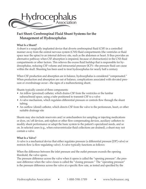
<strong>Fact</strong> <strong>Sheet</strong>: <strong>Cerebrospinal</strong> <strong>Fluid</strong> <strong>Shunt</strong> <strong>Systems</strong> <strong>for</strong> <strong>the</strong><br />
<strong>Management</strong> of Hydrocephalus<br />
What Is a <strong>Shunt</strong>?<br />
A shunt is a surgically implanted device that diverts cerebrospinal fluid (CSF) in a controlled<br />
manner away from <strong>the</strong> central nervous system (CNS) fluid compartments (<strong>the</strong> ventricles or fluid<br />
space near <strong>the</strong> spine) to an internal delivery site, such as <strong>the</strong> abdomen or heart. It thus provides an<br />
alternative pathway when CSF absorption is impaired, because of obstruction(s) in <strong>the</strong> CNS fluid<br />
compartments or o<strong>the</strong>r factors. This relieves <strong>the</strong> excess fluid buildup that is responsible <strong>for</strong> hydrocephalus,<br />
reducing CSF volume and intracranial pressure (ICP)—<strong>the</strong> pressure fluid can cause<br />
within <strong>the</strong> skull. <strong>Shunt</strong>ing has been used to treat hydrocephalus <strong>for</strong> nearly half a century.<br />
When CSF production and absorption are in balance, hydrocephalus is considered “compensated.”<br />
When production and absorption are out of balance, complications associated with elevated pressure<br />
or overdrainage occur—<strong>the</strong> signs of a malfunctioning shunt.<br />
<strong>Shunt</strong>s typically consist of three components:<br />
• An inflow (proximal) ca<strong>the</strong>ter, which drains CSF from <strong>the</strong> ventricles or <strong>the</strong> lumbar<br />
subarachnoid space, using a tube positioned to transmit CSF to a valve<br />
• A valve mechanism, which regulates differential pressure or controls flow through <strong>the</strong> shunt<br />
tubing;<br />
• An outflow (distal) ca<strong>the</strong>ter, which directs CSF from <strong>the</strong> valve to <strong>the</strong> peritoneum, heart, or o<strong>the</strong>r<br />
suitable drainage site<br />
<strong>Shunt</strong>s may also include reservoirs and/or antechambers <strong>for</strong> sampling or injecting medications<br />
or dyes, on/off devices, anti-siphon or o<strong>the</strong>r flow-compensating devices, auxiliary ca<strong>the</strong>ters to<br />
modify shunt per<strong>for</strong>mance or adapt <strong>the</strong> basic system to <strong>the</strong> patient’s specialized needs, and so<br />
on. In selected cases (e.g., when extraventricular fluid collections are drained), a shunt may not<br />
contain a valve.<br />
What Is a Valve?<br />
A valve is a mechanical device that ei<strong>the</strong>r regulates pressure (a differential pressure [DP] valve) or<br />
restricts flow (a flow-regulating valve). A valve typically functions as follows:<br />
When <strong>the</strong> difference between <strong>the</strong> inlet pressure and <strong>the</strong> outlet pressure exceeds <strong>the</strong> opening<br />
threshold, <strong>the</strong> valve opens.<br />
The pressure difference across <strong>the</strong> valve when it opens is called <strong>the</strong> “opening pressure”; <strong>the</strong> pressure<br />
difference when <strong>the</strong> valve closes is called <strong>the</strong> “closing pressure.” The “operating pressure”<br />
is <strong>the</strong> pressure difference across <strong>the</strong> valve at a specific flow rate, as tested and specified by <strong>the</strong><br />
Hydrocephalus Association • 1-888-598-3789 • www.hydroassoc.org
manufacturer.<br />
Valves’ opening and closing pressures may differ because of <strong>the</strong> nature of <strong>the</strong> materials used in<br />
valve design. For instance, silicone elastomer (used in slit, diaphragm, and miter valves) behaves<br />
differently when opening than it does when closing. Or slit and miter valves may stick, causing<br />
large deviations among opening, operating, and closing pressures.<br />
The difference between <strong>the</strong> pressure/flow curves at a valve’s opening and at its closing is called<br />
“hysteresis.” Miter and diaphragm valves demonstrate a larger degree of hysteresis compared to<br />
ball-in-cone valves. Depending on individual circumstances, a valve with more or less hysteresis<br />
might be more appropriate.<br />
What Is Siphoning/Overdrainage?<br />
Siphoning is <strong>the</strong> gravity effect of <strong>the</strong> hydrostatic (fluid) column on valve function when an individual<br />
is vertical. When <strong>the</strong> individual stands or sits, <strong>the</strong> fluid column height in <strong>the</strong> outflow<br />
ca<strong>the</strong>ter changes, and a negative pressure equal to <strong>the</strong> vertical height of <strong>the</strong> fluid column in <strong>the</strong><br />
ca<strong>the</strong>ter is produced. (Intracranial pressure [ICP], <strong>the</strong>re<strong>for</strong>e, can drop into <strong>the</strong> negative range, but<br />
even in persons without hydrocephalus and a shunt, upright ICP is slightly negative.) This gravity<br />
effect increases <strong>the</strong> differential pressure across <strong>the</strong> valve, keeping it open and allowing more<br />
CSF drainage. The effects of siphoning depend on multiple factors, occurring to a greater or lesser<br />
degree depending upon <strong>the</strong> type of valve implanted and present conditions.<br />
Ventricular overdrainage occurs when siphoning happens chronically. Overdrainage may in turn<br />
cause <strong>the</strong> brain to pull away from <strong>the</strong> inner surface of <strong>the</strong> skull, tearing <strong>the</strong> bridging scalp veins<br />
and causing associated bleeding and brain compression, debilitating headaches, and slit ventricles.<br />
What Types of Valves are Used to Treat Hydrocephalus?<br />
Most valves used in treating hydrocephalus are DP valves, operating on <strong>the</strong> principle of change<br />
in differential pressure—<strong>the</strong> difference between <strong>the</strong> pressure at <strong>the</strong> proximal ca<strong>the</strong>ter tip (inlet?)<br />
and <strong>the</strong> pressure at <strong>the</strong> drainage end (outlet?). Neurosurgeons select a specific DP valve based<br />
upon an individual’s age, <strong>the</strong> size of his or her ventricles, and o<strong>the</strong>r clinical factors. Sometimes<br />
<strong>the</strong> selected DP range does not adequately address <strong>the</strong> patient’s requirements, and a valve with<br />
a higher or lower DP range may be implanted. A number of newer shunts can be programmed<br />
noninvasively (<strong>the</strong> DP is changed magnetically), while o<strong>the</strong>rs have self-adjusting flow-regulating<br />
mechanisms.<br />
Most commercially available DP shunts are provided in three to five ranges: low, medium, and<br />
high pressure (and sometimes very low and very high), depending on a shunt’s response to<br />
<strong>the</strong> pressure differential between its upper and lower ends. The actual values of <strong>the</strong>se arbitrary<br />
ranges and <strong>the</strong> way valves are tested vary from manufacturer to manufacturer. Voluntary industry<br />
standards on measuring pressure/flow characteristics, such as those developed by ASTM<br />
and ISO, have not been adopted universally, nor are <strong>the</strong>re industry-defined values <strong>for</strong> each of <strong>the</strong><br />
nominal DP ranges. In addition, because of limitations in <strong>the</strong> technology related to quality control<br />
during <strong>the</strong> manufacturing process, it is not possible to make all shunts of a particular variety (e.g.,<br />
medium pressure) function within <strong>the</strong> same range.<br />
Hydrocephalus Association • 1-888-598-3789 • www.hydroassoc.org
Types of Differential Pressure (DP) Valves<br />
Slit valves. These valves, made of curved, silicone rubber material, are characterized by a cut,<br />
or slit. Flow on <strong>the</strong> concave side of <strong>the</strong> valve, if sufficient, will open <strong>the</strong> slit. The hydrodynamic<br />
properties of <strong>the</strong> valve depend on <strong>the</strong> thickness and stiffness of <strong>the</strong> elastomer and <strong>the</strong> number of<br />
slits. Some slit valves incorporate an internal spring to prevent slit inversion, which would lead to<br />
reflux (backward flow). These valves’ operating characteristics depend on slit integrity. The aging<br />
of silicone rubber materials and mishandling during surgery may significantly alter <strong>the</strong> per<strong>for</strong>mance<br />
of slit valves. The Codman Holter Valve, <strong>the</strong> Uni-shunt, <strong>the</strong> Phoenix Diamond, <strong>the</strong> Cruci<strong>for</strong>m,<br />
and <strong>the</strong> CRx are all slit valves.<br />
Duck-bill and miter valves. These valves are characterized by a round orifice that converges into<br />
two flat, horizontally opposed leaflets made of silicone. The valve leaflets open when <strong>the</strong> DP<br />
across <strong>the</strong>m increases. These valves operating characteristics depend on <strong>the</strong> size, shape, thickness,<br />
and length of <strong>the</strong> leaflets. The Integra Lifesciences (Heyer-Schulte) Mischler Valve is a miter valve.<br />
Spring-loaded, ball-in-cone valves. These valves incorporate a metallic coiled or flat spring that<br />
applies a calibrated <strong>for</strong>ce to a ball manufactured from a syn<strong>the</strong>tic ruby, located in a cone-shaped<br />
orifice. Such valves’ opening pressure is defined by <strong>the</strong> properties of <strong>the</strong> spring. The size of <strong>the</strong><br />
valve module opening changes according to DP across <strong>the</strong> mechanism: <strong>the</strong> higher <strong>the</strong> pressure<br />
differential, <strong>the</strong> larger <strong>the</strong> size of <strong>the</strong> opening. The ball moves away from <strong>the</strong> seat as DP increases,<br />
under control of <strong>the</strong> spring, <strong>the</strong>reby increasing <strong>the</strong> cross-sectional area through which CSF flows.<br />
When DP across <strong>the</strong> valve decreases, <strong>the</strong> ball moves toward <strong>the</strong> seat, reducing <strong>the</strong> cross-sectional<br />
area through which CSF flows.<br />
NMT Neuroscience produces a gravity-compensating lumboperitoneal valve (<strong>the</strong> HV Lumbar<br />
Valve) and a separate Gravity-Compensating Accessory that add resistance when an individual<br />
stands through <strong>the</strong> weight of several stainless steel balls on a ball-in-cone mechanism. Flow is not<br />
restricted when an individual is lying down.<br />
Ball-in-cone valves are less prone to <strong>the</strong> effects of <strong>the</strong> aging of materials than are miter or slit<br />
valves, and <strong>the</strong>y have been demonstrated to handle higher CSF protein levels. Examples of ball-incone<br />
valves include <strong>the</strong> Hakim Valve, available from NMT Neuroscience and Codman, a Johnson &<br />
Johnson Company, and <strong>the</strong> Phoenix Accura valves, available from Phoenix Biomedical Corp.<br />
Diaphragm valves. In <strong>the</strong>se valves, a mobile flexible membrane moves in response to pressure<br />
differences. The membrane may be held by a central piston that moves in a sleeve, or it may surround<br />
<strong>the</strong> piston and act as an occluder. The membrane can also be a dome that de<strong>for</strong>ms under<br />
pressure. These valves’ operating characteristics are based on <strong>the</strong> stiffness of <strong>the</strong> silicone rubber<br />
diaphragm, which is mounted beneath an integral pumping reservoir and has no metal parts.<br />
Pressure differentials cause <strong>the</strong> diaphragm to move, allowing CSF to flow around it. When pressure<br />
abates, <strong>the</strong> diaphragm seals <strong>the</strong> mechanism. The Radionics Contour Flex Valve and <strong>the</strong><br />
Medtronic PS Medical Delta Valve are examples of pressure-regulated diaphragm valves.<br />
Some DP valves include integral Anti-Siphon Devices (ASD) or Siphon Control Devices (SCD).<br />
Examples include <strong>the</strong> Medtronic PS Delta and <strong>the</strong> Integra Lifesciences Novus valves. Although<br />
Hydrocephalus Association • 1-888-598-3789 • www.hydroassoc.org
<strong>the</strong>ir mechanisms vary, <strong>the</strong> goal is <strong>the</strong> same: to counteract gravity’s effects on <strong>the</strong> fluid column<br />
below <strong>the</strong> valve when <strong>the</strong> patient sits or stands. For example, when pressure within <strong>the</strong> distal<br />
fluid column drops below atmospheric pressure, a diaphragm in <strong>the</strong> ASD might close <strong>the</strong> fluid<br />
passage, blocking CSF flow through <strong>the</strong> system, until pressure within <strong>the</strong> ventricles is again above<br />
atmospheric pressure. When this occurs, <strong>the</strong> diaphragm is pushed back, and flow resumes. The<br />
dependence on atmospheric reference, however, may cause such a mechanism to malfunction<br />
when tissue fibrosis occurs around it or when an individual’s head presses on it during sleep.<br />
Adjustable and programmable valves. Noninvasively adjustable valves incorporate a ball-in-cone<br />
mechanism regulated by a horseshoe-shaped spring that can be adjusted using a magnet.<br />
The resistance of programmable valves, such as those manufactured by Codman and Sophysa<br />
(not currently available in <strong>the</strong> U.S.), can be altered using a magnetic field transmitted through <strong>the</strong><br />
skin. These programmable valves, whose settings can be changed during a routine office visit,<br />
are not self-adjusting: <strong>the</strong>ir pressure settings must be reprogrammed until an acceptable level is<br />
reached. Because siphoning occurs with <strong>the</strong>se systems, a gravity-compensating or anti-siphon<br />
mechanism may sometimes be needed.<br />
The Hakim Programmable Valve includes a programmer and a transmitter. Depending on spring<br />
position on a series of steps within <strong>the</strong> valve, more or less tension is placed on <strong>the</strong> mechanism’s<br />
ball, adjusting DP to one of 18 settings between 30 and 200 mmH2O in 10 mmH2O increments.<br />
This mechanism, however, does not compensate <strong>for</strong> acute changes brought about by an individual’s<br />
daily activities. Also, because programmable valves contain metal, <strong>the</strong>y produce artifacts on<br />
scans. They also may be reprogrammed in strong magnetic fields such as an MRI.<br />
Multi-stage flow-regulating valves. These valves maintain <strong>the</strong> drainage flow rate at close to <strong>the</strong><br />
rate of CSF secretion, regardless of patient position and o<strong>the</strong>r conditions that normally promote<br />
overdrainage. They combine <strong>the</strong> features of a DP valve with <strong>the</strong> benefits of a variable flow restrictor.<br />
Examples include <strong>the</strong> NMT Orbis-Sigma Valve (OSV II) and <strong>the</strong> Phoenix Diamond Valve.<br />
Such valves typically are not used <strong>for</strong> drainage of extraventricular structures that require a valve<br />
with very low operating pressure—<strong>for</strong> example, a check valve with very little resistance—or no<br />
valve at all.<br />
What Are <strong>the</strong> Complications?<br />
First, some statistics: In <strong>the</strong> pediatric population, approximately 60 percent of shunts are still functioning<br />
one year after implantation. One pediatric study found that <strong>the</strong> average shunt functions<br />
<strong>for</strong> approximately 5.5 years; some shunts require revision much sooner, o<strong>the</strong>rs later. For an infant<br />
under one year of age, <strong>the</strong> likelihood of shunt revision during <strong>the</strong> first year is relatively high—approximately<br />
40 percent. Often, several revisions are required during childhood if hydrocephalus<br />
is treated from infancy.<br />
Generally, shunt malfunctions are caused by suboptimal surgical technique, shunt inadequacy,<br />
or <strong>the</strong> unique characteristics of an individual patient’s body. When shunts fail, <strong>the</strong> highest failure<br />
rate is during <strong>the</strong> acute postoperative period, sometimes because of complications of <strong>the</strong> surgical<br />
procedure itself, such as <strong>the</strong> presence of blood, debris, etc. As mechanical devices, shunts may<br />
Hydrocephalus Association • 1-888-598-3789 • www.hydroassoc.org
eak, migrate (move), or, more commonly, become blocked (occluded). Breakage causes a total or<br />
partial interruption in <strong>the</strong> shunt pathway, which completely or partially obstructs fluid flow and<br />
adds resistance to <strong>the</strong> system. Migration alters shunt function, causing ca<strong>the</strong>ters to move to locations<br />
that may restrict flow.<br />
As <strong>for</strong>eign bodies, shunts may become infected—colonized with bacteria or, more rarely, fungi,<br />
most often at <strong>the</strong> time of implantation. Infection occurs in about 1 to 15 percent of shunt operations.<br />
More rare complications include intestinal volvulus (twisting) around <strong>the</strong> shunt ca<strong>the</strong>ter,<br />
<strong>the</strong> <strong>for</strong>mation of encapsulated intraperitoneal CSF compartments, and adverse reactions to <strong>the</strong><br />
implanted materials. Seizures have also been associated with ventricular ca<strong>the</strong>ter implantation.<br />
Ventriculoatrial (VA) shunts have been associated with pulmonary hypertension, pulmonary tree<br />
embolization, and shunt nephritis.<br />
<strong>Shunt</strong> Revisions<br />
<strong>Shunt</strong> malfunction should be suspected when symptoms of hydrocephalus return (x-ref here).<br />
When a malfunction is suspected, a neurosurgeon must evaluate <strong>the</strong> implanted shunt system to<br />
determine its cause: complete or partial system blockage, <strong>the</strong> possibility of shunt disconnection or<br />
fracture, a mismatch between <strong>the</strong> individual’s requirements and shunt capabilities. Surgical shunt<br />
revisions may be required at any time to correct one or more of <strong>the</strong>se problems or to compensate<br />
<strong>for</strong> individual growth.<br />
Which Companies Manufacture <strong>Shunt</strong>s?<br />
During <strong>the</strong> past decade, <strong>the</strong> U.S. shunt industry has been consolidating. Presently, six companies<br />
serve <strong>the</strong> U.S. market:<br />
Codman, a Johnson and Johnson Company, sells several shunt lines: Holter, Medos, Hakim, Accuflow<br />
Unishunt, and Denver.<br />
Integra Neurocare produces Heyer Schulte and Novus.<br />
Medtronics PS Medical’s products include <strong>the</strong> Delta <strong>Shunt</strong>.<br />
NMT Neuroscience, which now includes Cordis, produces <strong>the</strong> Orbis-Sigma Valve.<br />
Phoenix Biomedical Corp., which now includes Holter-Hausner, Phoenix Bioengineering, and<br />
Theramedics, features <strong>the</strong> Diamond Valve.<br />
Radionics.<br />
Are There Alternatives <strong>for</strong> <strong>the</strong> <strong>Management</strong> of Hydrocephalus?<br />
Within this decade, neurosurgeons have made advancements in <strong>the</strong> development of surgical<br />
alternatives to implanted shunts <strong>for</strong> <strong>the</strong> treatment of hydrocephalus. These alternatives include<br />
endoscopic third ventriculostomy <strong>for</strong> aqueductal stenosis (narrowing of <strong>the</strong> passageway between<br />
<strong>the</strong> third and fourth ventricles); fenestration (cutting of <strong>the</strong> walls) of loculated ventricles; and microsurgical<br />
removal of tumors to try to restore normal CSF passageways. Despite great improvements<br />
in surgical technique, however, <strong>the</strong> long-term success of endoscopic third ventriculostomy<br />
has not been determined. For most people with hydrocephalus, shunt implantation is <strong>the</strong> only<br />
viable <strong>the</strong>rapeutic alternative.<br />
Hydrocephalus Association • 1-888-598-3789 • www.hydroassoc.org
Conclusion<br />
In <strong>the</strong> medical device industry, rarely has any o<strong>the</strong>r product had such a profound effect on patient<br />
care. Our understanding of shunts—how <strong>the</strong>y function, how <strong>the</strong>y might function better, which<br />
components best suit an individual’s given clinical condition—is constantly improving. However,<br />
<strong>the</strong> goal of most neurosurgeons involved in <strong>the</strong> treatment of hydrocephalus is to someday be able<br />
to provide an alternative to shunting, one without shunting’s relatively high rate of complications.<br />
Still, although shunting may not give us a perfect way to manage hydrocephalus, <strong>for</strong> now, <strong>for</strong> <strong>the</strong><br />
majority of patients, it is <strong>the</strong> best option. And it is improving: neurosurgeons are constantly evaluating<br />
<strong>the</strong> success of various techniques to improve our ability to safely manage hydrocephalus,<br />
while shunt manufacturers continue to work on new and improved shunts to address <strong>the</strong> limitations<br />
of existing designs.<br />
References<br />
Cinalli G., C. Sainte-Rose C, P. Chumas, et al. “Failure of Third Ventriculostomy in <strong>the</strong> Treatment<br />
of Aqueductal Stenosis in Children. Journal of Neurosurgery (U.S.) 90.3 (1999): 448–54.<br />
Drake, J.M. “Ventriculostomy <strong>for</strong> Treatment of Hydrocephalus.” Hydrocephalus 4.4 (1993): 657–<br />
67.<br />
Drake, J.M., and C. Sainte-Rose. The <strong>Shunt</strong> Book. 1995.<br />
Sainte-Rose C., J. H. Piatt, D. Renier, et al. “Mechanical Complications in <strong>Shunt</strong>s. Pediatric Neurosurgery<br />
(Switzerland) 17.1 (1991–92): 2–9.<br />
Toporek, C., K. Robinson, and L. Lamb. Hydrocephalus : A Guide <strong>for</strong> Patients, Families, and<br />
Friends. 1999.<br />
Patient In<strong>for</strong>mation Materials<br />
Hydrocephalus Association<br />
“About Hydrocephalus: A Book <strong>for</strong> Parents” (in English or Spanish).<br />
“About Normal Pressure Hydrocephalus: A Book <strong>for</strong> Adults and Their Families.”<br />
“Prenatal Hydrocephalus: A Book <strong>for</strong> Parents.”<br />
NMT Neurosciences, Inc.<br />
“Just Like Any O<strong>the</strong>r Beagle.” A coloring book on hydrocephalus <strong>for</strong> children with hydrocephalus<br />
and <strong>the</strong>ir families. Contact <strong>the</strong> Hydrocephalus Association or NMT Neurosciences, Inc.: 3450<br />
Corporate Way, Duluth GA, 30096.<br />
This in<strong>for</strong>mation sheet was produced by <strong>the</strong> Hydrocephalus Association, copyright © 2000. It may<br />
be reproduced provided a full citation of <strong>the</strong> source is given.<br />
Hydrocephalus Association • 1-888-598-3789 • www.hydroassoc.org
It was adapted from an article written by Marvin L. Sussman, Ph.D., entitled “<strong>Shunt</strong> Technology:<br />
An Update in <strong>the</strong> Newsletter of <strong>the</strong> Hydrocephalus Association: 17.2 (1999). Dr. Sussman’s article<br />
was reviewed by Drs. Marvin Bergsneider, Philip H. Cogen, Roger J. Hudgins, J. Gordon Mc-<br />
Comb, Donald H. Reigel, Marion L. Walker, and Michael A. Williams, along with industry representatives<br />
Thomas Tokas of Codman/Johnson and Johnson, Ralph A. Kistler of Medtronic, and<br />
Charles Hokanson of Phoenix Biomedical Corp.<br />
Marvin L. Sussman, Ph.D., has been involved with <strong>the</strong> hydrocephalus shunt industry <strong>for</strong> over<br />
20 years, holding a number of management positions with Cordis Corporation. He has served as<br />
industry co-chair of <strong>the</strong> ASTM <strong>Shunt</strong> Standards Committee and <strong>the</strong> HIMA Task Force on Device<br />
Tracking and as industry representative on <strong>the</strong> FDA Neurological Devices Panel. He holds a<br />
number of patents <strong>for</strong> shunt-related inventions and has written and edited books in <strong>the</strong> field of<br />
gerontology. He is <strong>the</strong> industry representative on <strong>the</strong> Board of Directors of <strong>the</strong> Hydrocephalus<br />
Association.<br />
For additional resources about hydrocephalus and in<strong>for</strong>mation about <strong>the</strong> services provided by <strong>the</strong><br />
Hydrocephalus Association, please contact:<br />
Hydrocephalus Association<br />
870 Market St., Suite 705<br />
San Francisco, CA 94102<br />
Telephone: (415) 732-7040 / (888) 598-3789<br />
Fax: (415) 732-7044<br />
www.hydroassoc.org<br />
info@hydroassoc.org<br />
Hydrocephalus Association • 1-888-598-3789 • www.hydroassoc.org


