Shunt Systems Factsheet - Hydrocephalus Association
Shunt Systems Factsheet - Hydrocephalus Association
Shunt Systems Factsheet - Hydrocephalus Association

You also want an ePaper? Increase the reach of your titles
YUMPU automatically turns print PDFs into web optimized ePapers that Google loves.
Fact Sheet<br />
<strong>Shunt</strong> <strong>Systems</strong> for the Management of <strong>Hydrocephalus</strong><br />
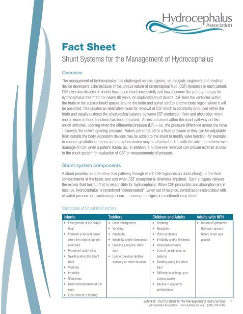
Overview<br />
The management of hydrocephalus has challenged neurosurgeons, neurologists, engineers and medical<br />
device developers alike because of the unique nature of cerebrospinal fluid (CSF) dynamics in each patient.<br />
CSF diversion devices or shunts have been used successfully and have become the primary therapy for<br />
hydrocephalus treatment for nearly 60 years. An implanted shunt diverts CSF from the ventricles within<br />
the brain or the subarachnoid spaces around the brain and spinal cord to another body region where it will<br />
be absorbed. This creates an alternative route for removal of CSF which is constantly produced within the<br />
brain and usually restores the physiological balance between CSF production, flow, and absorption when<br />
one or more of these functions has been impaired. Valves contained within the shunt pathway act like<br />
on-off switches, opening when the differential pressure (DP) – i.e., the pressure difference across the valve<br />
- exceeds the valve’s opening pressure. Valves are either set to a fixed pressure or they can be adjustable<br />
from outside the body. Accessory devices may be added to the shunt to modify valve function; for example,<br />
to counter gravitational forces an anti-siphon device may be attached in-line with the valve to minimize overdrainage<br />
of CSF when a patient stands up. In addition, a bubble-like reservoir can provide external access<br />
to the shunt system for evaluation of CSF or measurements of pressure.<br />
<strong>Shunt</strong> system components<br />
A shunt provides an alternative fluid pathway through which CSF bypasses an obstruction(s) in the fluid<br />
compartments of the brain, and acts when CSF absorption is otherwise impaired. Such a bypass relieves<br />
the excess fluid buildup that is responsible for hydrocephalus. When CSF production and absorption are in<br />
balance, hydrocephalus is considered “compensated”; when out of balance, complications associated with<br />
elevated pressure or overdrainage occur— causing the signs of a malfunctioning shunt.<br />
Symptoms of <strong>Shunt</strong> Malfunction:<br />
Infants Toddlers Children and Adults Adults with NPH<br />
• Enlargement of the baby’s<br />
head<br />
• Fontanel is full and tense<br />
when the infant is upright<br />
and quiet<br />
• Prominent scalp veins<br />
• Swelling along the shunt<br />
tract<br />
• Vomiting<br />
• Irritability<br />
• Sleepiness<br />
• Downward deviation of the<br />
eyes<br />
• Less interest in feeding<br />
• Head enlargement<br />
• Vomiting<br />
• Headache<br />
• Irritability and/or sleepiness<br />
• Swelling along the shunt<br />
tract<br />
• Loss of previous abilities<br />
(sensory or motor function)<br />
• Vomiting<br />
• Headache<br />
• Vision problems<br />
• Irritability and/or tiredness<br />
• Personality change<br />
• Loss of coordination or<br />
balance<br />
• Swelling along the shunt<br />
tract<br />
• Difficulty in waking up or<br />
staying awake<br />
• Decline in academic<br />
performance<br />
• Return of symptoms<br />
that were present<br />
before shunt was<br />
placed<br />
<strong>Factsheet</strong>: <strong>Shunt</strong> <strong>Systems</strong> for the Management of <strong>Hydrocephalus</strong><br />
<strong>Hydrocephalus</strong> <strong>Association</strong> www.hydroassoc.org (888) 598-3789<br />
1
<strong>Shunt</strong>s typically consist of three major components:<br />
• An inflow (proximal or closer to the inflow site) catheter, which drains CSF from the ventricles or the<br />
subarachnoid space; this tube leaves the brain through a small hole in the skull and then runs for a<br />
short distance under the skin.<br />
• A valve mechanism, which regulates differential pressure or controls flow through the shunt tubing;<br />
this device is connected to the proximal catheter and lies between the skin and the skull, usually on<br />
top of the head or just behind the ear.<br />
• An outflow (distal or farther away from the inflow site) catheter, which runs under the skin and directs<br />
CSF from the valve to the abdominal (or peritoneal) cavity, heart or other suitable drainage site.<br />
Other shunt components may include reservoirs and/or chambers for CSF sampling or injecting<br />
medications or dyes, on/off devices, anti-siphon or other flow-compensating devices, or auxiliary<br />
catheters to modify performance or adapt the basic system to the patient’s specialized needs. In<br />
selected cases (such as when cysts or subarachnoid fluid collections are drained), a shunt may not<br />
contain a valve or a very low resistance valve may be used.<br />
Catheters and tubing<br />
Catheters and tubing divert CSF from the site where there is an excess volume of CSF to a location<br />
within the body where the CSF will be absorbed. The proximal catheter (ventricular or lumbar catheter)<br />
drains excess CSF from the ventricles or the spinal lumbar sac through rows of small holes at its origin.<br />
Distal catheters are typically placed in the abdominal (or peritoneal) cavity, but may also be placed in the<br />
heart, pleural cavity (lungs) and other suitable locations where CSF is drained into the bloodstream. The<br />
type of shunt system is named by the inflow and outflow locations, e.g. if the proximal catheter is in the<br />
ventricle and the distal catheter is in the peritoneal cavity it is called a ventriculo-peritoneal (VP) shunt<br />
(Table 1). At times temporary CSF drainage is performed before a full shunt system is implanted; these<br />
short-term drainage systems are called external drains (ventricular or spinal) because the distal catheter<br />
is open or drains into a bag outside the body. <strong>Shunt</strong> tubing is made of flexible silicone, with short plastic<br />
tubes used at times as connectors to the valve mechanisms. Some shunt tubing is impregnated with<br />
antibiotics to reduce the incidence of infection during the post-operative period; examples include the<br />
Codman BactisealTM catheter and the Medtronic AresTM catheter.<br />
Table 1: Most common shunt systems<br />
<strong>Shunt</strong> Pathway <strong>Shunt</strong> Type CSF Inflow Location CSF Drainage Location<br />
Ventriculo-peritoneal VP Ventricle Peritoneal cavity<br />
Ventriculo-atrial VA Ventricle Right atrium of the heart<br />
Ventriculo-pleural VPL Ventricle Pleural cavity<br />
Lumbo-peritoneal LP Lumbar spine Peritoneal cavity<br />
Valve Mechanisms<br />
To assure that the rate of flow through the shunt is controlled, a valve is placed in the tubing system.<br />
In addition, the function of the valve may be modified with the addition of accessories such as siphon<br />
control, gravity compensating devices or flow regulating devices. Some shunt systems are a one-piece<br />
design from the manufacturer. Others are “custom-made” by the surgeon in the OR using connectors<br />
<strong>Factsheet</strong>: <strong>Shunt</strong> <strong>Systems</strong> for the Management of <strong>Hydrocephalus</strong><br />
<strong>Hydrocephalus</strong> <strong>Association</strong> www.hydroassoc.org (888) 598-3789<br />
2
to add components to the system. Most valves operate on the principles of change in differential pressure<br />
(DP)—the difference between the pressure at the proximal catheter tip and the pressure at the drainage<br />
end. Neurosurgeons select a DP valve based upon the age of the patient, the size of the ventricles, the<br />
amount of pressure, and other important clinical factors. Sometimes the selected DP range does not<br />
adequately address the patient’s requirements and a valve with a higher or lower DP range may need to<br />
be implanted; these are referred to as “high” or “low” pressure valves, respectively, based on the opening<br />
pressure of the valve. A number of newer shunts can be adjusted non-invasively (i.e. the DP is changed<br />
magnetically from outside the body), while others have self-adjusting flow-regulating mechanisms. When<br />
such valves are used, a second surgical operation is avoided as the valve’s operating characteristics may be<br />
changed non-invasively (programmable valves) or adjusted automatically (flow-regulated valves).<br />
Most commercially available fixed DP shunts are provided in three to five ranges: low, medium or high<br />
pressures (and very low and very high), depending on their response to the pressure differential between<br />
the shunt’s upper and lower ends. Voluntary industry standards on how to measure pressure/flow<br />
characteristics, such as those developed by independent or government standards, have not been adopted<br />
universally, nor are there industry-defined values for each of the nominal DP ranges. In addition, due to<br />
limitations in the technology related to quality control during the manufacturing process, it is not possible<br />
to make all shunts of a specific variety (e.g., medium pressure) function within the same range. These<br />
factors make the choice of the valve very complicated, and clinicians who are not familiar with the specific<br />
characteristics of different valves may have difficulty selecting the best one.<br />
The following shows examples of <strong>Shunt</strong> Valves:<br />
ProGAV Adjustable shunt<br />
Aesculap Inc.<br />
Toll free: 1-800-282-9000<br />
Website: www.aesculapusa.com<br />
Codman® CertasTM Programmable Valve<br />
Codman, a Johnson and Johnson Company<br />
Toll free: 1-800-225-0460<br />
Website: www.codman.com<br />
OSV II® Flow Regulating Valve<br />
Integra Life Sciences:<br />
Toll free: 1-800-654-2873<br />
Email: custsvcnj@integralife.com<br />
Website: www.integralife.com<br />
Strata Adjustable Valve<br />
Medtronic Neurologic Technologies:<br />
Toll free: 1-800-468-9710<br />
Website: www.medtronic.com<br />
Diamond Valve<br />
Polaris Adjustable Valve<br />
Sophysa<br />
Telephone: 219-663-7723<br />
Website: www.sophysa.com<br />
<strong>Factsheet</strong>: <strong>Shunt</strong> <strong>Systems</strong> for the Management of <strong>Hydrocephalus</strong><br />
<strong>Hydrocephalus</strong> <strong>Association</strong> www.hydroassoc.org (888) 598-3789<br />
3
Currently, there are three basic valve designs:<br />
Slit valves: These relatively old valves are characterized by a cut (slit) in the wall of the end of the distal<br />
catheter. Fluid pressure within the lumen of the catheter, if sufficient, will open the slit and allow CSF to<br />
flow out of the catheter. The operating pressure characteristics depend on the mechanical properties of the<br />
slit, i.e. the stiffness of the silicone, the thickness of the wall, and the length of the slit. Aging of silicone<br />
rubber materials and mishandling during surgery may significantly alter the performance of this type of<br />
valve.<br />
Diaphragm valves: For these valves, a flexible membrane moves in response to pressure differences.<br />
Pressure differentials cause the diaphragm to move, allowing CSF to flow around the diaphragm. When the<br />
pressure is reduced (after some CSF flows through the valve), the diaphragm returns to its original position<br />
and seals the mechanism.<br />
Spring-loaded, ball-in-cone valves: These valves incorporate a metallic coiled or flat spring that applies<br />
a calibrated force to a ball manufactured from a synthetic ruby located in a cone-shaped orifice. When<br />
the force (pressure) of CSF gets high enough it pushes the ball against the spring and opens the valve to<br />
allow flow. When the fluid pressure is reduced the ball returns to its original position and closes the valve.<br />
Ball-in-cone valves are less prone to the effects of the aging of materials than are slit valves, and have been<br />
demonstrated to not clog with higher CSF protein levels.<br />
Fixed vs. adjustable valves<br />
Adjustable valves incorporate an external adjustment tool, applied outside the body, to select a DP setting<br />
from a range of available settings. The number of adjustable settings depends on the manufacturer. Once<br />
adjusted to a specific setting, these valves operate like a fixed pressure valve until re-adjusted by a clinician.<br />
Since they are designed to be adjusted by a strong magnetic field, some of these valves may be susceptible<br />
to adjustment by strong environmental magnetic fields and care must be taken to keep toys with<br />
magnets and other sources of magnetic fields away from patients with these implanted valves.<br />
Some adjustable shunts incorporate mechanisms that cannot be adjusted by magnetic fields other than<br />
those produced by the programmer.<br />
Adjustable valves incorporate a ball-in-cone mechanism regulated by a spring that can be adjusted<br />
noninvasively using a magnet. So-called “programmable” valves are DP valves whose opening pressure<br />
can be altered using a magnetic field transmitted through the skin. These programmable valves, which can<br />
be changed during a routine office visit, are not self-adjusting; DP pressure settings must be reprogrammed<br />
until an acceptable setting is reached, which may take several trials. Because siphoning occurs with these<br />
systems, some gravity-compensating<br />
or anti-siphon mechanism may be<br />
needed in selected cases. These<br />
mechanisms do not compensate for<br />
acute changes brought about by the<br />
patient’s daily activities. Also, because<br />
they contain an amount of metal,<br />
some programmable valves produce<br />
artifacts on scans. They also may be<br />
reprogrammed in strong magnetic<br />
fields such as an MRI and must be<br />
checked after being exposed to a<br />
strong magnetic field.<br />
<strong>Factsheet</strong>: <strong>Shunt</strong> <strong>Systems</strong> for the Management of <strong>Hydrocephalus</strong><br />
<strong>Hydrocephalus</strong> <strong>Association</strong> www.hydroassoc.org (888) 598-3789<br />
4
Multi-stage flow-regulating valves: These valves maintain the drainage flow rate close to the rate of CSF<br />
secretion, regardless of patient position and other conditions that normally promote overdrainage. These<br />
valves combine the features of a DP valve with the benefits of a variable flow restrictor.<br />
Antechambers/Reservoirs<br />
An antechamber is a sampling chamber or reservoir located under the skin between the inflow catheter and<br />
the valve. They can be used to sample CSF in the shunt, inject drugs into the brain through the proximal<br />
catheter, and measure pressure. In addition, these “bubbles” can be felt through the skin and pumped<br />
manually to help keep the shunt open. In general, if one pushes on the reservoir and it does not spring<br />
back, then there might be an obstruction in the proximal catheter because the reservoir is not filling with<br />
CSF. On the other hand, if the reservoir feels rather stiff and more force is needed to depress it, then the<br />
distal catheter may be clogged. Some clinicians recommend pumping the reservoir periodically to help<br />
keep the shunt open.<br />
Connectors<br />
Short plastic tubes are used to connect catheters with tubing, tubing with valves or accessories, etc.<br />
Typically, such connectors require the surgeon to secure them with a suture tie during surgery and are weak<br />
points that can become loose and disconnect the catheter.<br />
Overdrainage Control Devices<br />
Ideal shunt pressure-flow characteristics must match the patient’s specific needs. However, postural<br />
(standing, sitting, or lying down) and vasogenic (blood flow) influences modify shunt function. Standing<br />
posture causes a siphon or sucking effect, essentially “pulling” fluid out of the brain or lumbar region<br />
when the patient stands. During the night, there are periodic, small increases in blood volume in the<br />
head. This nocturnal cerebral vasogenic wave activity occurs several times a night during sleep. Similar<br />
blood volume increases occur when the patient strains, as during a cough or a bowel movement. These<br />
volume increases, in turn, “push” CSF out of the ventricles through the shunt. The terms “siphoning”,<br />
“overdrainage” and “overshunting”, (often used interchangeably) are not the same. Siphoning is a purely<br />
postural phenomenon. Overdrainage and overshunting refer to shunting CSF in excess of that required and<br />
may be caused by postural and vasogenic effects. A variety of siphon and overdrainage regulating devices<br />
have been developed to mitigate the effects of CSF overdrainage. Many major shunting complications are<br />
due to shunt overdrainage, including, but not limited to:<br />
• proximal shunt obstruction, in which tissue (i.e. choroid plexus) can get sucked into the holes of the<br />
proximal catheter and reduce inflow,<br />
• headache and dizziness,<br />
• slit (or small) ventricle syndrome, a condition in which there is an absence of CSF within the ventricles<br />
combined with a growing brain. This can lead to a situation in which the intracranial pressure or brain<br />
pressure can rise to dangerous levels before the ventricles have time to expand.<br />
• subdural collections, or fluid accumulations between the external coverings of the brain (the arachnoid<br />
and dura mater membranes).<br />
• extradural collections, or fluid accumulations between the dura mater and the inner surface of the skull<br />
• secondary craniosynostosis, a cranial defect in which the bony sutures (the fibrous joints between the<br />
bones of the skull) of an infant close too early, causing problems with normal brain and skull growth.<br />
<strong>Factsheet</strong>: <strong>Shunt</strong> <strong>Systems</strong> for the Management of <strong>Hydrocephalus</strong><br />
<strong>Hydrocephalus</strong> <strong>Association</strong> www.hydroassoc.org (888) 598-3789<br />
5
Siphon-resistive devices (SRD)<br />
These so-called anti-siphon or siphon-control devices incorporate a silicone membrane that closes when<br />
conditions favoring postural overdrainage are present. Siphon-resistive devices react to hydrostatic<br />
pressure across the two ends of the catheter and close the valve (by increasing the valves opening<br />
pressure) when the patient assumes a vertical posture. Such devices tend to create a positive intracranial<br />
pressure in the standing position. Communication with atmospheric pressure is the basis for the function<br />
of such devices and thus their function can be impaired by the development of scar tissue over the devices<br />
diaphragm membrane. Some<br />
children may be too short to generate<br />
sufficient negative hydrostatic<br />
pressure to keep the SRDs closed;<br />
positive pressures above the SRD<br />
will keep the device open and allow<br />
excessive flow. Similarly, if either<br />
device is placed in the subcutaneous<br />
tissue of the upper neck, then tilting<br />
or turning of the neck can change<br />
the subcutaneous pressure over the<br />
device, causing it to malfunction.<br />
Hydrostatic mechanisms (gravitational devices) typically involve the use of metal balls that fall down<br />
into a cone-shaped seat when the patient is standing or in an upright position. When the mechanism is<br />
positioned vertically, this adds resistance (depending on the number of balls) to the flow resistance in the<br />
shunt flow pathway approximately equal to the height of the hydrostatic column. When the mechanism is<br />
horizontal, the balls rest away from the fluid pathway, adding little resistance to the CSF flow pathway.<br />
The difficulty in using such devices lies in choosing the proper valve (available in several resistances) since,<br />
in the case of children, growth requires replacement of the gravity compensating mechanism with one with<br />
a higher setting, necessitating another operative intervention. Such devices may create issues relative to<br />
the ability to evaluate the patients using MRI and creating artifacts on imaging scans.<br />
Flow-regulating devices (FRD) attempt to maintain a constant flow through the valve at different<br />
pressures. Valve resistance is increased as differential pressure across the valve is increased. Such valves<br />
have mechanisms that vary the<br />
size of the opening and associated<br />
resistance to CSF flow depending<br />
on the differential pressure across<br />
the valve. These valves have a lower<br />
incidence of early obstruction; the<br />
time to revision appears to be longer<br />
than that with conventional differential<br />
pressure valves (Sainte-Rose C.<br />
<strong>Shunt</strong> obstruction: a preventable<br />
complication? Pediatr Neurosurg.<br />
1993 May-Jun; 19(3):156-64).<br />
<strong>Factsheet</strong>: <strong>Shunt</strong> <strong>Systems</strong> for the Management of <strong>Hydrocephalus</strong><br />
<strong>Hydrocephalus</strong> <strong>Association</strong> www.hydroassoc.org (888) 598-3789<br />
6
What are the complications of shunting?<br />
This important topic is covered in more detail on our website, www.hydroassoc.org, nevertheless, a simple<br />
introduction is offered here.<br />
Malfunction<br />
As mechanical devices, shunts may break or become disconnected, migrate (move) or, more commonly,<br />
become blocked. Breakage causes a total or partial interruption in the shunt pathway, which may obstruct<br />
fluid flow and add resistance to the system. Sometimes a disconnection may occur, but the formation of a<br />
scar tissue tunnel around the subcutaneous catheter still allows fluid to flow. Migration may also alter shunt<br />
function, causing catheters to move to locations that may restrict flow.<br />
Infection<br />
<strong>Shunt</strong>s may be colonized with bacteria or, more rarely, fungi, most often at the time of shunt implantation.<br />
Most of these infections are caused by skin bacteria that enter or surround the shunt during surgical<br />
implantation. Infection rates vary from about 1 percent to 15 percent of shunt operations. Recently, some<br />
manufacturers have added anti-microbial coatings to the shunt (i.e., the Codman Bactiseal or Medtronic<br />
Arescatheters) which appear to lower the rate of infection during the immediate postoperative period.<br />
Material degradation<br />
Originally, barium sulfate was mixed with silicone to allow shunt catheters to be visible on x-ray. These<br />
barium sulfate crystals eventually dissolved, making the tubing surface rough. Tissue in-growth to<br />
the roughened surface caused binding of the tubing at specific locations which promoted breakage or<br />
deterioration of the tubing. <strong>Shunt</strong> tubing design has been changed and clear silicone elastomer now covers<br />
the surface greatly reducing the possibility of this to occur.<br />
Other complications<br />
The development of seizures has been associated with ventricular catheter implantation. Ventriculoatrial (VA)<br />
shunts have been associated with pulmonary hypertension, pulmonary tree embolization and shunt nephritis<br />
(an inflammation of the small round filters (glomeruli) located in the kidney).<br />
Rare Complications<br />
Rare complications include intestinal volvulus (twisting) around the shunt catheter, formation of<br />
encapsulated intra-peritoneal CSF compartments or development of reactions to the implanted materials.<br />
<strong>Shunt</strong> revisions<br />
Surgical revisions may be required at any time to correct one or more of the above problems or to<br />
compensate for the growth of individuals. <strong>Shunt</strong> malfunction should be suspected when any symptoms of<br />
hydrocephalus return. A neurosurgeon must evaluate the implanted shunt system to determine whether<br />
a complete or partial blockage has occurred in the system, the possibility of a disconnection or fracture,<br />
or whether the present shunt does not match the patient’s physiological requirements. In some patients,<br />
shunt malfunction is a medical emergency and must be treated promptly to avoid complications.<br />
<strong>Factsheet</strong>: <strong>Shunt</strong> <strong>Systems</strong> for the Management of <strong>Hydrocephalus</strong><br />
<strong>Hydrocephalus</strong> <strong>Association</strong> www.hydroassoc.org (888) 598-3789<br />
7
Conclusions<br />
CSF shunts are commonly used to treat hydrocephalus. If left unchecked, the CSF imbalance can lead to<br />
elevated intracranial pressure (ICP) which can lead to a variety of complications. CSF shunts can be used to<br />
alleviate these problems in patients who suffer from hydrocephalus. <strong>Shunt</strong>s can come in a variety of forms<br />
but all of them consist of an inflow catheter communicating with an outflow catheter under the control<br />
of a valve which regulates pressure (differential pressure valves) or controls flow (flow-regulated<br />
valves). The main differences between shunts are usually in:<br />
• the materials used to construct them,<br />
• the types of valves mechanism used,<br />
• the source of CSF (ventricular, lumbar, etc.) and location of the drainage end-point (peritoneal, atrial,<br />
pleural, etc.), and<br />
• whether or not the valve is externally adjustable.<br />
Routine treatment complications are infection, obstruction, and overdrainage to name a few. Although some<br />
(regrettably, the minority) of the patients with shunts can go for years without complications, even those<br />
lucky few are potentially one shunt malfunction away from a major crisis. At any time, and without warning,<br />
a shunt complication can require emergency intervention.<br />
We would like to thank J. P. McAllister, PhD and Marvin Sussman, PhD for their expert input.<br />
We also would like to credit the following shunt companies for their valuable assistance.<br />
Aesculap Inc.<br />
Codman, a Johnson and Johnson Company<br />
Integra Life Sciences<br />
Medtronic Neurologic Technologies<br />
Sophysa<br />
This Information Sheet was produced by the <strong>Hydrocephalus</strong> <strong>Association</strong>, copyright © 2012. It may be reproduced<br />
provided a full citation of the source is given. The trademarks referenced are owned by the companies with which<br />
they are associated.<br />
For additional resources about hydrocephalus and information about the services of the<br />
<strong>Hydrocephalus</strong> <strong>Association</strong>, please contact:<br />
4340 East West Highway, Suite 905, Bethesda, MD 20814<br />
Telephone: (301) 202-3811 (888) 598-3789 Toll-Free Fax: (301) 202-3813<br />
Website: www.HydroAssoc.org Email: info@HydroAssoc.org<br />
<strong>Factsheet</strong>: <strong>Shunt</strong> <strong>Systems</strong> for the Management of <strong>Hydrocephalus</strong><br />
8