Fact Sheet: Cerebrospinal Fluid Shunt Systems for the Management ...
Fact Sheet: Cerebrospinal Fluid Shunt Systems for the Management ...
Fact Sheet: Cerebrospinal Fluid Shunt Systems for the Management ...

Create successful ePaper yourself
Turn your PDF publications into a flip-book with our unique Google optimized e-Paper software.
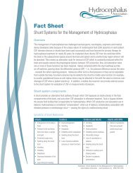
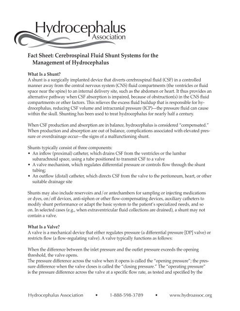
<strong>Fact</strong> <strong>Sheet</strong>: <strong>Cerebrospinal</strong> <strong>Fluid</strong> <strong>Shunt</strong> <strong>Systems</strong> <strong>for</strong> <strong>the</strong><br />
<strong>Management</strong> of Hydrocephalus<br />
What Is a <strong>Shunt</strong>?<br />
A shunt is a surgically implanted device that diverts cerebrospinal fluid (CSF) in a controlled<br />
manner away from <strong>the</strong> central nervous system (CNS) fluid compartments (<strong>the</strong> ventricles or fluid<br />
space near <strong>the</strong> spine) to an internal delivery site, such as <strong>the</strong> abdomen or heart. It thus provides an<br />
alternative pathway when CSF absorption is impaired, because of obstruction(s) in <strong>the</strong> CNS fluid<br />
compartments or o<strong>the</strong>r factors. This relieves <strong>the</strong> excess fluid buildup that is responsible <strong>for</strong> hydrocephalus,<br />
reducing CSF volume and intracranial pressure (ICP)—<strong>the</strong> pressure fluid can cause<br />
within <strong>the</strong> skull. <strong>Shunt</strong>ing has been used to treat hydrocephalus <strong>for</strong> nearly half a century.<br />
When CSF production and absorption are in balance, hydrocephalus is considered “compensated.”<br />
When production and absorption are out of balance, complications associated with elevated pressure<br />
or overdrainage occur—<strong>the</strong> signs of a malfunctioning shunt.<br />
<strong>Shunt</strong>s typically consist of three components:<br />
• An inflow (proximal) ca<strong>the</strong>ter, which drains CSF from <strong>the</strong> ventricles or <strong>the</strong> lumbar<br />
subarachnoid space, using a tube positioned to transmit CSF to a valve<br />
• A valve mechanism, which regulates differential pressure or controls flow through <strong>the</strong> shunt<br />
tubing;<br />
• An outflow (distal) ca<strong>the</strong>ter, which directs CSF from <strong>the</strong> valve to <strong>the</strong> peritoneum, heart, or o<strong>the</strong>r<br />
suitable drainage site<br />
<strong>Shunt</strong>s may also include reservoirs and/or antechambers <strong>for</strong> sampling or injecting medications<br />
or dyes, on/off devices, anti-siphon or o<strong>the</strong>r flow-compensating devices, auxiliary ca<strong>the</strong>ters to<br />
modify shunt per<strong>for</strong>mance or adapt <strong>the</strong> basic system to <strong>the</strong> patient’s specialized needs, and so<br />
on. In selected cases (e.g., when extraventricular fluid collections are drained), a shunt may not<br />
contain a valve.<br />
What Is a Valve?<br />
A valve is a mechanical device that ei<strong>the</strong>r regulates pressure (a differential pressure [DP] valve) or<br />
restricts flow (a flow-regulating valve). A valve typically functions as follows:<br />
When <strong>the</strong> difference between <strong>the</strong> inlet pressure and <strong>the</strong> outlet pressure exceeds <strong>the</strong> opening<br />
threshold, <strong>the</strong> valve opens.<br />
The pressure difference across <strong>the</strong> valve when it opens is called <strong>the</strong> “opening pressure”; <strong>the</strong> pressure<br />
difference when <strong>the</strong> valve closes is called <strong>the</strong> “closing pressure.” The “operating pressure”<br />
is <strong>the</strong> pressure difference across <strong>the</strong> valve at a specific flow rate, as tested and specified by <strong>the</strong><br />
Hydrocephalus Association • 1-888-598-3789 • www.hydroassoc.org
manufacturer.<br />
Valves’ opening and closing pressures may differ because of <strong>the</strong> nature of <strong>the</strong> materials used in<br />
valve design. For instance, silicone elastomer (used in slit, diaphragm, and miter valves) behaves<br />
differently when opening than it does when closing. Or slit and miter valves may stick, causing<br />
large deviations among opening, operating, and closing pressures.<br />
The difference between <strong>the</strong> pressure/flow curves at a valve’s opening and at its closing is called<br />
“hysteresis.” Miter and diaphragm valves demonstrate a larger degree of hysteresis compared to<br />
ball-in-cone valves. Depending on individual circumstances, a valve with more or less hysteresis<br />
might be more appropriate.<br />
What Is Siphoning/Overdrainage?<br />
Siphoning is <strong>the</strong> gravity effect of <strong>the</strong> hydrostatic (fluid) column on valve function when an individual<br />
is vertical. When <strong>the</strong> individual stands or sits, <strong>the</strong> fluid column height in <strong>the</strong> outflow<br />
ca<strong>the</strong>ter changes, and a negative pressure equal to <strong>the</strong> vertical height of <strong>the</strong> fluid column in <strong>the</strong><br />
ca<strong>the</strong>ter is produced. (Intracranial pressure [ICP], <strong>the</strong>re<strong>for</strong>e, can drop into <strong>the</strong> negative range, but<br />
even in persons without hydrocephalus and a shunt, upright ICP is slightly negative.) This gravity<br />
effect increases <strong>the</strong> differential pressure across <strong>the</strong> valve, keeping it open and allowing more<br />
CSF drainage. The effects of siphoning depend on multiple factors, occurring to a greater or lesser<br />
degree depending upon <strong>the</strong> type of valve implanted and present conditions.<br />
Ventricular overdrainage occurs when siphoning happens chronically. Overdrainage may in turn<br />
cause <strong>the</strong> brain to pull away from <strong>the</strong> inner surface of <strong>the</strong> skull, tearing <strong>the</strong> bridging scalp veins<br />
and causing associated bleeding and brain compression, debilitating headaches, and slit ventricles.<br />
What Types of Valves are Used to Treat Hydrocephalus?<br />
Most valves used in treating hydrocephalus are DP valves, operating on <strong>the</strong> principle of change<br />
in differential pressure—<strong>the</strong> difference between <strong>the</strong> pressure at <strong>the</strong> proximal ca<strong>the</strong>ter tip (inlet?)<br />
and <strong>the</strong> pressure at <strong>the</strong> drainage end (outlet?). Neurosurgeons select a specific DP valve based<br />
upon an individual’s age, <strong>the</strong> size of his or her ventricles, and o<strong>the</strong>r clinical factors. Sometimes<br />
<strong>the</strong> selected DP range does not adequately address <strong>the</strong> patient’s requirements, and a valve with<br />
a higher or lower DP range may be implanted. A number of newer shunts can be programmed<br />
noninvasively (<strong>the</strong> DP is changed magnetically), while o<strong>the</strong>rs have self-adjusting flow-regulating<br />
mechanisms.<br />
Most commercially available DP shunts are provided in three to five ranges: low, medium, and<br />
high pressure (and sometimes very low and very high), depending on a shunt’s response to<br />
<strong>the</strong> pressure differential between its upper and lower ends. The actual values of <strong>the</strong>se arbitrary<br />
ranges and <strong>the</strong> way valves are tested vary from manufacturer to manufacturer. Voluntary industry<br />
standards on measuring pressure/flow characteristics, such as those developed by ASTM<br />
and ISO, have not been adopted universally, nor are <strong>the</strong>re industry-defined values <strong>for</strong> each of <strong>the</strong><br />
nominal DP ranges. In addition, because of limitations in <strong>the</strong> technology related to quality control<br />
during <strong>the</strong> manufacturing process, it is not possible to make all shunts of a particular variety (e.g.,<br />
medium pressure) function within <strong>the</strong> same range.<br />
Hydrocephalus Association • 1-888-598-3789 • www.hydroassoc.org
Types of Differential Pressure (DP) Valves<br />
Slit valves. These valves, made of curved, silicone rubber material, are characterized by a cut,<br />
or slit. Flow on <strong>the</strong> concave side of <strong>the</strong> valve, if sufficient, will open <strong>the</strong> slit. The hydrodynamic<br />
properties of <strong>the</strong> valve depend on <strong>the</strong> thickness and stiffness of <strong>the</strong> elastomer and <strong>the</strong> number of<br />
slits. Some slit valves incorporate an internal spring to prevent slit inversion, which would lead to<br />
reflux (backward flow). These valves’ operating characteristics depend on slit integrity. The aging<br />
of silicone rubber materials and mishandling during surgery may significantly alter <strong>the</strong> per<strong>for</strong>mance<br />
of slit valves. The Codman Holter Valve, <strong>the</strong> Uni-shunt, <strong>the</strong> Phoenix Diamond, <strong>the</strong> Cruci<strong>for</strong>m,<br />
and <strong>the</strong> CRx are all slit valves.<br />
Duck-bill and miter valves. These valves are characterized by a round orifice that converges into<br />
two flat, horizontally opposed leaflets made of silicone. The valve leaflets open when <strong>the</strong> DP<br />
across <strong>the</strong>m increases. These valves operating characteristics depend on <strong>the</strong> size, shape, thickness,<br />
and length of <strong>the</strong> leaflets. The Integra Lifesciences (Heyer-Schulte) Mischler Valve is a miter valve.<br />
Spring-loaded, ball-in-cone valves. These valves incorporate a metallic coiled or flat spring that<br />
applies a calibrated <strong>for</strong>ce to a ball manufactured from a syn<strong>the</strong>tic ruby, located in a cone-shaped<br />
orifice. Such valves’ opening pressure is defined by <strong>the</strong> properties of <strong>the</strong> spring. The size of <strong>the</strong><br />
valve module opening changes according to DP across <strong>the</strong> mechanism: <strong>the</strong> higher <strong>the</strong> pressure<br />
differential, <strong>the</strong> larger <strong>the</strong> size of <strong>the</strong> opening. The ball moves away from <strong>the</strong> seat as DP increases,<br />
under control of <strong>the</strong> spring, <strong>the</strong>reby increasing <strong>the</strong> cross-sectional area through which CSF flows.<br />
When DP across <strong>the</strong> valve decreases, <strong>the</strong> ball moves toward <strong>the</strong> seat, reducing <strong>the</strong> cross-sectional<br />
area through which CSF flows.<br />
NMT Neuroscience produces a gravity-compensating lumboperitoneal valve (<strong>the</strong> HV Lumbar<br />
Valve) and a separate Gravity-Compensating Accessory that add resistance when an individual<br />
stands through <strong>the</strong> weight of several stainless steel balls on a ball-in-cone mechanism. Flow is not<br />
restricted when an individual is lying down.<br />
Ball-in-cone valves are less prone to <strong>the</strong> effects of <strong>the</strong> aging of materials than are miter or slit<br />
valves, and <strong>the</strong>y have been demonstrated to handle higher CSF protein levels. Examples of ball-incone<br />
valves include <strong>the</strong> Hakim Valve, available from NMT Neuroscience and Codman, a Johnson &<br />
Johnson Company, and <strong>the</strong> Phoenix Accura valves, available from Phoenix Biomedical Corp.<br />
Diaphragm valves. In <strong>the</strong>se valves, a mobile flexible membrane moves in response to pressure<br />
differences. The membrane may be held by a central piston that moves in a sleeve, or it may surround<br />
<strong>the</strong> piston and act as an occluder. The membrane can also be a dome that de<strong>for</strong>ms under<br />
pressure. These valves’ operating characteristics are based on <strong>the</strong> stiffness of <strong>the</strong> silicone rubber<br />
diaphragm, which is mounted beneath an integral pumping reservoir and has no metal parts.<br />
Pressure differentials cause <strong>the</strong> diaphragm to move, allowing CSF to flow around it. When pressure<br />
abates, <strong>the</strong> diaphragm seals <strong>the</strong> mechanism. The Radionics Contour Flex Valve and <strong>the</strong><br />
Medtronic PS Medical Delta Valve are examples of pressure-regulated diaphragm valves.<br />
Some DP valves include integral Anti-Siphon Devices (ASD) or Siphon Control Devices (SCD).<br />
Examples include <strong>the</strong> Medtronic PS Delta and <strong>the</strong> Integra Lifesciences Novus valves. Although<br />
Hydrocephalus Association • 1-888-598-3789 • www.hydroassoc.org
<strong>the</strong>ir mechanisms vary, <strong>the</strong> goal is <strong>the</strong> same: to counteract gravity’s effects on <strong>the</strong> fluid column<br />
below <strong>the</strong> valve when <strong>the</strong> patient sits or stands. For example, when pressure within <strong>the</strong> distal<br />
fluid column drops below atmospheric pressure, a diaphragm in <strong>the</strong> ASD might close <strong>the</strong> fluid<br />
passage, blocking CSF flow through <strong>the</strong> system, until pressure within <strong>the</strong> ventricles is again above<br />
atmospheric pressure. When this occurs, <strong>the</strong> diaphragm is pushed back, and flow resumes. The<br />
dependence on atmospheric reference, however, may cause such a mechanism to malfunction<br />
when tissue fibrosis occurs around it or when an individual’s head presses on it during sleep.<br />
Adjustable and programmable valves. Noninvasively adjustable valves incorporate a ball-in-cone<br />
mechanism regulated by a horseshoe-shaped spring that can be adjusted using a magnet.<br />
The resistance of programmable valves, such as those manufactured by Codman and Sophysa<br />
(not currently available in <strong>the</strong> U.S.), can be altered using a magnetic field transmitted through <strong>the</strong><br />
skin. These programmable valves, whose settings can be changed during a routine office visit,<br />
are not self-adjusting: <strong>the</strong>ir pressure settings must be reprogrammed until an acceptable level is<br />
reached. Because siphoning occurs with <strong>the</strong>se systems, a gravity-compensating or anti-siphon<br />
mechanism may sometimes be needed.<br />
The Hakim Programmable Valve includes a programmer and a transmitter. Depending on spring<br />
position on a series of steps within <strong>the</strong> valve, more or less tension is placed on <strong>the</strong> mechanism’s<br />
ball, adjusting DP to one of 18 settings between 30 and 200 mmH2O in 10 mmH2O increments.<br />
This mechanism, however, does not compensate <strong>for</strong> acute changes brought about by an individual’s<br />
daily activities. Also, because programmable valves contain metal, <strong>the</strong>y produce artifacts on<br />
scans. They also may be reprogrammed in strong magnetic fields such as an MRI.<br />
Multi-stage flow-regulating valves. These valves maintain <strong>the</strong> drainage flow rate at close to <strong>the</strong><br />
rate of CSF secretion, regardless of patient position and o<strong>the</strong>r conditions that normally promote<br />
overdrainage. They combine <strong>the</strong> features of a DP valve with <strong>the</strong> benefits of a variable flow restrictor.<br />
Examples include <strong>the</strong> NMT Orbis-Sigma Valve (OSV II) and <strong>the</strong> Phoenix Diamond Valve.<br />
Such valves typically are not used <strong>for</strong> drainage of extraventricular structures that require a valve<br />
with very low operating pressure—<strong>for</strong> example, a check valve with very little resistance—or no<br />
valve at all.<br />
What Are <strong>the</strong> Complications?<br />
First, some statistics: In <strong>the</strong> pediatric population, approximately 60 percent of shunts are still functioning<br />
one year after implantation. One pediatric study found that <strong>the</strong> average shunt functions<br />
<strong>for</strong> approximately 5.5 years; some shunts require revision much sooner, o<strong>the</strong>rs later. For an infant<br />
under one year of age, <strong>the</strong> likelihood of shunt revision during <strong>the</strong> first year is relatively high—approximately<br />
40 percent. Often, several revisions are required during childhood if hydrocephalus<br />
is treated from infancy.<br />
Generally, shunt malfunctions are caused by suboptimal surgical technique, shunt inadequacy,<br />
or <strong>the</strong> unique characteristics of an individual patient’s body. When shunts fail, <strong>the</strong> highest failure<br />
rate is during <strong>the</strong> acute postoperative period, sometimes because of complications of <strong>the</strong> surgical<br />
procedure itself, such as <strong>the</strong> presence of blood, debris, etc. As mechanical devices, shunts may<br />
Hydrocephalus Association • 1-888-598-3789 • www.hydroassoc.org
eak, migrate (move), or, more commonly, become blocked (occluded). Breakage causes a total or<br />
partial interruption in <strong>the</strong> shunt pathway, which completely or partially obstructs fluid flow and<br />
adds resistance to <strong>the</strong> system. Migration alters shunt function, causing ca<strong>the</strong>ters to move to locations<br />
that may restrict flow.<br />
As <strong>for</strong>eign bodies, shunts may become infected—colonized with bacteria or, more rarely, fungi,<br />
most often at <strong>the</strong> time of implantation. Infection occurs in about 1 to 15 percent of shunt operations.<br />
More rare complications include intestinal volvulus (twisting) around <strong>the</strong> shunt ca<strong>the</strong>ter,<br />
<strong>the</strong> <strong>for</strong>mation of encapsulated intraperitoneal CSF compartments, and adverse reactions to <strong>the</strong><br />
implanted materials. Seizures have also been associated with ventricular ca<strong>the</strong>ter implantation.<br />
Ventriculoatrial (VA) shunts have been associated with pulmonary hypertension, pulmonary tree<br />
embolization, and shunt nephritis.<br />
<strong>Shunt</strong> Revisions<br />
<strong>Shunt</strong> malfunction should be suspected when symptoms of hydrocephalus return (x-ref here).<br />
When a malfunction is suspected, a neurosurgeon must evaluate <strong>the</strong> implanted shunt system to<br />
determine its cause: complete or partial system blockage, <strong>the</strong> possibility of shunt disconnection or<br />
fracture, a mismatch between <strong>the</strong> individual’s requirements and shunt capabilities. Surgical shunt<br />
revisions may be required at any time to correct one or more of <strong>the</strong>se problems or to compensate<br />
<strong>for</strong> individual growth.<br />
Which Companies Manufacture <strong>Shunt</strong>s?<br />
During <strong>the</strong> past decade, <strong>the</strong> U.S. shunt industry has been consolidating. Presently, six companies<br />
serve <strong>the</strong> U.S. market:<br />
Codman, a Johnson and Johnson Company, sells several shunt lines: Holter, Medos, Hakim, Accuflow<br />
Unishunt, and Denver.<br />
Integra Neurocare produces Heyer Schulte and Novus.<br />
Medtronics PS Medical’s products include <strong>the</strong> Delta <strong>Shunt</strong>.<br />
NMT Neuroscience, which now includes Cordis, produces <strong>the</strong> Orbis-Sigma Valve.<br />
Phoenix Biomedical Corp., which now includes Holter-Hausner, Phoenix Bioengineering, and<br />
Theramedics, features <strong>the</strong> Diamond Valve.<br />
Radionics.<br />
Are There Alternatives <strong>for</strong> <strong>the</strong> <strong>Management</strong> of Hydrocephalus?<br />
Within this decade, neurosurgeons have made advancements in <strong>the</strong> development of surgical<br />
alternatives to implanted shunts <strong>for</strong> <strong>the</strong> treatment of hydrocephalus. These alternatives include<br />
endoscopic third ventriculostomy <strong>for</strong> aqueductal stenosis (narrowing of <strong>the</strong> passageway between<br />
<strong>the</strong> third and fourth ventricles); fenestration (cutting of <strong>the</strong> walls) of loculated ventricles; and microsurgical<br />
removal of tumors to try to restore normal CSF passageways. Despite great improvements<br />
in surgical technique, however, <strong>the</strong> long-term success of endoscopic third ventriculostomy<br />
has not been determined. For most people with hydrocephalus, shunt implantation is <strong>the</strong> only<br />
viable <strong>the</strong>rapeutic alternative.<br />
Hydrocephalus Association • 1-888-598-3789 • www.hydroassoc.org
Conclusion<br />
In <strong>the</strong> medical device industry, rarely has any o<strong>the</strong>r product had such a profound effect on patient<br />
care. Our understanding of shunts—how <strong>the</strong>y function, how <strong>the</strong>y might function better, which<br />
components best suit an individual’s given clinical condition—is constantly improving. However,<br />
<strong>the</strong> goal of most neurosurgeons involved in <strong>the</strong> treatment of hydrocephalus is to someday be able<br />
to provide an alternative to shunting, one without shunting’s relatively high rate of complications.<br />
Still, although shunting may not give us a perfect way to manage hydrocephalus, <strong>for</strong> now, <strong>for</strong> <strong>the</strong><br />
majority of patients, it is <strong>the</strong> best option. And it is improving: neurosurgeons are constantly evaluating<br />
<strong>the</strong> success of various techniques to improve our ability to safely manage hydrocephalus,<br />
while shunt manufacturers continue to work on new and improved shunts to address <strong>the</strong> limitations<br />
of existing designs.<br />
References<br />
Cinalli G., C. Sainte-Rose C, P. Chumas, et al. “Failure of Third Ventriculostomy in <strong>the</strong> Treatment<br />
of Aqueductal Stenosis in Children. Journal of Neurosurgery (U.S.) 90.3 (1999): 448–54.<br />
Drake, J.M. “Ventriculostomy <strong>for</strong> Treatment of Hydrocephalus.” Hydrocephalus 4.4 (1993): 657–<br />
67.<br />
Drake, J.M., and C. Sainte-Rose. The <strong>Shunt</strong> Book. 1995.<br />
Sainte-Rose C., J. H. Piatt, D. Renier, et al. “Mechanical Complications in <strong>Shunt</strong>s. Pediatric Neurosurgery<br />
(Switzerland) 17.1 (1991–92): 2–9.<br />
Toporek, C., K. Robinson, and L. Lamb. Hydrocephalus : A Guide <strong>for</strong> Patients, Families, and<br />
Friends. 1999.<br />
Patient In<strong>for</strong>mation Materials<br />
Hydrocephalus Association<br />
“About Hydrocephalus: A Book <strong>for</strong> Parents” (in English or Spanish).<br />
“About Normal Pressure Hydrocephalus: A Book <strong>for</strong> Adults and Their Families.”<br />
“Prenatal Hydrocephalus: A Book <strong>for</strong> Parents.”<br />
NMT Neurosciences, Inc.<br />
“Just Like Any O<strong>the</strong>r Beagle.” A coloring book on hydrocephalus <strong>for</strong> children with hydrocephalus<br />
and <strong>the</strong>ir families. Contact <strong>the</strong> Hydrocephalus Association or NMT Neurosciences, Inc.: 3450<br />
Corporate Way, Duluth GA, 30096.<br />
This in<strong>for</strong>mation sheet was produced by <strong>the</strong> Hydrocephalus Association, copyright © 2000. It may<br />
be reproduced provided a full citation of <strong>the</strong> source is given.<br />
Hydrocephalus Association • 1-888-598-3789 • www.hydroassoc.org
It was adapted from an article written by Marvin L. Sussman, Ph.D., entitled “<strong>Shunt</strong> Technology:<br />
An Update in <strong>the</strong> Newsletter of <strong>the</strong> Hydrocephalus Association: 17.2 (1999). Dr. Sussman’s article<br />
was reviewed by Drs. Marvin Bergsneider, Philip H. Cogen, Roger J. Hudgins, J. Gordon Mc-<br />
Comb, Donald H. Reigel, Marion L. Walker, and Michael A. Williams, along with industry representatives<br />
Thomas Tokas of Codman/Johnson and Johnson, Ralph A. Kistler of Medtronic, and<br />
Charles Hokanson of Phoenix Biomedical Corp.<br />
Marvin L. Sussman, Ph.D., has been involved with <strong>the</strong> hydrocephalus shunt industry <strong>for</strong> over<br />
20 years, holding a number of management positions with Cordis Corporation. He has served as<br />
industry co-chair of <strong>the</strong> ASTM <strong>Shunt</strong> Standards Committee and <strong>the</strong> HIMA Task Force on Device<br />
Tracking and as industry representative on <strong>the</strong> FDA Neurological Devices Panel. He holds a<br />
number of patents <strong>for</strong> shunt-related inventions and has written and edited books in <strong>the</strong> field of<br />
gerontology. He is <strong>the</strong> industry representative on <strong>the</strong> Board of Directors of <strong>the</strong> Hydrocephalus<br />
Association.<br />
For additional resources about hydrocephalus and in<strong>for</strong>mation about <strong>the</strong> services provided by <strong>the</strong><br />
Hydrocephalus Association, please contact:<br />
Hydrocephalus Association<br />
870 Market St., Suite 705<br />
San Francisco, CA 94102<br />
Telephone: (415) 732-7040 / (888) 598-3789<br />
Fax: (415) 732-7044<br />
www.hydroassoc.org<br />
info@hydroassoc.org<br />
Hydrocephalus Association • 1-888-598-3789 • www.hydroassoc.org