Heart disease in tHe fire service - West Valley City Firefighters, IAFF ...
Heart disease in tHe fire service - West Valley City Firefighters, IAFF ...
Heart disease in tHe fire service - West Valley City Firefighters, IAFF ...

Create successful ePaper yourself
Turn your PDF publications into a flip-book with our unique Google optimized e-Paper software.
<strong>Heart</strong> Disease <strong>in</strong> the Fire Service<br />
cHaPter 3 • anatOmy and functiOn Of <strong>tHe</strong> cardiOvascular system<br />
The cardiovascular or circulatory<br />
system consists of the heart and<br />
blood vessels responsible for the<br />
vital function of transport<strong>in</strong>g blood,<br />
oxygen and essential materials<br />
throughout the body necessary for<br />
survival. The follow<strong>in</strong>g summary of the<br />
anatomy and function of the circulatory<br />
system may be helpful <strong>in</strong> appreciat<strong>in</strong>g<br />
how <strong>disease</strong>s of the heart and blood<br />
vessels can lead to adverse health<br />
outcomes.<br />
THE CIRCULATORY SYSTEM<br />
AND NORMAL HEART<br />
The heart’s ma<strong>in</strong> function is to serve as<br />
a pump for blood that transports oxygen<br />
and essential materials throughout the<br />
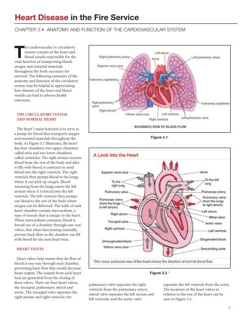
body. As Figure 3.1 illustrates, the heart<br />
has four chambers: two upper chambers<br />
called atria and two lower chambers<br />
called ventricles. The right atrium receives<br />
blood from the rest of the body and after<br />
it fills with blood, it contracts to send<br />
blood <strong>in</strong>to the right ventricle. The right<br />
ventricle then pumps blood to the lungs<br />
where it can pick up oxygen. Blood<br />
return<strong>in</strong>g from the lungs enters the left<br />
atrium where it is forced <strong>in</strong>to the left<br />
ventricle. The left ventricle then pumps<br />
out blood to the rest of the body where<br />
oxygen can be delivered. The walls of each<br />
heart chamber conta<strong>in</strong> myocardium, a<br />
type of muscle that is unique to the heart.<br />
When myocardium contracts, blood is<br />
forced out of a chamber through one-way<br />
valves, that when function<strong>in</strong>g normally,<br />
prevent back-flow so the chamber can fill<br />
with blood for the next heart beat.<br />
Figure 3.1 i<br />
HEART VALVES<br />
<strong>Heart</strong> valves help ensure that the flow of<br />
blood is one-way through each chamber,<br />
prevent<strong>in</strong>g back-flow that would decrease<br />
heart output. The sounds from each heart<br />
beat are generated from the clos<strong>in</strong>g of<br />
these valves. There are four heart valves;<br />
the tricuspid, pulmonary, mitral and<br />
aortic. The tricuspid valve separates the<br />
right atrium and right ventricle; the<br />
pulmonary valve separates the right<br />
ventricle from the pulmonary artery;<br />
mitral valve separates the left atrium and<br />
left ventricle; and the aortic valve<br />
Figure 3.2 ii<br />
separates the left ventricle from the aorta.<br />
The locations of the heart valves <strong>in</strong><br />
relation to the rest of the heart can be<br />
seen <strong>in</strong> Figure 3.2.<br />
7