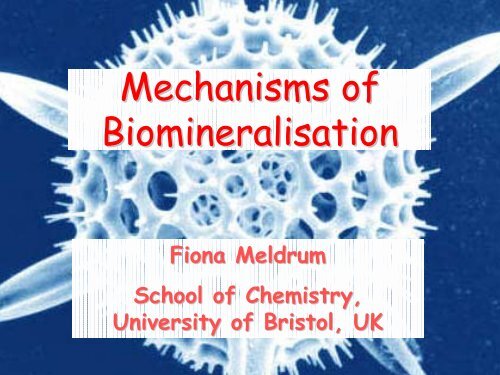
Mechanisms of Biomineralisation
Mechanisms of Biomineralisation
Mechanisms of Biomineralisation
You also want an ePaper? Increase the reach of your titles
YUMPU automatically turns print PDFs into web optimized ePapers that Google loves.
<strong>Mechanisms</strong> <strong>of</strong><br />
<strong>Biomineralisation</strong><br />
Fiona Meldrum<br />
School <strong>of</strong> Chemistry,<br />
University <strong>of</strong> Bristol, UK
<strong>Biomineralisation</strong><br />
• Describes processes by which organisms form minerals<br />
• Widespread phenomenon ⇒ over 60 different minerals formed,<br />
by members <strong>of</strong> all 5 kingdoms<br />
Eg. Iron Oxides/Hydroxides:<br />
Magnetite,<br />
Fe 3<br />
O 4<br />
Bacteria<br />
Chitons<br />
Intracellular<br />
Teeth<br />
Magnetotaxis<br />
Grinding<br />
Goethite<br />
α-Fe 2<br />
O 3<br />
Limpets<br />
Teeth<br />
Grinding<br />
Lepidocrocite<br />
γ-FeOOH<br />
Sponges<br />
Chitons<br />
Filaments<br />
Teeth<br />
Unknown<br />
Grinding<br />
Ferrihydrite<br />
5Fe 2<br />
O 3<br />
.9H 2<br />
O<br />
Animals/plants<br />
Beaver/ rat/ fish<br />
Ferritin<br />
Tooth surface<br />
Storage protein<br />
Mechanical<br />
strength<br />
• 80% <strong>of</strong> biominerals crystalline, the rest are amorphous
Characteristic Features <strong>of</strong> Biominerals:<br />
• Display remarkable and unique morphologies<br />
• Are <strong>of</strong>ten hierarchically organised on a scale from Angstroms<br />
to millimeters.<br />
• Are typically composite materials - intimately associated with<br />
organic macromolecules<br />
Show Properties Optimised for Their Function<br />
⇒<br />
⇒<br />
⇒<br />
⇒<br />
Skeletal materials show remarkable mechanical properties<br />
Magnetotactic bacteria exhibit permanent dipole moment<br />
Ferritin catalyses the formation <strong>of</strong> a soluble iron oxyhydroxide<br />
core within a soluble protein shell<br />
Single crystal calcite plates in brittle stars can act as lenses
Hierarchical Structures.. Silicaceous Sponge<br />
Euplectella sp.<br />
Shows seven hierarchical levels ... Outstanding mechanical stability<br />
Aizenberg, Weaver, Thanawala, Sundar, Morse, Fratz, Science, (2005) 309, 275-278.
Mechanical Properties <strong>of</strong> Mollusk Shell Nacre<br />
Mollusk shell nacre shows superior mechanical properties – why ?<br />
⇒<br />
Due to structural organisation <strong>of</strong> nacre<br />
• Comprises stacks <strong>of</strong> tabular<br />
aragonite crystals<br />
• Separated by interlamellar<br />
organic sheets<br />
c-axis oriented perpendicular to<br />
shell surface, organisation <strong>of</strong> a-<br />
and b- axes depends on organism
Mechanical Properties <strong>of</strong> Mollusk Shell Nacre<br />
Nacre has a very low organic content (≈ 1%), yet is superior to<br />
most other composite ceramics in stiffness, strength and toughness<br />
⇒ 3000 times more resistant to fracture than a single crystal <strong>of</strong><br />
pure aragonite<br />
Due to structural organisation <strong>of</strong> inorganic phase, and the nature<br />
<strong>of</strong> the organic sheets<br />
⇒ When a crack propagates through nacre it passes around the<br />
platelets by a tortuous path, causing the plates to spring apart,<br />
and extending the organic sheets<br />
⇒ The organic “adhesive” between the plates appears to be key<br />
in the fracture resistance <strong>of</strong> this material.
AFM Investigation into Mechanical Properties<br />
<strong>of</strong> Nacre<br />
• The organic molecules located between the aragonite plates were<br />
stretched using an AFM tip<br />
• The force required to stretch the fibres is measured as a<br />
function <strong>of</strong> the extension<br />
Shows a SAWTOOTH pr<strong>of</strong>ile
Behaviour can be explained by the organic molecules containing<br />
folded domains<br />
• When the fibres are pulled, the folded domains are extended<br />
• The force required to unfold these areas is less that the force<br />
required to break the fibre<br />
Smith et al, Nature, (1999), 399, 761-763;<br />
BL Smith, Prog. Biophys. Molec Biol. (2000) 74, 93-113.
Magnetotactic Bacteria<br />
• Single crystals <strong>of</strong> magnetite are used by<br />
bacteria, algae and many animals for navigation<br />
• In magnetotactic bacteria, single domain<br />
crystals <strong>of</strong> magnetite (Fe 3<br />
O 4<br />
) are aligned along<br />
the bacterium<br />
• The crystals are elongated to optimise the<br />
magnetic dipole
Structure and Function <strong>of</strong> Ferritin<br />
• Large storage capacity for iron ⇒ up to 4500 Fe<br />
atoms<br />
• The core is ferrihydrite, 5 Fe 2<br />
O 3<br />
.9H 2<br />
O ≈ 8 nm in<br />
diameter ⇒ a poorly crystalline mineral<br />
• The spherical protein shell is 12 nm in diameter<br />
and is formed from 24 protein subunits, <strong>of</strong> types H-<br />
chain and L-chain
Ferritin behaves catalytically towards the oxidation <strong>of</strong> Fe(II) and<br />
nucleation <strong>of</strong> iron oxide due to two key sites:<br />
⇒ a ferroxidase centre located in<br />
the intrahelical area <strong>of</strong> H-chain<br />
subunits<br />
⇒ a nucleation site comprising either<br />
four Glu residues on the cavity<br />
surface (L chain) or two Glu residues<br />
(H chain)<br />
These sites appear to act<br />
cooperatively in affecting the kinetics<br />
<strong>of</strong> iron oxide deposition within the<br />
protein.<br />
Oxidation site binds 2 Fe 3+ ions<br />
Chasteen and Harrison J. Struct. Biol. 126, 182–194 (1999)
Using the Optical Properties <strong>of</strong><br />
Calcite<br />
Certain species <strong>of</strong> brittle stars are light<br />
sensitive – why ?<br />
Looking at the structure <strong>of</strong> the<br />
skeletal elements <strong>of</strong> the brittle<br />
star, certain parts showed a unique<br />
structure<br />
Aizenberg and Hendler, J. Mater. Chem 2004, 14, 2066-2072.
Testing the Optical Behaviour…<br />
These structures behave as an array <strong>of</strong> micro-lenses, significantly<br />
enhancing the light intensity, and directing it within the animal
Bio-Inspired Crystal Growth<br />
Synthesis <strong>of</strong> many “advanced materials” require high temperatures<br />
and pressures<br />
In comparison, biology operates under ambient conditions<br />
⇒ achieves a degree <strong>of</strong> control over mineralisation that is (as yet!)<br />
difficult to reproduce synthetically<br />
Use <strong>of</strong> ambient conditions is very attractive:<br />
• Can employ heat-sensitive starting materials<br />
• Can prepare heat-sensitive products<br />
• Cheaper<br />
Ultimate goal <strong>of</strong> synthetic crystal growth must be to<br />
achieve the degree <strong>of</strong> control exhibited by nature<br />
⇒<br />
Many lessons can be learned from biology
<strong>Biomineralisation</strong> Processes<br />
Biologically Induced Mineralisation<br />
Adventitious mineralisation due to interactions between metabolic<br />
processes and environment<br />
⇒ No control over size, morphology, structure and organisation<br />
eg. mineralisation on bacteria cell walls<br />
Biologically Controlled Mineralisation<br />
Highly regulated process that has evolved to produce minerals with<br />
specific structures and functions. Characterised by:<br />
• Uniform particle sizes<br />
• Complex morphologies<br />
• Well-defined structures and organisation<br />
• Higher order assembly into hierarchical structures
CaCO 3 Polymorphism<br />
• Precipitates in 5 crystalline structures, and one amorphous<br />
form.<br />
• Crystalline anhydrous polymorphs:<br />
Calcite ⇒ most stable at RTP<br />
Aragonite ⇒ slightly less stable at RTP than calcite<br />
Vaterite ⇒ Less stable and quite rare<br />
• Crystalline hydrous polymorphs<br />
CaCO 3<br />
Monohydrate<br />
CaCO 3<br />
Hexahydrate<br />
Amorphous Calcium Carbonate (ACC) has recently also been<br />
recognised as an important biogenic mineral
Crystal Structures<br />
Aragonite and calcite have similar crystal structures:<br />
Calcite ⇒ rhombohedral (hexagonal) unit cell<br />
Aragonite ⇒ orthorhombic<br />
• Interactions are optimised in aragonite, giving better packing ⇒<br />
tends to form as needles as crystal growth is preferred along the<br />
c axis<br />
• Aragonite is more resistant to fracture as it has no cleavage<br />
planes. However crystals are small, needle-shaped and tend to<br />
form spherulitic clusters.<br />
• Synthetic calcite grows as rhombohedra<br />
exhibiting planar {10.4} faces.<br />
• Calcite forms large crystals but fractures<br />
readily.
Calcite<br />
Aragonite<br />
• Both have alternating layers <strong>of</strong> Ca 2+ and CO 3<br />
2-<br />
ions perpendicular to the c-axis<br />
(the ab plane).<br />
• The Ca ions occupy very similar lattice positions in the ab plane<br />
• The carbonate ions lie with their molecular planes parallel to the ab layer.<br />
• In aragonite, some <strong>of</strong> the carbonate ions are raised in the c direction to form<br />
2 layers separated by 0.96 Å, and their orientations in the two layers are<br />
different ⇒ main difference between the two structures
Control <strong>Mechanisms</strong> ⇒ Organic Macromolecules<br />
Strategies to control mineralisation rely on organic molecules:<br />
• Confining a space<br />
• Forming an organic matrix framework<br />
• Controlling ion input<br />
• Constructing a nucleation site<br />
• Controlling crystal orientation and growth<br />
• Terminating crystal growth<br />
Can categorise organic macromolecules as:<br />
• Insoluble matrix molecules, or<br />
• Soluble “control” macromolecules
Framework Macromolecules<br />
• Controlled crystal growth in organisms occurs within confined<br />
spaces<br />
⇒ Fabricated from framework macromolecules<br />
⇒ Usually relatively hydrophobic, are <strong>of</strong>ten cross-linked and<br />
provide a structured matrix<br />
eg. collagen in bone, chitin in crustaceans and mollusks<br />
• Organic framework <strong>of</strong>ten further functionalised<br />
macromolecules<br />
with soluble<br />
Growth <strong>of</strong> crystals within a RIGID COMPARTMENT is an<br />
important factor in controlling CRYSTAL MORPHOLOGIES
Control Macromolecules<br />
Can control crystal growth ⇒ Bound to a solid substrate<br />
⇒ As soluble additives<br />
Located within minerals, extracted by dissolution <strong>of</strong> mineral<br />
Control macromolecules from CaCO 3<br />
Typically rich in aspartic acid and glutamic acid, frequently<br />
contain bound polysaccharides<br />
Anionic groups ⇒ can interact with cations in solution eg. Ca 2+<br />
⇒ can interact with the surfaces <strong>of</strong> crystals
Control Macromolecules from Diatom Silica<br />
Dissolution <strong>of</strong> silica with HF or NH 4<br />
F yields:<br />
⇒ Set <strong>of</strong> low molecular mass proteins: SILAFINS<br />
⇒ Large quantities long chain polyamides<br />
Silaffins-1A and -1B isolated from Cylindrotheca fusiformis are<br />
polycationic and contain repeated pairs <strong>of</strong> lysine residues<br />
serine groups are<br />
phosphorylated<br />
ε–N-dimethylysine<br />
phosphorylated ε–Ntrimethyl–δ–hydroxylysine<br />
long-chain polyamines comprising 6 to<br />
11 units <strong>of</strong> N-methyl-propylamine Structure <strong>of</strong> natSil-1A<br />
Kroger, Lorenz, Brunner, Sumper, Science, 2002, 298, 584-586
A second silaffin protein, termed silaffin-2 has also been<br />
extracted from C. fusiformis<br />
⇒ POLYANIONIC and also bears unusual amino acid modifications<br />
Role <strong>of</strong> Silaffins and Long Chain Amines<br />
• NatSil-1A and the long-chain polyamines are extremely active<br />
in promoting silica precipitation in vitro<br />
• NatSil-2 alone does not precipitate silica from a silicic acid<br />
solution in vitro, but is active in combination with long-chain<br />
polyamines<br />
⇒ rapidly precipitates silica under conditions where neither <strong>of</strong><br />
these organic components would do so alone<br />
NatSil-2 may regulate the silica precipitation behaviour <strong>of</strong> long-chain<br />
polyamines and natSil-1A and may be active in silica morphogenesis.
Low<br />
natSil-1A /natSil-2<br />
High<br />
natSil-1A /natSil-2<br />
Intermediate<br />
natSil-1A /natSil-2<br />
Intermediate<br />
natSil-1A /natSil-2<br />
• Large interconnected spherical or pear-shaped silica particles were<br />
produced with low or high natSil-1A/natSil-2 ratios,<br />
• Intermediate ratios yielded porous silica blocks permeated with 0.1-1.0<br />
μm pores.<br />
• Assembly <strong>of</strong> the organic phase in the presence <strong>of</strong> polysilicic acid may<br />
provide a template for the ultimate form <strong>of</strong> the mineral phase<br />
Poulsen, Sumper and Kroger PNAS, 2003, 100(21), 12075-12080
Crystal Structure<br />
Organisms actively select mineral phases ⇒ frequently produce<br />
minerals highly undersaturated or unstable in environment<br />
Can be achieved through:<br />
• Presence <strong>of</strong> ion-specific pumps and channels<br />
• Control <strong>of</strong> composition and pH <strong>of</strong> solution<br />
• Interaction with organic matrix and soluble organic macromolecules<br />
eg. formation <strong>of</strong> ferrihydrite core, 5Fe 2<br />
O 3<br />
.9H 2<br />
O in ferritin<br />
⇒ In absence <strong>of</strong> protein, lepidocrocite is precipitated under<br />
identical conditions<br />
eg. formation <strong>of</strong> magnetite particles, Fe 3<br />
O 4<br />
bacteria<br />
in magnetotactic
Magnetite Precipitation in Magnetotactic Bacteria<br />
• Magnetite crystals are formed<br />
within separate vesicles termed<br />
magnetosomes<br />
• The magnetosome membrane contains at least one major protein<br />
which appears common to all strains.<br />
⇒ this protein may function in the accumulation <strong>of</strong> iron,<br />
nucleation <strong>of</strong> the iron oxides and in redox and pH control<br />
• Magnetosome vesicles form prior to magnetite formation<br />
Schueler and Frankel, Appl Microbiol Biotechnol (1999) 52: 464-473
• Mössbauer and HRTEM studies have suggested the magnetite<br />
crystals may not precipitate directly<br />
⇒ form via low-density hydrous Fe(III) oxide and ferrihydrite<br />
precursors.<br />
Proposed that:<br />
1) Fe(III) is taken up by the cell, where it is reduced to Fe(II)<br />
2) Subsequent re-oxidation within the magnetosome yields a lowdensity<br />
hydrous Fe(III) oxide which is dehydrated to form<br />
crystalline ferrihydrite.<br />
3) Finally, partial reduction and dehydration yields magnetite<br />
Recent experimental evidence has indicated that in M.<br />
gryphiswaldense, Fe(III) is taken up and rapidly converted into<br />
Fe 3<br />
O 4<br />
in the absence <strong>of</strong> a precursor phase.
Calcite/ Aragonite Polymorphism<br />
No examples <strong>of</strong> transformation between calcite and<br />
aragonite after precipitation ⇒ selection must occur at<br />
nucleation<br />
Good evidence that soluble macromolecules are involved in<br />
the selection <strong>of</strong> calcite or aragonite
Control <strong>of</strong> CaCO 3 Polymorphism by Soluble<br />
Mollusk-Shell Proteins<br />
• CaCO 3<br />
precipitated on abalone nucleating protein sheet<br />
• Proteins extracted from aragonitic, or calcitic layer <strong>of</strong> the shell<br />
• Spherulitic calcite crystals were produced in the presence <strong>of</strong><br />
the calcite-derived proteins<br />
• Aragonite crystals with needle morphologies and oriented in the<br />
plane <strong>of</strong> the nucleating protein layer were produced in the<br />
presence <strong>of</strong> the aragonite-derived proteins<br />
• Mixture <strong>of</strong> aragonitic and calcitic proteins induced precipitation<br />
<strong>of</strong> flat, polycrystalline plates <strong>of</strong> aragonite, oriented on (001) faces<br />
⇒ comprised oriented stacks <strong>of</strong> crystals, similar to molluscan nacre
Calcification in Abalone Shell Nacre..<br />
⇒ Nucleating protein sheet controlled the orientation <strong>of</strong><br />
the calcite primer layer<br />
⇒ Insoluble proteins<br />
thicknesses<br />
<strong>of</strong> the matrix define the plate<br />
⇒ Soluble proteins are then active in controlling further<br />
aspects <strong>of</strong> shell growth such as crystal polymorph and<br />
morphology<br />
Belcher, Wu, Christensen, Hansma, Stucky, Morse, Nature, (1996), 381, 56-58
Polymorph Selection on Artificial Molluscan Matrix<br />
The organic matrix in mollusc nacre mimicked using layers <strong>of</strong> β-<br />
chitin and silk fibroin and soluble macromolecules extracted<br />
either from the calcitic or aragonitic layers <strong>of</strong> mollusc shells<br />
⇒ Calcite precipitated inside the chitin when calcitic<br />
macromolecules were used<br />
⇒ Aragonite precipitated when the macromolecules had been<br />
extracted from an aragonite layer<br />
The specificity <strong>of</strong> these macromolecules was only achieved<br />
with the complete substrate assembly.<br />
Falini, Albeck, Weiner, Addadi, Science, 1996, 271, 67-69
• Polymorph selectivity appears to rely on both specialised<br />
macromolecules and a controlled microenvironment.<br />
• The β-chitin framework has a porous layered structure,<br />
allowing diffusion <strong>of</strong> ions and macromolecules into the structure<br />
• The silk is essential for the adsorption <strong>of</strong> the macromolecules<br />
• The macromolecules promoting calcite nucleation are strongly<br />
polyanionic and more strongly acidic than aragonite-inducing ones<br />
⇒ May provide a strong binding site for Ca 2+ ions, creating a<br />
high local supersaturation<br />
Levi, Albeck, Brack, Weiner, Addadi, Chem. Eur. J. (1998), 4(3), 389-396.
Crystal Orientation<br />
There are many examples <strong>of</strong> oriented crystals in nature<br />
⇒ Control over orientation must occur at nucleation, on a<br />
substrate with structural order (or maybe not !)<br />
⇒ IDEAS OF EPITAXIAL GROWTH<br />
Mollusk shell nacre ⇒ much studied system<br />
c<br />
⇒ comprises stacks <strong>of</strong> aligned<br />
tabular aragonite crystals which<br />
are separated by organic sheets<br />
All species c-axis perpendicular to<br />
shell surface<br />
Abalone nacre
Structure <strong>of</strong> Nacre<br />
Mollusk shell is characteristically layered, and is composed <strong>of</strong><br />
calcite, aragonite or both<br />
⇒ When both, the polymorphs are separated into different layers<br />
Mollusks form wide range <strong>of</strong> shell structures<br />
• Gastropod nacre ⇒ no preferred alignment <strong>of</strong> a- and b- axes<br />
within layer. Aragonite crystals in “stack <strong>of</strong> coins” structure<br />
• Bivalve nacre ⇒ typically good/ excellent alignment <strong>of</strong> a- and b-<br />
axes over large area. Has “brick wall” structure
Control over Nucleation in Mollusk Shell Nacre<br />
(Bivalve) - Old Model<br />
• Aragonite crystals nucleate at specific<br />
sites on a pre-deposited matrix<br />
• The organic matrix comprises thin<br />
layers <strong>of</strong> highly ordered β-chitin,<br />
sandwiched between two thicker layers <strong>of</strong><br />
silk fibroin-like proteins in a β-sheet<br />
conformation, onto which acidic<br />
macromolecules are adsorbed.<br />
• The chitin polymers and the proteinpolypeptide<br />
chains are orthogonally<br />
aligned and are aligned with the a and b<br />
axes <strong>of</strong> the aragonite tablets<br />
respectively.<br />
Weiner and Traub, Phil. Trans. R. Soc. Lond. B, 1984, 304, 425-434.
Epitaxial Growth<br />
• Binding <strong>of</strong> Ca 2+ ions to the oriented proteins could mimic the<br />
arrangement <strong>of</strong> ions in the ab face <strong>of</strong> the growing aragonite crystal<br />
⇒ causes the crystal to nucleate from the ab face<br />
This structural model was principally developed from TEM and<br />
X-ray diffraction analyses <strong>of</strong> dried samples<br />
Weiner and Traub, FEBS Letts, 1980, 111, 311-316.
New Model<br />
Cryo-TEM <strong>of</strong> mollusk nacreous layer in the hydrated state has<br />
suggested an alternative model<br />
• The silk may be in the form <strong>of</strong> a<br />
hydrated gel, located between,<br />
rather than within the sheets <strong>of</strong><br />
chitin fibrils<br />
• The acidic macromolecules may<br />
be situated in localised areas on<br />
the surfaces <strong>of</strong> the chitin layers<br />
which act as nucleation sites for<br />
the aragonite crystals, as well as<br />
within the silk gel<br />
⇒ Contradicts many <strong>of</strong> the previous ideas on oriented crystal growth in<br />
this organism<br />
Levi-Kalisman et al J Struc. Biol. 2001, 135, 8-17.
Epitaxial Growth – an Outdated Theory ?<br />
It is difficult to study oriented crystal growth directly in<br />
biological systems (very complicated!)<br />
⇒<br />
Use a MODEL system<br />
An excellent model system in which to study crystallization at<br />
an organised organic interface is provided by Langmuir<br />
monolayers
In has been suggested that the orientation and crystal polymorph<br />
are directed by the:<br />
• Spacing and geometry <strong>of</strong> the surfactant headgroups<br />
• Stereochemistry <strong>of</strong> the monolayer headgroups<br />
CdS<br />
CaCO 3
However ….<br />
• Many examples where monolayers with entirely different<br />
structures induce nucleation from IDENTICAL crystal faces<br />
• Studies <strong>of</strong> CaCO 3<br />
precipitation under monolayers <strong>of</strong> derivatised<br />
calixarenes has suggested that oriented nucleation may be<br />
controlled by:<br />
NON-SPECIFIC ELECTROSTATIC EFFECTS<br />
• Results suggest that it is the<br />
density <strong>of</strong> Ca 2+ ions associated<br />
with the monolayer which defines<br />
the polymorph and nucleation face<br />
Volkmer et al J. Mat. Chem. 2004, 14, 2249-2259.
Orientation Control in Abalone Nacre<br />
Examination <strong>of</strong> growth surface <strong>of</strong> abalone nacre suggests that<br />
successive layers form via MINERAL BRIDGES<br />
⇒ Bridges grow through pores in interlamellar organic sheets<br />
Growth <strong>of</strong> oriented stacks <strong>of</strong> aragonite crystals is due to<br />
mineral bridges between aragonite tablets<br />
Schaeffer et al. Chem. Mater. 1997, 9, 1731-1740
Mechanism:<br />
• Specialised sheet <strong>of</strong> protein governs initial nucleation <strong>of</strong><br />
oriented calcite crystals<br />
• Secretion <strong>of</strong> 2 different families <strong>of</strong> proteins induces a switch<br />
from calcite to aragonite production<br />
• The soluble proteins direct polymorph selection and orientation<br />
Further control <strong>of</strong> orientation over many layers occurs via mineral<br />
bridges ⇒ stacks <strong>of</strong> crystals effectively SINGLE CRYSTALS
Crystal Morphologies<br />
• One <strong>of</strong> most striking feature <strong>of</strong> many biominerals ⇒ remarkable<br />
morphologies.<br />
• Many <strong>of</strong> these unusual morphologies occur when the biomineral<br />
is amorphous ⇒ no preferred morphology<br />
• Organisms can also produce single crystals with complex shapes<br />
and curved surfaces<br />
⇒ Have developed mechanisms which override the basic growth<br />
form <strong>of</strong> a crystal<br />
⇒ Produce crystals whose overall morphologies <strong>of</strong>ten bear no<br />
relationship to the symmetry <strong>of</strong> the crystal lattice<br />
• Many biominerals with complex morphologies are polycrystalline<br />
structures – can be oriented or non-oriented
Biomineral Structures<br />
AMORPHOUS POLYCRYSTALLINE SINGLE CRYSTAL
Amorphous Silica<br />
Amorphous silica is very abundant - the polymeric structure<br />
allows it to be moulded into unusual structures eg. diatoms
Biosilica Morphogenesis in Diatoms<br />
SILAFFINS and LONG-CHAIN POLYAMINES induce formation <strong>of</strong><br />
silica nanospheres from silicic acid in vitro<br />
Cell wall <strong>of</strong> Coscinodiscus granii<br />
SB=20 mm<br />
Nanoscale architecture<br />
SB=1 mm<br />
Coscinodiscus ⇒ structure comprises a hierarchy <strong>of</strong> heaxagonal silica<br />
structures<br />
M. Sumper, Angew. Chem. Int. Ed. 2004, 43, 2251 –2254
Principal organic component <strong>of</strong> Coscinodiscus shells polyamines<br />
⇒ Model <strong>of</strong> pattern formation built on PHASE SEPARATION OF<br />
POLYAMINES<br />
• Phase separation occurs within SDV to form microdroplets <strong>of</strong><br />
polyamines<br />
• Close-packed arrangement <strong>of</strong> microdroplets ⇒ hexagonal<br />
monolayer <strong>of</strong> droplets<br />
• Aqueous interface between polyamine droplets contains silicic<br />
acid derivatives ⇒ promotes silica formation, catalysed by<br />
polyamine surfaces<br />
Repetition on smaller length scales gives intricate patterning <strong>of</strong><br />
diatom frustrules
(A) Monolayer <strong>of</strong> polyamine-containing droplets<br />
within the SDV guides silica deposition<br />
in close-packed arrangement<br />
(B and C) Consecutive segregations <strong>of</strong> smaller (about 300 nm) droplets open new<br />
routes for silica precipitation.<br />
(D) Dispersion <strong>of</strong> 300-nm droplets into 50-nm droplets guides the final stage <strong>of</strong><br />
silica deposition. Silica precipitation occurs only within the water phase<br />
(white areas). The repeated phase separations produce a hierarchy <strong>of</strong> selfsimilar<br />
patterns. (E to H)<br />
M. Sumper, Science, 2002, 295, 2430-2433.
Dispersion <strong>of</strong> droplets into smaller and smaller units may arise<br />
from:<br />
• Consumption <strong>of</strong> polyamines during precipitation<br />
• Creation <strong>of</strong> negatively charged silica surfaces with high<br />
affinities for positively charged polyamine-containing surfaces<br />
⇒ Should favour surface expansion by fragmentation <strong>of</strong> the<br />
polyamine-containing surface
Mechanism also supported by species-specific patterns...<br />
• Initial size <strong>of</strong> microdroplets limited by wall-to-wall distance <strong>of</strong><br />
areolae: 1 mm in C. granii and about 3 mm in C. asteromphalus<br />
• The number and arrangement <strong>of</strong> secondary droplets formed are<br />
then governed by the wall-to-wall distance <strong>of</strong> the areola<br />
⇒ Short distance in C. granii interferes with segregation <strong>of</strong><br />
secondary drops – appear to be positioned across areola wall
Amorphous Calcium Carbonate (ACC)<br />
• ACC can be prepared synthetically by mixing high<br />
concentrations <strong>of</strong> Ca 2+ and CO<br />
2-<br />
3<br />
ions<br />
• It is hydrated and is described as ≈ CaCO 3<br />
.1.5 H 2<br />
O<br />
• It is very unstable and rapidly crystallises<br />
ACC is also observed in biology...<br />
TRANSIENT ACC<br />
Can be short lived ⇒ acts as a precursor to a crystalline phase<br />
STABLE ACC<br />
Biogenic ACC can also be stable for long periods <strong>of</strong> time<br />
⇒<br />
organisms must actively stabilise it
Morphological Control <strong>of</strong> Single Crystals<br />
Structure <strong>of</strong> Skeletal Plates <strong>of</strong> Sea Urchin
<strong>Mechanisms</strong>:<br />
• Changes in the activity or positioning <strong>of</strong> ion pumps and channels<br />
during mineralisation ⇒ may lead to crystal growth in preferred<br />
directions<br />
Single crystals <strong>of</strong> magnetite in a<br />
magnetotactic bacterium<br />
⇒ equilibrium morphology cubic<br />
• Interaction <strong>of</strong> growing crystals with soluble additives, and<br />
which are occluded within many biominerals, can also produce<br />
subtle changes in morphology<br />
• More dramatic changes in morphology are imposed by the<br />
physical form <strong>of</strong> the compartment in which mineralisation occurs.
Effect <strong>of</strong> Additives on Crystal Growth<br />
• Additives can interact with specific crystal faces during<br />
crystal growth<br />
• These faces are then stabilised and grow slowly ⇒ they<br />
become the large faces in the product crystal<br />
• The final crystal morphology therefore differs from that <strong>of</strong> a<br />
crystal grown without additives<br />
During this process additives become incorporated within crystals<br />
⇒<br />
can modify mechanical properties
Soluble Additives in <strong>Biomineralisation</strong><br />
• Soluble macromolecules extracted from CaCO 3<br />
biominerals can<br />
adsorb to specific calcite crystal faces during precipitation<br />
⇒ modifies crystal morphologies<br />
• The macromolecules may possess structural motifs that match<br />
the atomic arrangement on one set <strong>of</strong> crystal planes, causing the<br />
additive to interact with and stabilise these faces<br />
• Following adsorption to specific crystal faces, the<br />
macromolecules become overgrown and are occluded within the<br />
crystals<br />
• The occluded macromolecules are unique to a particular<br />
biomineral
Macromolecules Extracted from Sea Urchin Spines...<br />
Whole assembly <strong>of</strong> macromolecules<br />
• Interact with planes approx parallel to the c-axis<br />
• Rough faces, not well-defined, produced<br />
• Indexed as {10l} where l ~ -1.5<br />
Control crystal<br />
Grown with total<br />
assembly <strong>of</strong><br />
macromolecules<br />
Aizenberg, Hanson, Koetzle, Weiner, Addadi J. Am. Chem. Soc., 1997, 119(5), 881-886.
Macromolecules further investigated to identify which moieties are<br />
involved in interaction with growing crystals<br />
Chemically and enzymatically treated to yield three fractions:<br />
• The polysaccharides were removed yielding deglycosylated<br />
protein<br />
• Isolated polysaccharide chains<br />
• Densely glycosylated peptide cores were produced, comprising<br />
short peptides with attached polysaccharides<br />
Epitaxial growth<br />
on young spine<br />
with released<br />
macromols<br />
Epitaxial growth<br />
on mature spine<br />
with released<br />
macromols<br />
Crystals grown in presence <strong>of</strong> macromolecules released on mild<br />
etching <strong>of</strong> biomineral
Polysaccharide fraction – interact non-specifically with a subset<br />
<strong>of</strong> planes oriented ~ parallel to c-axis ⇒ rough faces produced<br />
Deglycosylated proteins - interact specifically to produce large<br />
faces in addition to {104} faces. Many analysed as {-203}.<br />
Glycosylated peptides - interact specifically to produce large<br />
faces in addition to {104} faces. New faces close to {010}<br />
• Non specific interaction <strong>of</strong> isolated polysaccharides may<br />
result from loss <strong>of</strong> conformation on isolation.<br />
• Calcite crystals grown epitaxially in presence <strong>of</strong> proteins<br />
released under mild conditions show same faces as produced by<br />
entire assembly <strong>of</strong> proteins, but better defined<br />
Polysaccharides may enable fine-tuning <strong>of</strong> interactions<br />
Albeck, Weiner, Addadi, Chem. Eur. J. 1996, 2(3), 278-284.
Control <strong>of</strong> Morphology through Binding to Calcite<br />
Surface Steps<br />
AFM study <strong>of</strong> the growth <strong>of</strong> the calcite (10.4) face in the<br />
presence <strong>of</strong> the chiral molecules D-aspartic acid and L-aspartic<br />
acid showed that interaction with the crystal steps was<br />
asymmetric.<br />
• Growth hillock shows 2 acute and 2 obtuse steps to the cleavage<br />
plane<br />
• Addition <strong>of</strong> glycine – 2 acute steps become curved, 2 obtuse<br />
unaffected<br />
• Chiral D-Asp and L-Asp: growth hillocks mirror images <strong>of</strong> each<br />
other<br />
Orme et al, Nature, 2001, 411(6839), 775-779.
A pure calcite<br />
growth hillock<br />
Glycine -achiral<br />
amino acid<br />
L-Asp<br />
D-Asp<br />
OB<br />
Dissolution pits<br />
with L-Asp<br />
Dissolution pits<br />
with D-Asp<br />
001 oriented<br />
calcite grown<br />
with L-Asp<br />
001 oriented<br />
calcite grown<br />
with D-Asp
D-Asp binds to the (01–4) riser<br />
such that one <strong>of</strong> its negatively<br />
charged carboxyl groups<br />
completes the coordination <strong>of</strong><br />
calcium ions, while the positively<br />
charged (NH 3+<br />
) group remains in<br />
registry with the positive ions at<br />
the surface.<br />
The carboxyl group <strong>of</strong> does not<br />
closely match the carbonate<br />
groups on the other actute step<br />
Geometry <strong>of</strong> binding for Asp adsorbed on (104) steps <strong>of</strong> calcite. L-Asp (left)<br />
and D-Asp (right) binding to acute steps <strong>of</strong> calcite. Red=O, Blue=N, Green =Ca
The specific amino acid enantiomers therefore bind to the step<br />
edges that <strong>of</strong>fer the best geometric and chemical fit.<br />
This changes the step-edge free energies, which in turn results<br />
in macroscopic crystal shape modifications.<br />
Suggested that organic additives influence crystal morphologies<br />
by binding to the surface-step edges rather than single crystal<br />
faces.<br />
⇒ Crystal morphologies depend on stereochemical recognition and<br />
the effects <strong>of</strong> binding on the interfacial energies <strong>of</strong> the growing<br />
crystal
Texture <strong>of</strong> Biominerals<br />
How can large molecules such as proteins be incorporated within a<br />
single crystal ?<br />
Schematic diagram <strong>of</strong> the domain<br />
structure <strong>of</strong> a crystal<br />
Introduction <strong>of</strong> macromolecules causes defects in a single crystal<br />
⇒ The coherence length and domain spread in the biological<br />
crystals provides information on the location <strong>of</strong> macromolecules<br />
Coherence length - the size <strong>of</strong> perfect crystalline domains<br />
Domain spread - the misalignment <strong>of</strong> the crystalline blocks
Texture <strong>of</strong> Biominerals<br />
3D mapping <strong>of</strong> the distribution <strong>of</strong> defects in the curved monaxon<br />
spicule <strong>of</strong> the sponge Sycon reveals a domain shape resembling the<br />
overall morphology<br />
• Pure geological calcite ⇒ CL 800nm and DS 0.003 o<br />
• Sponge spicule<br />
⇒ plane perpendicular to long axis CL 130 nm and DS 0.07 o<br />
Aizenberg et al, JACS, 1997, 119, 881; Aizenberg et al, Chem. Eur. J, 1995, 1(7), 414.
Strong correlation between the distribution <strong>of</strong> defects within the<br />
spicules, and their macroscopic morphologies<br />
⇒ Defects arise from incorporation <strong>of</strong> macromolecules<br />
Suggested that the macromolecules play an important role in<br />
the modulation <strong>of</strong> morphologies<br />
HOWEVER....<br />
Soluble additives cannot be responsible for complex morphologies<br />
⇒ These must be defined by the shape <strong>of</strong> the environment in<br />
which the crystal forms<br />
⇒<br />
⇒<br />
Additives fine tune morphologies?<br />
Regulate mechanical properties?
Role <strong>of</strong> Occluded Macromolecules ?<br />
Biominerals ⇒ typically exhibit superior mechanical properties<br />
Conchoidal fracture<br />
surface<br />
Fractured sea urchin plate<br />
• Sea urchin calcite ~ 0.1 wt% organic macromolecules<br />
⇒ the single crystal plates are effectively a composite material<br />
• Macromolecules interfere with crack propagation along cleavage<br />
planes, reinforcing the crystal against fracture
Application <strong>of</strong> this Strategy to Synthetic Crystals<br />
Calcite crystals grown (a) in the presence <strong>of</strong> and (b) in the<br />
absence <strong>of</strong> sea urchin proteins<br />
a<br />
b<br />
Micro-indentation experiments show that the occluded proteins<br />
endow the crystal with far superior fracture resistance<br />
Weiner et al, Mat. Sci. Eng. C 2000, 11, 1-8.
Shape Constraint<br />
Crystals with unusual morphologies and curved surfaces are typical<br />
<strong>of</strong> growth within vesicles<br />
eg.<br />
calcite scales produced by coccoliths<br />
sea urchin larval spicules<br />
• Vesicles have defined shapes, and crystals grow until they<br />
impinge upon the vesicle, which effectively acts as a mould<br />
• The size and shape <strong>of</strong> the vesicle may be altered during the<br />
crystal growth process.<br />
• S<strong>of</strong>t organic membrane imposes a form on a crystal<br />
⇒ interaction <strong>of</strong> the crystal with the vesicle membrane may<br />
stabilise the high energy rounded crystal surfaces?
Control <strong>of</strong> Single Crystal Morphologies by Growing<br />
Crystals within a Rigid Template<br />
CaCO 3 crystal grown<br />
within a sponge-like<br />
polymer membrane<br />
Growth <strong>of</strong> single crystal within a rigid template ⇒ sufficient to<br />
control morphology<br />
Park R.J. et al J. Mater. Chem. 2004, 14, 2291-2296.
Poly-Crystalline<br />
Biominerals<br />
Many biominerals with complex morphologies are polycrystalline<br />
⇒ formed by organised assembly <strong>of</strong> single crystal components<br />
eg. coccosphere <strong>of</strong> the algae Emilania huxleyi<br />
The coccolith scales each comprise about 30-40 units which are<br />
organised in a ring to give a double-rimmed structure.
• Formation <strong>of</strong> coccoliths begins with<br />
assembly <strong>of</strong> vesicles along the rim <strong>of</strong><br />
an organic base plate scale<br />
• Nucleation then occurs within the<br />
vesicles to generate a proto-coccolith<br />
ring <strong>of</strong> interlinked calcite crystals<br />
Proto-coccolith ring<br />
• The crystals initially form as 40nm thick rhombohedral plates<br />
which are inclined to the plane <strong>of</strong> the ring<br />
• The plates grow to a height <strong>of</strong> 100 nm, and radial outgrowth<br />
along the c-axis from the top and bottom faces generates a Z-<br />
shape.<br />
• These units become inter-linked by selective growth along the<br />
inside rim, and further radial growths from the base and top <strong>of</strong><br />
the element produce the proximal and distal shield elements<br />
S.Mann “Biomineralization”, OUP, 2001.
Further Lectures ....<br />
• Crystallisation at Interfaces<br />
• Amorphous Biominerals<br />
• Mineral Morphologies<br />
20 µm


