Electrotherapy & Ultrasound Therapy Use - Implox
Electrotherapy & Ultrasound Therapy Use - Implox
Electrotherapy & Ultrasound Therapy Use - Implox

You also want an ePaper? Increase the reach of your titles
YUMPU automatically turns print PDFs into web optimized ePapers that Google loves.
Copyright:<br />
Enraf-Nonius B.V.<br />
P.O. Box 12080<br />
3004 GB ROTTERDAM<br />
The Netherlands<br />
Tel: +31 (0)10 – 20 30 600<br />
Fax: +31 (0)10 – 20 30 699<br />
info@enraf-nonius.nl<br />
www.enraf-nonius.com<br />
Part number: 1482.762 - 43<br />
December 2005
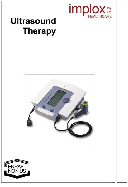
<strong>Ultrasound</strong><br />
therapy<br />
by<br />
R. Hoogland<br />
1
Table of copntents<br />
1 Preface.......................................................................................................................................... 5<br />
2 Basic information ........................................................................................................................ 5<br />
2.1 Definitions ........................................................................................................................... 5<br />
2.2 Physical fundamentals ........................................................................................................ 5<br />
2.2.1 Generation of <strong>Ultrasound</strong>......................................................................................................5<br />
2.2.2 Equipment.............................................................................................................................6<br />
2.3 Properties of the ultrasound beam...................................................................................... 8<br />
2.3.1 The near field ........................................................................................................................9<br />
2.3.2 The distant field.....................................................................................................................9<br />
2.3.3 The BNR value......................................................................................................................9<br />
2.4 Physical phenomena occurring in the medium ................................................................. 11<br />
2.4.1 The nature of the (ultra)sound wave ...................................................................................11<br />
2.4.2 The wavelength of ultrasound.............................................................................................11<br />
2.4.3 The mass density of the medium ........................................................................................12<br />
2.4.4 The specific acoustic impedance (Z s )..................................................................................12<br />
2.4.5 Compression and expansion of the media..........................................................................12<br />
2.4.6 Reflection and refraction of sound ......................................................................................12<br />
2.4.7 Scattering of ultrasound ......................................................................................................13<br />
2.4.8 Interference of ultrasound ...................................................................................................14<br />
2.4.9 Absorption and penetration of ultrasound ...........................................................................14<br />
2.5 The contact medium.......................................................................................................... 16<br />
2.6 Propagation properties of contact media .......................................................................... 16<br />
3 Biophysical effects of ultrasound............................................................................................ 17<br />
3.1 Introduction ....................................................................................................................... 17<br />
3.2 Mechanical effect .............................................................................................................. 17<br />
3.3 Thermal effect ................................................................................................................... 17<br />
3.4 Biologic effects .................................................................................................................. 19<br />
3.4.1 Promotion of blood-circulation.............................................................................................19<br />
3.4.2 Muscle relaxation ................................................................................................................20<br />
3.4.3 Increased membrane permeability......................................................................................20<br />
3.4.4 Promotion of tissue regenerative power..............................................................................20<br />
3.4.5 Effect on peripheral nerves .................................................................................................20<br />
3.4.6 Reduction of pain ................................................................................................................21<br />
3.5 Other effects...................................................................................................................... 21<br />
Tissue damage............................................................................................................................ 21<br />
Stasis of blood-cells .................................................................................................................... 21<br />
4 Technique................................................................................................................................... 22<br />
4.1 Modes of energy transfer and manipulation of the treatment head .................................. 22<br />
4.1.1 Direct contact between treatment head and body...............................................................22<br />
4.1.2 Treatment under water........................................................................................................22<br />
4.1.3 Manipulation of the treatment head.....................................................................................23<br />
4.2 Points of application.......................................................................................................... 23<br />
4.3 Dosage.............................................................................................................................. 24<br />
4.3.1 Introduction .........................................................................................................................24<br />
4.3.2 Intensity ..............................................................................................................................24<br />
4.3.3 Energy release....................................................................................................................24<br />
4.3.4 Duration of treatment ..........................................................................................................25<br />
4.3.5 Start and frequency of treatment.........................................................................................25<br />
4.4 Methods............................................................................................................................. 25<br />
4.4.1 Before treatment .................................................................................................................25<br />
4.4.2 During treatment .................................................................................................................25<br />
4.4.3 After treatment ....................................................................................................................26<br />
5 Special applications of ultrasound.......................................................................................... 26<br />
5.1 Combined therapy............................................................................................................. 26<br />
5.1.1 The combination of ultrasound with low-frequency electrotherapy......................................26<br />
5.1.2 The combination of ultrasound and medium-frequency electrotherapy...............................27<br />
5.1.3 Technique of combined therapy..........................................................................................28<br />
5.2 Indications and contra-indications..................................................................................... 28<br />
5.2.1 Indications...........................................................................................................................28<br />
5.2.2 Contra-indications ...............................................................................................................28<br />
5.3 Ultrasonophoresis therapy ................................................................................................ 28<br />
6 Indications.................................................................................................................................. 29<br />
6.1 Introduction ....................................................................................................................... 29<br />
3
6.1.1 The time factor ....................................................................................................................29<br />
6.1.2 The intensity factor..............................................................................................................30<br />
6.2 Disorders ...........................................................................................................................30<br />
6.2.1 Disorders of bony tissue, joints and muscles ......................................................................30<br />
6.2.2 Rheumatoid arthritis in a quiescent stage ...........................................................................30<br />
6.2.3 Disorders of the peripheral nerves ......................................................................................31<br />
6.2.4 Disorders of the circulation..................................................................................................31<br />
6.2.5 Internal organic disorders....................................................................................................31<br />
6.2.6 Disorders of the skin ...........................................................................................................31<br />
6.2.7 Dupuytren’s contracture ......................................................................................................31<br />
6.2.8 Open wounds......................................................................................................................31<br />
7 Contra-indications .....................................................................................................................32<br />
7.1 Absolute specific contra-indications ..................................................................................32<br />
7.2 Relative specific contra-indications ...................................................................................32<br />
8 Examples of treatment ..............................................................................................................33<br />
8.1 Introduction ........................................................................................................................33<br />
8.2 Some detailed examples of treatment...............................................................................35<br />
8.2.1 General ...............................................................................................................................35<br />
8.2.2 Specific ...............................................................................................................................35<br />
8.2.2.1 Subacromial bursitis ............................................................................................................................ 35<br />
8.2.2.2 Treatment of scar tissue after total hip surgery ................................................................................... 35<br />
8.2.2.3 Treatment of tendinitis of the extensor carpi radialis brevis muscle ................................................... 35<br />
8.2.2.4 Treatment of synovitis of the knee....................................................................................................... 36<br />
9 List of literature, recommended and consulted......................................................................38<br />
4
1 Preface<br />
This therapy manual is intended primarily to give background information in connection with the use of<br />
Enraf-Nonius ultrasound equipment.<br />
With a view of the demand for more information about ultrasound, we felt obliged to provide a more<br />
detailed explanation of this subject.<br />
We thank R. Hoogland for this description of the theory and the practical application.<br />
2 Basic information<br />
2.1 Definitions<br />
Definition: ‘Sound waves’ are mechanical vibrations in an elastic medium.<br />
These longitudinal waves can cause the tympanic membrane to vibrate. The are sound vibrations.<br />
Frequencies below about 20 Hz are subsonic, frequencies above 20.000 Hz are ultrasonic. This<br />
ultrasound frequency range is related to the human ear. It is subjective and arbitrary. This is evident from<br />
the fact that the range of audible sounds decreases with age.<br />
Definition: ‘<strong>Ultrasound</strong> therapy’ is medical treatment by means of mechanical vibrations with a frequency<br />
above 20 kHz.<br />
In practice, the frequencies used for treatment range between 0.7 and 3 MHz. However, some equipment<br />
exists for diagnosis and therapy which uses frequencies between 5 kHz and 10 MHz.<br />
Definition: ‘Ultrasonophorsis therapy’ is medical treatment with medical substances introduced into the body<br />
by means of ultrasound energy.<br />
Definition: ‘<strong>Ultrasound</strong> diagnosis’ is scanning of a portion of the body by means of ultrasound for<br />
pathological changes.<br />
If desired, this can be combined with various electrical currents. The sensitive zones in the tissues, which<br />
are quite easily found in this manner, can be used as application points for treatment. The technique will<br />
be described in Chapter 3.<br />
2.2 Physical fundamentals<br />
This book refers to standard manuals on the physics of ultrasound. The results only will be given. For the<br />
derivation of the formulae and background information, the relevant physics literature should be<br />
consulted.<br />
2.2.1 Generation of <strong>Ultrasound</strong><br />
Modes of generating ultrasound<br />
Any vibrating object is a source of sound. Sound waves can also be generated in different ways, e.g.<br />
mechanically by a tuning-fork or in medicine by means of electro-acoustic transducers.<br />
The piezo-electric effect<br />
If pressure is applied to crystals (quartz), and to some polycrystalline materials such as lead-zirconatetitanate<br />
(PZT) or barium titanate, electric charges develop on the outer surface of the material. This is<br />
called the piezo-electric effect.<br />
Piezo-electric effects are also seen in the human body, especially in bony tissue, collagen fibers and body<br />
proteins. Possibly, these piezo-electric phenomena are involved in the biologic effects of ultrasound.<br />
5
The reverse piezo-electric effect<br />
The piezo-electric effect is reversible. Thus, if the substances mentioned above are exposed to an<br />
alternating electric current, they will undergo changes in shape in the frequency of the alternating electric<br />
field. The material then becomes a source of sound. Currently, quartz, barium titanate and lead-zirconatetitanate<br />
(PZT) are used for generation of ultrasound via the reverse piezo-electric effect. The last two<br />
materials have the advantage that, because of their ferro-electric properties, only a small voltage is<br />
required to induce the acoustic energy. This, for instance, makes a transformer in the sound head<br />
superfluous, making it much smaller. Quartz requires a high voltage (several kV). On account of the<br />
necessary transformer, the treatment head becomes relatively large. PZT is preferable to barium titanate<br />
because it retains its marked piezo-electric properties up to much higher temperature thresholds. PZT is<br />
also less sensitive to mechanical shocks.<br />
2.2.2 Equipment<br />
The instrument consists of a high-frequency generator. This is connected to a piezo-electric crystal (the<br />
treatment head).<br />
The resonant frequency of the crystal is partly determined by the thickness of the piezo-electric material<br />
(PZT) and consequently the frequency of the ultrasound is so determined as well. Moreover, this implies<br />
that the sound head and the equipment must be mutually adjusted, so that the treatment head cannot be<br />
used with another piece of ultrasound equipment unless calibration is performed.<br />
Technical innovation has solved this problem in the Sonopuls® apparatus, where the treatment heads are<br />
fully interchangeable between different instruments of the same type and proper adjustment is<br />
automatically performed.<br />
As a result of the alternating current applied to the piezo-electric material, this generates sound waves.<br />
These will propagate in the neighboring media (e.g. tissues). Because the piezo-electric material<br />
generates sound waves bi-directional, ultrasound will also enter the treatment head (rebound effect).<br />
This is of little significance because of the air present in the treatment head.<br />
The transducer also vibrates laterally, consequently ultrasound energy in transferred to the sidewall of the<br />
treatment head via the transducer mounting (sidewall radiation).<br />
Picture 1: Sonopuls 492<br />
In the treatment heads of the Sonopuls® apparatus the sidewall radiation has been reduced to <<br />
10mW/cm². Various authorities quote < 10mW/cm² as an acceptable value. On protracted use of<br />
ultrasound sidewall radiation development of these symptoms depends on excessive intensity of this<br />
sidewall radiation.<br />
Therefore its value should be measured and specified for the equipment.<br />
® Registered trademark<br />
6
Continuous and pulsed ultrasound<br />
Most ultrasound equipment can generate both continuous and pulsed ultrasound energy. The maximum<br />
intensity that can be set for continuous ultrasound is 3W/cm²<br />
Picture 2: Sonopuls 190<br />
The Sonopuls® 190 permits adjustments of the intensity between 0 and 2 W/cm² for continuous and 3<br />
W/cm² for pulsed ultrasound.<br />
Pulsed ultrasound has the advantage that thermal sensations are suppressed. In addition, this mode<br />
permits a higher intensity, which for application of continuous ultrasound can cause undesirable effects.<br />
The higher intensity is probably the explanation for the nonthermal effects occurring in pulsed ultrasound<br />
therapy. Also due to the pulsation of the ultrasound beam the mechanical effects are more pronounced.<br />
Ratio (= ‘Duty cycle’) Pulse time (ms) Pulse pause (ms) Pulse repetition period (ms)<br />
1 : 5 (= 20%) 2 8 10<br />
1 : 10 (= 10%) 1 9 10<br />
1 : 20 (= 5%) 0,5 9,5 10<br />
Table 1.1: Example of the parameters for pulsed ultrasound with a pulse repetition frequency of 100 Hz.<br />
Fig. 1.1: Principle of ultrasound equipment<br />
7
The effective radiating area (ERA)<br />
The effective radiating area of the treatment head (ERA) is an important parameter determining the<br />
intensity. Because the piezo-electric element does not vibrate uniformly, the ERA is always smaller than<br />
the geometric area f the treatment head.<br />
To permit a true indication of the intensity on the instrument, determination of the ERA is essential<br />
because the effective intensity depends on this. Correct ultrasound dosage depends partly on the area to<br />
be treated and ERA, reason why the ERA should be known. Therefore the ERA should be measured and<br />
specified (see fig. 1.2).<br />
Fig. 1.2: Impression of a geometric area (A) and an effective radiating area (ERA).<br />
1- and 5-serie US heads ERA Geometric area<br />
1 MHz<br />
5,0 cm² 6,2 cm²<br />
0,8 cm² 1,4 cm²<br />
3 MHz<br />
5,0 cm² 6,2 cm²<br />
0,5 cm² 0,7 cm²<br />
9-serie US heads ERA Geometric area<br />
1 MHz and 3 MHz<br />
5,0 cm² 6,3 cm²<br />
0,8 cm² 1,3 cm²<br />
Table 1.2: Table of ERA for the Sonopuls® apparatus<br />
2.3 Properties of the ultrasound beam<br />
In the ultrasound beam two areas can be distinguished (see fig. 1.3):<br />
- the near field : the Fresnel zone<br />
- the distant field : the Fraunhofer zone.<br />
8<br />
Fig. 1.3: Longitudinal cross-section of the ultrasound beam.
2.3.1 The near field<br />
The near field is characterized by:<br />
- interference phenomena in the ultrasound beam, which may lead to marked variations in intensity<br />
- the absence of divergence, in fact here is slight convergence of the ultrasound beam<br />
2.3.2 The distant field<br />
The distant field is characterized by:<br />
- the near-absence of interference phenomena, so that the sound beam is uniform and the intensity<br />
gradually decreases with increasing distance to the transducer;<br />
- the ultrasound beam has a larger diameter. This size depends on the type of sound beam (divergent<br />
or collimating);<br />
- a wider spread of the sound energy due to both the divergence and the fact that the intensity<br />
distribution perpendicular to the longitudinal axis of the sound beam becomes increasingly bellshaped<br />
(see fig. 1.4).<br />
Fig. 1.4: Transverse cross-section of the ultrasound beam.<br />
The length of the near field depends on the diameter of the treatment head and the wavelength. With the<br />
usual treatment head of 5 cm² the near field is about 10 cm long. For a sound head of 1 cm² the near field<br />
is about 2 cm long at 1 MHz.<br />
At 3 MHz the near field is 3 times as long, because the wavelength is proportionally shorter.<br />
Because the depth effect of ultrasound is limited, the therapeutic effects occur mainly in the near field. It<br />
should be remembered that in the Fresnel zone interference phenomena occur in the ultrasound beam,<br />
resulting in its being non-homogeneous. These interference phenomena may cause intensity peaks 5-10<br />
times higher than the set value – in some cases even 30 times higher.<br />
This non-homogeneous behavior of the sound bundle is expresses as the Beam Non-uniformity Ratio<br />
(BNR).<br />
2.3.3 The BNR value<br />
Theoretically, the BNR cannot be smaller than 4, i.e. one should always allow for intensity peaks of at<br />
least 4 times the set value. For well-manufactured treatment heads the BNR lies between 5 and 6,<br />
depending on construction. The BNR value should be specified on the treatment head.<br />
1- and 5-serie US heads BNR<br />
1 MHz Treatment head 6.2 cm² 6.0 max.<br />
Treatment head 1.4 cm²<br />
6.0 max.<br />
3 MHz Treatment head 6.2 cm² 6.0 max.<br />
Treatment head 0.7 cm²<br />
6.0 max.<br />
9-serie US heads<br />
BNR<br />
1 & 3 MHZ Treatment head 6.3 cm² 6.0 max.<br />
Treatment head 1.3 cm²<br />
6.0 max.<br />
Table 1.3: BNR values for the Sonopuls® treatment heads<br />
See fig. 1.5.<br />
9
For safe treatment the head must always be kept in motion so that the ultrasound energy is<br />
properly spread.<br />
Rotation of the head in one position must be rejected because the intensity peaks in the sound beam are<br />
usually positioned symmetrical to the longitudinal axis of the treatment head (so-called rotation<br />
symmetry). Rotation of the treatment head causes intensity peaks in the same location, resulting in overdosage.<br />
By means of the underwater method the near field can be avoided by maintaining a sufficient distance to<br />
the body (viz. The length of the near field), depending on the size of the treatment head. The interference<br />
phenomena of the near field will then occur in the water. A disadvantage is the larger diameter of the<br />
ultrasound beam in the distant field, causing a reduction of the energy per cm². These aspects should be<br />
taken into account for dosage calculations. With the usual application of ultrasound therapy total reflection<br />
will not occur because the boundary angle is so large as not to be exceeded under normal conditions.<br />
Fig. 1.5:<br />
Beam diagram large head 1 MHz<br />
Beam diagram large head 3 MHz<br />
Beam diagram small head 1 MHz<br />
Beam diagram small head 3 MHz<br />
Divergence of the ultrasound beam<br />
Divergence of the ultrasound beam occurs only in the distant field.<br />
The divergence is determined by the angle of spread (α) (see fig. 1.3) according to the formula:<br />
Sin α = 1.22 λ/D, where<br />
λ = wavelength of the ultrasound<br />
D = diameter of the treatment head<br />
5 cm² 1 cm²<br />
1 MHz 4.2º 9.3º<br />
3 MHz 4.4º 3.1º<br />
Table 1.4: Angles of spread at 1 and 3 MHz for various treatment heads<br />
It has been said earlier that the near field is shorter for a small treatment head, so that divergence occurs<br />
earlier and the ultrasound energy is spread over a large area. It will be clear that divergence of the<br />
ultrasound beam is markedly less at 3 MHz.<br />
10
2.4 Physical phenomena occurring in the medium<br />
2.4.1 The nature of the (ultra)sound wave<br />
The ultrasound wave is of a longitudinal nature, i.e. the direction of propagation is the same as the<br />
direction of oscillation. Longitudinal waves require an elastic medium for propagation. In principle, every<br />
medium is elastic with the exception of a vacuum. The longitudinal elastic waves (sound waves) cause<br />
compression and expansion of the medium at half a wavelength’s distance, leading to pressure variations<br />
of the medium (see fig. 1.6).<br />
In this context the medium is the contact substance and the tissues of the body in which the ultrasound<br />
energy propagates.<br />
Fig. 1.6: Elastic waves in a spring and a liquid.<br />
2.4.2 The wavelength of ultrasound<br />
This is expressed by the relationship:<br />
λ * f = c, where:<br />
λ = the wavelength (m)<br />
f = the frequency (Hz)<br />
c = the speed of propagation (metres/second)<br />
Because the frequency of the equipment is set and the speed of propagation is determined by the<br />
medium, the wavelength also depends on the latter.<br />
In soft tissue and in water the wavelength at 1 MHz is approximately 1.5 mm and in bony tissue about 3<br />
mm. The effect on the speed of propagation in the tissues at 3 MHz is small. Thus there is a linear<br />
reduction of the wavelength, which is about 0.5 mm in soft tissues and about 1 mm in bony tissue.<br />
Medium c (m/s) ρ (kg/m³ λ (mm)<br />
1 MHz<br />
λ (mm)<br />
3 MHz<br />
Aluminium 5100 2.7 x 10³ 5,10 1,70<br />
Blood 1566 1.0 x 10³ 1,57 0,52<br />
Blood-vessel 1530 1.1 x 10³ 1,53 0,51<br />
Bony tissue 3445 1.8 x 10³ 3,44 1,14<br />
Skin 1519 ----- 1,51 0,50<br />
Cartilage 1665 ----- 1,75 0,58<br />
Air at 20 ºC 343 0.0012 x 10³ 0,34 0,11<br />
Tendon tissue 1750 ----- 1,75 0,58<br />
Muscle tissue 1552 1.0 x 10³ 1,55 0,52<br />
Fatty tissue 1478 0,9 x 10³ 1,48 0,49<br />
Water at 20 ºC 1492 1,0 x 10³ 1,49 0,50<br />
Table 1.5<br />
Survey of media and their speed of propagation (c), mass density (ρ) and wavelength<br />
(λ) for ultrasound at a frequency of 1 MHz and 3 MHz.<br />
11
2.4.3 The mass density of the medium<br />
The mass density of the medium (ρ) is a parameter expressed in kg/m³. Together with the specific<br />
acoustic impedance (Zs), this determines the resistance of the tissues to sound waves. The mass density<br />
also partly determines the speed of propagation (c). The higher the mass density, the higher the speed of<br />
propagation (see Table 1.5). the value of the mass density is required for determination of the specific<br />
acoustic impedance and consequently for the reflection.<br />
2.4.4 The specific acoustic impedance (Z s )<br />
Because the specific acoustic impedance is a material parameter it depends on the mass density and the<br />
speed of propagation: Z s = ρ * c.<br />
Medium<br />
Z s (Kg/m²s)<br />
Aluminium 13,8 - 10 6<br />
Blood 1,6 - 10 6<br />
Bone 6,3 - 10 6<br />
Blood-vessel 1,7 - 10 6<br />
Gel circa 1,8 - 10 6<br />
Skin circa 1,6 - 10 6<br />
Air 0,0004 - 10 6<br />
Muscle tissue 1,6 - 10 6<br />
Fatty tissue 1,4 - 10 6<br />
Water (20 ºC) 1,5 - 10 6<br />
Table 1.6<br />
The specific acoustic impedance<br />
2.4.5 Compression and expansion of the media<br />
The medium (tissue) is a compressed and expanded at the same frequency as that of the ultrasound, i.e<br />
approximately 1 . 106 times per second. The resultant pressure changes are fairly large. For instance, at<br />
an intensity of 1 W-cm² the pressure variation is about 1.7 bar (at 1 MHz and c = 1500 m/s).<br />
At a wavelength of 1.5 mm this implies a pressure gradient of 3.4 bar over a distance of 0.75 mm in view<br />
of the fact that the points of high and low pressure are half a wavelength apart. At 3 MHz the pressure<br />
variation is greater; it increases as the square law. Therefore it may be assumed that the pressure<br />
variation increases by a factor of 9! Due to attenuation of the sound beam the pressure variation<br />
decreases with depth. However, due to interference in the near field and reflection at the various<br />
boundaries a large increase in pressure can develop.<br />
2.4.6 Reflection and refraction of sound<br />
Reflection of ultrasound<br />
Reflection occurs at the boundaries between different tissues. The amount of reflected energy depends<br />
on the specific acoustic impedance (Zs) of various media according to the formula:<br />
R = the amount of reflected energy<br />
This formula applies to a sound beam with perpendicular incidence where Zs 1 is the specific acoustic<br />
impedance of medium 1 and Zs that of medium 2 at a boundary surface measured from the treatment<br />
head.<br />
In practice this means that the reflection decreases as the difference between the two specific acoustic<br />
impedances becomes smaller. In the body, significant reflection occurs for transitions between tissue and<br />
bone (30%).<br />
The specific acoustic impedance of the sound head is practically identical to that of the contact<br />
medium (gel). Consequently there is hardly any reflection between these media. Theoretically reflection<br />
between aluminium and contact medium would be about 60%.<br />
12
Aluminium – air 100,0%<br />
Aluminium – contact medium 60,0%<br />
Treatment head – contact medium<br />
Nil<br />
Contact medium – skin 0,1%<br />
Skin – fatty tissue 0,9%<br />
Water – fatty tissue 0,2%<br />
Fatty tissue – muscle tissue 0,8%<br />
Muscle tissue – bone tissue 34,5%<br />
Skin - air 100,0%<br />
Table 1.7<br />
Survey of reflection at some medium boundaries.<br />
Refraction of sound<br />
In addition to reflection, a non-perpendicular incidence of the sound waves causes refraction of the sound<br />
beam according to the formula:<br />
c1 = speed of sound in medium 1<br />
c² = speed of sound in medium 2<br />
In n > 1, refraction takes place towards the perpendicular;<br />
If n < 1, then refraction is away from the perpendicular.<br />
Only the latter is significant because of the deflection of the ultrasound beam when the critical angle is<br />
reached and the ultrasound beam begins to run parallel to the boundary between the two media. The<br />
speed of sound in the various body tissues is such that for normal applications of ultrasound the critical<br />
angle will not be exceeded.<br />
Reflection and refraction will not be appreciably different at 1 and 3 MHz, because the mass density of<br />
the various tissues is constant and the effect of the sound frequency on the speed of propagation is small.<br />
2.4.7 Scattering of ultrasound<br />
Scattering of ultrasound in the body occurs due to two phenomena:<br />
- divergence in the far field<br />
- reflection<br />
Especially because of reflection, the ultrasound beam may spread in the body, so that effects can<br />
develop not only in the direction of the sound beam, but also outside it (see fig. 1.7). as pointed out<br />
before, reflection needs to be taken into account only if highly reflective materials and/or substances such<br />
as metals, air and bony tissue are placed in the ultrasound beam. In addition, it should be remembered<br />
that ultrasound can hardly leave the body as a result of the reflection form air, which is virtually 100%<br />
(see table 1.7). the total dosage of ultrasound applied to the body is converted to other forms of energy.<br />
An exception is the underwater method, where the ultrasound energy can leave the body.<br />
If the sound beam hits bony tissue, reflection amounts to about 30%. Then the ultrasound beam is<br />
attenuated by absorption of energy, depending on the thickness of the surrounding tissue layers (e.g.<br />
muscle tissue).<br />
Fig. 1.7 Scattering of the ultrasound beam by reflection.<br />
13
The reflected energy again enters the original tissue layer and will again enters the original tissue layer<br />
and will again be attenuated by absorption. At the boundary between skin and air almost complete<br />
reflection occurs, and so over again. The ultrasound beam rebounds between bony tissue and air.<br />
The same is possibly true for the 70% of sound energy propagated in the bony tissue. The sound beam<br />
will be markedly attenuated in this tissue on account of the very high absorption of the ultrasound energy.<br />
(see fig. 1.7)<br />
2.4.8 Interference of ultrasound<br />
Interference of ultrasound waves occurs due to two phenomena:<br />
a. Interference in the ultrasound beam in the near field leading to at least a four-fold local increase<br />
in intensity compared to the value set on the instrument (see BNR value).<br />
b. Interference phenomena as a result of reflection. The incident and reflected sound beams can<br />
overlap, leading the two wave motions which may attenuate or enhance each other.<br />
Interference resulting in enhancement leads to an increase in the intensity of the sound beam.<br />
(see fig. 1.8)<br />
In practice, problems result only if the tissue layer extending down to the bone is thin or absorbs<br />
little sound energy. This is the case for treatment near the wrist area, the ankles, patella and<br />
similar locations. Especially with application of continuous ultrasound, this phenomenon causes<br />
irritation of the periostium with a sensation of heat and/or pain. This once more illustrates the<br />
importance of motion of the treatment head.<br />
Fig. 1.8 Interference by reflection<br />
2.4.9 Absorption and penetration of ultrasound<br />
As the (mechanical) ultrasound energy penetrates into the body tissues, biologic effects can be expected<br />
to occur only if the energy is absorbed by the tissues. Due to the absorption the intensity of the sound<br />
waves will decrease as they penetrate further into the tissues. The absorption of ultrasound energy by<br />
biologic tissues varies. The absorption coefficient (a) is used as a measure of the absorption in various<br />
tissues. The absorption in the tissues is lower than for high frequencies. This relation ship is linear for all<br />
tissues except bone between the frequency, absorption and action at depth of ultrasound. In effect, the<br />
absorption coefficient together with reflection determines the spread of ultrasound in the body.<br />
For ultrasound, among other things, the following formula applies. This formula is true for ultrasound<br />
consisting of longitudinal waves with perpendicular incidence on homogeneous tissues:<br />
l(x) = lo * e-ax<br />
where:<br />
l(x) = the intensity in W/cm² at a depth x in cm<br />
Io = the intensity in W/cm² at the surface of the body, but IN the body tissue<br />
e = 2.7 (base of natural logarithms)<br />
a = absorption coefficient (cm –1).<br />
14<br />
From this formula it emerges that the intensity of ultrasound at a certain depth depends on the absorption<br />
coefficient (a).
Medium<br />
Absorption coefficient (a)<br />
1 MHz 3 MHz<br />
Blood 0,028 0,084<br />
Blood-vessel 0,4 1,2<br />
Bony tissue 3,22<br />
Skin 0,62 1,86<br />
Cartilage 1,16 3,48<br />
Air (20 ºC) 2,76 8,28<br />
Tendon tissue 1,12 3,36<br />
Muscle tissue 0,76 2,28*<br />
0,28 0,84**<br />
Fatty tissue 0,14 0,42<br />
Water (20 ºC) 0,0006 0,0018<br />
Nerve tissue 0,2 0,6<br />
* sound beam perpendicular to fibers<br />
** sound beam parallel to fibers<br />
Table 1.8<br />
Absorption coefficient (a) at 1 and 3 MHz<br />
From the table it appears that two values are used for the absorption in muscle tissue. The marked<br />
difference is caused by the direction of the sound beam in relation to the muscle fibers. The former value<br />
applies if the sound beam is perpendicular to the muscle fibers. This is by far the most usual situation in<br />
the practical application of ultrasound. The latter value applies if the sound beam runs parallel to the<br />
muscle fibers. In the latter case the absorption is almost a factor 3 smaller.<br />
A more practical value relating to absorption is the half-value depth (D1/2).<br />
Definition: the ‘half-value depth’ in the distance in the direction of the sound beam in which the intensity in<br />
a certain medium decreases by half.<br />
The half-value depth (D1/2) as determined by the absorption coefficient can be calculated with the<br />
formula:<br />
Half-value depth (D1/2) 0,69/a<br />
1 MHz 3 MHz<br />
Bone tissue 2,1 mm ----<br />
Skin 11,1 mm 4,0 mm<br />
Cartilage 6,0 mm 2,0 mm<br />
Air 2,5 mm 0,8 mm<br />
Tendon tissue 6,2 mm 2,0 mm<br />
Muscle tissue 9,0 mm 3,0 mm *<br />
24,6 mm 8,0 mm**<br />
Fatty tissue 50,0 mm 16,5 mm<br />
Water 11500,0 mm 3833,3 mm<br />
* sound beam perpendicular to fibers<br />
** sound beam parallel to fibers<br />
Table 1.9<br />
Half-value depth (D1/2) of various media<br />
Only the most practically significant values have been included in the table.<br />
Until now it has been generally assumed that the half-value depth for muscle tissue is about 3 cm. This is<br />
correct if the sound beam runs parallel to the muscle fibers, which in practice will hardly ever be the case.<br />
If the sound beam is perpendicular to the muscle fibers – as is mostly the case during treatment – the<br />
half-value depth is found to be 0.9 cm.<br />
The consequence of the greater absorption is that the action in depth decreases.<br />
In addition it is seen that much ultrasound energy is absorbed in tendon tissue and cartilage. Possibly this<br />
is an explanation for the favorable therapeutic results of treatment of these tissues.<br />
15
The greatest depth at which a therapeutic effect can still be expected is called the penetration depth (p).<br />
this is the point where 10% of the applied sound intensity remains. It should be noted that this value only<br />
specifies the depth, not the local intensity of the ultrasound. The intensity of the ultrasound at the<br />
penetration depth determines whether a therapeutic effect will really no longer result at this depth.<br />
The value of p is approximated by:<br />
p 2.3/a<br />
1 MHz 3 MHz<br />
Bone tissue 7 mm ----<br />
Skin 37 mm 12 mm<br />
Cartilage 20 mm 7 mm<br />
Air 8 mm 3 mm<br />
Tendon tissue 21 mm 7 mm<br />
Muscle tissue 30 mm 10 mm *<br />
82 mm 27 mm**<br />
Fatty tissue 165 mm 55 mm<br />
Water 38330 mm 12770 mm<br />
* sound beam perpendicular to fibres<br />
** sound beam parallel to fibres<br />
Table 1.10 Penetration depths of some media<br />
2.5 The contact medium<br />
As appears from chapter 3, it is necessary to use a contact medium between the treatment head and the<br />
body in order to transfer the ultrasound energy to the body. Air is wholly unsuitable as a contact medium<br />
because of the almost complete reflection of the ultrasound. Water, however, is a good contact medium<br />
and is also cheap. If water is used as a contact medium it must have been degassed by boiling as far as<br />
possible and in some cases it must be sterile, e.g. for treatment of open wounds. By degassing,<br />
deposition of air bubbles on the treatment head and the treated part of the body is prevented. In practice<br />
gel, oil and ointment – sometimes with other substances added (ultrasonophoresis) – are used in addition<br />
to water.<br />
In random order, the following requirements can be specified for the contact medium.<br />
The contact medium must be:<br />
- sterile, if there is a risk of (cross) infection<br />
- not too liquid (except for the underwater method)<br />
- not too quickly absorbed by the skin<br />
- incapable of causing marked staining<br />
- free from a marked cooling or irritant effect on the skin<br />
- chemically inert<br />
- cheap<br />
- endowed with good propagation properties<br />
- free from (micro) gas bubbles<br />
- transparent<br />
- free from micro-organisms or fungi.<br />
2.6 Propagation properties of contact media<br />
16<br />
In propagation of ultrasound energy to the body various media and boundary surfaces are encountered,<br />
viz. from transducer plate to contact medium and from contact medium to the body. For the transition<br />
between two media a so-called transmission factor is specified, which indicates the fraction of the<br />
energy transmitted.<br />
The intensity of the ultrasound indicated on the equipment is specified for water at 5 mm from the<br />
treatment head.<br />
As a rule of thumb one may assume 100% transmission of ultrasound in the body tissues.<br />
According to current data on transmission losses in the contact substances, the gels in present use as<br />
contact medium do not have a significant effect on the amount of energy that reaches the body.
3 Biophysical effects of ultrasound<br />
3.1 Introduction<br />
The effects of ultrasound are not yet completely clear. It is clear, however, that application of ultrasound<br />
to biologic tissues has a number of effects. The primary consideration with ultrasound is that it is a form of<br />
mechanical therapy. It is also evident that mechanical energy can be converted to thermal energy, for<br />
instance, but this is a consequence of the mechanical effect of ultrasound.<br />
The effects of 1 MHz and 3 MHz are probably not different, however, certain effects may be more<br />
emphasized at the respective frequencies.<br />
As far back as the fifties, Pohlman stated that no other effects are observed for ultrasound between 1 and<br />
10 MHz. The special nature of ultrasound at 3 MHz is therefore rather the much greater mechanical effect<br />
and the markedly higher absorption of the ultrasound energy in the superficial tissue layers (see table 1.7<br />
and 1.8). This also spares the deeper tissues, because the intensity greatly decreases as a consequence<br />
of the greater absorption.<br />
3.2 Mechanical effect<br />
The first effect to occur in body tissue as a result of ultrasound is of a mechanical nature. Sound<br />
vibrations require an elastically deformable medium for their propagation. In principle, any medium except<br />
a vacuum is deformable. The ultrasound vibrations cause compression and expansion in the tissues at<br />
the same frequency as the ultrasound, leading to pressure variations in the tissue.<br />
Therefore the mechanical effect is also called micro-massage.<br />
At 3 MHz the pressure maxima and minima are closer together than at 1 MHz because the wavelength<br />
decreases to 0.5 mm approximately.<br />
Due to reflection in the sound beam and at the boundaries between the tissues the intensity in W/cm²<br />
may increase, so that the greatest pressure variations occur at the boundary between two different<br />
media. Therefore one may assume the most pronounced therapeutic effects to occur at the boundaries.<br />
<strong>Ultrasound</strong> therapy is therefore also called a therapy of boundary surfaces.<br />
These pressure differences have the following consequences:<br />
- changes in volume of the body cells of the order of 0.02%<br />
- changes in permeability of the cell and tissue membranes<br />
- an improved exchange of metabolic products.<br />
Micro massage is of great therapeutic importance. All effects of ultrasound therapy are caused by it.<br />
These effects occur with both continuous and pulsed ultrasound energy. Depending on the intensity used<br />
for treatment, these effects may have a favorable or unfavorable influence on the tissues. The<br />
unfavorable effects will be discussed in paragraph 2.5.<br />
3.3 Thermal effect<br />
Micro massage of the tissues leads to generation of frictional heat. This thermal effect has been<br />
frequently described in the literature and is the best-known effect of ultrasound.<br />
The amount of heat generated differs for the various tissues. It depends on a number of factors, some of<br />
which can be adjusted, e.g. the form of ultrasound (continuous or pulsed), the intensity and the duration<br />
of treatment. In addition, the absorption coefficient plays an important part (see table 1.8). Lehmann<br />
states that the temperature in muscle tissues increases by 0.07°C per second for continuous ultrasound<br />
of 1 W/cm². This value has been calculated for a muscle phantom, i.e. without the regulating effects of the<br />
bloodstream. Therefore this appears to indicate the value of the maximum increase in temperature in<br />
muscle tissue. In a therapeutic situation involving ischemia such a marked rise in temperature might<br />
increase and lead to unfavorable effects. In a study on the medial side of the knee of pigs this author<br />
showed that the increase in temperature in the soft parts is relatively small in comparison with the deeper<br />
articular structures. For continuous ultrasound at 1.5W/cm² for 5 minutes with a transducer plate of 12.5<br />
cm² the average increase in temperature of the capsule is 6.3°C, that of the soft tissues 3.3°C. The<br />
medial part of the meniscus shows an average increase of 8.2°C, whereas in the bony tissue an increase<br />
in temperature of 9.3°C occurs.<br />
Measurements in dogs have shown that the temperature in the bone-marrow increase by 0.4°C at a<br />
dosage of 0.5 W/cm² continuous ultrasound and by 5°C for 2.5 W/cm² continuous ultrasound, in both<br />
cases applied for 5 minutes (Payton et al. 1975).<br />
17
Heat is especially generated at sites of reflection of ultrasound. This reflection occurs particularly at<br />
boundaries between tissues with different specific acoustic impedance. Because of this reflection,<br />
interference phenomena may result that lead to an increase in intensity. Reflection takes place mainly at<br />
bony tissue (35%). The generation of heat as a result of this increase in intensity is marked in the<br />
periostium and may lead to periostial pain. This problem is much less marked when pulsed ultrasound<br />
energy is used because the generated heat is wholly or partly dissipated between the pulses. Thus the<br />
thermal effect is low.<br />
To summarize:<br />
because of differences in absorption coefficient,<br />
- as a result of reflection on tissue boundaries, and<br />
- as a consequence of interference peaks and troughs, generation of heat in the ultrasound field will be<br />
non-uniform.<br />
By keeping the treatment head in motion an effort is made to minimize this non-uniformity. The<br />
distribution of the heat in the various tissues is unique in comparison with other forms of treatment such<br />
as short-wave therapy and thermo therapy.<br />
Heat is especially generated in bony tissue, cartilage, tendons, muscle tissue and skin.<br />
Because the ultrasound beam is almost parallel, the area where the thermal effect occurs will<br />
approximate that of the treatment head (ERA). If heat is expected to have a favorable effect on the<br />
healing of lesions in the tissues mentioned above, ultrasound therapy in continuous form is indicated. It is<br />
important to ensure that the patient at most senses a small thermal effect. At a high intensity (more than 2<br />
W/cm²) and with the continuous form of ultrasound, a marked increase in the blood circulation results in<br />
order to keep the body temperature as constant as possible. It has been noted that a lower intensity can<br />
also promote circulation. The mechanisms involved will be dealt with later. The significance of heat as a<br />
part of ultrasound therapy has been variously assessed. Many diseases are accompanied by disturbed<br />
circulation. The body is often incapable of dissipating the heat generated by ultrasound. This leads to an<br />
increase in temperature that may have an adverse effect on the disease. In the case of an acute injury,<br />
e.g. a sprain of the ankle, the heat generated (in combination with the mechanical irritation) may have an<br />
adverse effect on the recovering blood-vessels. Bleeding may easily recur. Therefore it is advisable to<br />
wait a couple of days before local ultrasound therapy is started in such cases. From the rheumatologic<br />
angle a warning has also been issued about the consequences of raised intra-articular temperatures.<br />
The heat generated in arthritis is found to have a noxious effect on the internal articular structure,<br />
especially the articular cartilage. The collagen fibers in hyaline cartilage are destroyed and are replaced<br />
by inferior collagen fibers. The enzyme collagenase initiates this process, other enzymes becoming<br />
involved in the destruction of the joint.<br />
Articular disorders in which this process in particularly manifest are inflammations of the joints (including<br />
rheumatoid arthritis and arthorosis, often characterized by synovitis).<br />
The conclusion from these findings is that ultrasound therapy resulting in an increase in intra-articular<br />
temperature is contraindicated, especially in disorders where this temperature is already higher than<br />
normal. The interesting question now arises whether anything is known about the effect of heat on<br />
collagen fibers other than in hyaline cartilage. Viidik et al. Have shown that under the influence of heat a<br />
softening of collagen fibers in tendons and articular capsules can occur, leading to hyper mobility. The<br />
loading of the newly formed fibers only becomes optimum if specific kinesio-therapy is also given.<br />
Yet it is incorrect to deny that heat generation is of any value. Lehmann has shown that a rise in<br />
temperature is an important factor in the development of some physiologic processes.<br />
18
3.4 Biologic effects<br />
As stated, the effects of ultrasound therapy are all the result of micromassage (mechanical effect).<br />
Depending on the form, continuous or pulsed, this micromassage results in a predominance of either<br />
thermal or other effects. The following biologic effects can be seen as a physiologic response to the<br />
mechanical and thermal effects mentioned.<br />
<strong>Ultrasound</strong> energy<br />
Micromassage<br />
(mechanical effect)<br />
heat<br />
(thermal effect)<br />
- promotion of blood-circulation<br />
- muscle relaxation<br />
- increased membrane permeability<br />
- increased regenerative power of tissues<br />
- effect on peripheral nerves<br />
- reduction of pain<br />
- other effects<br />
3.4.1 Promotion of blood-circulation<br />
The possibility of promoting the blood-circulation by means of ultrasound is mentioned in many<br />
publications. The absorption of ultrasound energy results in a thermal effect, to which the body responds<br />
by vasodilation. It is important to remember that the thermal effect is not limited to the continuous form of<br />
ultrasound. With pulsed ultrasound there is also a thermal effect, although much smaller. The vasodilation<br />
occurring as a result of treatment with ultrasound can be partly regarded as a protective measure aimed<br />
at keeping the body temperature within the narrowest possible limit.<br />
The vasodilation is caused by:<br />
a. The release of tissue stimulants. This is the consequence of cellular damage resulting from<br />
mechanical vibration.<br />
b. Stimulation – possibly direct – of (thick myelinated?) afferent nerve-fibers. This leads to a postexcitatory<br />
depression of orthosympathetic activity.<br />
c. Reduction of muscle tone as a result of the mechanism mentioned above.<br />
Although several authors have demonstrated a reflex effect of ultrasound, it is not yet clear what afferent<br />
fibers are being stimulated.<br />
An increased muscle tone leads to an impeded blood circulation, with a simultaneous increase in the<br />
energy demands of the hypertonic tissue. Thus the concentration of tissue stimulants increases fairly<br />
rapidly, leading to an increased nociceptive afferent activity of thin nerve-fibers.<br />
The consequences of this are: increased pain, increased muscle (tissue) tone and a further disturbance<br />
of the circulation.<br />
To break this vicious circle, promotion of the circulation will be an important step towards recovery. The<br />
possibility of promoting the blood-circulation via the reflex route, using ultrasound, has been<br />
demonstrated by Becker and others. He described an improved circulation, especially in the acral blood<br />
vessels of patients with vascular disorders, as a consequence of segmentally applied ultrasound therapy.<br />
Pohlmann recommends routine inclusion of segmental therapy in the therapeutic plan, in addition to local<br />
treatment of disorders.<br />
In the literature, this refers almost exclusively to Para vertebral application, although other localizations<br />
may also be useful. Lota described the effect of ultrasound at low intensity (0.5-1 W/cm²) on the<br />
peripheral blood-circulation and on the temperature of the skin and muscles. The effects of both local and<br />
segmental (Para vertebral) treatment were recorded.<br />
19
The conclusion was reached that continuous application of only 1 W/cm² resulted in both an improvement<br />
of the blood-circulation and a rise in temperature of the skin and muscles on local application. Para<br />
vertebral application resulted in improvement of the circulation in the skin.<br />
Otherwise, the effect of ultrasound on the blood-vessels has been a subject of controversy. Some authors<br />
observed vasodilation while others described vasoconstriction. In a remarkable study by Hogan et al.<br />
more light was shed on the significance of these apparently controversial results given by different<br />
authors. It is pointed out that the effect of ultrasound (on arterioli in the skeletal muscle) usually leads to<br />
vascoconstriction. One phenomenon described by these authors is highly interesting.<br />
In most tissues the arterioli are not at rest under normal physiologic conditions, but show slow peristaltic<br />
movements (2 or 3 per minute). On application of pulsed ultrasound the frequency of this vascular<br />
mobility is seen to increase enormously (to 31 per minute). An interesting finding is that the frequency of<br />
these vascular motions hardly increases (7 to 8 per minute) on ordinary heating of the tissue.<br />
The authors point out that such movements of the arteriolar walls are more important for nutrition of the<br />
tissues than arteriolar dilation alone.<br />
3.4.2 Muscle relaxation<br />
In the preceding paragraph it has been pointed out how improvement in the blood-circulation can lead to<br />
muscle relaxation because tissue irritants can be carried away.<br />
In addition, it may be possible that the ultrasound directly stimulates afferent nerve fibers and that the<br />
muscle relaxation is the consequence of post-excitatory depression of orthosympathetic activity.<br />
3.4.3 Increased membrane permeability<br />
<strong>Ultrasound</strong> vibrations have been found to increase the permeability of membranes. This effect is seen for<br />
both continuous and pulsed application of ultrasound. As a result of the mechanical vibrations, tissue fluid<br />
is forced through the cell membrane. This may have an altered ion concentration as a consequence,<br />
which might result in altered cell excitability. In the cells the protoplasmic flow is seen to increase so that<br />
processes of physiologic exchange are promoted. Due to the circulation of tissue fluid the pH becomes<br />
less acidic. This is called the anti-acidotic effect of ultrasound and is useful for the treatment of<br />
rheumatoid inflammation (soft tissue rheumatism) in which there is acidosis of the tissues (see paragraph<br />
2.2).<br />
3.4.4 Promotion of tissue regenerative power<br />
<strong>Ultrasound</strong> has been shown to promote the process of regeneration in various tissues. Dyson and Pond<br />
described the effect of ultrasound on artificially-induced small wounds of the ears of rabbits. The<br />
favorable effect of ultrasound was equaled only by that of drugs. The most effective intensity was found to<br />
be 0.5 W/cm² with pulsed application (1:5) at a frequency of 3.5 MHz. Electron microscope studies<br />
showed that the mechanical forces produced a stream of freely mobile particles. The thermal effect plays<br />
a subordinate role in this process.<br />
3.4.5 Effect on peripheral nerves<br />
Some authors postulate that ultrasound can depolarize afferent nerve fibers. If the intensity is so chosen<br />
as to result in mild stimulation this is evidently the case. How, and to what extent ultrasound can also<br />
directly act on nerve fibers at lower intensities, and what afferent nerve fibers are then excited, is not yet<br />
clear. The sound pressure is probably not responsible for this because the net pressure is almost zero<br />
and the frequency of the pressure changes is so high that mechanosensors cannot react to it.<br />
Once more it must be pointed out that depolarization of many afferent nerve fibers is an obscure<br />
phenomenon and that the function of many of these fibers is not yet known.<br />
20<br />
Continuous ultrasound at an intensity of 0.5-3 W/cm² has been shown to affect the conduction velocity of<br />
peripheral nerves. Both an increase and a decrease of conduction velocity have been described. Almost<br />
without exception, the thermal effect has been regarded as responsible for this alteration. In this<br />
connection the mechanical aspect is not thought to be significant.<br />
At higher intensities a conduction block can occur. Although this is not specifically evident from Table 1.8,<br />
nerve tissue is especially sensitive to the effect of ultrasound. In a study of the effect of continuous<br />
ultrasound for 5-10 minutes at an intensity of 2-3 W/cm²on the sciatic nerve, swelling of the axon<br />
cylinders to total severance of the nerve were found. At a lower intensity (0.25-0.5 W/cm²) the myelin<br />
sheath showed minimal changes that became more serious on repeated application.<br />
In the central nervous system an effect of ultrasound can also be demonstrated. An increased serotonin<br />
release has been found, the significance of which is as yet unclear.
3.4.6 Reduction of pain<br />
Experience shows that ultrasound therapy results in a reduction of pain which is difficult to explain. The<br />
complexity of the processes leading to a sensation of pain is responsible for this difficulty. In addition, little<br />
is known about the effect of ultrasound energy on pain sensation. However, some factors can be<br />
distinguished that contribute to reduction of pain. These are:<br />
- Improvement of tissue circulation<br />
The favorable effect of improved blood-circulation has already been discussed in paragraph 2.4. An<br />
improved circulation leads to a better drainage of tissue irritants (pain mediators), so that fewer<br />
nociceptive nerve fibers are excited.<br />
- Normalization of muscle tone<br />
Because there is less chemical excitation of muscle afferents, a reduction of reflexly raised tone<br />
occurs.<br />
- Reduction of tissue tension<br />
An improved circulation of the blood (an lymph) has a favorable effect on the resorption of oedema<br />
fluid.<br />
Reduction of the oedema leads to a fall in tissue tension, which in turn results in a reduction of pain<br />
and promotion of tissue circulation.<br />
- Reduction of pH<br />
The improvement in circulation results in an increase of the tissue pH. It is not exactly known how<br />
this results in a reduction of pain.<br />
- Stimulation of afferent nerve fibers<br />
Possibly ultrasound can directly depolarize (thick?) afferent nerve fibers. As with electrotherapy, this<br />
can result in a reduction of pain.<br />
3.5 Other effects<br />
Other effects have been observed as a consequence of ultrasound application. At present their<br />
therapeutic significance is not clear, while many of the effects in this category are known to have a<br />
negative influence.<br />
Tissue damage<br />
Although the use of pulsed ultrasound has reduced the thermal effect it should be remembered that high<br />
intensity causes a marked mechanical peak loading of the tissue. This may even lead to tissue damage.<br />
The extreme pressure differences developing as a consequence of exposure to ultrasound may cause<br />
cavitation in the tissues. Although the output of equipment in current use is such that this phenomenon<br />
can hardly occur or not at all, it is wise to adjust the intensity such that the patient does not sense a<br />
painful excitation.<br />
Stasis of blood-cells<br />
Dyson and Pond described stasis of blood-cells in the blood vessels running parallel to the ultrasound<br />
beam after application of ultrasound to chick embryos. The minimum energy at which this phenomenon<br />
still occurred was 0.5 W/cm² of continuous ultrasound. This phenomenon was generally reversible. After<br />
conclusion of the experiment the circulation was undisturbed.<br />
Continuous movement of the treatment head certainly appears sufficient to eliminate this phenomenon.<br />
Other side-effects described:<br />
- reduction of the blood-sugar level<br />
- fatigue<br />
- nervousness<br />
- irritation<br />
- anorexia<br />
- constipation<br />
- tendency to catch cold<br />
These side-effects are all thought to result from over-dosage.<br />
21
4 Technique<br />
4.1 Modes of energy transfer and manipulation of the treatment head<br />
Transfer of energy can in principle be applied in two ways:<br />
4.1.1 Direct contact between treatment head and body<br />
This mode of energy transfer is the most frequently used.<br />
The treatment head is applied directly to the skin. It is known that air reflects ultrasound almost<br />
completely. Therefore it is absolutely necessary to ensure application of a highly conductive medium<br />
between the treatment head and the skin.<br />
For the requirements in relation to this medium see paragraph 1.5.<br />
In principle, water is an excellent and cheap medium meeting the requirements. However, application<br />
often encounters practical difficulties. Underwater treatment is therefore limited to some specific fields<br />
and indications, as will become clear later. The many types of contact media currently available for<br />
ultrasound transmission can be broadly classified as follows:<br />
- oils<br />
- water-oil emulsions<br />
- aqueous gels<br />
- ointments<br />
For transmission of ultrasound, gels are the most suitable. The gel must be somewhat dissolved by the<br />
skin salts, so that it can be rubbed effectively into the skin (pores). Some manufacturers add a<br />
medicament to the contact medium, often a substance that promotes blood-circulation (hyperemiainducing<br />
gels).<br />
The Sonopuls® units include a normal treatment head and a small head. The advantage of this small<br />
head is that body parts of irregular shape such as the joints of the hand, wrist, foot and ankle, and<br />
structures such as the Achilles tendon, can be properly treated because the small head is in full contact<br />
with the part of the body involved.<br />
The treatment head has an (optical) monitoring system that gives a warning indication if the ultrasound<br />
energy differs too much from the set value. If the amount of ultrasound energy reaching the tissue<br />
becomes less than 80% of the set intensity the intensity is automatically reduced to 0.05 W/cm².<br />
The clock stops if the energy transfer is insufficient and starts again when transfer of energy is resumed.<br />
Then the intensity initially set will be automatically supplied by the instrument. In this manner the set time<br />
for treatment is effectively used.<br />
4.1.2 Treatment under water<br />
If the surface of the body is very irregularly shaped, and good contact between the treatment head and<br />
the skin is complicated in consequence, the so-called underwater method can be chosen as well as<br />
treatment with the small treatment head mentioned above. Underwater treatment can also be chosen if<br />
direct contact is not possible, e.g. because of pain.<br />
22<br />
The part of the body in question is submerged in a trough filled with water at a pleasant temperature. The<br />
treatment head is also submerged and positioned at some distance from the part to be treated.<br />
Preferably, the water should have been boiled previously because otherwise any air present may be<br />
deposited in the form of bubbles on the transducer plate and the skin of the treated area. As is known, air<br />
greatly impedes the transfer of energy; therefore air-bubbles must always be removed. Some areas of the<br />
body are difficult to reach, e.g. the under surface of the toes. In such cases a metal plate can be used,<br />
placed on the bottom of the trough, which will reflect the ultrasound to reach the body area from below.<br />
Some sense of geometry is desirable for this technique. Although the literature often mentions a mirror as<br />
a reflecting surface, it is better to use a metal plat in view of the higher reflectivity of metal.<br />
A third technique for the treatment of irregularly shaped surfaces is the so-called water pillow. This is a<br />
plastic or rubber bag three-quarters filled with boiled and cooled water. The bag closely conforms to the<br />
area to be treated. The treatment head and the side of the bag in contact with the skin are covered with a<br />
sufficient quantity of contact medium and the treatment head is then applied to the bag. However, an<br />
appreciable loss of energy is involved. The introduction of the small treatment head has rendered the<br />
above-mentioned technique essentially superfluous.
4.1.3 Manipulation of the treatment head<br />
In the ultrasound beam two areas are distinguished (see Chapter 1). The reduction in intensity as a result<br />
of absorption in the body is such that most effects are assumed to take place in the near field. This region<br />
is characterized by marked differences in intensity. The resulting peaks in intensity may cause thermal<br />
and mechanical tissue lesions. This is more marked for 3 MHz than for 1 MHz. Although the energy<br />
distribution in the distant field shows intensity peaks to a lesser extent, the highest intensity is still<br />
measured in the center of the ultrasound beam. The energy peaks occurring at membranes separating<br />
different tissue layers may cause excessive heating of relatively small areas (hot spots).<br />
To ensure treatment of an area as evenly as possible it is necessary to keep the treatment head in<br />
continuous motion, at an even rate. In this way the position of the intensity variations is continuously<br />
altered. Movement of the treatment head – sometimes called the dynamic method – is also necessary to<br />
avoid changes in the blood-circulation. <strong>Ultrasound</strong> may cause stasis of the blood cells in the bloodvessels<br />
running parallel to the ultrasound beam.<br />
With the underwater method the treatment head can be kept at a distance from the body corresponding to<br />
at least the length of the near field. The greater variations in intensity then occur in the water rather than<br />
in the body. This suggests that this technique deserves more widespread application than has so far been<br />
the case. It must be noted, however, that as a result of reflection at the walls of the water-trough the<br />
ultrasound may return to the body. This also implies that it is unwise for the therapist to keep his hand in<br />
the trough.<br />
The treatment head can be moved in two ways:<br />
1. by means of short, stroking movements of a few centimeters that always overlap to ensure even<br />
treatment of the area.<br />
2. by means of small circular motions. These should also overlap, resulting in an essentially spiral<br />
movement.<br />
In both cases the rate of movement is very slow.<br />
Even if relatively small areas are treated, such as trigger points, portions of scars and portions of tendons,<br />
continuous movement is necessary although the movement may be very small.<br />
From the above it may be evident that treatment with a stationary treatment head (formerly called the<br />
static method) should be abandoned. At present the expression semi-stationary method is used in this<br />
context.<br />
4.2 Points of application<br />
As in the case with all other forms of physiotherapy, ultrasound can be used with the aim of treating<br />
tissues. These can be situated at the point of application itself (i.e.: direct effect) or at other sites within<br />
the segment (i.e.: indirect effect). In the literature the indirect effect is often called ‘segmental treatment’,<br />
mostly referring to Para vertebral application. Other localizations within the same segment are also<br />
suitable for segmental treatment. For instance, the trigger-points of the ribs (periostium) occurring in<br />
patients with gastric and intestinal ulcers are also typical points of application for segmental treatment.<br />
<strong>Ultrasound</strong> can only have a therapeutic effect if it is absorbed. From the above it may be clear that the<br />
points of application for treatment are especially situated in tissues which receive a sufficient amount of<br />
energy and have a fair absorption coefficient (see table 1.8). Important points of application are therefore<br />
bony tissue, cartilage, tendons, muscles and skin.<br />
The tissues mentioned above therefore constitute points of application for ultrasound therapy, in view of<br />
their favorable absorption coefficient. Although nerve tissue absorbs relatively little energy it is found to be<br />
highly sensitive to ultrasound energy. The aim can be a direct as well as an indirect effect.<br />
Some authors recommend a combination of local and Para vertebral application in all cases. This is the<br />
old ‘somatic’ line of reasoning, which postulates that in a patient with symptoms at the elbow, for instance,<br />
local treatment should be combined with treatment at the Para vertebral levels C6-T1. This has been<br />
supplanted by the ‘autonomic’ philosophy. For Para vertebral application, this implies that the levels C8-<br />
T9 are also (or exclusively) treated.<br />
An effect on sites of linkage found at these levels may lead to a post-excitatory depression of<br />
orthosympathetic activity. In addition to the more or less specific localizations of the trigger points, other<br />
localizations are detectable by periostial massage which are candidates for an indirect effect.<br />
23
4.3 Dosage<br />
4.3.1 Introduction<br />
Dosage is the product of stimulus strength (intensity) and duration of treatment.<br />
In the application of ultrasound energy the following should be taken into account, however:<br />
a. The possibility of treatment with two frequencies: the higher the frequency, the higher its energy.<br />
b. The possibility of periodic interruption of the oscillation. Within the same period, pulsed ultrasound<br />
leads to a lower dosage than continuous ultrasound.<br />
c. The fact that the intensity is given as power per surface are (W/cm²) on most instruments.<br />
d. The use of treatment heads of different size. The dosage is then also different.<br />
The factors of intensity and duration of treatment will now be discussed successively.<br />
4.3.2 Intensity<br />
The intensity is expressed in W/cm².<br />
The Sonopuls® 590 permits the use of continuous ultrasound up to 2 W/cm² and pulsed ultrasound up to<br />
3 W/cm². When the large treatment head with an area (ERA) of 5 cm² is used, the maximum power<br />
generation of these instruments is therefore 15W. With the small treatment head (ERA of 0.8 cm²) the<br />
maximum power released is 2.4W. The area of the treatment head should always be taken to mean the<br />
effective radiating area (ERA), not the geometric area of the treatment head (see chapter 1).<br />
Opinions differ widely on the intensity to be used. Lehmann advocated high-power production, while Edel<br />
and Lange claim that a low power yields better effects. Conradi considers and intensity of 0.6 W/cm² as<br />
high for continuous ultrasound under some circumstances.<br />
This is taken to mean the intensity at the site of the affected tissue. This implies that the energy applied to<br />
the body-surface must often be considerably higher. The question arises whether the superficial tissue<br />
layers may not be exposed to an unacceptably high intensity.<br />
The values mentioned have been found empirically over the years, an experience gained mainly with<br />
ultrasound frequencies between 800 kHz and 1 MHz. On theoretic grounds it is assumed that the<br />
therapeutic intensity is lower at 3 MHz than at 1 MHz. This conclusion is based mainly on the greater<br />
absorption and greater mechanical effect of ultrasound at 3 MHz.<br />
In any case, during treatment the patient may not feel unpleasant sensations amounting to pain. A mild<br />
sensation of excitation is permissible.<br />
If, as a result of treatment, headache, vertigo, fatigue and/or other (autonomic nervous) reactions<br />
develop, subsequent treatment should be given at a lower intensity. With continuous and pulsed<br />
ultrasound at high intensity a sensation of heat may be felt. Only a mild sensation of warmth is<br />
acceptable. If the Arndt-Schultz rule is kept in mind it will be clear that in general the intensity of the<br />
ultrasound energy applied should be low.<br />
The following guidelines are given for continuous ultrasound:<br />
< 0.3 W/cm²<br />
0.3 – 1.2 W/cm² is a medium intensity<br />
1.2 – 3 W/cm² is a high intensity.<br />
For pulsed ultrasound the mean value must be considered. For instance, an intensity of 1 W/cm² in<br />
position 1:5 pulsed ultrasound is equivalent to 0.2 W/cm² continuous ultrasound. This is approximately the<br />
case for the thermal effect of ultrasound. However, the peak intensity of the pulse must also be<br />
considered because of the mechanical effect.<br />
4.3.3 Energy release<br />
24<br />
The release of energy can be either continuous or pulsed. The reason for treatment with pulsed<br />
ultrasound is to avoid the effects of heat generation. For the physical aspect reference may be made to<br />
chapter 1. Besides elimination of the thermal effect, enhancement of the mechanical effect is sought.<br />
Summer and Patric, as well as Edel, refer to the specific sensitivity of nerve fibres to pulsed energy. Edel<br />
also points out that pulsed ultrasound has a greater muscle relaxant effect than the continuous form. At a<br />
frequency of 3 MHz these specific effects of pulsed application are even greater.
Some instruments permit a variation of the ratio of pulse time and pulse pause (see chapter 1). A<br />
reduction of the dosage – and consequently of the heat generated – for the pulsed form of ultrasound<br />
permits an increase in intensity at the body surface and thus an enhanced effect of ultrasound in more<br />
deeply located tissue structures.<br />
However, one should remain aware of the adverse effects that may occur, as discussed in the previous<br />
chapters.<br />
4.3.4 Duration of treatment<br />
Opinions in the literature on the duration of treatment also vary. The duration of treatment depends on the<br />
size of the body area to be treated.<br />
Lehmann fixes the maximum duration of treatment at 15 minutes. This refers to a treated area of 75-100<br />
cm², which he considers the maximum area that can reasonably be treated (see chapter 5). Naturally the<br />
ERA of the treatment head is of importance in this respect.<br />
Areas no larger than the treatment head are in general treated for a few minutes (3-5 minutes) by means<br />
of the semi-static method. Larger areas treated by the dynamic method require a longer duration of<br />
treatment.<br />
4.3.5 Start and frequency of treatment<br />
In fact, the frequency of treatment is not a part of the dosage but a consequence. The start of ultrasound<br />
therapy for acute trauma is usually set at 24-36 hours after injury.<br />
The reason is that direct (local) treatment by means of ultrasound energy might damage the recovering<br />
blood vessels.<br />
However, in addition to other physio-technical applications such as cryotherapy and pulsed shortwave<br />
therapy, indirect ultrasound application may be considered or treatment of the area around the lesion to<br />
promote the regional circulation.<br />
The acuteness of the condition determines the dosage and the latter determines the frequency of<br />
treatment. Very acute disorders should be treated at least daily. The more chronic conditions, which are<br />
generally less severe, should be treated two or three times a week.<br />
4.4 Methods<br />
4.4.1 Before treatment<br />
The therapist begins by taking the patient’s history in relation to ultrasound therapy, and investigates<br />
absolute and relative contraindications.<br />
- The patient is informed of the treatment and its purposes<br />
- The site of the disorder is located as accurately as possible<br />
- Then the therapist tests the thermal sensitivity<br />
- In the meantime the therapist has chosen either:<br />
o the direct contact method, or<br />
o the underwater method<br />
and the contact medium or the water is brought to the correct temperature.<br />
- The patient is placed in correct initial position, relaxed and free from pain as far as possible.<br />
- The skin of the area in question is cleaned (removal of grease) with soap or 70% alcohol to<br />
permit optimal transmission of the ultrasound.<br />
- If the skin is hairy shaving is best<br />
- The parts of the body not treated are covered to avoid cooling.<br />
4.4.2 During treatment<br />
- The therapist sets the parameters on the instrument, for instance:<br />
o The frequency (1 or 3 MHz)<br />
o Pulsed ultrasound (incl. Pulse time/pulse pause ratio)<br />
o The intensity<br />
- The contact medium is applied to the area to be treated. With the underwater method the part to be<br />
treated is submerged in water at a pleasant temperature. Air bubbles on the skin are removed.<br />
- In the meantime the therapist has chosen the small or large treatment head and this is applied to the<br />
skin, or with the underwater method – at a distance from the body at least as large as the near field.<br />
This depends on the frequency and the size of the treatment head.<br />
- The duration of treatment is set.<br />
- The treatment head is kept in slow continuous motion, also for the semi-static method.<br />
25
- The patient is regularly asked to report any sensations felt. If necessary, the treatment is modified;<br />
the intensity can be reduced or a switch is made from continuous to pulsed ultrasound.<br />
- If there are indications of poor transfer of the ultrasound energy the contact medium can be reviewed<br />
if necessary, or it can be moved with the treatment head. Especially for patients with a dry (scaly)<br />
skin, contact medium must be regularly added.<br />
4.4.3 After treatment<br />
- The equipment is switched off<br />
- The patient’s skin and the treatment head are cleaned. The residual contact medium can be easily<br />
removed with a towel or tissue. The treatment head is also cleaned with 70% alcohol<br />
- The expected effects are checked (e.g. pain, circulation and mobility). Attention is given to the<br />
occurrence of side-effects<br />
- The patient is asked to comment subsequently on any reactions that may occur<br />
5 Special applications of ultrasound<br />
5.1 Combined therapy<br />
In physiotherapy, combined therapy refers to simultaneous application of ultrasound and low- or mediumfrequency<br />
electrical stimuli.<br />
Combination of stimuli is only purposeful if:<br />
- The combination of stimuli has an effect different from those of the stimuli separately. These<br />
effects must have a diagnostic and/or therapeutic value<br />
- The combination of stimuli saves time, because otherwise they would have to be given separately<br />
in which case the effects of the separate stimuli remain the same<br />
5.1.1 The combination of ultrasound with low-frequency electrotherapy<br />
In general, the combination is with diadynamic currents. However, it is not at all necessary to use this<br />
form of current exclusively. Trabert’s called 2-5 current is also suitable and, any low-frequency interrupted<br />
direct current.<br />
Gierlich was the first to introduce this method. He had noted that sensitive points (e.g. trigger points,<br />
tender points and tendomyoses) react very strongly to this combination of stimuli. In analogy with<br />
Kahane’s observations (galvanopalpation), it is noted that in addition to the hyper-esthesia a<br />
(circumscribed) skin rash also develops over the sensitive point.<br />
Gierlich mentions the advantage – in contrast to galvanopalpation – that more deeply situated points can<br />
also be diagnosed. In this way, not only can sensitive points be discovered but larger hyper-esthetic<br />
areas, dermatomes or parts thereof (Head’s zones) can also be tracked. They show an analogous<br />
reaction to trigger-points, with hyper-esthesia and rash. However, the rash is not as sharply outlined.<br />
Hoogland (1980) reported that the combination of these stimuli yields effects different from those of the<br />
stimuli separately.<br />
This is seen, for instance, from:<br />
- The fact that, for localization of application points for treatment by means of combined therapy, a<br />
very low current intensity is sufficient. With this low intensity, localization of such a point exclusively<br />
by electrical current is not possible.<br />
- The fact that ultrasound has a sensitizing effect on nerve fibers. In combined therapy this becomes<br />
evident because the intensity of the current must be repeatedly reduced during treatment; otherwise<br />
the excitatory sensation becomes excessive for the patient.<br />
- The fact that switching off the ultrasound equipment leads to a reduced current sensation. This often<br />
occurs immediately, or after a short interval, because adaptation of the nerve-fiber occurs. This leads<br />
to the conclusion that combined therapy is especially suitable for diagnosis. Certainly, this is true in<br />
those cases where the disorder is not very acute and the application points are not immediately<br />
traceable.<br />
26
Therapeutically, ultrasound complements the effect of electrotherapy because the ultrasound prevents or<br />
markedly reduces adaptation, so that the electrical stimulus becomes more effective and can be applied<br />
for a longer period without such unsiderable features as unacceptably high current and etching of the<br />
skin. It may be clear that, for various reasons, the intensity of the ultrasound must be low when this form<br />
of therapy is employed (Hoogland 1985).<br />
The method as advocated by Gierlich has a number of major disadvantages. These can be summarized<br />
as follows:<br />
- The method is very aggressive because of the galvanic effects of the diadynamic current. Therefore<br />
it is suitable almost exclusively for conditions which are not very acute<br />
- Very soon, etching develops. This is probably the consequence of the very thin layer of contact<br />
medium used as a conductor<br />
- The effect at depth is slight because use is made of rectified forms of current and in addition of a low<br />
frequency, so that application points for electrotherapy can be found only in the skin and superficial<br />
muscle layer<br />
- The treatment head, which is also the active electrode, must be regularly lifted from the body surface<br />
to add contact medium. The same intensity must always be used in order to demonstrate differences<br />
in sensitivity, so that it is almost impossible to reduce the current to zero. Lifting and replacing the<br />
treatment head is unpleasant for the patient when the circuit is opened and, especially, closed<br />
To eliminate or reduce a number of these ‘negative’ phenomena the Sonopuls® uses chopping of the<br />
electrical pulse of the low-frequency current (fig. 4.1b). This results in a current with a frequency of 4000<br />
Hz and pulse pauses of 0.125 ms each.<br />
This halves the galvanic effect of the direct current and in addition the action in depth of the current<br />
increases on account of the higher frequency. The electro-physiologic consequences of increasing the<br />
frequency will not be further considered in this connection.<br />
Fig. 4.1a Electric pulse of the low-frequency current<br />
Fig. 4.1b Pulse shape from the ‘chopper’<br />
5.1.2 The combination of ultrasound and medium-frequency electrotherapy<br />
- Suppression of the adverse effects of combined therapy has been obtained by combining ultrasound<br />
with medium-frequency alternating currents (Hoogland 1980). The advantages can be summarized<br />
as follows:<br />
- There is no aggressive excitation<br />
- With a sine shape alternating current etching is virtually excluded<br />
- With correct electrode positioning the action in depth is greater, so that action points situated at a<br />
greater depth are also localized.<br />
- Opening the electrical circuit does not cause unpleasant sensations for the patient. Closing the<br />
circuit is still unpleasant on account of the sudden electrical sensation, but is much less unpleasant<br />
than with interrupted direct current.<br />
27
5.1.3 Technique of combined therapy<br />
Any discussion of the technique can only be approximative because of individual differences. For<br />
instance, the current or frequency of the electrotherapy may e chosen differently as the situation changes.<br />
Diagnosis<br />
<strong>Ultrasound</strong> setting<br />
Diadynamic current<br />
Medium-frequency current<br />
: 0.5 W/cm² continuous<br />
: current form DF or<br />
: AMF 100 Hz bipolar method.<br />
For the localization of superficially situated structures, use is made of an indifferent electrode positioned<br />
in the same plane as the active electrode.<br />
For more deeply situated application points the indifferent electrode must be positioned opposite the<br />
active electrode. The active electrode is formed by the metal plate of the treatment head (see fig. 4.2 and<br />
4.3).<br />
In combination with rectified forms of current the indifferent electrode is connected to the anode (+) and<br />
the treatment head with the cathode (-). Attention should always be given to supplying an ample amount<br />
of contact medium to minimize the risk of etching. The intensity of the current is so adjusted that the<br />
patient can just sense its passage. This intensity is determined in an area where pathologic excitability of<br />
the tissues may be assumed to be absent. With this combination of current and 0.5 W/cm² a search is<br />
made for points that:<br />
- have a (markedly) increased sensitivity. (Beware of etching at sites of skin defects)<br />
- radiate into the affected area<br />
- radiate into an area at some distance from the point found (referred sensation).<br />
Fig. 4.2 Diagnosis with combined therapy and treatment of deep structures.<br />
Fig. 4.3 Combined therapy for superficial conditions.<br />
<strong>Therapy</strong><br />
At the site found, therapy is given by means of the semi-stationary method. As a safety precaution, but<br />
also on account of the effect of pulsed ultrasound on nerve tissue (Edel) the pulsed mode of ultrasound is<br />
now chosen with the same intensity as used for diagnosis.<br />
The duration of treatment is 5 to 10 minutes per point, depending on the excitability of the point involved.<br />
In general, it can be said that if the excitability decreases a therapeutic effect has been obtained.<br />
In some cases the excitation sensed markedly increases during treatment. The intensity should then be<br />
reduced until it is well tolerated by the patient.<br />
5.2 Indications and contra-indications<br />
5.2.1 Indications<br />
As pointed out earlier, the combined therapy is unique on account of the sensitizing effect of ultrasound<br />
on nerve tissue. This is a complementary effect to that of the application of the electrotherapy. The range<br />
of indications for combined therapy therefore corresponds to that of low- and medium frequency<br />
electrotherapy.<br />
5.2.2 Contra-indications<br />
There are no specific contra-indications to combined therapy. Any contra-indications refer to the stimuli<br />
separately, i.e. both the ultrasound (see chapter 6) and the electrical current. A discussion of the latter is<br />
outside the scope of this book.<br />
5.3 Ultrasonophoresis therapy<br />
28<br />
By ultrasonophoresis therapy is meant medical treatment with substances introduced into the body by<br />
means of ultrasound energy.<br />
Ultrasonophoresis is a rational supplement to the classic method of introducing substances into the skin<br />
by means of massage. The possible disadvantages of massage are that hypersensitive tissue cannot be<br />
treated easily and that penetration of the active substances is not deep.<br />
Iontophoresis therapy makes it possible to introduce the ions of certain active substance into the body by<br />
means of direct current. The penetration depth of the substances used with this method is distinctly<br />
greater than with massage.
It has been found that active substances can also be introduced into the body through the intact skin by<br />
means of ultrasound energy. Riffin and Touchstone demonstrated this for hydrocortisone ointment. They<br />
were able to demonstrate the presence of corticosteroid to a depth of 6 cm. It can be clearly shown that<br />
solid particles at the bottom of a glass vessel are vigorously agitated if the treatment head is aimed at<br />
them. In addition to the well-known oscillating motion of solid particles induced by ultrasound there is also<br />
a ‘sound pressure’ that pushes the particles away.<br />
<strong>Ultrasound</strong> energy has the advantage that the particles to be introduced into the body need not be<br />
electrically charged and that etching cannot develop. In addition, the increased permeability of the various<br />
membranes under the influence of ultrasound makes it possible for the substances to penetrate rather<br />
deeply into the body. Because the substances in question (e.g. ointments) greatly absorb ultrasound a<br />
good contact medium should be employed. Some substances are soluble in water (or alcohol) and water<br />
is known to be an excellent contact medium. The viscosity of water can be increased by adding glycerol.<br />
In the resulting thixotropic gel the active substances are therefore present in solution. After conclusion of<br />
treatment the contact medium containing the active agents need not be removed, but can be covered with<br />
a bandage.<br />
Substances not present in the contact medium can be rubbed into the skin (e.g. with a swab of cotton<br />
wool), after which the contact medium is applied to the area to be treated.<br />
For determination of the dosage of the ultrasound no specific directions can be given. Reference may be<br />
made to Chapter 1 and Chapter 3, paragraph 3.3.<br />
The active agent used in ultrasonophoresis can be classified according to their effect. The best-known<br />
and most frequently used are briefly summarized here.<br />
Drugs affecting the circulation<br />
These substances, such as histamine, methyl nicotinate, mecholyl etc., are known to be potent<br />
vasodilators and are used in cases of peripheral circulatory disorders, rheumatic conditions and aseptic<br />
inflammations.<br />
Drugs promoting wound healing<br />
<strong>Use</strong>d are substances with a resorption promoting and fibrinolytic action. These substances are available<br />
under different trade-marks.<br />
Anti-inflammatory drugs<br />
These mostly contain corticosteroids and are used in (aseptic) inflammations such as (peri)tendinopathy,<br />
bursitis and other conditions of the soft tissues (soft tissue rheumatism).<br />
The reports of two studies indicate that ultrasonophoresis is a valuable tool in physiotherapy.<br />
Griffin (1967) treated 66 patients with ultrasound and hydrocortisone. 68% of the patients thereafter<br />
showed normal mobility without pain. In 36 patients treated with ultrasound and a placebo 55% showed<br />
no improvement. Moll (1979) treated various disorders and used the following forms of treatment:<br />
Improvement<br />
1. lidocaine/decadron in combination with ultrasound 88.1%<br />
2. placebo in combination with ultrasound 56 %<br />
3. placebo with ultrasound of zero intensity 23.1%<br />
6 Indications<br />
6.1 Introduction<br />
The Range of indications for ultrasound is very wide. It includes nearly all the conditions encountered in<br />
physiotherapy. The question arises as to the conditions for which ultrasound is the most suitable form of<br />
treatment. This becomes clearer when the factors restricting the use of ultrasound are considered. These<br />
factors are: time and intensity.<br />
6.1.1 The time factor<br />
The maximum duration of therapy is set at 15 minutes per treatment. It is improbable that (specific)<br />
effects will still occur with treatment of longer duration. However, the side-effects described in Chapter 2<br />
will do so. Lehmann states that during the maximum period of treatment of 15 minutes the area treated<br />
can be at most 75-100 cm². This amounts to 1 to 1.3 minutes per cm². The present author fixes the<br />
minimum duration of treatment at 1 min/cm². This means that the maximum treatable area is 75 cm²<br />
(treatment head of 5 cm² and 15 minutes treatment). The ERA should be taken into account!<br />
29
6.1.2 The intensity factor<br />
In determining the intensity the essential question is: what intensity is desired at the site of the affected<br />
tissues? Then, bye means of the half/value depth an estimate can be made of the intensity to be applied<br />
to the body-surface. On the basis of the penetration depth it can be determined whether a therapeutic<br />
effect may be expected for the location in question.<br />
The required intensity at the surface may then become so high that over dosage will occur for the<br />
superficial tissues – especially the skin, subcutaneous connective and fatty tissue and superficial muscle<br />
layers.<br />
This policy leads to two criteria of importance for ultrasound therapy and thus for defining the indications:<br />
a. the maximum area for treatment is small. This implies that for many disorders included in the range<br />
of indications for ultrasound therapy only partial treatment is possible. For instance, in anykylosing<br />
spondylitis the sacroiliac joints can be treated but not the entire vertebral column.<br />
b. the action in depth of ultrasound is not great. Although it depends on the absorption in the<br />
surrounding layers of tissue, the action in depth of ultrasound is limited to 3-4 cm (at 1 MHz, see<br />
table 1.10).<br />
Against this background the range of indications will now be described, with some comments.<br />
6.2 Disorders<br />
From the effects as described in Chapter 2 it might be determined under what conditions ultrasound is<br />
indicated in some pathological states. The indication is in fact independent of the medical diagnosis. A<br />
tissue-specific analysis for the nature and position of the affected tissues determines the choice of<br />
ultrasound, taking account of the position laid down in paragraph 5.1.<br />
Example: in the diagnosis of a sprained ankle the indication for ultrasound is the swelling of the joint.<br />
A specific classification of the range of indications is obtained by classification of pathological conditions:<br />
6.2.1 Disorders of bony tissue, joints and muscles<br />
Post-traumatic conditions after contusion, sprain, luxation, fracture.<br />
In these cases there is a relative contra-indication for 24-36 hours after the injury. Treatment is mainly<br />
aimed at control of swelling and pain and also at the promotion of wound-healing internally and externally.<br />
Only small joints (surface < 75 cm²) are candidates for treatment.<br />
Various effects of ultrasound have a favorable influence on fracture healing, including the resorption of<br />
calcium. In fractures, the piezo-electric effects may be important for healing. The indication depends on<br />
the location of the fracture.<br />
A distal radial fracture can be more easily treated, because of the thin surrounding tissue layers, than a<br />
fracture of the femur where there is absorption in the surrounding tissues.<br />
6.2.2 Rheumatoid arthritis in a quiescent stage<br />
- Arthrosis/arthritis<br />
Here also it is true that if the condition is very acute, i.e. if the joint is hot, there is a relative contraindication.<br />
A retropatellar chondropathy can be treated satisfactorily with ultrasound, whereas lumbar<br />
spondylarthrosis cannot, on account of the deep location of the facetal joints. Reflex muscular<br />
hypertonia is a good indication, however.<br />
- Ankylosing spondylitis<br />
Local effect only. See paragraph 5.1.<br />
- Bursitis/capsulitis/tendonitis<br />
Aspects mentioned previously are also of importance here. In addition, piezo-electric effects also<br />
appear to be involved, especially for the collagen tissue. Viidik indicates that the ordered<br />
arrangement of these fibers might be determined by piezo-electric phenomena. This might imply that<br />
in the application of ultrasound the direction of the sound beam is of importance. The direction<br />
should then be parallel to the arrangement of the collagen fibers.<br />
30
6.2.3 Disorders of the peripheral nerves<br />
- Neuropathy<br />
In many entrapment neuropathies a swelling around the nerve is assumed to exist. On account of<br />
the specific localization with a small area, the treatment of nerve fibers with ultrasound is a good<br />
indication. The high sensitivity of nerve fibers for ultrasound should be taken into account.<br />
- Phantom pain<br />
The action is assumed to be based on the mechanical effect of ultrasound, leading to a ‘numbing’ of<br />
the nerve. However, it is not impossible that the neurinoma is damaged by ultrasound.<br />
- Intervertebral disc prolapse<br />
In fact, this is a special form of entrapment neuropathy. The favorable effect of ultrasound has<br />
recently been demonstrated once more. However, the thermal effect may lead to swelling of the<br />
protrusion.<br />
6.2.4 Disorders of the circulation<br />
- Raynaud’s disease<br />
- Buerger’s disease<br />
- Sudeck’s dystrophy<br />
- Oedema<br />
Various authors indicate that little improvement is obtained with local treatment of these diseases,<br />
segmental treatment being preferable. In this connection the application points are especially the triggerpoints<br />
in the muscles.<br />
6.2.5 Internal organic disorders<br />
Much clinical experience has been gained on he influence of ultrasound on the internal organs. Local<br />
application of ultrasound to organs such as the stomach (anti-acidotic effect) is now hardly used.<br />
However, this field of indication should not be forgotten.<br />
In suitable cases, ultrasound may be a substitute for chemotherapy. Typical application points for this<br />
indication are muscle and periosteal points in the corresponding segmental areas.<br />
6.2.6 Disorders of the skin<br />
- Scar tissue<br />
o Surgical scars<br />
o Traumatic scars<br />
Both form a very good indication for ultrasound therapy. In dosage, the time factor is set at 1 to 1.5<br />
min/cm² scar tissue. The intensity depends on the depth of the scar. Both the rate of healing and the<br />
quality of the scar are improved.<br />
For scars of wounds that have not yet closed, sterility of the contact medium is an absolute prerequisite.<br />
The possibility of cross-infection from the treatment head may be a problem in such cases.<br />
6.2.7 Dupuytren’s contracture<br />
Just as with scar tissue, Dupuytren’s contracture is a good indication because of the effect of ultrasound<br />
on collagen fibres, leading to a reduction of the contracture. Sometimes the underwater method is<br />
necessary because of the marked flexion of the fingers.<br />
Ultrasonophoresis with hyaluronidase is valueable in such cases, as in the treatment of scar tissue.<br />
6.2.8 Open wounds<br />
- Decubitus ulcers<br />
- Post-traumatic<br />
Accelerated wound-healing is a well known effect of ultrasound. The reasons have been described under<br />
the effects of ultrasound.<br />
31
7 Contra-indications<br />
7.1 Absolute specific contra-indications<br />
Because ultrasound therapy, when the intensity is too high, can induce a potent thermal effect, all the<br />
contra-indications that apply to thermal therapy are also applicable here. For reasons of safety some<br />
tissues or organs are not treated, such as:<br />
- Eyes<br />
Because cavitation in the ocular fluid may possibly lead to irreversible damage, the eyes should not<br />
be treated with ultrasound<br />
- Heart<br />
With direct treatment, changes in the action potential have been described<br />
- Pregnant uterus<br />
Although the intensity reaching the uterus is minimal, the abdomen of a pregnant woman should not<br />
be treated for reasons of safety. The effect of ultrasound on rapidly growing tissue (fetus) is<br />
uncertain. To avoid any disturbance the treatment of segmentally corresponding tissues is also<br />
inadvisable<br />
- Epiphyseal plates<br />
These regions formerly ranked high among the contra-indications. With pulsed application of<br />
ultrasound (at low intensity) these areas can now be treated in patients younger than 18.<br />
- Brain tissue<br />
No literature data known<br />
- Testicles<br />
Because the influence of ultrasound on these organs is unpredictable, they are not treated.<br />
7.2 Relative specific contra-indications<br />
Status after laminectomy<br />
Care is necessary in the case of patients who have undergone laminectomy for disc prolapse. Treatment<br />
of scar tissue or the facetal joints of the vertebral column – on insufficiently careful indications – can lead<br />
to damage to the nerve-roots within the spinal membranes.<br />
Loss of sensitivity<br />
Areas where loss of sensitivity exists should be treated carefully. This is especially true for the application<br />
of continuous ultrasound.<br />
Endoprostheses<br />
Bone cement (methyl methacrylate) has a high absorption coefficient. Plastic parts of prostheses may<br />
suffer from the thermal effects of continuous application of ultrasound.<br />
Note: osteosynthesis material shows little increase in temperature, either on account of the good<br />
conduction of metals or because metal reflects ultrasound energy. In this case the surrounding tissues<br />
are hazardous.<br />
Studies have shown that internal fixation by means of bolts does not form a contra-indication to<br />
ultrasound if this is applied at low intensity and by the dynamic method.<br />
Tumors<br />
After some initial success, ultrasound therapy of tumors has been discontinued.<br />
Post-traumatic sequelae<br />
As described previously, in these cases the circulation is often not capable of reacting adequately to the<br />
thermal input. As a result of both the thermal and mechanical effects the recovering blood-vessels may<br />
rupture, leading to recurrent bleeding. Local treatment of low intensity may be given only after 24-36<br />
hours.<br />
Thrombophlebitis and varices<br />
The mechanical vibrations may cause an embolism.<br />
32
Septic inflammations<br />
In such cases there is the hazard of accelerated growth and dissemination of the bacteria throughout the<br />
body.<br />
Diabetes mellitus<br />
<strong>Ultrasound</strong> may lead to slight depression of the blood-sugar level. In patients with diabetes this may lead<br />
to symptoms such as fatigue. These usually disappear after reduction of the dosage.<br />
8 Examples of treatment<br />
8.1 Introduction<br />
From the discussion of ultrasound therapy in this book it may be apparent that the therapist can<br />
determine whether ultrasound will be an effective therapeutic means or not, on the basis of the effects of<br />
ultrasound.<br />
The nature and position – deep or superficial – of the tissues determine this, as do a large number of<br />
secondary and tertiary factors such as:<br />
The actual temperature of a joint, which may alter from one session to another.<br />
- A patient with diabetes mellitus where decompensation occurs, with sudden sensitivity to small<br />
variations in the blood-glucose level.<br />
- Deterioration of the circulation.<br />
These are a few examples, taken at random, where ultrasound is not suitable despite the fact that the<br />
indication, as such, is correct.<br />
The choice of frequency – 1 or 3 MHz – is also determined by the localization of the tissues affected. As a<br />
consequence of the high absorption at 3 MHz, penetration into the tissue is relatively low. High intensities<br />
are required fairly soon if some ultrasound energy is to reach the affected tissue and produce a<br />
therapeutic effect.<br />
<strong>Ultrasound</strong> dosage<br />
Dose I.t<br />
Treatment surface cm 2<br />
Minimum 1 min/cm 2<br />
Total treatment max. 15 min.<br />
Treatment<br />
head<br />
1 cm 2 5 cm 2<br />
Max. 15 cm 2<br />
treatment surface<br />
Max. 75 cm 2<br />
treatment surface<br />
Diagram 7.1.<br />
Determination of time factor<br />
33
Determine nature and acuteness of<br />
the condition<br />
Thermal effects mainly<br />
Acute or Chronic<br />
condition<br />
Mechanic effects mainly<br />
Set continuous mode<br />
Set pulsed mode<br />
deep 1 MHz<br />
max. 3-4 cm<br />
Position of<br />
the affected<br />
tissue<br />
Determine nature and thickness<br />
of surrounding tissues<br />
superficial 3 MHz<br />
max. 1cm<br />
Determine surface of<br />
the tissues to be<br />
treated<br />
large treatment head<br />
small treatment head<br />
Determine absorption of<br />
surrounding tissues<br />
Preset desired intensity<br />
Set desired treatment time<br />
(see diagram 7.1)<br />
Start treatment<br />
Diagram 7.2<br />
Treatment scheme<br />
34<br />
N.B.<br />
If the intensity is too high, it must be changed and/or the choice of thermal or mechanical<br />
effect must be reconsidered.
8.2 Some detailed examples of treatment<br />
8.2.1 General<br />
First, determine the intensity required at the site of the affected tissue. Then calculate – on the basis of<br />
the half-value depth (D1/2) – the intensity to be applied to the body-surface. The duration of treatment is<br />
referred to the area in cm² to be treated (see diagrams 7.1 and 7.2).<br />
8.2.2 Specific<br />
8.2.2.1 Subacromial bursitis<br />
Duration of treatment<br />
Surface area: say 15 cm².<br />
For a large treatment head (5 cm²) a minimum treatment time of 3 minutes. For a treatment head of 1 cm²<br />
the corresponding minimum would be 15 minutes. A small treatment head is therefore excluded.<br />
Intensity and frequency (1 or 3 MHz)<br />
Assume a thickness of the deltoid muscle of 2 cm.<br />
Half-value depth (D1/2): at 1 MHz 1 cm.<br />
At 3 MHz 0.3 cm.<br />
Desired intensity at the location of the bursa:<br />
0.5 W/cm² pulsed ultrasound for acute conditions<br />
1 W/cm² continuous ultrasound for chronic conditions<br />
Required intensity at the body surface:<br />
2 W/cm² for acute conditions (1 MHz)<br />
8 W/cm² for acute conditions (3 MHz)<br />
4 W/cm² for chronic conditions (1 MHz)<br />
16 W/cm² for chronic conditions (3 MHz)<br />
Conclusion<br />
Under these circumstances subacromial bursitis can only be treated at a frequency of 1 MHz and only in<br />
the acute phase. Also note the contra-indication if the joint is hot.<br />
8.2.2.2 Treatment of scar tissue after total hip surgery<br />
Duration of treatment<br />
Assume: length of scar 14 cm.<br />
Time: 6 minutes minimum with the large treatment head (diameter 2.5 cm).<br />
Frequency (1 or 3 MHz)<br />
3 MHz is ideal on account of the high absorption in the skin and superficial tissue layers to a maximum of<br />
1 cm depth in the muscle tissue (see table 1.10 of penetration depths). The advantage is that the<br />
prosthesis and the cement are spared, because the intensity at these sites is generally negligible.<br />
If the deeper parts of the scar are also to be treated, this can be done at 1 MHz. A combination of both<br />
approaches is a logical consequence for deep scars.<br />
8.2.2.3 Treatment of tendinitis of the extensor carpi radialis brevis muscle<br />
Basic assumptions<br />
Situation: just below the extensor carpi radialis longus muscle at the origin of the tendon from the muscle<br />
belly.<br />
Thickness of tissue layer: < 1 cm<br />
Area of pathological tissue:<br />
the origin of the tendon is mostly involved (1-2 cm²).<br />
Technique: semi-stationary<br />
Duration of treatment<br />
1 to 2 minutes (small treatment head of 1 cm²)<br />
35
Intensity and frequency (1 or 3 MHz)<br />
0.2 W/cm² pulsed ultrasound for an acute condition at the site of the lesion<br />
- 1 W/cm² pulsed ultrasound for a chronic condition at the site of the lesion.<br />
Required intensity at the body surface:<br />
0.6 w/cm² for acute conditions (3 MHz)<br />
0.4 W/cm² for acute conditions (1 MHz)<br />
3 W/cm² for chronic conditions (3 MHz)<br />
2 W/cm² for chronic conditions (1 MHz)<br />
These high values are the results of absorption in the overlying tissue layers.<br />
Conclusion<br />
An acute disorder is a good indication for both 1 and 3 MHz and for application of the small treatment<br />
head. With decreasing acuteness the condition becomes less suitable for ultrasound. In addition, the<br />
question arises as to whether the required dosage of ultrasound for a chronic condition is permissible for<br />
the superficial tissues.<br />
8.2.2.4 Treatment of synovitis of the knee<br />
Basic assumptions<br />
The area of the knee in one plane is 15 x 15 cm.<br />
It is usual to treat 3 planes (anterior, medial, lateral).<br />
The total area to be treated is therefore 675 cm².<br />
The large treatment head has an area of 5 cm².<br />
The duration of treatment for this area is at least 135 minutes. This is unacceptably high regardless of the<br />
intensity used. Even if a single plane is treated per session the duration of treatment is unacceptably long,<br />
viz. 45 minutes.<br />
From the above examples two general postulations can be derived relating to ultrasound therapy.<br />
- ultrasound therapy is mainly suitable for highly acute conditions<br />
- ultrasound therapy is mainly suitable for localizations with a small area<br />
Figure 7.1 Treatment of arthritis of the finger joint<br />
Figure 7.2 Treatment of supraspinal tendinitis<br />
36
Figure 7.3 Treatment of an enthesopathy of the<br />
patellar ligament.<br />
Figure 7.4 Treatment of the attachment of the<br />
extensor carpi radialis brevis muscle<br />
Figure 7.5 Treatment of tendonitis of the Achilles<br />
tendon.<br />
Notice the towel for the protection of the treater<br />
(layer of air)<br />
Figure 7.6 Underwater treatment of a Dupuytren’s<br />
muscle.<br />
A large distance has been chosen in order to<br />
avoid the near field of the small 3 MHz head as<br />
much as possible<br />
Figure 7.7 Underwater treatment of a Dupuytren’s<br />
muscle<br />
Note the small distance of the large 1 MHz head<br />
Figure 7.8 Underwater treatment of a<br />
frictionsyndrom of the tibialis anterior muscle<br />
Note for the therapist: for underwater treatment it is advised to use a glove.<br />
37
9 List of literature, recommended and consulted<br />
Arnold, W., ‘Temperatureffekte an chirurgischen Metallimplantaten unter Elektrotherapie’. Zeitschrift für<br />
Physiotherapie 35 (1983), 253-258.<br />
Allen, K.G.R. and C.K. Battye, ‘Performance of Ultrasonic <strong>Therapy</strong> Instruments’. Physiotherapy (Eng.) 64<br />
(1978) 6, 174-179.<br />
Antich, T.J., ‘Phonophoresis’. Journal of orthop. and sports phys. therapy, 4 (1982) 2, 99-103.<br />
Bantjes, A. and R. Klomp, ‘Onderzoek naar de overdracht van ultrageluidenergie door kontaktstoffen’.<br />
Ned. tijdschrift voor fysiotherapie 88 (1978) nr. 11.<br />
Basset, C.A.L., ‘Electrical effects in bone’. Scient. Am. 213 (1965) 10, 18-25.<br />
Basset, C.A.L., ‘biologic significance of piezoelectricity’. Calif. tissue res, 1 (1968) 252-272.<br />
Becker, S., ‘Untersuchungen mit der Infrarotoszillographie über die segmentale Wirkung des Ultraschalls<br />
bei Gefäkranken’. Inaugurale Dissertation Medizinische Akademie ‘Carl Gustav Carus’ Dresden (1969).<br />
Behari, J. and S. Singh, ‘<strong>Ultrasound</strong> propagation in ‘in vivo’ bone’. Ultrasonics (1981).<br />
Beier, W. and E. Dorner, Der Ultraschall in Biologie und Medizin. VEB Georg Thieme, Leipzig 1954.<br />
Bergmann, L., Der Ultraschall. S. Hirzel Verlag 5th ed. 1949.<br />
Bombicki K. and M. Kwasnieuwska – Blaszczyk, ‘Experimentelle Schädigung des peripheren<br />
Nervensystems durch Ultraschallanwendungen’. Zeitschrift für Physiotherapie 28 (1976) 5, 351-355.<br />
Born, Strahlentherapie, 79 (1949), 513.<br />
Callies, R., ‘Differenzierte Ultraschalltherapie un ihr Einsatz in der Sportmedizin’. Nach einem Vortrag auf<br />
der Tagung der Gesellschaft für Sportmedizin der DDR, Bezirksgruppe Gera Jena (1978).<br />
Callies, R., Danz J., Smolenski, U. ‘Dosierungsstrategie einer Ultraschalltherapie’. Zeitschrift für<br />
Physiotherapie 35 (1983), 259-164.<br />
Child, S.Z. e.a., ‘Ultrasonic treatment of tumors III’. <strong>Ultrasound</strong> in med. and boil. 8 (1982) 1, 41-44.<br />
Coakley, W.T., ‘Biophysical effects of ultrasound at therapeutic intensities’. Physiotherapy (Eng.) 64<br />
(1978) 6, 166-169.<br />
Cohen Stuart Sv. And E.G. Leffelaar, Compendium fysiotechniek. De Tijdstroom Lochem 1969.<br />
Conradi, E. e.a., ‘Zum gegenwärtigen Stand der Therapie mit Impulsultraschall’. Zeitschrift für<br />
Physiotherapie 28 (1982) 6, 371-376.<br />
Conradi, E., Fritze, U., Hoffmann, B., ‘Untersuchungen zur Verteilung der Wärmeenergie in<br />
verschiedenen Gewebsschichten beim Schwein nach Ultraschalltherap[ie im Gleich- und Impulsbetrieb’.<br />
Zeitschrift für Physiotherapie 35 (1983), 271-280.<br />
Cordes, J. and Zeibig, B., Physiotherapie. VEB Verlag Volk und Gundheit Berlin (1981).<br />
Cosentino, A.B., Cross, S.L., Harrington, R.J., Soderberg, G.L. ‘<strong>Ultrasound</strong> effects on electroneurographic<br />
measures in sensory fibers of the median nerve’. Physical therapy’ 63 (1983) 11.<br />
Danz, J. and Geske, G. ‘Ultrageluidreflexie bij de toepassing van verschillende kontaktstoffen’.<br />
Ned.Tijdschrift voor Fysiotherapie (1978) 7/8.<br />
Dumoulin, J. and de Bisschop, G. Electrotherapie 4 th ed. Maloine SA Paris.<br />
38<br />
Dyson, M, Franks, C., Suckling, J., ‘Stimulation of healing of varicose ulcera by ultrasound;. Ultrasonics<br />
(1976), 9.
Dyson, M., Suckling, J., ‘Stimulation of tissue repair by ultrasound: A survey of the mechanism involved’.<br />
Physiotherapy 64 (1978), 4.<br />
Dyson, M. and Pond, J.B., ‘Biological effects of therapeutic ultrasound’. Reumatol. and rehab. (1973) 12,<br />
209-213.<br />
Edel, H., Fibel der Elektrodiagnosit und Elektrotherapie 3rd ed., Verlag Theodor Steinkopff Dresden,<br />
1975.<br />
Edel, H., Fibel der Elektrodiagnostik und Elektrotherapie 5th ed., Verlag Theodor Steinkopf Dresden,<br />
1983.<br />
Edel, H. And Lange, A., ‘Schmerzmodulation durch elektrische Reize und Ultraschall’. Zeitschrift für<br />
Physiotherapie 31, (1979) 4.<br />
Fukada, E., ‘Mechanical deformation and electrical polarization in biological substances’. Biorheology<br />
(1968) 5, 199-208.<br />
Fukada, E. and Yasuda, I., ‘Piezoelectric effects in collagen’. Japan J. Appl. Phys, (1964) 3, 117-121.<br />
Gierlich, K., and Jung, A., ‘Die kombinierte Anwendung von Ultraschall und Reizströme’. Zeitschrift für<br />
Physikalische Medizin und Rehabilitation (1968) 9, 258-261.<br />
Gorkiewicz, R., ‘<strong>Ultrasound</strong> for subacromial bursitis’. Physical therapy 64 (1978) 4.<br />
Griffin, J.E. and Touchstone, J.C., ‘Ultrasonic movement of cortical into pig tissues, 2, Movement into<br />
skeletal muscle’. Am. J. Phys. Med. 43 (1963) 77.<br />
Haar, ter G., ‘ Basic physiscs of therapeutic ultrasound’. Physiotherapy 64 (1978) 4.<br />
Haimovici, N. and Feibel, A., ‘Kritische Bemerkungen zum Thema ‘Therapeutische Erwärmung der<br />
Gelenke’. Zeitschrift für Physikalische Medizin (1980) 10, 113-123.<br />
Halle, J.S., Scoville, C.R., Greathouse, D.G., ‘ <strong>Ultrasound</strong>’s effect on the conduction latency of the<br />
superficial radial nerve in man’. Physical therapy (USA), 61 (1981) 3, 345-350.<br />
Harms, P., ‘Behandlungserfahrungen bei Sportverletzungen und Sportschaden durch<br />
Ultraphonophoreses mit Alpha-Chymocutan.’ Münchener Medizinischer Wochenschrift Jahrgang 23 Nr. 2.<br />
Heidenreich, E.M., ‘ Erfahrungen mit der synchronischen Kombination von Ultraschall und Reizstrom in<br />
der Behandlung verscheidener Schmerzsyndrome des Bewegungsapparates’. Zeitschrift für<br />
Physiotherapie 30 (1978).<br />
Hill, C.R., Manual on health aspects of exposure to non-ionizing radiation. Chapter on ultrasound.<br />
ICP/CEP 803. Regional office for Europe, WHO (1977).<br />
Hippe, H., and Uhlmann, J., Zbl. Chir., 84 (1959), 1105-1109.<br />
Hogan, R.D., e.a., ‘The effects of ultrasound on microvascular hemodynamics in skeletal muscle: effect<br />
on arterioles’. <strong>Ultrasound</strong> in med. and boil. 8 (1982) 1, 45-55.<br />
Hogenkamp, M., Mittelmeijer, E., Smits I., e.a., Interferentietherapie ENRAF-NONIUS Holland 1983.<br />
Hussy, M.B.E., Diagnositc ultrasound Blackie, Thomson Litho Scotland 1975.<br />
Kalenda, Z., Het mucusprobleem in de kliniek, Den Haag 1974.<br />
Koel, G., ‘Dosering bij ultrageluidtherapie’. Ned. Tijdschrift voor fysiotherapie 94 (1984) 2.<br />
Koeppen, S., Die Anwendung des Ultraschalls in der Medizin. Hippokrates Verlag Marquardt & Cie<br />
Stuttgart 1951.<br />
Korst, J.K. van der, ‘Warmtetherapie bij reumatoide arthritis’. Ned. tijdschrift voor fysiotherapie, 84 (1974)<br />
10, 328-329.<br />
Krusen, F.H., Handbook of physical medicine and rehabilitation, 3 rd ed. W.B. Saunders & Co 1982.<br />
39
Lange, A., ‘Moderne Ultraschalldosierung Übersicht über die sowjetische Literatur’. Zeitschrift für<br />
Physiotherapie, 80 (1978) 2, 117-124.<br />
Laudien, K., ‘Charakterisierung einiger handelsüblicher medizinischer Verneblungsgeräte hisichtlich der<br />
Teilchengrössenverteilung des erzeugten Aerosols’. Zeitschrift für Physiotherapie, 26 (1974) 3, 183-198.<br />
Lehmann, J.F., Therapeutic heat and cold, 3e druk. Williams and Wilkins Baltimore, London 1982.<br />
Lehmann, J.F., diathermie. In: Krusen F.H. and F.J. Kottke and P.M. Elwood jr., Handbook of physical<br />
medicine and rehabilitation Philadelphia 1965.<br />
Lehmann, J.F. e.a., ‘Heating produced by ultrasound in bone and soft tissue’. Archives of physical<br />
medicine and rehabilitation, 78 (1967) 397-401.<br />
Lota, M.J.L., ‘Electronic plethysmographic and tissue temperature studies of effect of ultrasound on blood<br />
flow’. Archives of physical medicine and rehabilitation, (1965) 4, 315-322.<br />
Makaloluwe, R.T.B., Mouzas, G.L., ‘<strong>Ultrasound</strong> in the treatment of sprained ankles’.<br />
Manthey, von J., Callies, R., ‘Zur Änderung des elektrischen Hautwiderstandes nach einmaliger<br />
Ultraschallapplikation bei Patienten mit Rheumatoid-Arthritis’. Zeitschrift für Physiotherapie 36 (1984),<br />
219-221.<br />
Markham, D.E. and Wood, M.R., ‘<strong>Ultrasound</strong> for Dupuytrens contracture’. Physiotherapy (Eng.) 2, 55-58.<br />
Martin, C.J. e.a., ‘Observations of ultrasound-induced effects in the fish xiphophorcus maculates;.<br />
<strong>Ultrasound</strong> in med. and boil. 9 (1983)2, 177-183.<br />
Moraldo, M., Schramm, G., ‘Die Effektivität des Ultrascahlls mit Alph-Chymocutan bei Epicondylopathia<br />
humeri radialis’. Orthopaedische Praxis 9 (1982).<br />
Niegel, J., Schepper, P., ‘Kombinationstherapie, Ultraschall und Reizstrom’. Zeitschrift für Physiotherapie<br />
4 (1976).<br />
Nwuga, V.C.B., ‘ <strong>Ultrasound</strong> in treatment of back pain resulting from prolapsed intervertebral disc’. Arch.<br />
Phys. Med. Rehabil. 64 (1983).<br />
Oakley, E.M., ‘Dangers and contra-indications of therapeutic ultrasound’. Physiotherapy (Eng.) 64 (1978)<br />
6, 173-174.<br />
Oakley, E.M., ‘Application of continuous beam ultrasound at therapeutic levels’. Physiotherapy 64 (1978)<br />
6.<br />
Oosterbaan, W., Hekkenberg, R., van Bemmel, R., Noeck, B., ‘An ultrasound transducer with a low<br />
parasitical radiation for use in physiotherapy’. Medisch fysisch institutt TNO, Utrecht 1980.<br />
Paaske, W.P., Hovind, H., Sejrsen, P., ‘Influence of therapeutic ultrasonic irradiation on blood flow in<br />
human cutaneous, subcutaneous and muscular tissues’. Scan. J. clin. Lab. Invest. 31 (1973) 389-394.<br />
Patrick, M.K., ‘Applications of therapeutic pulsed ultrasound’. Physiotherapy 64 (1978) 4.<br />
Patzold, J., ‘Ultraschalltherapie und Ultrachalldiagnostik’, In: Compendium Elektromedizin,<br />
Berlin/München 1976.<br />
Payton, O.D., Lamb, R.L., Kasey, M.E.,; Effects on therapeutic ultrasound on bone marrow in dogs’.<br />
Physical therapy (USA) 55 (1975) 1, 20-27.<br />
Pohlman, R., Die Ultraschalltherapie. Verlag Hans Huber, Bern 1951.<br />
Reid, D.C. and Cummings, G.E., ‘Effectiviteit van contactstoffen bij ultrageluid’. Ned. tijdschrift voor<br />
fysiotherapie, 88 (1978) 6, 165-168.<br />
40<br />
Rott, D. e.a., ‘Chromosomenuntersuchungen nach Einwirkung von Ultraschall auf menslichen<br />
Lymphocyten in vitro’. Elektromedizin Siemens (1972).