Orthodontic Diagnosis - IneedCE.com
Orthodontic Diagnosis - IneedCE.com
Orthodontic Diagnosis - IneedCE.com

Create successful ePaper yourself
Turn your PDF publications into a flip-book with our unique Google optimized e-Paper software.
Earn<br />
2 CE credits<br />
This course was<br />
written for dentists,<br />
dental hygienists,<br />
and assistants.<br />
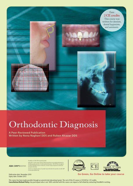
<strong>Orthodontic</strong> <strong>Diagnosis</strong><br />
A Peer-Reviewed Publication<br />
Written by Nona Naghavi DDS and Ruben Alcazar DDS<br />
Publication date: November 2010<br />
Expiry date: October 2013<br />
Go Green, Go Online to take your course<br />
This course has been made possible through an unrestricted educational grant. The cost of this CE course is $39.00 for 2 CE credits.<br />
Cancellation/Refund Policy: Any participant who is not 100% satisfied with this course can request a full refund by contacting PennWell in writing.
Educational Objectives<br />
The overall goal of this article is to provide the reader with<br />
information on orthodontic diagnosis. Upon <strong>com</strong>pletion of<br />
this article, the reader will be able to:<br />
1. List and describe the areas that need to be addressed in<br />
the patient interview/consultation<br />
2. List and describe the steps involved in the extraoral<br />
examination of patients presenting for orthodontic<br />
diagnosis and treatment<br />
3. List and describe the steps involved in the intraoral<br />
examination of patients presenting for orthodontic<br />
diagnosis and treatment<br />
4. List and describe the types of malocclusions and their genesis<br />
Abstract<br />
<strong>Orthodontic</strong> diagnosis must be performed thoroughly prior<br />
to orthodontic treatment planning. A number of steps are<br />
involved in the diagnostic process, all of which must be<br />
performed to reach an accurate diagnosis. The overall steps<br />
involved include the patient interview/consultation, clinical<br />
examination and use of diagnostic records. Only after these<br />
steps have been performed and analyzed can a treatment plan<br />
be developed for the individual patient.<br />
Introduction<br />
An orthodontic diagnosis must be carried out in a series of logical<br />
steps. The <strong>com</strong>bination of three sources of information will<br />
lead to a proper orthodontic diagnosis: the patient interview/<br />
consultation; the clinical examination by the clinician; and the<br />
evaluation of the diagnostic records that include, but may not<br />
be limited to, dental casts, radiographs and clinical images.<br />
Each of these sources of information is critical to the diagnosis<br />
and, ultimately, the patient’s orthodontic treatment. 1<br />
The Patient Interview/Consultation<br />
The three main areas that need to be addressed during the patient<br />
interview/consultation appointment are the chief <strong>com</strong>plaint,<br />
medical and dental history, and growth potential prediction.<br />
Chief Complaint<br />
The clinician must identify the main reason why the patient is<br />
seeking treatment, and this should be noted and documented<br />
in the chart in the patient’s own words. This does not have to be<br />
limited to one item only. The list of chief concerns should be<br />
established and noted in order of importance to the patient,<br />
and nothing should ever be assumed. 1 Some leading questions<br />
that will uncover the patient’s chief <strong>com</strong>plaint(s) follow:<br />
“Do you think you need braces?” and “What don’t you like<br />
about your smile/teeth/face?” If the patient is attending the<br />
appointment with one or both parents/guardians, it is always<br />
a good idea to first address the patient and determine his or<br />
her chief concern prior to addressing the ac<strong>com</strong>panying party.<br />
This will both establish a positive rapport with the patient and<br />
let you know whether or not the patient will be <strong>com</strong>pliant with<br />
treatment. It is extremely helpful to have a motivated child/<br />
adult, since the orthodontic results are directly affected by<br />
<strong>com</strong>pliance. Both you and the patient will be more satisfied<br />
at the end of treatment if you take the time at the consultation<br />
appointment to assess the patient’s motivation level and<br />
discuss realistic expectations. It is important to know whether<br />
the patient recognizes the need for treatment.<br />
Medical and Dental History<br />
A careful and full medical and dental history is necessary to<br />
provide a thorough background on the patient’s overall health<br />
status and to ascertain whether the patient is currently under a<br />
physician’s care. It is important to discuss any medications the<br />
patient may be taking, since some may have an effect on orthodontic<br />
treatment. Some examples of conditions and medications<br />
that impact orthodontic treatment include uncontrolled<br />
diabetes, which can exacerbate periodontal breakdown in<br />
response to orthodontic forces, and bisphosphonates, which<br />
can result in very slow orthodontic tooth movement. Similarly,<br />
chronic use of high-dose prostaglandin inhibitors for management<br />
of arthritis in adults may interfere with orthodontic tooth<br />
movement. 1 Extractions may be contraindicated in patients<br />
with hemophilia, while patients with attention deficit hyperactivity<br />
disorder (ADHD) may have less than ideal <strong>com</strong>pliance.<br />
In addition, latex allergic patients must be identified and<br />
appropriate measures taken to avoid any incidents. 2<br />
Growth Potential Prediction<br />
The patient (or ac<strong>com</strong>panying adult(s)) should be asked<br />
questions about recent changes in clothes/shoe sizes, signs of<br />
sexual maturity (achievement of menarche in girls) and age of<br />
sexual maturation in older siblings. Look for signs of secondary<br />
sexual characteristics, and take note of the patient’s height<br />
and weight <strong>com</strong>pared to siblings and parents, as this will tell<br />
you whether the patient has reached the onset of puberty, is<br />
at the peak of his or her growth spurt, or if the growth spurt<br />
has ceased altogether. <strong>Orthodontic</strong> correction can benefit from<br />
rapid growth during adolescence, whereas growth modification<br />
may not be feasible if a child is over the peak of the growth<br />
spurt. Cervical vertebral assessment can be made from the patient’s<br />
cephalometric X-ray (Fig. 1). It is important to note that<br />
one’s chronological age does not always coincide with skeletal<br />
or dental age. Serial cephalometric X-rays are the best way to<br />
determine whether growth has stopped or is still ongoing. 1,3<br />
Figure 1. Cephalometric X-ray and cervical vertebral assessment<br />
Stage II-III peak growth, Stage V is at least 2 years post peak growth*<br />
*(Angle Orthod. 2002 Aug;72(4):316-23. Baccetti, Franci, McNamara)<br />
2 www.ineedce.<strong>com</strong>
Clinical Examination<br />
Extraoral Examination<br />
The facial analysis is conducted with the patient either sitting<br />
upright or standing, not reclining in a dental chair. The<br />
analysis must consider the frontal plane, facial midlines and<br />
lip <strong>com</strong>petency.<br />
Frontal Plane<br />
The proportional relationship between facial height and width<br />
is the first step in facial evaluation. The three characteristic<br />
categories of facial type are dolichofacial (facial height > facial<br />
width, long faces), mesofacial (facial height proportional to<br />
width) and brachyfacial (facial width > facial height, square<br />
faces). The facial thirds are determined by evaluating the distances<br />
from the hairline (trichion) to the prominent ridge between<br />
the eyebrows (gl = glabella), the glabella to the bottom<br />
of the nose (sn = subnasale), and the bottom of the nose to the<br />
chin point (me = menton) (Fig. 2). These distances should be<br />
equal. The mouth should be a third of the way between the<br />
base of the nose and the chin (Fig. 3). The facial one-fifths<br />
are determined by vertical lines going through the helix of the<br />
outer ear, the outer canthus of the eye and the inner canthus of<br />
the eye. The line through the inner canthus of the eye should<br />
pass through the lateral aspect of the alar base of the nose, and<br />
all five segments should be one eye distance in width. This<br />
can also aid in evaluation of facial symmetry (Fig. 4). 1,4<br />
Figure 2. Facial thirds<br />
Figure 4. Evaluation of facial symmetry<br />
Facial Midlines<br />
First and foremost, the presence of any nasal deviation<br />
must be identified because this will affect your perception<br />
of dental midlines. If a deviation exists, then the midlines<br />
should be examined relative to an imaginary straight line<br />
(or an actual piece of string held vertical in front of the face)<br />
from the soft-tissue glabella. Ideally, this piece of string or<br />
imaginary line should pass through the soft-tissue glabella,<br />
the philtrum of the upper lip and the soft-tissue chin point.<br />
This will aid in determining any asymmetry of the face<br />
(Figs. 5, 6, 7).<br />
Figure 5. Relationship of facial to dental midlines before treatment<br />
Figure 3. Mouth-nose-chin relationship<br />
Note: This patient does not show lower midline upon smiling<br />
Figure 6. Relationship of upper to lower dental midlines<br />
www.ineedce.<strong>com</strong> 3
Figure 7. Relationship of facial to dental midlines after treatment<br />
Note: If the patient does not show her lower dental midline when smiling naturally,<br />
any dental correction in the lower arch will not be visible<br />
Lip Competency<br />
The upper and lower lips should ideally be touching or remain<br />
apart up to 3-4 mm while the patient is in a relaxed position<br />
(i.e., with no straining of lips or chin to close the mouth). Patients<br />
with a short upper lip (short philtrum) tend to “strain”<br />
their lips in order to close them and have an interlabial gap of<br />
more than 4 mm at rest. Besides indicating a short philtrum,<br />
this can also be indicative of protrusive incisors (while jaws<br />
are in their normal position), normally inclined teeth but<br />
mandibular retrognathism (the mandible being farther back<br />
than the maxilla), normally inclined teeth but maxillary prognathism<br />
(the maxilla being farther forward than mandible), a<br />
<strong>com</strong>bination of both mandibular retroprognathism and maxillary<br />
prognathism, or a longer than normal lower face with or<br />
without an anterior open bite. In addition to lip strain, these<br />
patients can present with a deep mentolabial sulcus and an<br />
ac<strong>com</strong>panying hyperactive mentalis. Hyperactive mentalis<br />
typically shows up as an “orange peel” appearance of the soft<br />
tissue around the chin point (Fig. 8). 1,3,4<br />
Figure 8. Orange peel appearance<br />
Smile Analysis, Smiling View and Dental Midlines<br />
Typically, the relationship between maxillary dental midline<br />
and facial midline can be determined with this view. If the<br />
patient shows lower teeth upon smiling, then the relationship<br />
of the maxillary dental midline to the mandibular dental midline,<br />
as well as mandibular dental midline to facial midline,<br />
can also be determined. Note that any nasal deviations may<br />
affect perception of the facial midline. The maxillary dental<br />
midline should coincide with the facial midline (see above),<br />
and the maxillary and mandibular dental midlines should<br />
coincide with each other. Finally, the mandibular dental<br />
midline should coincide with the soft-tissue chin point. Deviated<br />
chin points may also exist, and this should be taken into<br />
consideration (Figs. 5 - 7).<br />
Gingival display can also be noted in this view. Ideally, there<br />
should be about 1-2 mm of soft tissue apparent on smiling in<br />
this view with 100% of the upper incisor’s crown. Document in<br />
millimeters the upper incisor visible at rest and when smiling,<br />
and the amount of gingivae shown at rest and when smiling.<br />
Note that with the aging process, the upper lip will lengthen<br />
and the amount of incisor visible will decrease. 4 This can have<br />
a definitive effect on what orthodontic treatment plan is eventually<br />
undertaken. An above-average gingival display may<br />
indicate short clinical crowns (dental), short upper lip/short<br />
philtrum (soft tissue) or vertical maxillary excess (skeletal). A<br />
below-average gingival display may indicate vertical maxillary<br />
deficiency or long philtrum. Recording lip height at the philtrum<br />
and the <strong>com</strong>missures can help clarify the problem. 1<br />
Buccal corridors (the dark space between the buccal<br />
mucosa of the cheeks and the posterior maxillary dentition)<br />
should also be evaluated. Obliterated corridors can indicate<br />
wide arches. Conversely, excessive corridors can indicate<br />
crossbites or transverse jaw discrepancies. At any rate, the<br />
width of the dental arches should be related to the width of<br />
that individual’s face for optimum esthetics. Lay persons<br />
can detect this difference and have shown a preference for<br />
narrower buccal corridors. 5 The smile arc is basically the<br />
contour of the incisal edges of the maxillary incisors relative<br />
to the curvature of the lower lip while smiling. If these two<br />
lines match each other, the smile arc is called “consonant”<br />
(Fig. 9). 4 It has been shown that lay people prefer a consonant<br />
smile arc to one that is considered flat. 6 The golden proportion<br />
of teeth width when viewed from the front is another aspect<br />
of dental appearance to take note of. In an attractive smile, the<br />
apparent width of the lateral incisor is 62% of the central and<br />
the apparent width of the canine is 62% of the lateral and so<br />
on. The width of the maxillary central incisor should ideally<br />
be 80% of its height. Obviously, in<strong>com</strong>plete tooth eruption<br />
in children and dental attrition in adults will affect this ratio.<br />
In terms of gingival heights, the contour of gingival height<br />
of the central incisors and canines should be equal, with this<br />
gingival height being about 1.5 mm higher than that of the<br />
lateral incisor. The contact points of the maxillary teeth move<br />
up gingivally, progressively from central incisor to premolars<br />
with the incisal embrasures also getting larger. It is important<br />
to inform patients with triangular-shaped incisors that once<br />
the teeth are aligned and overlaps cleared, “black triangles”<br />
will appear as the contact points move incisally. 1<br />
4 www.ineedce.<strong>com</strong>
Figure 9. Consonant smile arc<br />
The ¾ View<br />
This view best aids assessment of the relative projections of<br />
the upper and lower jaw and gives an impression of the depth<br />
of the face. The patient must be positioned at a 45-degree angle.<br />
Some features that can be studied in this view are midface<br />
deformity, including nasal deformity; prominence of gonial<br />
angle; length and definition of the border of the mandible; lip<br />
fullness; and vermilion display. 3<br />
Profile<br />
The same three lines drawn on the frontal plane can be<br />
extended to this photograph. Additionally, the Esthetic<br />
line of Ricketts (E-line) should be drawn from the tip of<br />
the nose to the chin. This helps determine the positions of<br />
the upper and lower lip in relation to the E-line. Note that<br />
this relationship is directly affected by the size of the nose<br />
and chin anteroposteriorly. Patients should be asked to have<br />
their lips relaxed when taking this image. Typically, the upper<br />
lip should be 4 mm, and the lower lip 2 mm, behind the<br />
E-line. 1,3 The prominence of the incisors can affect the patient’s<br />
profile appearance. Bimaxillary dentoalveolar protrusion<br />
explains the situation where the incisors are protruded<br />
beyond their normal inclination, while the jaws are in their<br />
normal position (Fig. 10).<br />
Figure 10. Bimaxillary dentoalveolar protrusion<br />
Lip strain can also be seen in these cases as the patient struggles<br />
to achieve a lip seal (see above). In these patients, retracting<br />
the protruded teeth into a normal position improves lip posture.<br />
What is interesting to note is that if the incisors are protruded<br />
in the absence of lip strain, retraction of the incisors has little<br />
effect on lip function or prominence. To establish whether the<br />
jaws are proportionally positioned in the anteroposterior plane,<br />
a line is drawn on the profile from the bridge of the nose to the<br />
base of the upper lip, and another one from that point down<br />
to the chin. These two lines should form a straight line. If the<br />
angle formed between these is less than 180 degrees, the patient<br />
has a convex profile with the chin being behind the bridge of<br />
the nose (posterior divergence), while a wider angle indicates<br />
a concave profile (anterior divergence). Facial divergence is<br />
directly influenced by ethnic background, with American<br />
Indians and Asians presenting with anteriorly divergent faces<br />
while Northern Europeans typically present with posterior<br />
divergence. Vertical facial proportions can also be assessed with<br />
the profile image. By placing a finger or an instrument along<br />
the lower border of the mandible, the mandibular plane angle<br />
(the angle formed by the inclination of the mandibular plane<br />
to true horizontal) can be evaluated. Patients with long vertical<br />
facial dimensions (dolichofacial) usually have steep mandibular<br />
plane angles and a skeletal open bite tendency. Conversely,<br />
patients with short vertical facial dimensions (brachyfacial)<br />
usually have flat plane angles and deep bite malocclusions. 1<br />
The nasolabial angle (NLA) is very helpful in determining<br />
the final treatment plan customized for the patient. This<br />
angle is produced by two lines: one tangential to the columella<br />
of the nose (the part of the nose between the base of nose and<br />
the nasal tip) and the other tangential to the stomion superius<br />
(the highest point on the upper lip). Wherever these two lines<br />
meet forms the NLA. This angle relates the upper lip to the<br />
columella line. Typically, the measurement in a Caucasian<br />
patient is between 90 and 120 degrees. Anything less than<br />
90 degrees is considered an acute NLA and anything greater<br />
than 90 degrees an obtuse NLA (Fig. 11). 4<br />
Figure 11. Nasolabial angle<br />
www.ineedce.<strong>com</strong> 5
Intraoral Examination<br />
Oral Health<br />
Ascertain whether the patient is currently under a dentist’s<br />
care. The patient must have clearance from the general dentist<br />
stating that a full clinical examination, including any needed<br />
X-rays, has been conducted; that any dental caries has been<br />
treated; and that a cleaning as well as fluoride treatment, if<br />
needed, has been <strong>com</strong>pleted. All teeth must be accounted<br />
for to rule out any missing or supernumerary teeth. A thorough<br />
examination of the lips, oral mucosa, tongue and floor<br />
of the mouth and visual caries detection must be performed<br />
for every patient. Any disease or pathology (medical issues,<br />
caries, pulpal pathology, periodontal disease, or soft-tissue<br />
disease or conditions) must be under control prior to the<br />
<strong>com</strong>mencement of orthodontic services. Generalized probing<br />
is typically performed to evaluate bleeding on probing, and<br />
inadequately attached gingival areas must be noted to avoid<br />
treatment that could result in further dehiscence. Any history<br />
of prior orthodontic treatment must be explored and will help<br />
determine a more precise chief concern of the patient as well<br />
as provide insight about the patient’s attitude and <strong>com</strong>pliance<br />
with orthodontic treatment. Any oral habits such as digit or<br />
object sucking, as well as tongue thrust, must be evaluated,<br />
as these can be associated with the etiology and have a direct<br />
effect on the prognosis of orthodontic treatment (Fig. 12). 1<br />
Figure 12. Tongue thrust<br />
shifts (although determining CR in children is not easy, due<br />
to undeveloped articular eminences). Detection of a CO-CR<br />
discrepancy is needed to rule out “Sunday bites.” A Sunday<br />
bite can exist in two situations: 1) a patient who shifts his or her<br />
mandible forward into a Class I to get closure when there truly<br />
exists a Class II mandibular deficiency if he or she were to bite<br />
down on the posterior teeth in CO; or 2) a patient who shifts<br />
his or her mandible forward into a Class III to get closure but<br />
does so in order to bypass an incisor interference when there<br />
truly exists an end-on relationship if the patient were to bite<br />
down on his or her posterior teeth. This latter condition is also<br />
called a Pseudo Class III. Any history of trauma to the face,<br />
jaws or teeth must be explored and further followed up on. 1,3<br />
The patient’s overbite and overjet must be determined.<br />
Overbite – the vertical distance in millimeters between the<br />
incisal edges of the lower incisors and the incisal edges of the<br />
upper incisors (Fig. 13) – can be measured using a periodontal<br />
probe or ruler. In open-bite cases, the resulting number<br />
is negative. Overjet is the horizontal distance in millimeters<br />
between the facial surface of the lower anterior teeth and the<br />
lingual surface of the upper anterior teeth (Fig. 14). Based on<br />
the amount of overlap, you may get different overbite and<br />
overjet values, depending on which incisor you do your measurement<br />
from. Typically, the largest number is recorded.<br />
Figure 13. Overbite measurement<br />
Occlusion<br />
Mastication, speech and temporomandibular joint disorder<br />
(TMD) must be evaluated. Although it is difficult to evaluate<br />
masticatory efficiency, some patients report better chewing<br />
ability after orthodontic treatment. In children with speech<br />
problems, speech therapy in conjunction with orthodontics<br />
may help. The most important indicator of joint function is<br />
the amount of maximum opening, since restricted opening<br />
usually indicates a functional problem. 7 Therefore, any pain<br />
and/or click on opening and/or closing, as well as crepitation<br />
on movement, must be evaluated and assessed. If the jaws lock<br />
on opening and closing, this must be confirmed and followed<br />
up on. The muscles of mastication must be palpated as part of<br />
the routine examination. Any anterior or lateral shift on closure<br />
must be recorded, as it may have an effect on orthodontic<br />
diagnosis (true unilateral vs. bilateral crossbites). It is important<br />
to determine centric occlusion-centric relation (CO-CR)<br />
Figure 14. Overjet measurement<br />
6 www.ineedce.<strong>com</strong>
Figure 15. Total amount of crowding per arch<br />
Figure 17. Skeletal deep bite<br />
The amount of crowding or spacing in each arch must be<br />
measured and documented in millimeters. A gauge or intraoral<br />
ruler as well as visual analysis only can be used for this<br />
purpose. In crowded cases, each area of overlap between two<br />
teeth must be measured in millimeters and added together<br />
to give the sum total amount of crowding per arch (Fig. 15). 1<br />
The presence or absence of a crossbite can be evaluated<br />
by bringing the teeth into occlusion. Posterior crossbite explains<br />
the position of the upper molars in relation to the lower,<br />
and in a bilateral posterior crossbite, both upper molars<br />
are lingual to the lower molars. In unilateral crossbite, only<br />
one side manifests this problem. A crossbite can be either<br />
purely dental or skeletal in nature. A skeletal crossbite exists<br />
due to inadequate palatal widths of the maxilla – this can be<br />
seen by examining the palatal vault on the casts; if the vault<br />
is narrow and maxillary teeth lean out to reach the mandibular<br />
teeth, the problem is skeletal. Conversely, a normal-sized<br />
vault with tipped molars signifies a dental crossbite (Fig.<br />
16). With teeth in occlusion, vertical problems such as anterior<br />
or posterior open bites, and deep bites, can be evaluated.<br />
Once again the origin could be dental only or skeletal (for<br />
which the cephalometric X-ray needs to be evaluated). A<br />
patient with a skeletal open bite will usually have excessive<br />
eruption of the posterior teeth but may or may not have an<br />
anterior open bite (if the anterior teeth have super-erupted<br />
in order to <strong>com</strong>pensate, the patient will not have an anterior<br />
open bite). In a skeletal deep-bite patient, the posterior teeth<br />
are usually under-erupted and the patient presents with a<br />
deep anterior dental bite (Fig. 17). 1,3<br />
Figure 16. Anterior and posterior crossbites<br />
Diagnostic Records<br />
It is important to recognize that records are considered an<br />
adjunct and are not to be used as a replacement for clinical<br />
examination. 8 Cephalograms are usually not required<br />
as adjuncts for orthodontic diagnosis and treatment in<br />
adults, or for cases involving the correction of minor problems<br />
in children. However, if jaw relationships and incisor<br />
positions are being changed with treatment, one should<br />
definitely consider a cephalogram an integral part of the<br />
diagnostic records. Trimmed dental casts (or electronic<br />
casts), a panoramic X-ray supplemented with appropriate<br />
periapicals and facial form analysis constitute the minimum<br />
records needed. 1<br />
Cast Analysis<br />
Symmetry<br />
A transparent ruled grid is the simplest tool to use to establish<br />
symmetry. When it is placed over the maxillary cast and<br />
lined up with the midpalatal raphe, any distortion of arch<br />
form and shifts of dental units can be determined quickly<br />
(Fig. 18). 3<br />
Figure 18. Establishing symmetry<br />
www.ineedce.<strong>com</strong> 7
Space Analysis<br />
Space analysis is essentially the difference between “space<br />
available” and “space needed.” Space available is measured<br />
as arch perimeter from the mesial of one first molar in an arch<br />
to the opposite first molar in the same arch. There are two<br />
ways of doing this – either by measuring the contact point<br />
by contact point of each tooth and adding all the numbers<br />
or by placing a wire/string on the line of occlusion molar to<br />
molar and then measuring its length. Space required is measured<br />
by estimating the size of the unerupted permanent<br />
teeth and <strong>com</strong>paring that to the size of the erupted primary<br />
teeth. If the space required for the unerupted teeth exceeds<br />
that of the erupted teeth or space available, space deficiency<br />
exists and crowding is imminent (and vice versa) (Fig. 15).<br />
The size of unerupted teeth can be estimated by measuring<br />
the teeth on individual periapical radiographs (the enlargement<br />
factor of the X-ray must be taken into account) or<br />
by using proportionality tables fabricated using data from<br />
white American children by Moyers as well as the Tanaka<br />
and Johnston table. 1<br />
Tooth Size Analysis<br />
Also known as the Bolton analysis, this measurement identifies<br />
any discrepancy between the sizes of the upper teeth<br />
and those of the lower teeth. If the teeth themselves are mismatched<br />
in size between the two arches, it is not possible to<br />
achieve an ideal occlusion and anterior coupling of the teeth.<br />
An anomaly in size of the maxillary lateral incisors is the most<br />
<strong>com</strong>mon cause of Bolton discrepancy, but variations in the<br />
size of premolars or other teeth can also be present. Typically,<br />
upper lateral incisors should be larger than lower lateral incisors<br />
and all second premolars must be of equal size. Ideally,<br />
the sum of the mesiodistal width of the lower six anterior teeth<br />
is about 77.2% that of the upper six anterior teeth (anterior<br />
Bolton) and the sum of the mesiodistal width of all the lower<br />
teeth (excluding second and third molars) is about 91.3% that<br />
of the upper teeth (overall Bolton). 9<br />
Angle’s Classification of Malocclusion<br />
Angle’s classification is based on the relationship of the first<br />
molars and the alignment of the teeth relative to the line of<br />
occlusion. Normal occlusion consists of a Class I molar relationship<br />
– the mesiobuccal cusp of the upper first molar fits<br />
in the buccal groove of the lower first molar, with teeth on the<br />
line of occlusion (Fig. 20). A Class I malocclusion–Class I<br />
molar relationship consists of crowded and rotated teeth (Fig.<br />
21). In a Class II, division 1 malocclusion, the mesiobuccal<br />
groove of the upper molars is mesial to the buccal groove of<br />
the lower molars and the anterior teeth are protruded (Fig.<br />
22), while in a Class II, division 2 malocclusion, the upper<br />
central incisors are more retroclined than the lateral incisors<br />
(Fig. 23). Last, in a Class III malocclusion, the mesiobuccal<br />
groove of the upper molars is distal to the buccal groove of<br />
the lower molars (Fig. 24). 1,2<br />
Figure 19. Relationship of first molars and tooth alignment<br />
Figure 20. Normal Class I molar relationship<br />
Figure 21. Class I malocclusion-Class I molar relationship<br />
Figure 22. Class II, div 1 relationship<br />
8 www.ineedce.<strong>com</strong>
Figure 23. Class II, div 2 relationship<br />
radiographs, it is immensely helpful when a facial asymmetry<br />
is observed in a patient and an underlying skeletal <strong>com</strong>ponent<br />
is suspected and needs verification (Figs. 25-27). 10<br />
Figure 25. Orthognathic maxilla, mandible and dental arches<br />
Figure 24. Class III malocclusion<br />
Figure 26. Maxillary dental protrusion, normal maxilla and mandible<br />
Cephalometric Analysis<br />
The cephalogram helps with the analysis of the relationship<br />
of the major functional <strong>com</strong>ponents of the face, namely<br />
cranial base, jaws and teeth. For every malocclusion, there<br />
may exist a dental and a skeletal contributor, and it is possible<br />
to have identical dental relationships but very different<br />
skeletal discrepancies (the dental cast analysis is incapable of<br />
telling the clinician anything about the skeletal relationship<br />
of the patient that can be pertinent in the ultimate treatment<br />
plan chosen for that case). The Steiner Analysis has been the<br />
most widely used cephalometric analysis to date, and while<br />
not perfect, it can certainly help the clinician understand the<br />
underlying basis for a patient’s malocclusion. A Class II or<br />
III Angle malocclusion can be the result of a skeletal discrepancy<br />
or just a displacement within dental units with ideal<br />
jaw relationships; it is also possible to have a <strong>com</strong>bination<br />
of jaw discrepancy and dental displacement. 1 It is important<br />
to realize that solely <strong>com</strong>paring individual measurements to<br />
a norm is not as important as also looking at the soft-tissue<br />
presentation of that patient. Measurements are a means to an<br />
end, not an end unto themselves. One other type of cephalogram,<br />
a posteroanterior or frontal cephalogram, is used to<br />
evaluate whether skeletal asymmetry exists. Although this<br />
radiograph is not considered a part of the routine diagnostic<br />
Figure 27. Prognathic mandible, protrusive mandibular arch,<br />
normal maxilla<br />
Panoramic X-ray<br />
An overview of all the tissues present in a panoramic X-ray<br />
should confirm or eliminate the possible presence of any<br />
pathology. The sinuses, nasal airways, coronoid and condyle<br />
www.ineedce.<strong>com</strong> 9
processes, and hyoid bone area as well as the maxillary and<br />
mandibular bone proper must be checked to rule out abnormalities.<br />
Any dental pathology such as cysts, traumatic<br />
fractures, or abnormal bone pattern or destruction should be<br />
evaluated. The number of teeth present must be confirmed<br />
and supernumerary or missing teeth accounted for. The<br />
location of impacted canines is best viewed in a panoramic<br />
radiograph (Fig. 28), and can be backed up with a periapical<br />
radiograph (Fig. 29) of that area. 1,3 Lately, even better evaluation<br />
has be<strong>com</strong>e possible with the use of a Cone Beam CT<br />
scan (Fig. 30). Any retained primary teeth and/or congenital<br />
absence of the succedaneous teeth can be confirmed using a<br />
panoramic radiograph (Fig. 31). Next, the condition of the<br />
roots and the presence of periodontal ligament should be<br />
noted. The presence of already short roots should instill caution<br />
in the clinician. In addition, the status of the wisdom teeth<br />
and unerupted second molars must be determined and taken<br />
into account in the patient’s overall treatment plan. 1 Posterior<br />
crowding can be readily viewed on a panoramic radiograph<br />
and must be confirmed with additional data from the occlusal<br />
casts and intraoral examination.<br />
Figure 28. Panoramic radiograph showing impacted canines<br />
Figure 29. Periapical showing impacted canines<br />
Figure 31. Congenitally missing teeth<br />
Summary<br />
The overall steps involved in orthodontic diagnosis are the<br />
patient interview/consultation, clinical examination and<br />
use of diagnostic records. All are crucial in the attainment of<br />
an accurate diagnosis, which is a prerequisite for successful<br />
orthodontic planning and treatment. The automatic <strong>com</strong>pilation<br />
of all diagnostic findings helps the clinician create the<br />
list of problems present, from which the treatment plan will<br />
be developed.<br />
References and Resources<br />
1. Proffit WR, Fields Jr. HW, Sarver DM. Contemporary <strong>Orthodontic</strong>s. 4th ed.<br />
St. Louis: Mosby; 2007. Chapter 6.<br />
2. Patel A, Burden DJ, Sandler J. Medical disorders and orthodontics. J Orthod.<br />
36:1-21, 2009.<br />
3. Grabber TM, Vig KWL, Vanarsdall Jr. RL. <strong>Orthodontic</strong>s: Current Principles<br />
and Techniques. 4th ed. Elsevier Health Sciences; 2005. Chapter 1.<br />
4. Ackerman MB. Enhancement <strong>Orthodontic</strong>s, Theory and Practice. 1st ed.<br />
Ames: Blackwell Munksgaard; 2007. Chapters 3, 4.<br />
5. Moore T, Southard KA, Casko JS, et al. Buccal corridors and smile esthetics.<br />
Am J Orthod Dentofac Orthop. 127:208-213, 2005.<br />
6. Parekh J, Fields HW, Beck FM, et al. Attractiveness of variations in the smile<br />
arc and buccal corridor space as judged by orthodontists and laymen. Angle<br />
Orthod. 76:557-563, 2005.<br />
7. Okeson JP. Management of Temporomandibular Disorders and Occlusion,<br />
ed. St. Louis: Mosby; 2002.<br />
8. Atchison KA, Luke LS, White SC. An algorithm for ordering pretreatment<br />
orthodontic radiographs. Am J Orthod Dentofac Orthop. 102:29-44, 1992.<br />
9. Bolton WA. The clinical application of a tooth-size analysis. Am J Orthod.<br />
48:504-529, 1962.<br />
10. Trpkova B, Prasad NG, Lam EW, et al. Assessment of facial asymmetries<br />
from posteroanterior cephalograms: Validity of reference lines. Am J Orthod<br />
Dentofac Orthop. 123:512-520, 2003.<br />
Author Profiles<br />
Nona Naghavi DDS<br />
Dr. Naghavi graduated from the University of Toronto Dental School in<br />
2004. She <strong>com</strong>pleted an AEGD residency at the University of Maryland,<br />
Baltimore in 2005 and a Clinical Research Fellowship at Jacksonville<br />
University School of <strong>Orthodontic</strong>s in 2008. She is currently a second year<br />
resident at Jacksonville University School of <strong>Orthodontic</strong>s.<br />
Figure 30. Cone beam CT scan<br />
Ruben Alcazar DDS<br />
Dr. Alcazar obtained his dental degree from the University of San Martin,<br />
Peru in 1995.He received his training in <strong>Orthodontic</strong>s from the University<br />
of San Marcos, Peru, earning a Certificate in <strong>Orthodontic</strong>s in 2003. Dr.<br />
Alcazar is currently a resident at Jacksonville University, School of <strong>Orthodontic</strong>s,<br />
Class of 2011.<br />
Disclaimer<br />
The author(s) of this course has/have no <strong>com</strong>mercial ties with the sponsors<br />
or the providers of the unrestricted educational grant for this course.<br />
Reader Feedback<br />
We encourage your <strong>com</strong>ments on this or any PennWell course. For your convenience,<br />
an online feedback form is available at www.ineedce.<strong>com</strong>.<br />
10 www.ineedce.<strong>com</strong>
Online Completion<br />
Use this page to review the questions and answers. Return to www.ineedce.<strong>com</strong> and sign in. If you have not previously purchased the program select it from the “Online Courses” listing and <strong>com</strong>plete the<br />
online purchase. Once purchased the exam will be added to your Archives page where a Take Exam link will be provided. Click on the “Take Exam” link, <strong>com</strong>plete all the program questions and submit your<br />
answers. An immediate grade report will be provided and upon receiving a passing grade your “Verification Form” will be provided immediately for viewing and/or printing. Verification Forms can be viewed<br />
and/or printed anytime in the future by returning to the site, sign in and return to your Archives Page.<br />
Questions<br />
1. The three main areas that need to be<br />
addressed during the patient interview/<br />
consultation appointment are ________.<br />
a. all <strong>com</strong>plaints, the medical history and the <strong>com</strong>pliance<br />
potential prediction<br />
b. the chief <strong>com</strong>plaint, the medical and dental history,<br />
and the <strong>com</strong>pliance potential prediction<br />
c. the chief <strong>com</strong>plaint, the medical and dental history,<br />
and the growth potential prediction<br />
d. all <strong>com</strong>plaints, the medical and dental history, and<br />
the growth potential prediction<br />
2. The main reason why the patient is<br />
seeking orthodontic treatment should be<br />
noted and documented _________.<br />
a. in the chart in the clinician’s words<br />
b. in a separate file in the clinician’s words<br />
c. in a separate file in the patient’s own words<br />
d. in the chart in the patient’s own words<br />
3. If the patient is attending the appointment<br />
with one or both parents/guardians, it is<br />
________ to first address the patient and<br />
determine his or her chief concern prior to<br />
addressing the ac<strong>com</strong>panying party.<br />
a. sometimes a good idea<br />
b. always a good idea<br />
c. never a good idea<br />
d. not necessary<br />
4. It is important to know whether _______<br />
recognizes the need for treatment.<br />
a. the patient himself or herself<br />
b. the parent or guardian<br />
c. friends<br />
d. all of the above<br />
5. ________ can impact orthodontic<br />
treatment by resulting in very slow<br />
orthodontic movement.<br />
a. The use of antihistamines<br />
b. The use of bisphosphonates<br />
c. Uncontrolled diabetes<br />
d. all of the above<br />
6. Chronic use of ________ may interfere<br />
with orthodontic tooth movement.<br />
a. high-dose prostaglandins<br />
b. low-dose prostaglandins<br />
c. high-dose prostaglandin inhibitors<br />
d. low-dose prostaglandin inhibitors<br />
7. Chronological age ________.<br />
a. always coincides with skeletal or dental age<br />
b. always coincides with skeletal age<br />
c. always coincides with dental age<br />
d. does not always coincide with skeletal or dental age<br />
8. Serial ________ are the best way to<br />
determine whether growth has stopped or<br />
is still ongoing.<br />
a. periapical X-rays<br />
b. occlusal X-rays<br />
c. cephalometric X-rays<br />
d. panoramic X-rays<br />
9. The facial analysis is conducted with the<br />
patient ________.<br />
a. sitting upright<br />
b. standing<br />
c. reclining in a chair<br />
d. a or b<br />
10. The three characteristic categories of<br />
facial type are ________.<br />
a. dolichofacial, mesofacial and brachyfacial<br />
b. mesotheliofacial, distofacial and brachyfacial<br />
c. mesiocclusal, distobuccal and brachyfacial<br />
d. none of the above<br />
11. The presence of any nasal deviation will<br />
________.<br />
a. determine the position of dental midlines<br />
b. affect your perception of dental midlines<br />
c. determine the amount of medial tooth movement<br />
that is required<br />
d. all of the above<br />
12. An above-average gingival display may<br />
indicate _________.<br />
a. short clinical crowns<br />
b. short upper lip/short philtrum<br />
c. vertical maxillary excess<br />
d. all of the above<br />
13. The width of the maxillary central<br />
incisor should ideally be ________ of its<br />
height.<br />
a. 60%<br />
b. 70%<br />
c. 88%<br />
d. 90%<br />
14. The ¾ view _________.<br />
a. best aids assessment of the relative projections of<br />
the upper and lower jaw<br />
b. must be performed with the patient positioned at a<br />
45-degree angle<br />
c. gives an impression of the depth of the face<br />
d. all of the above<br />
15. ________ explains the situation where<br />
the incisors are protruded beyond their<br />
normal inclination, while the jaws are in<br />
their normal position.<br />
a. Maxillary dentoalveolar protrusion<br />
b. Maxillary dentoalveolar retrusion<br />
c. Bimaxillary dentoalveolar protrusion<br />
d. Bimaxillary dentoalveolar retrusion<br />
16. If the incisors are protruded in the<br />
absence of lip strain, retraction of the<br />
incisors has _________.<br />
a. little effect on lip function but a great effect on<br />
prominence<br />
b. a prominent effect on lip function<br />
c. little effect on lip function or prominence<br />
d. none of the above<br />
17. American Indians and Asians present<br />
with _________while Northern Europeans<br />
typically present with _________.<br />
a. anteriorly divergent faces; posterior divergence<br />
b. medially divergent faces; posterior divergence<br />
c. posteriorly divergent faces; anterior divergence<br />
d. anteriorly divergent faces; distal divergence<br />
18. Patients who are brachyfacial usually<br />
have _________ .<br />
a. steep plane angles and overjet malocclusions<br />
b. flat plane angles and overjet malocclusions<br />
c. flat plane angles and deep bite malocclusions<br />
d. steep plane angles and deep bite malocclusions<br />
19. _________ can have a direct effect on the<br />
prognosis of orthodontic treatment.<br />
a. Digit sucking<br />
b. Object sucking<br />
c. Tongue thrust<br />
d. all of the above<br />
20. The most important indicator of joint<br />
function is the amount of _________.<br />
a. maximum protrusion<br />
b. maximum retrusion<br />
c. maximum opening<br />
d. maximum overbite<br />
21. In open-bite cases, the overbite number<br />
is ________.<br />
a. greater<br />
b. positive<br />
c. negative<br />
d. none of the above<br />
22. In a bilateral posterior crossbite, both<br />
upper molars are ________ to the lower<br />
molars.<br />
a. distal<br />
b. inferior<br />
c. lingual<br />
d. none of the above<br />
23. In a skeletal deep-bite patient, the<br />
posterior teeth are usually ________.<br />
a. over-erupted<br />
b. under-erupted<br />
c. early to erupt<br />
d. late to erupt<br />
24. _________constitutes the minimum<br />
orthodontic record needed.<br />
a. Dental casts<br />
b. A panoramic X-ray with appropriate supplemental<br />
periapicals<br />
c. A facial form analysis<br />
d. all of the above<br />
25. Space required is measured by estimating<br />
the size of the ________ and <strong>com</strong>paring<br />
that to the size of the ________.<br />
a. erupted permanent teeth; unerupted permanent<br />
teeth<br />
b. erupted permanent teeth; erupted primary teeth<br />
c. unerupted permanent teeth; erupted primary teeth<br />
d. all of the above<br />
26. The _________ analysis identifies any<br />
discrepancy between the sizes of the upper<br />
teeth and those of the lower teeth.<br />
a. Munsell<br />
b. Morton<br />
c. Boston<br />
d. Bolton<br />
27. In a Class I molar relationship, the<br />
mesiobuccal cusp of the upper first molar<br />
fits in the buccal groove of the _________,<br />
with teeth on the line of occlusion.<br />
a. lower second molar<br />
b. lower second bicuspid<br />
c. lower first molar<br />
d. any of the above<br />
28. The ________ analysis has been the most<br />
widely used cephalometric analysis to date.<br />
a. Stettler<br />
b. Steiner<br />
c. Scheiner<br />
d. Steiger<br />
29. Posterior crowding can be readily viewed<br />
on a panoramic radiograph and must be<br />
confirmed with additional data from the<br />
_________.<br />
a. occlusal casts and extraoral examination<br />
b. occlusal casts and intraoral examination<br />
c. occlusal casts and films<br />
d. all of the above<br />
30. The overall step involved in orthodontic<br />
diagnosis is the _________.<br />
a. patient interview/consultation<br />
b. use of diagnostic records<br />
c. clinical examination<br />
d. all of the above<br />
www.ineedce.<strong>com</strong> 11
ANSWER SHEET<br />
<strong>Orthodontic</strong> <strong>Diagnosis</strong><br />
Name: Title: Specialty:<br />
Address:<br />
E-mail:<br />
City: State: ZIP: Country:<br />
Telephone: Home ( ) Office ( ) Lic. Renewal Date:<br />
Requirements for successful <strong>com</strong>pletion of the course and to obtain dental continuing education credits: 1) Read the entire course. 2) Complete all<br />
information above. 3) Complete answer sheets in either pen or pencil. 4) Mark only one answer for each question. 5) A score of 70% on this test will earn<br />
you 2 CE credits. 6) Complete the Course Evaluation below. 7) Make check payable to PennWell Corp. For Questions Call 216.398.7822<br />
Educational Objectives<br />
1. List and describe the areas that need to be addressed in the patient interview/consultation<br />
2. List and describe the steps involved in the extraoral examination of patients presenting for orthodontic diagnosis<br />
and treatment<br />
3. List and describe the steps involved in the intraoral examination of patients presenting for orthodontic diagnosis<br />
and treatment<br />
4. List and describe the types of malocclusions and their genesis<br />
Course Evaluation<br />
Please evaluate this course by responding to the following statements, using a scale of Excellent = 5 to Poor = 0.<br />
1. Were the individual course objectives met? Objective #1: Yes No Objective #3: Yes No<br />
Objective #2: Yes No Objective #4: Yes No<br />
2. To what extent were the course objectives ac<strong>com</strong>plished overall? 5 4 3 2 1 0<br />
3. Please rate your personal mastery of the course objectives. 5 4 3 2 1 0<br />
If not taking online, mail <strong>com</strong>pleted answer sheet to<br />
Academy of Dental Therapeutics and Stomatology,<br />
A Division of PennWell Corp.<br />
P.O. Box 116, Chesterland, OH 44026<br />
or fax to: (440) 845-3447<br />
For immediate results,<br />
go to www.ineedce.<strong>com</strong> to take tests online.<br />
Answer sheets can be faxed with credit card payment to<br />
(440) 845-3447, (216) 398-7922, or (216) 255-6619.<br />
Payment of $39.00 is enclosed.<br />
(Checks and credit cards are accepted.)<br />
If paying by credit card, please <strong>com</strong>plete the<br />
following: MC Visa AmEx Discover<br />
Acct. Number: ______________________________<br />
Exp. Date: _____________________<br />
Charges on your statement will show up as PennWell<br />
4. How would you rate the objectives and educational methods? 5 4 3 2 1 0<br />
5. How do you rate the author’s grasp of the topic? 5 4 3 2 1 0<br />
6. Please rate the instructor’s effectiveness. 5 4 3 2 1 0<br />
7. Was the overall administration of the course effective? 5 4 3 2 1 0<br />
8. Do you feel that the references were adequate? Yes No<br />
9. Would you participate in a similar program on a different topic? Yes No<br />
10. If any of the continuing education questions were unclear or ambiguous, please list them.<br />
___________________________________________________________________<br />
11. Was there any subject matter you found confusing? Please describe.<br />
___________________________________________________________________<br />
___________________________________________________________________<br />
12. What additional continuing dental education topics would you like to see?<br />
___________________________________________________________________<br />
___________________________________________________________________<br />
AGD Code 734<br />
PLEASE PHOTOCOPY ANSWER SHEET FOR ADDITIONAL PARTICIPANTS.<br />
AUTHOR DISCLAIMER<br />
The author(s) of this course has/have no <strong>com</strong>mercial ties with the sponsors or the providers of<br />
the unrestricted educational grant for this course.<br />
SPONSOR/PROVIDER<br />
This course was made possible through an unrestricted educational grant. No<br />
manufacturer or third party has had any input into the development of course content.<br />
All content has been derived from references listed, and or the opinions of clinicians.<br />
Please direct all questions pertaining to PennWell or the administration of this course to<br />
Machele Galloway, 1421 S. Sheridan Rd., Tulsa, OK 74112 or macheleg@pennwell.<strong>com</strong>.<br />
COURSE EVALUATION and PARTICIPANT FEEDBACK<br />
We encourage participant feedback pertaining to all courses. Please be sure to <strong>com</strong>plete the<br />
survey included with the course. Please e-mail all questions to: macheleg@pennwell.<strong>com</strong>.<br />
INSTRUCTIONS<br />
All questions should have only one answer. Grading of this examination is done<br />
manually. Participants will receive confirmation of passing by receipt of a verification<br />
form. Verification forms will be mailed within two weeks after taking an examination.<br />
EDUCATIONAL DISCLAIMER<br />
The opinions of efficacy or perceived value of any products or <strong>com</strong>panies mentioned<br />
in this course and expressed herein are those of the author(s) of the course and do not<br />
necessarily reflect those of PennWell.<br />
Completing a single continuing education course does not provide enough information<br />
to give the participant the feeling that s/he is an expert in the field related to the course<br />
topic. It is a <strong>com</strong>bination of many educational courses and clinical experience that<br />
allows the participant to develop skills and expertise.<br />
COURSE CREDITS/COST<br />
All participants scoring at least 70% on the examination will receive a verification<br />
form verifying 2 CE credits. The formal continuing education program of this sponsor<br />
is accepted by the AGD for Fellowship/Mastership credit. Please contact PennWell for<br />
current term of acceptance. Participants are urged to contact their state dental boards<br />
for continuing education requirements. PennWell is a California Provider. The California<br />
Provider number is 4527. The cost for courses ranges from $29.00 to $110.00.<br />
Many PennWell self-study courses have been approved by the Dental Assisting National<br />
Board, Inc. (DANB) and can be used by dental assistants who are DANB Certified to meet<br />
DANB’s annual continuing education requirements. To find out if this course or any other<br />
PennWell course has been approved by DANB, please contact DANB’s Recertification<br />
Department at 1-800-FOR-DANB, ext. 445.<br />
RECORD KEEPING<br />
PennWell maintains records of your successful <strong>com</strong>pletion of any exam. Please contact our<br />
offices for a copy of your continuing education credits report. This report, which will list<br />
all credits earned to date, will be generated and mailed to you within five business days<br />
of receipt.<br />
CANCELLATION/REFUND POLICY<br />
Any participant who is not 100% satisfied with this course can request a full refund by<br />
contacting PennWell in writing.<br />
© 2010 by the Academy of Dental Therapeutics and Stomatology, a division<br />
of PennWell<br />
12 Customer Service 216.398.7822 www.ineedce.<strong>com</strong>