The Response of Oral Submucous Fibrosis to Lycopene – A ... - JCDR
The Response of Oral Submucous Fibrosis to Lycopene – A ... - JCDR
The Response of Oral Submucous Fibrosis to Lycopene – A ... - JCDR

You also want an ePaper? Increase the reach of your titles
YUMPU automatically turns print PDFs into web optimized ePapers that Google loves.
Original Article<br />
<strong>The</strong> <strong>Response</strong> <strong>of</strong> <strong>Oral</strong> <strong>Submucous</strong> <strong>Fibrosis</strong><br />
<strong>to</strong> <strong>Lycopene</strong> – A Carotenoid Antioxidant:<br />
A Clinicopathological Study<br />
Dentistry Section<br />
Bhagavan Banige Komary Gowda, Yathish T.R., Sinhasan Sankappa P., Kumar Naik H., Purushotham Somayaji, Anand D.<br />
ABSTRACT<br />
Introduction: <strong>The</strong> oral submucous fibrosis (OSMF) is a precancerous<br />
condition <strong>of</strong> the oral cavity. Untreated and neglected cases<br />
<strong>of</strong> OSMF might end up as invasive squamous cell carcinoma.<br />
Some pernicious habits like smoking, pan chewing and <strong>to</strong>bacco<br />
addiction for which human being has succumbed <strong>to</strong> are causing<br />
various dreaded diseases, the important among them being oral<br />
submucous fibrosis, leukoplakia and erythroplakia. Free radicals<br />
have more recently emerged as media<strong>to</strong>rs <strong>of</strong> other phenotypic<br />
and genotypic changes that lead from mutation <strong>to</strong> neoplasia.<br />
<strong>The</strong> role <strong>of</strong> free radicals in the various oxidation processes in the<br />
body has lead <strong>to</strong> the identification <strong>of</strong> antioxidants in inhibiting and<br />
reversing the disease process. <strong>Lycopene</strong> is a safe antioxidant<br />
<strong>of</strong> utmost importance. <strong>Lycopene</strong> is a bright red carotene and<br />
carotenoid pigment and phy<strong>to</strong>chemical found in <strong>to</strong>ma<strong>to</strong>es and<br />
other red fruits and vegetables, such as red carrots, watermelons<br />
and papayas.<br />
Objectives: <strong>The</strong> main aim and objective <strong>of</strong> this study is <strong>to</strong><br />
evaluate the clinical and his<strong>to</strong>pathological response <strong>of</strong> oral<br />
submucous fibrosis <strong>to</strong> <strong>Lycopene</strong>-a carotenoid antioxidant.<br />
Methods and Materials: <strong>The</strong> material for this study constitutes<br />
12 adult patients picked from the regular outpatient attending the<br />
Department <strong>of</strong> Dentistry, Hassan institute <strong>of</strong> Medical Sciences,<br />
Hassan, Karnataka, India. Clinically, they are diagnosed as<br />
having oral submucous fibrosis.<br />
Conclusions: <strong>Response</strong>s are assessed clinically and his<strong>to</strong>pathologically.<br />
We observed clinical and his<strong>to</strong>logical improvement<br />
in various parameters used in our study. No significant <strong>to</strong>xicity<br />
attri butable <strong>to</strong> <strong>Lycopene</strong> is encountered in this study.<br />
Key Words: <strong>Lycopene</strong>, Antioxidant, <strong>Submucous</strong> <strong>Fibrosis</strong>, Free Radical theory<br />
Introduction<br />
<strong>Oral</strong> submucous fibrosis (OSMF) has a very interesting his<strong>to</strong>ry.<br />
Sushrutha, a renowned Indian physician who lived in the era from<br />
2500 <strong>to</strong> 3000BC, had already recognized it as a mouth and throat<br />
melady and had labeled it as Vidhari [1]. <strong>The</strong> features <strong>of</strong> which were<br />
described as a progressive narrowing <strong>of</strong> the mouth, blanching<br />
<strong>of</strong> the oral mucosa, pain and burning sensation on taking food,<br />
hypomobility <strong>of</strong> the s<strong>of</strong>t palate and <strong>to</strong>ngue, loss <strong>of</strong> gusta<strong>to</strong>ry<br />
sensation and occasional mild hearing impairment due <strong>to</strong> the<br />
blockage <strong>of</strong> the Eustachian tube [2, 3]. <strong>The</strong>re has been nearly no<br />
change in these symp<strong>to</strong>ms till <strong>to</strong>day. This condition has a reference<br />
in modern literature due <strong>to</strong> the works <strong>of</strong> Schwartz in 1952. Joshi<br />
was the first <strong>to</strong> describe this condition in India in 1952 and he<br />
coined the name ‘oral submucous fibrosis’ [4]. <strong>The</strong> aetiological<br />
studies on this condition still remain obscure. Various reasons<br />
have been put forth as the predisposing fac<strong>to</strong>rs, some <strong>of</strong> which<br />
are capsaicin, betel nut alkaloids, hypersensitivity, au<strong>to</strong>immunity,<br />
genetic predisposition and perhaps malnutrition [2, 3].<br />
Neoplasm is a multistage disease process, where a single cell can<br />
develop from an otherwise normal tissue in<strong>to</strong> malignancy that can<br />
eventually destroy the very base. <strong>The</strong> series <strong>of</strong> cellular and molecular<br />
changes that occur through the development <strong>of</strong> cancers can be<br />
mediated by a diversity <strong>of</strong> endogenous and other free radicals<br />
which have long been known <strong>to</strong> be mutagenic. Further, these free<br />
radicals have more recently emerged as the media<strong>to</strong>rs <strong>of</strong> the other<br />
phenotypic and genotypic changes that can lead from mutation <strong>to</strong><br />
neoplasia. It has therefore been felt that free radicals may have a<br />
major contribution <strong>to</strong> the cancer development in humans [4, 5].<br />
<strong>The</strong> role <strong>of</strong> free radicals in the various oxidation processes in the<br />
body has led <strong>to</strong> the identification <strong>of</strong> antioxidants in inhibiting and<br />
reversing the disease process. <strong>The</strong> mode <strong>of</strong> action <strong>of</strong> antioxidants<br />
may involve the stimulation <strong>of</strong> the immune system or direct action<br />
on the tumour cells. <strong>The</strong>y are antimutagenic and antimi<strong>to</strong>genic and<br />
operate by the common mechanism <strong>of</strong> breaking the free radical<br />
chain reactions [6].<br />
<strong>Lycopene</strong> is a safe antioxidant <strong>of</strong> utmost importance. <strong>Lycopene</strong> is<br />
a bright red carotene and carotenoid pigment and a phy<strong>to</strong>chemical<br />
found in <strong>to</strong>ma<strong>to</strong>es and other red fruits and vegetables, such as<br />
red carrots, watermelons and papayas. Preliminary research has<br />
shown an inverse correlation between the consumption <strong>of</strong> <strong>to</strong>ma<strong>to</strong>es<br />
and cancer risk. It has been shown <strong>to</strong> have several potent anticarcinogenic<br />
and antioxidant properties and has demonstrated<br />
pr<strong>of</strong>ound benefits in precancerous lesions such as leukoplakia.<br />
<strong>Lycopene</strong> exhibits the highest physical quenching rate constant<br />
with singlet oxygen [7].<br />
OSMF has a similarity in behaviour and malignant changes <strong>to</strong> other<br />
premalignant lesions <strong>of</strong> the oral cavity. So, it has been felt that the<br />
disease process in OSMF also could be reversed and inhibited<br />
by the use <strong>of</strong> anti oxidants, as is observed in other pre malignant<br />
lesion therapies <strong>of</strong> the oral cavity [8, 9].<br />
<strong>The</strong> main aim and objective <strong>of</strong> this study was <strong>to</strong> evaluate the<br />
clinical and his<strong>to</strong>pathological response <strong>of</strong> OSMF <strong>to</strong> the antioxidant,<br />
lycopene.<br />
Materials and methods: <strong>The</strong> material for this study constituted 12<br />
adult patients selected from the regular outpatients who attended<br />
882<br />
Journal <strong>of</strong> Clinical and Diagnostic Research. 2011 August, Vol-5(3): 000-000
www.jcdr.net<br />
Bhagavan Banige Komary Gowda ety al., <strong>Response</strong> <strong>of</strong> <strong>Oral</strong> <strong>Submucous</strong> <strong>Fibrosis</strong> <strong>to</strong> <strong>Lycopene</strong><br />
the Department <strong>of</strong> Dentistry, Hassan Institute <strong>of</strong> Medical Sciences,<br />
Hassan, Karnataka, India, who were clinically diagnosed as having<br />
oral submucous fibrosis.<br />
<strong>The</strong> his<strong>to</strong>ry and the clinical findings <strong>of</strong> each patient who were<br />
recorded as having submucous fibrosis was diagnosed by the presence<br />
<strong>of</strong> pallor <strong>of</strong> the oral mucous membrane, presence <strong>of</strong> palpable<br />
fibrous bands, lack <strong>of</strong> pliability <strong>of</strong> the oral mucous membrane and<br />
limited mouth opening. Consent was taken from each participant.<br />
<strong>The</strong> required investigations are carried out. All the patients were<br />
biopsied, both <strong>to</strong> confirm the diagnosis and <strong>to</strong> reveal any dysplasia<br />
which was present.<br />
All the selected cases who were in the habit <strong>of</strong> chewing arecanuts,<br />
were advised <strong>to</strong> give up the habit and they were kept under observation<br />
for six weeks. No improvement was observed and later,<br />
the treatment was instituted.<br />
Examination <strong>of</strong> the oral cavity was carried out under artificial light.<br />
Any abnormality and variation in the colour and the pliability <strong>of</strong> the<br />
oral mucosa was noted. <strong>The</strong> inter arch distance was measured in<br />
millimeters by using a graduated calipers kept between the incisal<br />
tips <strong>of</strong> the upper and lower central incisor with the mouth opening<br />
being maximum. <strong>The</strong> above measurement was taken bimonthly<br />
and it was recorded.<br />
Several fac<strong>to</strong>rs should be considered when assessing these trials.<br />
1. Choice <strong>of</strong> the agent: Although oral submucous fibrosis is premalignant,<br />
only chemo-preventive agents with minimal <strong>to</strong>xicity<br />
are justifiable for clinical testing (such as the antioxidant,<br />
lycopene)<br />
2. Assessment response: It is difficult <strong>to</strong> apply oral pho<strong>to</strong>graphy<br />
consistently. <strong>The</strong> response can be best assessed, based on<br />
the examination <strong>of</strong> the condition by two examiners who can<br />
independently agree on a category with pho<strong>to</strong>graphs as a<br />
help ful adjunct [Table/Fig-1, 2, 3, 4]. <strong>The</strong> increase in the mouth<br />
opening may be measured by using calipers [Table/Fig-5, 6, 7, 8].<br />
3. Evaluation <strong>of</strong> the his<strong>to</strong>pathological response: <strong>The</strong> comparison<br />
<strong>of</strong> the pre treatment biopsy with a biopsy specimen<br />
obtained after 3 <strong>to</strong> 6 months <strong>of</strong> the treatment was done. This<br />
is done <strong>to</strong> confirm a clinical response where a biopsy may<br />
show whether normalization has also occurred at the microscopic<br />
level [Table/Fig-9, 10 , 11, 12].<br />
An incisional biopsy was performed in each patient at entry. <strong>The</strong><br />
biopsies were taken from the buccal mucosa at the level <strong>of</strong> the<br />
cervical margin <strong>of</strong> the lower second molar, so as <strong>to</strong> avoid frictional<br />
kera<strong>to</strong>sis at the level <strong>of</strong> the occlusal line.<br />
In this study, the following parameters were evaluated:<br />
1. Clinical parameters:<br />
a) Symp<strong>to</strong>matic relief in the form <strong>of</strong> absence <strong>of</strong> burning<br />
sensation in the mouth and spontaneous healing <strong>of</strong> the<br />
ulcers when present.<br />
b) Change in the colour <strong>of</strong> the mucosa and its texture.<br />
c) Improvement in mouth opening<br />
2. His<strong>to</strong>logical parameters:<br />
a) Surface mucosa showing non keratinization<br />
b) Rete-ridge formation with an increase in the epithelial layer<br />
c) Less infiltration <strong>of</strong> the inflamma<strong>to</strong>ry cells, mainly polymorphs,<br />
in the connective tissue.<br />
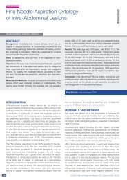
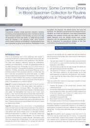
[Table/Fig-1]: Buccal Mucosa among group II Patients before treatment<br />
Study Design<br />
After the clinical examination, investigation and biopsy confirmed<br />
the diagnosis as oral submucous fibrosis, each patient was given<br />
the commercially available anti-oxidant which was used in this study,<br />
LycoRed (Jagsonpal Pharmaceutical Company, New Delhi, India)<br />
capsule containing 100% natural lycopene with zinc, selenium and<br />
added phy<strong>to</strong>nutrients. <strong>The</strong> zinc and selenium are essential for the<br />
proper assimilation <strong>of</strong> carotenes in our metabolism. This capsule<br />
was given orally, twice daily for 3 months, where each capsule<br />
contained 2000 µg <strong>of</strong> lycopene. <strong>The</strong> responding patients continued<br />
<strong>to</strong> take lycopene for an additional 3 months. <strong>The</strong> responses were<br />
assessed clinically and by bimonthly evaluation with examination<br />
and pho<strong>to</strong>graphs. <strong>The</strong> increase in the mouth opening was measured<br />
by using graduated calipers.<br />
All the biopsies were reviewed by a single pathologist at the entry<br />
level before the start <strong>of</strong> the treatment. After 3 months <strong>of</strong> treatment,<br />
a second biopsy was done as close as possible <strong>to</strong> the initial site<br />
and a his<strong>to</strong>logical evaluation was performed <strong>to</strong> confirm a clinical<br />
response.<br />
[Table/Fig-2]: Buccal Mucosa among group II Patients after treatment<br />
Journal <strong>of</strong> Clinical and Diagnostic Research. 2011 Aug, Vol-5(4): 000-000<br />
883
Bhagavan Banige Komary Gowda ety al., <strong>Response</strong> <strong>of</strong> <strong>Oral</strong> <strong>Submucous</strong> <strong>Fibrosis</strong> <strong>to</strong> <strong>Lycopene</strong><br />
www.jcdr.net<br />
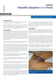
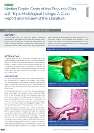
[Table/Fig-3]: Buccal Mucosa among group III Patients before treatment<br />
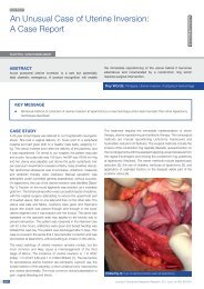
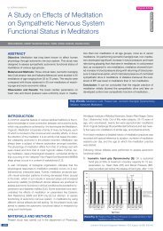
[Table/Fig-6]: Mouth opening among group II Patients after treatment<br />
[Table/Fig-7]: Mouth opening among group III Patients before treatment<br />
[Table/Fig-4]: Buccal Mucosa among group III Patients after treatment<br />
[Table/Fig-5]: Mouth opening among group II Patients before treatment<br />
[Table/Fig-8]: Mouth opening among group III Patients after treatment<br />
884<br />
Journal <strong>of</strong> Clinical and Diagnostic Research. 2011 August, Vol-5(4): 000-000
www.jcdr.net<br />
Bhagavan Banige Komary Gowda ety al., <strong>Response</strong> <strong>of</strong> <strong>Oral</strong> <strong>Submucous</strong> <strong>Fibrosis</strong> <strong>to</strong> <strong>Lycopene</strong><br />
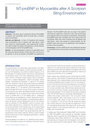
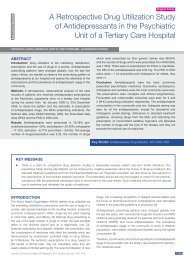
[Table/Fig-9]: Pho<strong>to</strong>micrograph among group II Patients before treatment<br />
showing atrophy <strong>of</strong> epithelium and juxtra epithelial hyalinization<br />
[Table/Fig-12]: Pho<strong>to</strong>micrograph among group III Patients after treatment<br />
showing basal cell proliferation and reteridge formation, decrease<br />
in keratin layer.<br />
[Table/Fig-10]: Pho<strong>to</strong>micrograph among group II Patients after treatment:<br />
appreciable reteridge formation and increase in epithelial layers.<br />
4. Effect <strong>of</strong> habit changes on the response: Patients who were<br />
in the habit <strong>of</strong> chewing betel nuts alone or in combination,<br />
were strictly advised <strong>to</strong> give up the habit and were kept under<br />
observation for six weeks. No improvement was observed<br />
and later, the treatment was initiated with lycopene.<br />
5. Study duration: At least 3 months <strong>of</strong> treatment should be<br />
planned. <strong>The</strong> response can occur 3 <strong>to</strong> 6 months after the<br />
beginning <strong>of</strong> the treatment.<br />
All the patients were grouped in<strong>to</strong> four categories, based on the<br />
clinical and his<strong>to</strong>logical criteria (as described by Khanna J.N [10-12]:<br />
Group1: Very early case<br />
Clinical: A burning sensation in the mouth was reported. Acute<br />
ulceration and recurrent s<strong>to</strong>matitis were seen. <strong>The</strong> mouth opening<br />
was normal.<br />
His<strong>to</strong>logical: A fine fibrillar collagen network interspersed with<br />
marked oedema. Young fibroblasts were seen, with abundant<br />
cy<strong>to</strong>plasm. Inflamma<strong>to</strong>ry cells mainly polymorphonuclear leucocytes<br />
were seen. <strong>The</strong> epithelium was essentially normal.<br />
Group 2: Early cases<br />
Clinical: <strong>The</strong> limitation <strong>of</strong> mouth opening was evident. <strong>The</strong> s<strong>of</strong>t<br />
palate and the faucial pillars were the areas primarily affected.<br />
<strong>The</strong> buccal mucosa appeared <strong>to</strong> be mottled, with varying degrees<br />
<strong>of</strong> fibrosis. His<strong>to</strong>logical: <strong>The</strong> results showed varing degrees <strong>of</strong><br />
keratinisation, atropy <strong>of</strong> the epithelium and juxta epithelial earely<br />
hyalinization, decrease in fibroblasts and increase in fibrocytes.<br />
<strong>The</strong>re was a dense infiltration <strong>of</strong> mixed inflamma<strong>to</strong>ry cells predominantly<br />
polymorphs.<br />
[Table/Fig-11]: Pho<strong>to</strong>micrograph among group III Patients before<br />
treatment showing marked epithelial atrophy, thickness is very much<br />
decreased, dense inflamma<strong>to</strong>ry cell infiltration.<br />
Journal <strong>of</strong> Clinical and Diagnostic Research. 2011 Aug, Vol-5(4): 000-000<br />
Group 3: Moderately advanced cases<br />
Clinical: Trismus was evident, ranging from 9mm <strong>to</strong> 15mm. <strong>The</strong><br />
buccal mucosa was pale and was firmly attached <strong>to</strong> the underlying<br />
tissues. <strong>The</strong> vertical fibrous bands were palpated in the premolar area.<br />
885
Bhagavan Banige Komary Gowda ety al., <strong>Response</strong> <strong>of</strong> <strong>Oral</strong> <strong>Submucous</strong> <strong>Fibrosis</strong> <strong>to</strong> <strong>Lycopene</strong><br />
www.jcdr.net<br />
His<strong>to</strong>logical: <strong>The</strong> results showed a marked reduction <strong>of</strong> the thickness<br />
<strong>of</strong> the epithelium and basal layer proliferation. <strong>The</strong> reteridges<br />
showed flattening and intercellular oedema in the focal areas. All<br />
the cases showed varing degrees <strong>of</strong> keratinization. <strong>The</strong> connective<br />
tissue showed moderate hyalinization with amorphous changes.<br />
Thick collagen bundles were seen, which were separated by slight<br />
oedema and they were infiltrated by lymphocytes and plasma cells,<br />
also occasionally by eosinophils.<br />
Group 4: Advanced cases with pre malignant and<br />
malignant changes<br />
Clinical: Trismus was severe . <strong>The</strong> mouth opening varied from 2-9<br />
mm. <strong>The</strong> uvula was involved .<strong>The</strong> <strong>to</strong>nsils which were compressed<br />
could have been associated with leukoplakia and squamous cell<br />
carcinoma.<br />
His<strong>to</strong>logical: <strong>The</strong> collagen hyalization had a smooth sheath which<br />
eliminated all evidence <strong>of</strong> the individual bundles. Fibroblasts were<br />
markedly absent within the hyalinized zone. <strong>The</strong>re was <strong>to</strong>tal loss<br />
<strong>of</strong> the epithelial rete pegs. <strong>The</strong>re was mild <strong>to</strong> moderate atypia.<br />
Extensive degeneration <strong>of</strong> the muscle fibers was seen.<br />
In our study, the groups 2 and 3 were selected: group 1 had normal<br />
mouth opening and in group 4, the disease was advanced, with<br />
malignant changes. Hence, the groups 1 and 4 were omitted.<br />
Results: <strong>The</strong> responses are assessed clinically by bi-monthly<br />
evalu ation with examination and pho<strong>to</strong>graphy. <strong>The</strong> responses were<br />
classified as:<br />
1. Complete<br />
• When the colour <strong>of</strong> the mucosa turned from blanched<br />
white <strong>to</strong> normal pale pink.<br />
• When there was a definite improvement in the burning<br />
sensation, which reduced <strong>to</strong> an appreciable extent.<br />
• When there was an increase in the mouth opening which<br />
ranged from 2mm <strong>to</strong> 3mm.<br />
2. Partial<br />
• When there was a partial improvement in the above<br />
said signs and symp<strong>to</strong>ms and the mouth opening was<br />
increased by 0.4mm <strong>to</strong> 1mm.<br />
3. Stable<br />
• When there was no response and no improvement.<br />
4. Disease progression<br />
• When there was a progressive increase in the signs and<br />
symp<strong>to</strong>ms in spite <strong>of</strong> having undergone the treatments.<br />
Patient characteristics: A <strong>to</strong>tal <strong>of</strong> 12 eligible patients were included<br />
in this study, 3 females and 9 males. <strong>The</strong> age group <strong>of</strong> the<br />
study sample ranged from 18 <strong>to</strong> 52 years. Among the 12 patients,<br />
6 were in the habit <strong>of</strong> pan chewing. Two patients were in the habit<br />
<strong>of</strong> smoking. Four patients were using pan with <strong>to</strong>bacco and slaked<br />
lime. No one was alcoholic. Four patients were using chillies in<br />
excess, whereas 8 patients had a normal intake <strong>of</strong> chillies. Seven<br />
patients were in group II (early cases). <strong>The</strong>ir mouth opening ranged<br />
between 15mm <strong>to</strong> 35mm. Five patients were in group III (moderately<br />
advanced) and their mouth opening was between 9mm <strong>to</strong>14mm<br />
[Table./Fig-13 and 14].<br />
Post Treatment Evaluation: <strong>The</strong> number <strong>of</strong> cases in which the<br />
following improvement was seen after the treatment with lycopene,<br />
2000 µg twice daily for 3 months, is shown in [Table/Fig-13 & 14].<br />
<strong>The</strong> responses were assessed clinically and his<strong>to</strong>logically as described<br />
below:<br />
IN GROUP II (EARLY CASES)<br />
Clinical evaluation<br />
1. Disappearance <strong>of</strong> the burning sensation was noticed.<br />
2. <strong>The</strong>re is a change in the colour <strong>of</strong> the mucosa from pale<br />
blanching <strong>to</strong> normal pink [Table/Fig-1, 2].<br />
3. Spontaneous healing <strong>of</strong> the ulcers was seen.<br />
4. <strong>The</strong>re was a definite improvement in the mouth opening,<br />
ranging from 1mm <strong>to</strong> 3mm [Table/Fig-5, 6, 13].<br />
His<strong>to</strong>logical Evaluation<br />
1. Showed a mild degree <strong>of</strong> inflamma<strong>to</strong>ry infiltration in the connective<br />
tissue [Table/Fig-10].<br />
2. <strong>The</strong> epithelial changes were more appreciable where in, they<br />
showed rete ridge formation with an increase in the epithelial<br />
layer.<br />
3. In few cases, the surface mucosa almost showed a loss <strong>of</strong><br />
keratinization.<br />
IN GROUP III (MODERATELY ADVANCED<br />
CASES)<br />
Clinical Evaluation:<br />
1. Disappearance <strong>of</strong> the burning sensation was observed.<br />
2. Healing <strong>of</strong> the ulcers when present, was noticed.<br />
3. <strong>The</strong>re was a definite change in the colour <strong>of</strong> the mucosa from<br />
pale blanching <strong>to</strong> normal pink [Table/Fig-3, 4].<br />
4. <strong>The</strong>re was improvement in the mouth opening, ranging from<br />
1mm <strong>to</strong> 3mm [Table/Fig-7, 8, 14].<br />
His<strong>to</strong>logical Evaluation<br />
1. Showed a decrease in the keratinization layer.<br />
2. An increase in the number <strong>of</strong> layers which reformed the rete<br />
ridges, with basal cell proliferation were seen [Table/Fig-12].<br />
Sl no<br />
Age/ Sex<br />
Mouth opening<br />
Prior <strong>to</strong><br />
treatment<br />
Post<br />
treatment<br />
<strong>Response</strong><br />
1 18/ M 27 mm 30 mm 3 mm Complete<br />
2 20/ M 30 mm 32 mm 2 mm Complete<br />
3 50/ M 20 mm 22 mm 2 mm Complete<br />
4 25/ M 25 mm 26 mm 1 mm Partial<br />
5 23/ M 18 mm 20 mm 2 mm Complete<br />
6 40/ M 16 mm 18 mm 2 mm Complete<br />
7 52/ M 35 mm 36 mm 1 mm Partial<br />
Table/Fig-13: Post Treatment Evaluation Observed in Group II<br />
Sl no<br />
Age/ Sex<br />
Mouth opening<br />
Prior <strong>to</strong><br />
treatment<br />
Post<br />
treatment<br />
Improvement<br />
Improvement<br />
<strong>Response</strong><br />
1 35/ F 11 mm 12 mm 1 mm Partial<br />
2 24/ M 12 mm 15 mm 3 mm Complete<br />
3 32/ M 14 mm 16 mm 2 mm Complete<br />
4 45/ F 9 mm 10 mm 1 mm Partial<br />
5 35/ F 14 mm 15 mm 1 mm Partial<br />
[Table/Fig-14]: Post treatment evaluation observed in group III<br />
886<br />
Journal <strong>of</strong> Clinical and Diagnostic Research. 2011 August, Vol-5(4): 000-000
www.jcdr.net<br />
Bhagavan Banige Komary Gowda ety al., <strong>Response</strong> <strong>of</strong> <strong>Oral</strong> <strong>Submucous</strong> <strong>Fibrosis</strong> <strong>to</strong> <strong>Lycopene</strong><br />
<strong>Response</strong> Number <strong>of</strong> patients Percentage (%)<br />
Number evaluable 12<br />
Complete response 7 58.5%<br />
Partial response 5 41.5%<br />
Stable – Nil<br />
Progression – Nil<br />
[Table/Fig-15]: <strong>Response</strong> Data<br />
RESPONSE DATA<br />
Often, the first change which was noted was a decrease in the<br />
burning sensation and the appearance <strong>of</strong> the normal pink colouration<br />
<strong>of</strong> the mucosa. Subsequently, a definite reduction in the<br />
burning sensation and an increase in the mouth opening were<br />
noted. In a majority <strong>of</strong> the responding patients, the first signs <strong>of</strong> the<br />
response were visible within the second and third follow up visits<br />
i.e. 4 patients at 1 month, 5 at 1½ months and 3 at 2 months. See<br />
[Table/Fig-15].<br />
DISCUSSION AND CONCLUSION<br />
<strong>Oral</strong> submucous fibrosis (OSMF) is a disease associated with the<br />
habit <strong>of</strong> betel nut chewing and it is characterized by extensive<br />
collagen deposition in the s<strong>of</strong>t tissues <strong>of</strong> the mouth [8]. Various<br />
researchers, in their studies on the mutagenic properties <strong>of</strong><br />
arecanuts have found that the constituents and the extracts <strong>of</strong> the<br />
nuts cause chromosomal aberrations and DNA damage[8]. In the<br />
review <strong>of</strong> literature, starting from Pay Master, 1959 <strong>to</strong> J.N. Khanna,<br />
1995, every one is <strong>of</strong> the opinion that submucous fibrosis is a<br />
precancerous condition with a malignant transformation rate which<br />
varies from 3 <strong>to</strong> 7.6% [9]. <strong>The</strong> development <strong>of</strong> cancers can be<br />
mediated by a diversity <strong>of</strong> endogenous and environmental stimuli.<br />
Free radicals have long been known <strong>to</strong> be mutagenic. Further,<br />
these agents have more recently emerged as the media<strong>to</strong>rs <strong>of</strong> the<br />
other phenotypic and genotypic changes that lead from mutation<br />
<strong>to</strong> neoplasia.<br />
OSMF is an incurable disease. No treatment modality, either surgical<br />
or medical has been successful in completely eliminating the<br />
disease. In view <strong>of</strong> the strong relationship between oral cancer and<br />
pre cancerous lesions, chemo-prevention is said <strong>to</strong> be feasible and<br />
practicable. A safe and simple mode <strong>of</strong> treatment as described in<br />
this study, along with proper habit restriction is required in OSMF<br />
<strong>to</strong> ensure that the progression <strong>of</strong> the disease is retarded and that<br />
maximum relief is obtained by the patient.<br />
Many authors are <strong>of</strong> the opinion that conservative treatment is<br />
preferable than the conventional ones [3]. Hazardous treatments<br />
like the submucosal injections <strong>of</strong> steroids, hyaluranidase and<br />
placental extracts should be avoided. Several studies in humans<br />
have confirmed the cancer preventive nature <strong>of</strong> antioxidants. <strong>The</strong><br />
oral intake <strong>of</strong> retinoids has a significant <strong>to</strong>xic effect on the normal<br />
tissue. A less <strong>to</strong>xic group <strong>of</strong> micronutrients are the carotenoids,<br />
which include lycopene. Its mode <strong>of</strong> action may involve stimulation<br />
<strong>of</strong> the immune system or a direct action on the tumour cells. <strong>Lycopene</strong><br />
has been shown <strong>to</strong> inhibit hepatic fibrogenesis in LEC rats<br />
by Kitade et al., and it may also exert a similar inhibition on the<br />
abnormal fibroblasts in submucous fibrosis. <strong>The</strong> findings <strong>of</strong> this<br />
study are in concurrence with those <strong>of</strong> another study on OSMF,<br />
which was performed by Haque et al, who reported a positive result<br />
in the OSMF cases with a therapeutic modality which was used for<br />
liver fibrosis. <strong>Lycopene</strong> also upregulates the lymphocyte resistance<br />
<strong>to</strong> stress and suppresses the inflamma<strong>to</strong>ry response [10].<br />
<strong>The</strong> results <strong>of</strong> the recent studies are encouraging. <strong>The</strong> unifying<br />
mechanism which underlies these diseases is cumulative oxidative<br />
damage. So, antioxidants can influence or prevent seemingly<br />
unrelated conditions [11].<br />
It is clear from this, that a long term maintenance treatment is necessary,<br />
if there has <strong>to</strong> be an impact on oral cancer incidence.<br />
<strong>Lycopene</strong> may be more suitable for this purpose than the more<br />
<strong>to</strong>xic retinoids. <strong>Lycopene</strong> has been shown <strong>to</strong> inhibit various types<br />
<strong>of</strong> cancers and it has been shown <strong>to</strong> have potent benefits in oral<br />
pre malignant lesions such as oral leukoplakia, where it has been<br />
reported <strong>to</strong> modulate dysplastic changes. Another point in favour <strong>of</strong><br />
the use <strong>of</strong> lycopene for the prevention <strong>of</strong> OSMF is that it is relatively<br />
non <strong>to</strong>xic and can be easily supplemented in the diet.<br />
This was a short term study with a small sample size and the patients<br />
in this study were advised <strong>to</strong> give up the habit <strong>of</strong> pan and<br />
betel nut chewing. As we could not have a control group due <strong>to</strong> the<br />
limited number <strong>of</strong> patients, it was felt that a long term study with<br />
a larger sample size with all the above variables being taken in<strong>to</strong><br />
consideration would be necessary <strong>to</strong> get a clear picture about the<br />
utility <strong>of</strong> the drug.<br />
References<br />
[1] Mukherji AL, Biswas SK. <strong>Oral</strong> submucous fibrosis- A search for<br />
etiology. Indian J O<strong>to</strong>laryngol. 1972; 24:11-5.<br />
[2] Anil S, Beena VT. <strong>Oral</strong> submucous fibrosis in a 12 year-old girl: case<br />
report. Pediatr Dent. 1993; 15:116.<br />
[3] Borle RM, Borle SR. Management <strong>of</strong> oral submucosal fibrosis, a<br />
conservative approach. Journal <strong>of</strong> <strong>Oral</strong> and Maxill<strong>of</strong>acial Surgery.<br />
1991; 49:788-91.<br />
[4] Desa JV. <strong>Submucous</strong> fibrosis <strong>of</strong> the palate and cheek. Ann O<strong>to</strong>l Rhin<br />
and Laryng 1957; 66:1043-59.<br />
[5] Guy<strong>to</strong>n KZ, Kensler TW. Oxidative mechanisms in carcinogenesis. Br<br />
Med Bull 1993; 49:523-44.<br />
[6] Boone CW, Gary J, Kell<strong>of</strong>f LS, Freedman. Intraepithelial and postinvasive<br />
neoplasia as a s<strong>to</strong>chastic continuum mechanism <strong>of</strong> chemopreventive<br />
drug action. Journal <strong>of</strong> Cellular Biochemistry. 1993; 53:<br />
14-25.<br />
[7] Armstron GA, Hearst JE. Carotenoids 2: Genetics and molecular<br />
biology <strong>of</strong> carotenoid pigment biosynthesis. Faseb J. 1996;10:228-37<br />
[8] Van WCK, Oliver A, Miranda D, Vander CM. Observations on the effect<br />
<strong>of</strong> areca nut extracts on oral fibroblast proliferation. Journal <strong>of</strong> <strong>Oral</strong><br />
Path and Medicine. 1993;21: 41-9.<br />
[9] Hayes PA. <strong>Oral</strong> submucous fibrosis in a 4-year old girl. Journal <strong>of</strong> <strong>Oral</strong><br />
Surgery. 1985; 59:475-8.<br />
[10] Kumar A, Bagewadi A, Keluskar V, Singh M. Efficacy <strong>of</strong> lycopene. <strong>Oral</strong><br />
Surg <strong>Oral</strong> Med <strong>Oral</strong> Pathol <strong>Oral</strong> Radiol. 2007; 103:207-13.<br />
[11] Garewal HS. <strong>Response</strong> <strong>of</strong> oral leukoplakia <strong>to</strong> betacarotene. J Clin<br />
Oncology. 1990; 8:1715-20.<br />
[12] Khanna JN , Andrade NN. <strong>Oral</strong> submucosal fibrosis: a new concept<br />
in surgical management. Inter J <strong>Oral</strong> and Maxill<strong>of</strong>acial Surgery.<br />
1995;24(6):433-39.<br />
Journal <strong>of</strong> Clinical and Diagnostic Research. 2011 Aug, Vol-5(4): 000-000<br />
887