Light Regulation of Protein Phosphorylation in Blepharisma japonicum
Light Regulation of Protein Phosphorylation in Blepharisma japonicum
Light Regulation of Protein Phosphorylation in Blepharisma japonicum
Create successful ePaper yourself
Turn your PDF publications into a flip-book with our unique Google optimized e-Paper software.
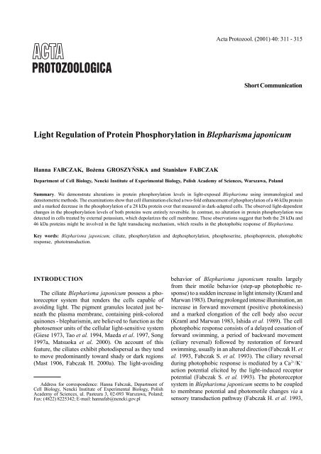
Acta Protozool. (2001) 40: 311 - 315<br />
Short Communication<br />
<strong>Light</strong> <strong>Regulation</strong> <strong>of</strong> <strong>Prote<strong>in</strong></strong> <strong>Phosphorylation</strong> <strong>in</strong> <strong>Blepharisma</strong> <strong>japonicum</strong><br />
Hanna FABCZAK, Bo¿ena GROSZYÑSKA and Stanis³aw FABCZAK<br />
Department <strong>of</strong> Cell Biology, Nencki Institute <strong>of</strong> Experimental Biology, Polish Academy <strong>of</strong> Sciences, Warszawa, Poland<br />
Summary. We demonstrate alterations <strong>in</strong> prote<strong>in</strong> phosphorylation levels <strong>in</strong> light-exposed <strong>Blepharisma</strong> us<strong>in</strong>g immunological and<br />
densitometric methods. The exam<strong>in</strong>ations show that cell illum<strong>in</strong>ation elicited a two-fold enhancement <strong>of</strong> phosphorylation <strong>of</strong> a 46 kDa prote<strong>in</strong><br />
and a marked decrease <strong>in</strong> the phosphorylation <strong>of</strong> a 28 kDa prote<strong>in</strong> over that measured <strong>in</strong> dark-adapted cells. The observed light-dependent<br />
changes <strong>in</strong> the phosphorylation levels <strong>of</strong> both prote<strong>in</strong>s were entirely reversible. In contrast, no alteration <strong>in</strong> prote<strong>in</strong> phosphorylation was<br />
detected <strong>in</strong> cells treated by external potassium, which depolarizes the cell membrane. These observations suggest that both the 28 kDa and<br />
46 kDa prote<strong>in</strong>s might be <strong>in</strong>volved <strong>in</strong> the light transduc<strong>in</strong>g mechanism, which results <strong>in</strong> the photophobic response <strong>of</strong> <strong>Blepharisma</strong>.<br />
Key words: <strong>Blepharisma</strong> <strong>japonicum</strong>, ciliate, phosphorylation and dephosphorylation, phosphoser<strong>in</strong>e, phosphoprote<strong>in</strong>, photophobic<br />
response, phototransduction.<br />
INTRODUCTION<br />
The ciliate <strong>Blepharisma</strong> <strong>japonicum</strong> possess a photoreceptor<br />
system that renders the cells capable <strong>of</strong><br />
avoid<strong>in</strong>g light. The pigment granules located just beneath<br />
the plasma membrane, conta<strong>in</strong><strong>in</strong>g p<strong>in</strong>k-colored<br />
qu<strong>in</strong>ones - blepharism<strong>in</strong>, are believed to function as the<br />
photosensor units <strong>of</strong> the cellular light-sensitive system<br />
(Giese 1973, Tao et al. 1994, Maeda et al. 1997, Song<br />
1997a, Matsuoka et al. 2000). On account <strong>of</strong> this<br />
feature, the ciliates exhibit photodispersal as they tend<br />
to move predom<strong>in</strong>antly toward shady or dark regions<br />
(Mast 1906, Fabczak H. 2000a). The light-avoid<strong>in</strong>g<br />
Address for correspondence: Hanna Fabczak, Department <strong>of</strong><br />
Cell Biology, Nencki Institute <strong>of</strong> Experimental Biology, Polish<br />
Academy <strong>of</strong> Sciences, ul. Pasteura 3, 02-093 Warszawa, Poland;<br />
Fax: (4822) 8225342; E-mail: hannafab@nencki.gov.pl<br />
behavior <strong>of</strong> <strong>Blepharisma</strong> <strong>japonicum</strong> results largely<br />
from their motile behavior (step-up photophobic response)<br />
to a sudden <strong>in</strong>crease <strong>in</strong> light <strong>in</strong>tensity (Kraml and<br />
Marwan 1983). Dur<strong>in</strong>g prolonged <strong>in</strong>tense illum<strong>in</strong>ation, an<br />
<strong>in</strong>crease <strong>in</strong> forward movement (positive photok<strong>in</strong>esis)<br />
and a marked elongation <strong>of</strong> the cell body also occur<br />
(Kraml and Marwan 1983, Ishida et al. 1989). The cell<br />
photophobic response consists <strong>of</strong> a delayed cessation <strong>of</strong><br />
forward swimm<strong>in</strong>g, a period <strong>of</strong> backward movement<br />
(ciliary reversal) followed by restoration <strong>of</strong> forward<br />
swimm<strong>in</strong>g, usually <strong>in</strong> an altered direction (Fabczak H. et<br />
al. 1993, Fabczak S. et al. 1993). The ciliary reversal<br />
dur<strong>in</strong>g photophobic response is mediated by a Ca 2+ /K +<br />
action potential elicited by the light-<strong>in</strong>duced receptor<br />
potential (Fabczak S. et al. 1993). The photoreceptor<br />
system <strong>in</strong> <strong>Blepharisma</strong> <strong>japonicum</strong> seems to be coupled<br />
to membrane potential and photomotile changes via a<br />
sensory transduction pathway (Fabczak H. et al. 1993,
312 H. Fabczak et al.<br />
Song 1997b). It <strong>in</strong>volves, as is the case <strong>in</strong> <strong>in</strong>vertebrate or<br />
vertebrate photoreceptor cells (Rayer et al. 1990), cGMP<br />
and <strong>in</strong>ositol trisphosphate as the possible second<br />
messengers (Fabczak H. et al. 1993, 1998, 1999;<br />
Fabczak H. 2000b).<br />
One <strong>of</strong> the most important mechanisms <strong>in</strong> the regulation<br />
<strong>of</strong> cellular metabolism or cell function is the<br />
process <strong>of</strong> prote<strong>in</strong> phosphorylation (Greengard 1978,<br />
Cohen 1982, Bünemann and Hosey 1999, Dickman and<br />
Yarden 1999, Graves and Krebs 1999). In the photoreceptor<br />
rod <strong>of</strong> vertebrate ret<strong>in</strong>a, there is evidence that<br />
phosphorylated prote<strong>in</strong>s may participate <strong>in</strong> the regulation<br />
<strong>of</strong> visual function. Of particular relevance are prote<strong>in</strong>s<br />
that exhibit light-dependent changes <strong>in</strong> the extent <strong>of</strong> their<br />
phosphorylation (Polans et al. 1979, Hermol<strong>in</strong> et al.<br />
1982, Lee et al. 1984, Hamm 1990). In the present study<br />
us<strong>in</strong>g immunological and densitometer methods, we demonstrate<br />
that prote<strong>in</strong> phosphorylation or dephosphorylation<br />
occurs <strong>in</strong> the ciliate when light is applied. These<br />
observations <strong>in</strong>dicate that the light-dependent processes<br />
<strong>of</strong> prote<strong>in</strong> phosphorylation may be <strong>in</strong>volved <strong>in</strong> the control<br />
<strong>of</strong> the photophobic behavior <strong>of</strong> <strong>Blepharisma</strong> <strong>japonicum</strong>.<br />
MATERIALS AND METHODS<br />
Cell culture<br />
Cells <strong>of</strong> <strong>Blepharisma</strong> <strong>japonicum</strong> were grown <strong>in</strong> 100 ml glass<br />
vessels conta<strong>in</strong><strong>in</strong>g Pr<strong>in</strong>gsheim solution (pH 7.0) at room temperature<br />
<strong>in</strong> darkness (Fabczak H. et al. 1993). The cells were fed with axenically<br />
cultured cells <strong>of</strong> Tetrahymena pyriformis. Prior to experiments,<br />
the cells were starved for at least 24 h before the organisms were<br />
collected, washed <strong>in</strong> an excess <strong>of</strong> fresh culture medium and subsequently<br />
used for assays after a further 12 h <strong>in</strong>cubation <strong>in</strong> fresh culture<br />
medium <strong>in</strong> darkness.<br />
Cell stimulation<br />
Before each experiment, 200 µl samples <strong>of</strong> dark-adapted cells<br />
were first left at rest for about 10 m<strong>in</strong> to avoid mechanical disturbances.<br />
They were subsequently exposed to light or ionic stimulation.<br />
The cell samples were illum<strong>in</strong>ated for 2 s with light afforded by<br />
a 150 W fiberoptic light source (MLW, Germany) equipped with an<br />
electromagnetic programmable shutter (model 22-841, Eal<strong>in</strong>g Electro-<br />
Optics, England). Ionic stimulation was performed by cell <strong>in</strong>cubation<br />
for 2 s <strong>in</strong> external 4 mM K + solution prepared by gentle addition <strong>of</strong><br />
the proper amount 0.1 M KCl to the cell suspension.<br />
Immunoblott<strong>in</strong>g assay<br />
In order to quantify prote<strong>in</strong> phosphorylation levels, samples <strong>of</strong><br />
control (dark-adapted) cells and cells follow<strong>in</strong>g treatment with light<br />
or potassium ions were immediately mixed after exposure with sample<br />
buffer conta<strong>in</strong><strong>in</strong>g 2% SDS, 5% 2-mercaptoethanol, 10% glycerol,<br />
1.0 mM EDTA and 62.5 mM Tris at pH 6.8 (Laemmli 1970)<br />
supplemented with protease or phosphatase <strong>in</strong>hibitors (50 mM NaF,<br />
2 mM phenylmethylsulfonylfluoride, 1 µM okadaic acid, 10 µg/ml<br />
aprot<strong>in</strong><strong>in</strong> or 50 µM leupept<strong>in</strong>) and then boiled for 5 m<strong>in</strong>. The<br />
equal amounts <strong>of</strong> cell prote<strong>in</strong>s (30 µg) were separated on<br />
10% SDS-polyacrylamide gels (SDS-PAGE) with the use <strong>of</strong> the<br />
Hoefer System (Amersham, USA) and transferred to nitrocellulose<br />
filters for 1 h at 100 V <strong>in</strong> transfer buffer consist<strong>in</strong>g <strong>of</strong> 192 mM<br />
glyc<strong>in</strong>e, 20% methanol and 25 mM Tris at pH 8.3 (Towb<strong>in</strong> at al.<br />
1979). For detection <strong>of</strong> phosphoser<strong>in</strong>e-conta<strong>in</strong><strong>in</strong>g prote<strong>in</strong>s, the blots<br />
were <strong>in</strong>cubated <strong>in</strong> block<strong>in</strong>g solution composed <strong>of</strong> 2% BSA <strong>in</strong> TBS (50<br />
mM NaCl, 25 mM Tris, pH 8.0) and 0.05% Tween-20 (BSA-Tris-<br />
Tween) for 2 h at room temperature. After the block<strong>in</strong>g procedure,<br />
the blots were <strong>in</strong>cubated with the primary antibody (Alexis, Switzerland)<br />
aga<strong>in</strong>st phosphoser<strong>in</strong>e overnight at a concentration <strong>of</strong> 0.05 µg/<br />
ml <strong>in</strong> 0.3% BSA-TBS-Tween at 4 0 C. The nitrocellulose filters were<br />
f<strong>in</strong>ally washed several times <strong>in</strong> TBS with 0.05% Tween (TBS-<br />
Tween). After wash<strong>in</strong>g, the blots were treated for 1h with anti-mouse<br />
IgG-horseradish peroxidase conjugates at a dilution <strong>of</strong> 1:10 000<br />
(Calbiochem) prepared <strong>in</strong> the block<strong>in</strong>g buffer. Immunoreactive bands<br />
were developed us<strong>in</strong>g an Amersham Enhanced Chemilum<strong>in</strong>escence<br />
(ECL) detection system. Their <strong>in</strong>tensities were quantified us<strong>in</strong>g a<br />
molecular dynamics personal laser densitometer and ImageQuant<br />
s<strong>of</strong>tware. Molecular weights <strong>of</strong> polypeptides were determ<strong>in</strong>ed based<br />
on their relative electrophoretic mobilities us<strong>in</strong>g presta<strong>in</strong>ed molecular<br />
weight standards (BioRad). In a control set <strong>of</strong> experiments, <strong>in</strong>cubation<br />
with primary antibodies was omitted. The concentration <strong>of</strong> cell<br />
prote<strong>in</strong>s <strong>in</strong> <strong>in</strong>dividual samples was estimated by a method reported<br />
elsewhere (Bradford 1976).<br />
RESULTS AND DISCUSSION<br />
Detection <strong>of</strong> phosphorylated prote<strong>in</strong>s <strong>in</strong> cell samples<br />
were made us<strong>in</strong>g three different monoclonal antibodies,<br />
Pser-7F12, Pser-4A9 and Pser-1C8, which may specifically<br />
recognize phosphorylated ser<strong>in</strong>e epitopes. Two <strong>of</strong><br />
the applied antibodies, Pser-7F12 and Pser-4A9, revealed<br />
marked prote<strong>in</strong> phosphorylation levels <strong>of</strong> similar<br />
patterns (Figs 1 A; 2 A), while the Pser-1C8 antibody<br />
<strong>in</strong>dicated a lower number <strong>of</strong> phosphoprote<strong>in</strong>s (Fig. 3 A),<br />
possibly due to lower am<strong>in</strong>o acid sequence specificity. It<br />
is well known that antibody specificity depends on<br />
phosphorylation <strong>of</strong> the am<strong>in</strong>o acid and on the surround<strong>in</strong>g<br />
am<strong>in</strong>o acid sequence. If one <strong>of</strong> these two criteria is<br />
unfulfilled, the antibody will not detect the phosphorylation<br />
site. Most labeled cell prote<strong>in</strong>s were unaffected by<br />
illum<strong>in</strong>ation, except for prote<strong>in</strong>s <strong>of</strong> a molecular weight <strong>of</strong><br />
about 46 kDa (Figs 1 A, B; 2 A) and prote<strong>in</strong>s below<br />
28 kDa (Figs 1 A, B; 2 A; 3 A). The <strong>in</strong>creased<br />
phosphorylation <strong>of</strong> the 46 kDa prote<strong>in</strong> on illum<strong>in</strong>ation<br />
(Figs 1 A; 2 A and lanes 1, 2) lasts up to 180 s after<br />
illum<strong>in</strong>ation (Figs 1 A, B; 2 A and lanes 1 to 4). In the
<strong>Blepharisma</strong> light-sensitive prote<strong>in</strong> phosphorylation 313<br />
Figs 1 A-C. Detection <strong>of</strong> light-<strong>in</strong>duced prote<strong>in</strong> phosphorylation <strong>in</strong><br />
<strong>Blepharisma</strong> by monoclonal antibody, Pser-7F12. Lane 1: cells<br />
adapted to darkness (control); Lane 2: cells exposed to light for 2 s;<br />
Lanes 3, 4: cells exposed to light for 2 s and then kept <strong>in</strong> darkness for<br />
180 s and 300 s, respectively; Lane 5: dark adapted cells stimulated<br />
by 4 mM K + for 2 s; Lane 6: dark-adapted cells stimulated by 4 mM<br />
K + and subsequently exposed to light for 2 s. The arrows <strong>in</strong>dicate<br />
46 kDa and 28 kDa prote<strong>in</strong>s, <strong>of</strong> which phosphorylation levels were<br />
altered when illum<strong>in</strong>ated. Immunoblot exposure to X-ray film for<br />
10 s (A) and 2 s (B); C - quantification <strong>of</strong> prote<strong>in</strong> phosphorylation<br />
<strong>in</strong>tensity <strong>of</strong> bands from B. The phosphorylation values found for<br />
control cells were def<strong>in</strong>ed as 100%<br />
dark, the most extensively labeled phosphoprote<strong>in</strong> had an<br />
apparent molecular weight <strong>of</strong> 28 kDa (Figs 1 A, B; 2 A;<br />
3 A and lane 1) and the phosphorylation level <strong>of</strong> this<br />
prote<strong>in</strong> was markedly reduced by illum<strong>in</strong>ation (Figs 1 A,<br />
Figs 2 A, B. Detection <strong>of</strong> prote<strong>in</strong> phosphorylation by light <strong>in</strong><br />
<strong>Blepharisma</strong> by monoclonal antibody, Pser-4A9. A - 30 s exposure <strong>of</strong><br />
immunoblot to X-ray film. B -quantification <strong>of</strong> ser<strong>in</strong>e phosphorylation<br />
<strong>of</strong> 46 kDa and 28 kDa prote<strong>in</strong>s. Other details as <strong>in</strong> Fig. 1<br />
B; 2 A; 3 A and lane 2). The light-<strong>in</strong>duced dephosphorylation<br />
<strong>of</strong> 28 kDa prote<strong>in</strong> was entirely reversible, as<br />
progressive phosphorylation <strong>of</strong> the prote<strong>in</strong> was observed<br />
<strong>in</strong> dark-adapted cells for 180 s (Figs 1 A, B; 2 A; 3 A<br />
and lane 3) and after about 300 s the phosphorylation<br />
level reached the level observed <strong>in</strong> control cells (Figs 1<br />
A, B; 2 A; 3 A and lane 4).<br />
To test whether phosphorylation and dephosphorylation<br />
<strong>of</strong> cellular prote<strong>in</strong>s <strong>in</strong> <strong>Blepharisma</strong> is specifically<br />
<strong>in</strong>duced by light or is simply elicited by cell membrane
314 H. Fabczak et al.<br />
Figs 3 A, B. Detection <strong>of</strong> light-<strong>in</strong>duced prote<strong>in</strong> phosphorylation <strong>in</strong><br />
<strong>Blepharisma</strong> by monoclonal antibody, Pser-1C8. A - 20 m<strong>in</strong>. exposure<br />
<strong>of</strong> immunoblot to X-ray film. B - quantification <strong>of</strong> ser<strong>in</strong>e phosphorylation<br />
<strong>of</strong> the 28 kDa prote<strong>in</strong>. Other details as <strong>in</strong> Fig. 1<br />
depolarization, the effect <strong>of</strong> ionic stimulation was exam<strong>in</strong>ed.<br />
These experiments <strong>in</strong>dicate that the phosphorylation<br />
level <strong>of</strong> prote<strong>in</strong>s <strong>of</strong> 28 kDa and 46 kDa was<br />
unaffected by membrane depolarization compared to<br />
cell samples exposed to light (Figs 1 A, B; 2 A; 3 A and<br />
lane 5). The pattern <strong>of</strong> prote<strong>in</strong> phosphorylation <strong>in</strong> cell<br />
samples, which were first treated with K + and subsequently<br />
exposed to light (Figs 1 A, B; 2 A; 3 A and<br />
lane 6), was similar to that obta<strong>in</strong>ed for cells that were<br />
only illum<strong>in</strong>ated (Figs 1 A; 2 A; 3 A and lane 2).<br />
The results <strong>of</strong> semi-quantitative analysis <strong>of</strong> prote<strong>in</strong><br />
phosphorylation <strong>in</strong>dicated that the level <strong>of</strong> phosphorylation<br />
<strong>of</strong> 46 kDa prote<strong>in</strong>s by 2 s illum<strong>in</strong>ation caused a tw<strong>of</strong>old<br />
<strong>in</strong>crease over the values found <strong>in</strong> dark-adapted cells<br />
(Figs 1 C; 2 B). In the case <strong>of</strong> the 28 kDa polypeptide,<br />
the level <strong>of</strong> phosphorylation is lower <strong>in</strong> all cell samples<br />
exposed to light. The phosphorylation levels for both<br />
these prote<strong>in</strong>s returned to the control levels after about<br />
300 s <strong>of</strong> cell <strong>in</strong>cubation under dark conditions (Figs 1 C,<br />
2 B, 3 B).<br />
It has been shown that phosphorylation and dephosphorylation<br />
<strong>of</strong> cellular prote<strong>in</strong>s play a crucial role <strong>in</strong> the<br />
regulation <strong>of</strong> various sensory transduction pathways<br />
(Greengard 1978, Cohen 1982, Bünemann and Hosey<br />
1999, Dickman and Yarden 1999, Graves and Krebs<br />
1999). In the visual system, light <strong>in</strong>duces phosphorylation<br />
<strong>of</strong> photoreceptors (rhodops<strong>in</strong>) by a prote<strong>in</strong> k<strong>in</strong>ase<br />
(Bownds et al. 1972, Kühn 1974, Frank et al. 1973). The<br />
higher light <strong>in</strong>tensity causes a marked dephosphorylation<br />
<strong>of</strong> prote<strong>in</strong>s <strong>of</strong> low molecular weight <strong>in</strong> <strong>in</strong>tact rods (Polans<br />
et al. 1979, Lee et al. 1984, Bownds and Brewer 1986).<br />
In the present study, we showed that light is capable <strong>of</strong><br />
<strong>in</strong>fluenc<strong>in</strong>g the level <strong>of</strong> prote<strong>in</strong> phosphorylation <strong>in</strong><br />
<strong>Blepharisma</strong> <strong>japonicum</strong>, as it takes place <strong>in</strong> the photoreceptor<br />
cells <strong>of</strong> vertebrates. In this ciliate, however, the<br />
prote<strong>in</strong>s be<strong>in</strong>g phosphorylated or dephosphorylated by<br />
light have not yet been identified. It was recently shown<br />
that the photosensory pigment blepharism<strong>in</strong> <strong>in</strong><br />
<strong>Blepharisma</strong> <strong>japonicum</strong> is associated with a 200 kDa<br />
membrane prote<strong>in</strong> (Matsuoka et al. 2000). It is likely<br />
that the polypeptide <strong>of</strong> molecular weight <strong>of</strong> 46 kDa,<br />
which underwent highly specific phosphorylation on<br />
ser<strong>in</strong>e after light stimulation, is associated with the<br />
200 kDa complex photoreceptor prote<strong>in</strong> or it simply<br />
represents a fragment <strong>of</strong> the high molecular weight<br />
prote<strong>in</strong> result<strong>in</strong>g from digestion by endogenous cellular<br />
proteases dur<strong>in</strong>g detergent solubilization (Matsuoka et<br />
al. 2000). Further <strong>in</strong>vestigations are necessary regard<strong>in</strong>g<br />
the identification <strong>of</strong> this prote<strong>in</strong> and elucidation <strong>of</strong> the<br />
mechanism that governs the observed prote<strong>in</strong> phosphorylation<br />
and dephosphorylation by light <strong>in</strong> protozoan<br />
ciliate <strong>Blepharisma</strong>.<br />
Acknowledgements. This work was supported <strong>in</strong> part by grant no.<br />
6P04C-057-18 from the Committee for Scientific Research and the<br />
statutory fund<strong>in</strong>g for the Nencki Institute <strong>of</strong> Experimental Biology.<br />
REFERENCES<br />
B<strong>in</strong>der B. M., Brewer E., Bownds M. D. (1989) Stimulation <strong>of</strong><br />
prote<strong>in</strong> phosphorylation <strong>in</strong> frog rod outer segments by prote<strong>in</strong><br />
k<strong>in</strong>ase activators. J. Biol. Chem. 264: 8857-8864<br />
Bownds M. D., Brewer E. (1986) Changes <strong>in</strong> prote<strong>in</strong> phosphorylations<br />
and nucleotide triphosphates dur<strong>in</strong>g phototransduction. In:<br />
The Molecular Mechanism <strong>of</strong> Photoreception. (Ed. H. Stieves),<br />
Spr<strong>in</strong>ger Verlag, New York Inc., New York, 159-169<br />
Bownds M. D., Bawes J., Miller J., Stahlman M. (1972) <strong>Phosphorylation</strong><br />
<strong>of</strong> frog photoreceptor membranes <strong>in</strong>duced by light.<br />
Nature New Biol. 237: 125-127<br />
Bradford M. M. (1976) A rapid and sensitive method for the<br />
quantitation <strong>of</strong> microgram quantities <strong>of</strong> prote<strong>in</strong> utiliz<strong>in</strong>g the<br />
pr<strong>in</strong>ciple <strong>of</strong> prote<strong>in</strong>-dye b<strong>in</strong>d<strong>in</strong>g. Anal. Biochem. 72: 248-254<br />
Bünemann M., Hosey M. M. (1999) G-prote<strong>in</strong> coupled receptor<br />
k<strong>in</strong>ases as modulators <strong>of</strong> G-prote<strong>in</strong> signal<strong>in</strong>g. J. Physiol. 517:<br />
5-23<br />
Cohen P. (1982) The role <strong>of</strong> prote<strong>in</strong> phosphorylation <strong>in</strong> neural and<br />
hormonal control <strong>of</strong> cellular activity. Nature 296: 613-620<br />
Dickman M. B., Yarden O. (1999) Ser<strong>in</strong>e/threon<strong>in</strong>e prote<strong>in</strong> k<strong>in</strong>ases<br />
and phosphatases <strong>in</strong> filamentous fungi. Fungal Gen. Biol. 26:<br />
99-117
<strong>Blepharisma</strong> light-sensitive prote<strong>in</strong> phosphorylation 315<br />
Fabczak H. (2000a) Protozoa as model system for studies <strong>of</strong> sensory<br />
light transduction: photophobic response <strong>in</strong> the ciliate Stentor and<br />
<strong>Blepharisma</strong>. Acta Protozool. 39: 171-181<br />
Fabczak H. (2000b) Contribution <strong>of</strong> the phospho<strong>in</strong>ositide-dependent<br />
signal pathway to photo motility <strong>in</strong> <strong>Blepharisma</strong>.<br />
J. Photochem. Photobiol. B. 55:120-127<br />
Fabczak H., Tao N., Fabczak S., Song P.-S. (1993) Photosensory<br />
transduction <strong>in</strong> ciliates. IV Modulation <strong>of</strong> the photomovement<br />
response <strong>of</strong> <strong>Blepharisma</strong> <strong>japonicum</strong> by cGMP. Photochem.<br />
Photobiol. 57: 889-892<br />
Fabczak H., Walerczyk M., Fabczak S. (1998) Identification <strong>of</strong><br />
prote<strong>in</strong> homologous to <strong>in</strong>ositol trisphosphate receptor <strong>in</strong> ciliate<br />
<strong>Blepharisma</strong>. Acta Protozool. 37: 209-213<br />
Fabczak H., Walerczyk M., Groszyñska B., Fabczak S. (1999) <strong>Light</strong><br />
<strong>in</strong>duces <strong>in</strong>ositol trisphosphate elevation <strong>in</strong> <strong>Blepharisma</strong> <strong>japonicum</strong>.<br />
Photochem. Photobiol. 69: 254-258<br />
Fabczak S., Fabczak H., Song P.-S. (1993) Photosensory transduction<br />
<strong>in</strong> ciliates. III. The temporal relation between membrane<br />
potentials and photomototile responses <strong>in</strong> <strong>Blepharisma</strong> <strong>japonicum</strong>.<br />
Photochem. Photobiol. 57: 872-876<br />
Frank R. N., Cavanagh H. D., Kenyon K. R. (1973) <strong>Light</strong> stimulated<br />
phosphorylation <strong>of</strong> bov<strong>in</strong>e visual pigments by adenos<strong>in</strong>e triphosphate.<br />
J. Biol. Chem. 248: 596-609<br />
Giese A. C. (1973) <strong>Blepharisma</strong>. The Biology <strong>of</strong> a <strong>Light</strong>-sensitive<br />
Protozoan. Stanford Univ. Press, Stanford<br />
Grave J. D., Krebs E. G. (1999) <strong>Prote<strong>in</strong></strong> phosphorylation and signal<br />
transduction. Pharmacol. Ther. 82: 111-121<br />
Greengard P. (1978) Phosphorylated prote<strong>in</strong>s as physiological effectors.<br />
Science 199: 146-152<br />
Hamm H. (1990) <strong>Regulation</strong> by light <strong>of</strong> cyclic nucleotide-dependent<br />
prote<strong>in</strong> k<strong>in</strong>ases and their substrates <strong>in</strong> frog rod outer segments.<br />
J. Gen. Physiol. 95: 545-567<br />
Hermol<strong>in</strong> J., Karell M. A., Hamm H. E., Bownds M. D. (1982)<br />
Calcium and cyclic GMP regulation <strong>of</strong> light-sensitive prote<strong>in</strong><br />
phosphorylation <strong>in</strong> frog photoreceptor membranes. J. Gen.<br />
Physiol. 79: 633-655<br />
Ishida M., Shigenaka Y., Taneda K. (1989) Studies on the mechanism<br />
<strong>of</strong> cell elongation <strong>in</strong> <strong>Blepharisma</strong> <strong>japonicum</strong>. I. Physiological<br />
mechanism how light stimulation evokes cell elongation. Europ.<br />
J. Physiol. 25: 182-186<br />
Kraml M., Marwan W. (1983) Photomovement responses <strong>of</strong> the<br />
heterotrichous ciliate <strong>Blepharisma</strong> <strong>japonicum</strong>. Photochem.<br />
Photobiol. 37: 313-319<br />
Kühn H. (1974) <strong>Light</strong> dependent-phosphorylation <strong>of</strong> rhodops<strong>in</strong> <strong>in</strong><br />
liv<strong>in</strong>g frogs. Nature (Lond). 250: 588-590<br />
Laemmli U. K. (1970) Cleavage <strong>of</strong> structural prote<strong>in</strong>s dur<strong>in</strong>g the<br />
assembly <strong>of</strong> the head <strong>of</strong> bacteriophage T4. Nature 277: 680-685<br />
Lee R. H., Brown B. M., Lolley R. N. (1984) <strong>Light</strong>-<strong>in</strong>duced<br />
dephosphorylation <strong>of</strong> a 33K prote<strong>in</strong> <strong>in</strong> rod outer segments <strong>of</strong> rat<br />
ret<strong>in</strong>a. Biochemistry 23: 1972-1977<br />
Matsuoka T., Tokumori D., Kotsuki H., Ishida M., Matsushita M.,<br />
Kimura S., Itoh T., Checcucci G. (2000) Analyses <strong>of</strong> structure <strong>of</strong><br />
photoreceptor organelle and blepharism<strong>in</strong>-associated prote<strong>in</strong> <strong>in</strong><br />
unicellular eukaryote <strong>Blepharisma</strong>. Photochem. Photobiol. 72:<br />
709-713<br />
Maeda M., Haoki H., Matsuoka T., Kato Y., Kotsuki H., Utsumi K.,<br />
Tanaka T. (1997) Blepharism<strong>in</strong> 1-5, novel photoreceptor from the<br />
unicellular organism <strong>Blepharisma</strong> <strong>japonicum</strong>. Tetrahedron Lett.<br />
38: 7411-7414<br />
Mast S. O. (1906) <strong>Light</strong> reactions <strong>in</strong> lower organisms. 1. Stentor<br />
coeruleus. J. Exp. Biol. 3: 359-399<br />
Polans A. S., Hermol<strong>in</strong> J., Bownds M. D. (1979) <strong>Light</strong>-<strong>in</strong>duced<br />
dephosphorylation <strong>of</strong> two prote<strong>in</strong>s <strong>in</strong> frog rod outer segments.<br />
J. Gen Physiol.74: 595-613<br />
Rayer B., Naynert M., Stieve H., (1990) Phototransduction; Different<br />
mechanisms <strong>in</strong> vertebrates and <strong>in</strong>vertebrates. J. Photochem.<br />
Photobiol. Part B. 7: 107-148<br />
Song P.-S., (1997a). <strong>Light</strong> signal transduction <strong>in</strong> ciliate Stentor and<br />
<strong>Blepharisma</strong>. 1. Structure and function <strong>of</strong> the photoreceptors. In:<br />
Biophysics <strong>of</strong> Photoreception Molecular and Phototransductive<br />
Events (Ed. C. Taddei-Ferretti), World Sci., S<strong>in</strong>gapore, New<br />
Jersey, London, Hong Kong, 48-66<br />
Song P.-S., (1997b). <strong>Light</strong> signal transduction <strong>in</strong> ciliate Stentor and<br />
<strong>Blepharisma</strong>. 2. Transduction mechanism. In: Biophysics <strong>of</strong><br />
Photoreception Molecular and Phototransductive Events (Ed.<br />
C. Taddei-Ferretti), World Sci., S<strong>in</strong>gapore, New Jersey, London,<br />
Hong Kong, 76-82<br />
Tao N., Diforce L., Romanowski M., Meza-Keuthen S., Song P.-S.,<br />
Furuya M. (1994) Stentor and <strong>Blepharisma</strong> photoreceptors:<br />
structure and function. Acta Protozool. 33: 199-211<br />
Towb<strong>in</strong> H., Staehel<strong>in</strong> T., Gordon J. (1979) Electrophoretic transfer<br />
<strong>of</strong> prote<strong>in</strong>s from polyacrylamide gels to nitrocellulose sheet:<br />
Procedure and some applications. Proc. Natl. Acad. Sci. USA 76:<br />
4350-4354<br />
Received and accepted on 10th October, 2001


