Alteration of Cryptosporidium parvum (Apicomplexa: Eucoccidiorida ...
Alteration of Cryptosporidium parvum (Apicomplexa: Eucoccidiorida ...
Alteration of Cryptosporidium parvum (Apicomplexa: Eucoccidiorida ...

Create successful ePaper yourself
Turn your PDF publications into a flip-book with our unique Google optimized e-Paper software.
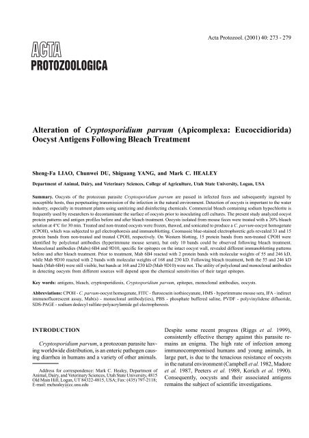
<strong>Alteration</strong> <strong>of</strong> C. <strong>parvum</strong> oocyst antigens 275Fig. 1. Bleach-treated <strong>Cryptosporidium</strong> <strong>parvum</strong> oocysts examinedby an indirect immun<strong>of</strong>luorescent assay using Mab 6B4. Oocystsappear brightly stained with hollow centers. Scale bar - 12 µmIndirect immun<strong>of</strong>luorescent assay (IFA)A modified Mab-based IFA (Garcia et al. 1987, Arrowood andSterling 1989) was used to examine oocysts and intermediate stages<strong>of</strong> C. <strong>parvum</strong> by employing different Mabs generated in our laboratory.Individual samples (either oocysts in feces, purified oocysts, orCPOH) were applied to the surface <strong>of</strong> pre-cleaned microscope slides,air-dried at 25°C for 30 min, and fixed by gentle flaming. Sampleswere incubated with one <strong>of</strong> the candidate Mabs (60 µl/application)for 30 min in a 100% humidified chamber at 37°C, followed by anadditional 30 min with fluorescein isothiocyanate (FITC)-conjugatedgoat anti-mouse immunoglobulin (Sigma Chemical Company, St. Louis,MO, USA) diluted at 1:100 (50 µl/application). Following incubation,samples were thoroughly washed with PBS, covered with 2drops <strong>of</strong> non-drying mounting medium (50% glycerol in PBS), andexamined under a Zeiss epifluorescence microscope equipped with anultraviolet lamp. To evaluate Mabs against intermediate parasiticstages, C. <strong>parvum</strong>-infected cell cultures growing on coverslips weresubjected to the same procedure as described above for oocysts.Dot blot assayMabs and HMS reacting with respective native and denaturedCPOH were discerned by a dot-blot micr<strong>of</strong>iltration apparatus inaccordance with the manufacturer’s instructions (Bio-Rad Laboratories,Richmond, CA, USA). Denaturation <strong>of</strong> CPOH was done by thesame procedure as described for SDS-PAGE (below). The intensity<strong>of</strong> reaction was scored as: - = negative, 1+ = light, 2+ = moderate,3+ = strong, and 4+ = very strong. Mabs for use in Western blotanalysis were selected by the results <strong>of</strong> this assay.Sodium dodecyl sulfate-polyacrylamide gel electrophoresis(SDS-PAGE)A commercially available 4 ~ 20% gradient gel and Mini-PRO-TEAN ® II dual slab cell (Bio-Rad Laboratories) were employed inSDS-PAGE. One part <strong>of</strong> solubilized CPOH was diluted with 2 parts(v/v) <strong>of</strong> sample buffer [2% SDS, 5% (v/v) β-mercaptoethanol,Fig. 2. Pr<strong>of</strong>ile <strong>of</strong> <strong>Cryptosporidium</strong> <strong>parvum</strong>-oocyst homogenate(CPOH) proteins resolved by SDS-PAGE and stained by GelCode ®blue reagent. Molecular weight standards are shown in lane SD andlabeled in kilodaltons (kD) in the left-hand margin. Non-bleachtreatedCPOH demonstrate at least 33 protein bands, with molecularweights ranging from 6.5 to 187 kD (Lane N). Beach-treated CPOHreduced the number <strong>of</strong> protein bands to 15, with molecular weightsranging from 6.5 to 55 kD (Lane T)0.0125% bromophenol blue, and 0.25% (v/v) glycerol in 62.5 mMTris-HCl, pH 6.8] and boiled for 5 min before loading 30 µl into eachgel lane. Prestained and non-prestained molecular weight standards(broad range, Bio-Rad Laboratories) were incorporated into the controlgel lanes to determine the relative molecular weights <strong>of</strong> theresolved proteins. Electrophoresis was performed with a discontinuousbuffer system (Laemmli 1970) at 175 V until the dye frontreached the bottom <strong>of</strong> the slab gel (approximately 50 min). Followingelectrophoresis, GelCode ® blue stain reagent (Pierce Chemical Company,Rockford, IL, USA), which employs the colloidal properties <strong>of</strong>Coomassie ® blue G-250 for protein staining, was used to visualizeCPOH protein bands in the slab gels.Western blot analysisFollowing SDS-PAGE, CPOH protein bands were electrophoreticallytransferred (Towbin et al. 1979) to the polyvinylidene difluoride(PVDF) membrane using a Mini Trans-Blot ® Transfer Cell (Bio-Rad). Transfer was carried out overnight at 4°C at 30 V, followed by60 V for 2 h. Transfer efficiency was monitored by observing theprestained molecular weight standards. Following transfer, the membranesheets were rinsed with ddH 2O and dried with a cold air stream.Nonspecific sites on the sheets were blocked for 18 h with a solution
276 S-F. Liao et al.Table 1. Dot-blot assay scores for polyclonal and monoclonal antibodies against native and denatured <strong>Cryptosporidium</strong> <strong>parvum</strong>-oocysthomogenate (CPOH)Antibody 1 HMS 9D10 2C5 2F8 3F11 9G12 2B7 2C12 8B1 D8 6B4 8C6 9F11Isotype 2 ND 3 IgM ND IgM IgA ND IgG2b IgA ND ND IgM ND NDNative 4 4+ 3+ 3+ 3+ 3+ 3+ 4+ 4+ 4+ 4+ 4+ 4+ NDDenatured 5 4+ 2+ 3+ 1+ 1+ 1+ 1+ 1+ 1+ 1+ 2+ 2+ 1+1Polyclonal (HMS - hyperimmune mouse serum) and monoclonal antibody designations; 2 Isotyping was done on only select Mabs;3ND - not done; 4 Native - score for non-denatured CPOH; 5 Denatured - score for denatured CPOH.RESULTSFig. 3. Western blot analysis <strong>of</strong> bleach and non-bleach-treated<strong>Cryptosporidium</strong> <strong>parvum</strong>-oocyst homogenate (CPOH) using HMSand Mabs (6B4 and 9D10). Prior to bleach treatment, HMS recognized15 protein bands with molecular weights ranging from 21.5 to246 kD (Lane 2). Following bleach treatment, 10 protein bandsremain with molecular weights ranging from 21.5 to 230 kD (Lane 1).Prior to and following bleach treatment, Mab 6B4 recognized 2protein bands having molecular weights <strong>of</strong> 55 kD (**) and 246 kD (*)(Lanes 4 and 3, respectively). Similarly, Mab 9D10 recognized twoprotein bands before bleach treatment. The respective molecularweights are 168 kD and 230 kD (arrowheads) (Lane 6). Followingbleach treatment, Mab 9D10 failed to recognize either band (Lane 5)<strong>of</strong> 5% bovine serum albumin in 0.05 M Tris Buffered Saline (BSA-TBS, pH 7.4). The blocked sheet was cut into strips and incubatedwith either HMS (1:3000) or a Mab for 2 h, washed with PBS +0.05% Tween 20 (PBS-T), and incubated for 1.5 h with biotinylatedanti-mouse IgG goat serum (Sigma) diluted at 1:1000 in 1% BSA-TBS. The strips were further reacted with ExtrAvidin-Alkaline Phosphatase(Sigma) for 1.5 h after washing with PBS-T. Color developmentwas accomplished by using an Alkaline Phosphatase ConjugateSubstrate Kit (Bio-Rad) and stopped by washing with ddH 2O.Antibody screeningELISA showed that more than 70 hybridomas producingMabs against C. <strong>parvum</strong> were generated. IFAdetermined that some Mabs (6B4 and 9D10) wereagainst epitopes present on the oocyst stage (Fig. 1),while others (such as 2C5) were against epitopes onintermediate parasitic stages (data not shown).Bleach treatment on oocystsPrior to bleach treatment, oocysts reacted stronglywith Mabs 6B4 and 9D10 as determined by IFA.Oocysts appeared brightly stained with hollow centers(Fig. 1). Following bleach treatment, oocysts lost theirreaction with Mab 9D10, but retained their reaction withMab 6B4 showing bright apple green fluorescence.Dot blottingDot blot assay revealed that HMS had very strongreactions with both native and denatured CPOH (Table1). The optimal concentration <strong>of</strong> HMS used in Westernblotting was 1:3000, as determined by serial-dilution indot blotting. Almost all Mabs reacted with native (nondenatured)CPOH antigens, with reaction intensity varyingfrom 1+ to 4+. Following CPOH denaturation, only12 Mabs continued to react with CPOH antigens (Table1). Based on reaction intensity (2+ ~ 3+), 4 Mabs (6B4,9D10, 2C5, and 8C6) were selected for further analysisin Western blotting.SDS-PAGE <strong>of</strong> CPOHThe pr<strong>of</strong>ile <strong>of</strong> CPOH proteins resolved bySDS-PAGE and stained by GelCode ® blue reagent isshown in Fig. 2. Non-bleach-treated CPOH demonstratedat least 33 protein bands (Lane N). The molecularweights <strong>of</strong> these proteins ranged from 6.5 kD to187 kD. Proteins with molecular weights below 6.5 kD
<strong>Alteration</strong> <strong>of</strong> C. <strong>parvum</strong> oocyst antigens 277or over 200 kD could not be clearly discerned. Bleachtreatment <strong>of</strong> oocysts reduced the number <strong>of</strong> proteinbands to 15, with molecular weights ranging from 6.5 kDto 55 kD (Lane T).Western blottingThe majority <strong>of</strong> CPOH protein bands in the gradientgels were transferred to PVDF membranes as noted bythe disappearance <strong>of</strong> prestained standards in the gelsand also by light staining <strong>of</strong> gels with GelCode ® bluereagent following transfer. Western blot analysis <strong>of</strong>bleach- and non-bleach-treated CPOH using HMS andMabs (6B4 and 9D10) are illustrated in Fig. 3. Prior tobleach treatment, HMS recognized 15 protein bandswith molecular weights ranging from 21.5 kD to 246 kD(Lane 2). Following bleach treatment, 10 protein bandsremained with molecular weights ranging from 21.5 kDto 230 kD (Lane 1). No bands were observed whennormal mouse serum was used (data not shown).Prior to and following bleach treatment <strong>of</strong> oocysts,Mab 6B4 recognized 2 protein bands from CPOHhaving molecular weights <strong>of</strong> 55 kD and 246 kD (Lanes4 and 3, respectively). The band having the lowestmolecular weight (55 kD) appeared much more intense(Lanes 4 and 3, respectively). Similarly, Mab 9D10recognized two protein bands before bleach treatment.The respective molecular weights were 168 kD and230 kD (Lane 6). However, following bleach treatment,Mab 9D10 failed to recognize either band (Lane 5). Noprotein bands were visible following incubation with Mab2C5, Mab 8C6, and cell culture medium (negative control)(data not shown).DISCUSSION<strong>Cryptosporidium</strong> <strong>parvum</strong> is an undisputed waterbornepathogen <strong>of</strong> humans and other animals. Thisintestinal protozoan has been responsible for outbreaks<strong>of</strong> cryptosporidiosis traced back to surface and potablewater sources (MacKenzie et al. 1994, Wallis et al.1996). Because low numbers <strong>of</strong> oocysts present apotential risk for infection, the challenge has been todevelop immun<strong>of</strong>luorescent and flow cytometric assayscapable <strong>of</strong> detecting only a few oocysts (Vesey andSlade 1990, Vesey et al. 1993). A confounding factor isthe effect that water disinfectants may have on oocystdetection. It has been reported that oocyst viability orinfectivity is not affected by exposure <strong>of</strong> oocysts to1.05 ~ 3% sodium hypochlorite (equivalent to 20 ~ 60%commercial bleach) for up to 18 h at 4 or 37°C, and only70 ~ 100% commercial bleach can destroy oocystinfectivity (Campbell et al. 1982, Korich et al. 1990,Sterling 1990). Our results regarding the alteration <strong>of</strong>oocyst antigens following bleach treatment not onlysupport the notion that the routine chlorination <strong>of</strong> drinkingand recreation water has little or no effect on oocystviability (Madore et al. 1987, Peeters et al. 1989,Current and Garcia 1991, Moore et al. 1998), but alsosuggest that routine chlorination may lead to a falsenegativedetection if an improper antibody is used in theassay. Moore et al. (1998) reported that 4 commerciallyavailable antibodies recognized a similar set <strong>of</strong>immunodominant epitopes on the oocyst wall. Unfortunately,these epitopes were labile to chlorine treatmentand oxidizing conditions. Specifically, sodium hypochloriteand sodium meta-periodate reduced the ability <strong>of</strong>antibodies to detect the oocysts. This problem couldhave been circumvented by using <strong>Cryptosporidium</strong>specificantibodies that recognize non-labile antigens onthe oocyst wall. We report herein the generation <strong>of</strong> apanel <strong>of</strong> Mabs, the relative molecular weights <strong>of</strong> theirtarget antigens, and the ability <strong>of</strong> at least one Mab toreact with an epitope on the oocyst wall before and aftertreatment with 20% commercial bleach (1.05% sodiumhypochlorite) for 30 min.From 17 to 51 protein bands have been identified inelectrophoretic pr<strong>of</strong>iles <strong>of</strong> freeze-thawed C. <strong>parvum</strong>oocysts, with molecular weights ranging from less than6.5 to larger than 330 kD (Lazo et al. 1986, Luft et al.1987, Lumb et al. 1988, Tilley and Upton 1990, Bonninet al. 1991, Nina et al. 1992, Peeters et al. 1992). Wedemonstrated at least 33 protein bands in the electrophoreticanalysis <strong>of</strong> non-bleach-treated CPOH, withmolecular weights ranging from 6.5 to 187 kD (Fig. 2).Nine additional weak bands appeared in Western blotting,extending the band number to 42 and the molecularweight to 246 kD (Fig. 3). Our findings are, for the mostpart, in agreement with those reported by investigatorscited above. Slight differences are likely due to thevarying percentage <strong>of</strong> acrylamide gels used and thestaining methods employed. In addition, the number <strong>of</strong>protein bands counted can be somewhat arbitrary. Weenumerated protein bands by linear and exponentialregression analysis. Bleach treatment reduced the number<strong>of</strong> protein bands in CPOH from 33 to 15, underscoringthe effect that the sodium hypochlorite treatment hason oocyst wall proteins (Fig. 2).Luft et al. (1987) demonstrated that antisera fromorally infected mice consistently recognized 4 oocyst
278 S-F. Liao et al.antigens with molecular weights ranging from 72 kD togreater than 100 kD. Current and Garcia (1991) reportedseveral Mabs specific for C. <strong>parvum</strong> oocyst/sporozoite antigens, with molecular weights ranging fromapproximately 48 kD (Nina et al. 1992) to more than 200kD (Arrowood and Sterling 1989, Bonnin et al. 1991).However, the sensitivity <strong>of</strong> these antigens to bleachtreatment was not evaluated. In the present study, HMSrecognized 15 CPOH protein bands before and 10 bandsafter oocysts were treated with bleach, again signalingprotein sensitivity to sodium hypochlorite exposure(Fig. 3). Not surprisingly, epitopes recognized by Mabsproduced to CPOH showed variable susceptibility tobleach. Whereas Mabs 9D10, 2C5, and 8C6 reactedwith labile epitopes, the epitope recognized by Mab 6B4was clearly not sensitive to bleach treatment (Fig. 3).Moore et al. (1998) predicted that the development <strong>of</strong>antibodies targeted to antigens that were not sensitive tosodium hypochlorite treatment was unlikely. Althoughwe agree that commercially available antibodies specificfor <strong>Cryptosporidium</strong> antigens either fail to react orreact very weakly with oocysts following sodium hypochloritetreatment, Mab 6B4 from our panel recognizesan epitope that resists exposure to 20% bleach forat least 30 min, while Mab 9D10 recognizes an epitopethat does not resist such an exposure. This is particularlyrelevant for investigators studying the life cycle <strong>of</strong>C. <strong>parvum</strong> in vitro because there is a measured advantagein discriminating between oocysts used to inoculatecell culture (decontaminated with bleach) and oocystsproduced in cell culture as the parasite completes its lifecycle (not exposed to bleach) (Healey et al. 1997, Upton1997). Moreover, the water industry has a dedicatedinterest in antibodies capable <strong>of</strong> detecting C. <strong>parvum</strong>oocysts that have been exposed to various disinfectionprocedures (Campbell et al. 1982). Such proceduresfrequently reduce oocyst detection by antigenic alteration,but rarely reduce oocyst viability (Korich et al.1990).The importance <strong>of</strong> C. <strong>parvum</strong> oocyst antigenic liabilityto disinfectants, sanitizing treatments, and naturallyoccurring oxidizing conditions is uncontested from thevantage point <strong>of</strong> antibody-mediated oocyst detection.The next step will be to develop Mabs capable <strong>of</strong>specifically identifying C. <strong>parvum</strong> in samples containingdifferent species <strong>of</strong> <strong>Cryptosporidium</strong>.Acknowledgements. This research was supported in part by theUtah Agricultural Experiment Station and approved as journal paperno. 7223. The protocol for this study was approved by the UtahState University Institutional Animal Care and Use Committee.REFERENCESArrowood M. J., Sterling C. R. (1989) Comparison <strong>of</strong> conventionalstaining methods and monoclonal antibody-based methods for<strong>Cryptosporidium</strong> oocyst detection. J. Clinic. Microbiol. 27:1490-1495Bonnin A., Dubremetz J. F., Camerlynck P. (1991) Characterizationand immunolocalization <strong>of</strong> an oocyst wall antigen <strong>of</strong><strong>Cryptosporidium</strong> <strong>parvum</strong> (Protozoa: <strong>Apicomplexa</strong>). Parasitology103: 171-177Campbell I., Tzipori S., Hutchison G., Angus K. W. (1982) Effect <strong>of</strong>disinfectants on survival <strong>of</strong> <strong>Cryptosporidium</strong> oocysts. Vet. Rec.111: 414-415Current W. L., Garcia L. S. (1991) Cryptosporidiosis. Clin. Microbiol.Rev. 4: 325-358Forney J. R., Yang S., Healey M. C. (1996) Interaction <strong>of</strong> the humanserine protease inhibitor α-1-antitrypsin with <strong>Cryptosporidium</strong><strong>parvum</strong>. J. Parasitol. 82: 469-502Garcia L. S., Brewer T. C., Bruckner D. A. (1987) Fluorescencedetection <strong>of</strong> <strong>Cryptosporidium</strong> oocysts in human fecal specimensby using monoclonal antibodies. J. Clin. Microbiol. 25: 119-121Healey M. C., Yang S., Du C., Liao S-F. (1997) Bovine fallopian tubeepithelial cells, adult C57BL/6 mice, and non-neonatal pigs asmodels for cryptosporidiosis. J. Euk. Microbiol. 44: 64S-65SKilani R. T., Sekla L. (1987) Purification <strong>of</strong> <strong>Cryptosporidium</strong> <strong>parvum</strong>oocysts and sporozoites by cesium chlorite and Percoll gradients.J. Trop. Med. Hyg. 36: 505-508Korich D. G., Mead J. R., Madore M. S., Sinclair N. A., Sterling C.R. (1990) Effects <strong>of</strong> ozone, chlorine dioxide, chlorine, andmonochloramine on <strong>Cryptosporidium</strong> <strong>parvum</strong> oocyst viability.Appl. Environ. Microbiol. 56: 1423-1428Laemmli U. K. (1970) Cleavage <strong>of</strong> structural proteins during theassembly <strong>of</strong> the head <strong>of</strong> bacteriophage T4. Nature (London) 227:680-685Lazo A., Barriga O. O., Redman D. R., Bech-Nielsen S. (1986)Identification by transfer blot <strong>of</strong> antigens reactive in the enzymelinkedimmunosorbent assay (ELISA) in rabbits immunized anda calf infected with <strong>Cryptosporidium</strong> sp. Vet. Parasitol. 21: 151-163Luft B. J., Payne D., Woodmansee D., Kim C. W. (1987) Characterization<strong>of</strong> the <strong>Cryptosporidium</strong> antigens from sporulated oocysts<strong>of</strong> <strong>Cryptosporidium</strong> <strong>parvum</strong>. Infec. Immun. 55: 2436-2441Lumb R., Lanser J. A., O’Donoghue P. J. (1988) Electrophoretic andimmunoblot analysis <strong>of</strong> <strong>Cryptosporidium</strong> oocysts. Immunol. CellBiol. 66: 369-376MacKenzie W. R., Hoxie N. J., Gradus M. E., Blair K. A., PetersonD. E., Kazmierczak J. J., Addiss D. G., Fox K. R., Rose J. B.,Davis J. P. (1994) A massive outbreak in Milwaukee <strong>of</strong><strong>Cryptosporidium</strong> infection transmitted through the public watersupply. N. Engl. J. Med. 331: 161-167Madore M. S., Rose J. B., Gerba C. P., Arrowood M. J., Sterling C.R. (1987) Occurrence <strong>of</strong> <strong>Cryptosporidium</strong> oocysts in sewageeffluents and selected surface waters. J. Parasitol. 73: 702-705Moore A. G., Vesey G., Champion A., Scandizzo P., Deere D., VealD., Williams K. L. (1998) Viable <strong>Cryptosporidium</strong> <strong>parvum</strong>oocysts exposed to chlorine or other oxidizing conditions maylack identifying epitopes. Int. J. Parasitol. 28: 1205-1212Nina J. M. S., McDonald V., Deer R. M. A., Wright S. E., Dyson D.A., Chiodini P. L., McAdam K. P. W. J. (1992) Comparativestudy <strong>of</strong> the antigenic composition <strong>of</strong> oocyst isolates <strong>of</strong><strong>Cryptosporidium</strong> <strong>parvum</strong> from different hosts. Parasite Immunol.14: 227-232Peeters J. E., Mazás E. A., Masschelein W. J., Martinez de MatueanaI. V., Debacker E. (1989) Effect <strong>of</strong> disinfection <strong>of</strong> drinking waterwith ozone or chlorine dioxide on survival <strong>of</strong> <strong>Cryptosporidium</strong><strong>parvum</strong> oocysts. Appl. Environ. Microbiol. 55: 1519-1522Peeters J. E., Villacorta I., Vanopdenbosch E., Vandergheynst D.,Naciri M., Ares-Mazás E., Yvoré P. (1992) <strong>Cryptosporidium</strong><strong>parvum</strong> in calves: kinetics and immunoblot analysis <strong>of</strong> specificserum and local antibody responses (Immunoglobulin A [IgA],
<strong>Alteration</strong> <strong>of</strong> C. <strong>parvum</strong> oocyst antigens 279IgG, and IgM) after natural and experimental infections. Infec.Immun. 60: 2309-2316Petry F., Robinson H. A., McDonald V. (1995) Murine infectionmodel for maintenance and amplification <strong>of</strong> <strong>Cryptosporidium</strong><strong>parvum</strong> oocysts. J. Clin. Microbiol. 33: 1922-1924Riggs M. W., Stone A. L., Yount P. A., Langer R. C., Arrowood M.J., Bentley D. L. (1997) Protective monoclonal antibodydefines a circumsporozoite-like glycoprotein exoantigen <strong>of</strong><strong>Cryptosporidium</strong> <strong>parvum</strong> sporozoites and merozoites. J. Immunol.158: 1787-1795Riggs M. W., McNeil M. R., Perryman L. E., Stone A. L., SchermanM. S., O’Connor R. M. (1999) <strong>Cryptosporidium</strong> <strong>parvum</strong> sporozoitepellicle antigen recognized by a neutralizing monoclonalantibody is a β-mannosylated glycolipid. Infec. Immun. 67: 1317-1322Sterling C. R. (1990) Waterborne cryptosporidiosis. In:Cryptosporidiosis <strong>of</strong> Man and Animals, (Eds. J. P. Dubey, C. A.Speer and R. Fayer.). CRC Press, Inc., Boca Raton, FL, USA, 51-58Tilley M., Upton S. J. (1990) Electrophoretic characterization <strong>of</strong><strong>Cryptosporidium</strong> <strong>parvum</strong> (KSU-1 isolate) (<strong>Apicomplexa</strong>:Cryptosporidiidae). Can. J. Zool. 68: 1513-1519Towbin H., Staehelin T., Gordon J. (1979) Electrophoretic transfer<strong>of</strong> proteins from polyacrylamide gels nitrocellulose sheets: Procedureand some applications. Proc. Nat. Acad. Sci. USA 76:4350-4354Upton S. J. (1997) In vitro cultivation. In: <strong>Cryptosporidium</strong> andCryptosporidiosis, (Ed. R. Fayer). CRC Press, Inc., Boca Raton,FL, USA, 181-207Vesey G., Slade J. S. (1990). Isolation and identification <strong>of</strong><strong>Cryptosporidium</strong> from water. Water Sci. Technol. 24: 165-167Vesey G., Slade J. S., Byrne M., Shepard K., Dennis P. J., FrickerC. R. (1993). Routine monitoring <strong>of</strong> <strong>Cryptosporidium</strong> oocysts inwater using flow cytometry. J. Appl. Bacteriol. 75: 87-90Wallis P. M., Erlandsen S. L., Isaac-Renton J. L., Olson M. E.,Robertson W. J., van Keulen H. (1996) Prevalence <strong>of</strong> Giardiacysts and <strong>Cryptosporidium</strong> oocysts and characterization <strong>of</strong>Giardia spp. isolated from drinking water in Canada. Appl.Environ. Microbiol. 66: 2789-2797Yang S., Healey M. C. (1993) The immunosuppressive effects <strong>of</strong>dexamethasone administered in drinking water to C57BL/6N miceinfected with <strong>Cryptosporidium</strong> <strong>parvum</strong>. J. Parasitol. 79: 626-630Yang S., Healey M. C., Du C., Zhang J. (1996) Complete development<strong>of</strong> <strong>Cryptosporidium</strong> <strong>parvum</strong> in bovine fallopian tube epithelialcells. Infec. Immun. 64: 349-354Zhang J., Yang S., Healey M. C. (1998) Effect <strong>of</strong> bleach pretreatmenton the in vitro excystation <strong>of</strong> <strong>Cryptosporidium</strong> <strong>parvum</strong> oocysts.Chin. J. Zoonoses. 14: 53-55Received on 2nd March, 2001; accepted on 19th July, 2001