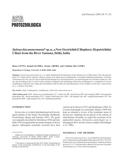
Stylonychia ammermanni* sp. n., a New Oxytrichid (Ciliophora ...
Stylonychia ammermanni* sp. n., a New Oxytrichid (Ciliophora ...
Stylonychia ammermanni* sp. n., a New Oxytrichid (Ciliophora ...

You also want an ePaper? Increase the reach of your titles
YUMPU automatically turns print PDFs into web optimized ePapers that Google loves.
Acta Protozool. (2001) 40: 75 - 82<br />
<strong>Stylonychia</strong> <strong>ammermanni*</strong> <strong>sp</strong>. n., a <strong>New</strong> <strong>Oxytrichid</strong> (<strong>Ciliophora</strong>: Hypotrichida)<br />
Ciliate from the River Yamuna, Delhi, India<br />
Renu GUPTA, Komal KAMRA, Seema ARORA and Gulshan Rai SAPRA<br />
Department of Zoology, University of Delhi, Delhi, India<br />
Summary. <strong>Stylonychia</strong> ammermanni <strong>sp</strong>. n. is widely distributed in the backwaters of the Yamuna river of Delhi region. The cell measures<br />
about 135 x 50µm and the vegetative ciliature consists of an adoral zone of membranelles, two parallel undulating membranes, 18 frontalventral<br />
transverse cirri, one row each of right and left marginal cirri, four dorsal kineties, two dorsomarginal rows and three caudal cirri. The<br />
new <strong>sp</strong>ecies belongs to the S. mytilus-lemnae complex as judged by the morphology and division morphogenesis. A comparison of various<br />
morphometric characters (size, shape and cirral pattern) with the other two described <strong>sp</strong>ecies of the complex reveals that S. ammermanni<br />
<strong>sp</strong>. n. is distinct from them.<br />
Key words: ciliate, morphogenesis, morphometry, <strong>Stylonychia</strong> ammermanni <strong>sp</strong>. n.<br />
Abbreviations used: AZM - adoral zone of membranelles, CC - caudal cirri, DK - dorsal kinety, DM - dorsomarginal, DMP - dorsomarginal<br />
primordium, DP - dorsal primordium, FVT - frontal ventral transverse, LMC - left marginal cirri, MP - marginal primordium, OP - oral<br />
primordium, RMC - right marginal cirri, UM - undulating membrane<br />
INTRODUCTION<br />
<strong>Stylonychia</strong> is a widely distributed and well investigated<br />
member of the family <strong>Oxytrichid</strong>ae (Subfamily<br />
Oxytrichinae; Berger and Foissner 1997). The genus<br />
was originally described by Ehrenberg (1830) and later<br />
Kahl (1935) recognized the taxonomic integrity of eleven<br />
<strong>sp</strong>ecies. Subsequent systematic revisions have been<br />
carried out by Borror (1972) and Hemberger (1982). In<br />
a recent monograph on oxytrichids, Berger (1999) has<br />
done an exhaustive review of the systematic status of<br />
<strong>Stylonychia</strong> validating eleven <strong>sp</strong>ecies in his scheme of<br />
classification. Presently, we report the occurrence of an<br />
undescribed <strong>sp</strong>ecies, <strong>Stylonychia</strong> ammermanni <strong>sp</strong>. n.<br />
belonging to the S. mytilus-lemnae complex (Wirnsberger<br />
et al. 1986).<br />
Address for corre<strong>sp</strong>ondence: Gulshan Rai Sapra, Department of<br />
Zoology, University of Delhi, Delhi-110007, India; E-mail:<br />
duzdel@del2.vsnl.net.in; sapradu@mantraonline.com<br />
* The <strong>sp</strong>ecies is being named after Prof. Dieter Ammermann in<br />
honour of his pioneering contributions to the knowledge of the<br />
biology of <strong>Stylonychia</strong><br />
MATERIALS AND METHODS<br />
<strong>Stylonychia</strong> ammermanni <strong>sp</strong>. n. was isolated from the backwaters<br />
of river Yamuna flowing through the Delhi region (28°34’N, 76°07’E).<br />
Isolated cells were acclimatized to the laboratory conditions and then<br />
grown at 23 ±1°C with axenically cultured Chlorogonium elongatum<br />
(Ammermann et al. 1974) as the food organism.
76 R. Gupta et al.<br />
Morphometric characterization was done on randomly selected<br />
non-dividing cells after staining with the modified protargol method<br />
(Kamra and Sapra 1990). The terminology employed was that of<br />
Wallengren (1900), Borror (1972), Hemberger (1982) and Martin<br />
(1982).<br />
Most of the cells exhibiting better details have orientation with<br />
their ventral sides facing downwards so that the AZM appears on<br />
viewers left.<br />
RESULTS<br />
<strong>Stylonychia</strong> ammermanni <strong>sp</strong>. n. (Fig. 1) feeds voraciously<br />
on the alga Chlorogonium elongatum and<br />
healthy cells are very motile. With its rigid body and<br />
broad anterior and narrow posterior ends it shows<br />
morphological similarities to the S. mytilus-lemnae complex.<br />
However, S. ammermanni <strong>sp</strong>. n. appears comparatively<br />
flattened as it lacks the peristomial bulge. This<br />
feature distinguishes it from the other two related <strong>sp</strong>ecies<br />
viz. S. mytilus and S. lemnae (Fig. 2). On an<br />
average S. ammermanni <strong>sp</strong>. n. measures 135 x 50 µm<br />
with length to width ratio of 2.5:1. The generation time<br />
under laboratory conditions is 11±0.5 hrs. Encystment<br />
and excystment are occasional in the laboratory as well<br />
as in wild cultures. Each cell possesses two macronuclei<br />
and two to four micronuclei. The right and left marginal<br />
cirral rows (RMC and LMC) are straight and posteriorly<br />
well separated. Morphometric characterization of the<br />
<strong>sp</strong>ecies is shown in Table 1.<br />
<strong>Stylonychia</strong> ammermanni <strong>sp</strong>. n. (Fig. 1) has a large<br />
peristome with the adoral zone of membranelles (AZM)<br />
extending slightly more than fifty percent of the body<br />
length. Two parallel undulating membranes (UMs) are<br />
situated on the right wall of the peristome which appear<br />
crossed in stained preparations due to flattening. Of the<br />
18 FVT cirri, the eight frontals (F 1-8<br />
) are di<strong>sp</strong>osed<br />
characteristically. The posterior two pairs (F 5, 6<br />
and F 7, 8<br />
)<br />
are separated from the anterior hypertrophied frontals<br />
(F 1-4<br />
) and also from each other by distinct gaps. Posterior<br />
frontals (F 5, 6<br />
) are in linear orientation and parallel to<br />
the RMC. The posteriormost pair (F 7, 8<br />
) is separated<br />
apart from (F 5, 6<br />
) and is situated near the cytostome. The<br />
transverse cirri (T 1-5<br />
) are placed in two groups of three<br />
and two cirri. Among the five ventrals two are close to<br />
the oral region and are termed the left (V 1<br />
) and right<br />
(V 2<br />
) postoral ventral cirri, while the other three (V 3-5<br />
)<br />
are more posteriad. Marginal cirri are evenly <strong>sp</strong>aced in<br />
one row each near the right and left margins of the cell.<br />
These right and left marginal cirral rows (RMC and<br />
LMC) are widely separated posteriorly.<br />
Dorsally, S. ammermanni <strong>sp</strong>. n. shows the presence<br />
of six dorsal rows (DK 1-4<br />
and DM 1, 2<br />
) and three caudal<br />
cirri. Three of the dorsal kineties (DK 1-3<br />
) are curved<br />
apically. The first dorsomarginal (DM 1<br />
) terminates at the<br />
posterior quarter region of the cell and the DM 2<br />
before<br />
the mid region. There are three caudal cirri (CC 1-3<br />
), one<br />
each at the end of dorsal kineties DK 1, 2, 4<br />
and are equally<br />
<strong>sp</strong>aced (Fig.1). The right caudal cirrus (CC 3<br />
) is positioned<br />
between, terminal 2-3 cirri of the RMC row.<br />
The morphogenetic pattern during division (Figs. 3-6)<br />
is essentially similar to that in S. lemnae and S. mytilus<br />
(Wirnsberger et al. 1986).<br />
A small group of basal bodies evolves very close to<br />
the uppermost transverse cirrus (T 1<br />
). The kinetosomes<br />
proliferate linearly to form an anarchic field, the oral<br />
primordium (OP). A new AZM for the opisthe (posterior<br />
daughter cell) is formed from the OP. The parental AZM<br />
is retained for the proter (anterior daughter cell).<br />
The FVT cirri for the two daughter cells develop from<br />
two sets of six cirral primordia (I-VI) each. In the proter,<br />
the parental UMs function as primordium I. The other<br />
primordia originate from the OP (primordium II), F 8<br />
(III),<br />
F 7<br />
(IV) and right postoral ventral cirrus (V and VI). In<br />
the opisthe, three primordia (I-III) originate in conjunction<br />
with the OP and the other three (IV-VI) from the<br />
right postoral ventral cirrus.<br />
The origin of the two sets of six primordia involves<br />
three parental cirri (2 frontals and 1 ventral) and they<br />
differentiate into 18 FVT cirri by the cleavage pattern 1,<br />
3, 3, 3, 4, 4.<br />
The marginal and dorsal ciliature are formed in a<br />
manner similar to that described for other <strong>Stylonychia</strong><br />
<strong>sp</strong>ecies (Hemberger 1982; Wirnsberger et al. 1985,<br />
1986; Berger and Foissner 1997).<br />
In the proter, marginal primordia are formed at the<br />
anterior ends of the marginal rows by reorganization of<br />
2-3 parental marginal cirri. In the opisthe, marginal<br />
primordia originate similarly slightly below the pro<strong>sp</strong>ective<br />
division furrow.<br />
Dorsal kineties DK 1-3<br />
are generated by intrakinetal<br />
proliferation of kinetosomes in parental kineties<br />
DK 1-3<br />
. The kinety DK 4<br />
originates by unequal<br />
fragmentation of the third dorsal primordium (DP 3<br />
).<br />
Dorsomarginals (DM 1, 2<br />
) are generated at the anterior<br />
end of the new right marginal rows which shift to the
<strong>Stylonychia</strong> ammermanni <strong>sp</strong>. n., from India 77<br />
Fig. 1. Line diagrams of protargol impregnated cells and photomicrograph of S. ammermanni <strong>sp</strong>. n. A - nuclear apparatus and ventral ciliature.<br />
B - dorsal ciliature. C - live cell. AZM - adoral zone of membranelles, CC 1-3<br />
- caudal cirri, DK - dorsal kineties, DM - dorsomarginal rows,<br />
1-4 1, 2<br />
F 1-8<br />
-frontal cirri, LMC - left marginal cirri, Ma - macronucleus, Mi - micronucleus, RMC - right marginal cirri, T 1-5<br />
- transverse cirri,<br />
UM - undulating membranes, V 1-5<br />
- ventral cirri. Scale bars - A, B - 25 µm, C - 30 µm<br />
Fig. 2. Line diagrams of protargol impregnated cells of: A - S. ammermanni <strong>sp</strong>. n, B - S. lemnae and C - S. mytilus showing the ventral ciliature<br />
(Figures B and C have been modified from Wirnsberger et al. 1986). The differences in the position of the posterior frontal cirri (F 5-8<br />
). Scale<br />
bar - 47 µm
78 R. Gupta et al.<br />
Table 1. Morphometric characterization of <strong>Stylonychia</strong> ammermanni <strong>sp</strong>. n. (Data from protargol impregnated cells. All measurements are<br />
in µm)<br />
Character Mean Minimum Maximum SD CV n<br />
Body length 133.98 125.65 146.48 5.00 3.73 25<br />
Body width 52.12 47.25 57.75 3.01 5.78 25<br />
Body length/Body width 2.58 2.33 2.97 0.15 5.94 25<br />
Macronuclear, number 2.00 2.00 2.00 0.00 0.00 25<br />
Macronuclear, length 25.27 24.33 26.08 0.61 2.41 12<br />
Macronuclear, width 10.33 9.98 10.68 0.53 5.72 12<br />
Micronuclear, number 2.00 2 4 - - 25<br />
Micronuclear diameter 2.62 2.45 2.97 0.20 7.91 12<br />
Adoral membranelles, number 51.43 46 55 2.24 4.35 30<br />
Adoral length 70.80 65.10 80.50 3.95 5.58 25<br />
Adoral length/Body length 0.53 0.48 0.61 0.03 5.05 25<br />
Frontal cirri, number 8.00 8 8 0.00 0.00 25<br />
Ventral cirri, number 5.00 5 5 0.00 0.00 25<br />
Transverse cirri, number 5.00 5 5 0.00 0.00 25<br />
Left marginal cirri, number 17.17 13 18 1.12 6.51 30<br />
Right marginal cirri, number 24.20 22 27 1.45 5.98 30<br />
Dorsal kineties (DK), number 4.00 4 4 0.00 0.00 25<br />
Dorsomarginal (DM), number 2.00 2 2 0.00 0.00 25<br />
Dorsal bristles, number in DK 1<br />
52.67 44 58 4.19 7.95 12<br />
DK 2<br />
40.58 36 45 3.20 7.89 12<br />
DK 3<br />
28.83 24 33 2.66 9.22 12<br />
DK 4<br />
20.83 19 25 1.99 9.56 12<br />
DM 1<br />
29.33 26 32 2.10 7.17 12<br />
DM 2<br />
17.00 15 20 1.86 10.93 12<br />
Dorsal bristles, total number 189.25 176 200 8.01 4.23 12<br />
Caudal cirri, number 3.00 3 3 0.00 0.00 25<br />
Abbreviations: CV - coefficient of variance, SD - standard deviation, n - number<br />
Table 2. Morphometric comparison of S. ammermanni <strong>sp</strong>. n. with S. mytilus and S. lemnae. Measurements are in µm<br />
Characters S. ammermanni <strong>sp</strong>. n. S. mytilus S. lemnae<br />
(Present investigation) (German population, (German population,<br />
Wirnsberger et al. 1986) Wirnsberger et al. 1986)<br />
Appearance No bulge in the Bulge in the Bulge in the postperistomial<br />
region peristomial region peristomial region<br />
Body length 130 227 188<br />
Body width 50 110 82<br />
Macronuclear, size 25 x 10 37 x 11 27 x 11<br />
Micronuclear, size 2.6 3.6 4<br />
Adoral length ≥ 50% of body length ≥ 50% of body length ≤ 50% of body length<br />
AM, number 50 71 63<br />
Posterior frontal cirri (F 5-8<br />
) Arranged in two pairs; Arranged in a ‘L’ Arranged in a ‘J’<br />
posterior pair close shaped manner shaped manner<br />
to cytostome<br />
Left marginal cirri (LMC) Straight Anterior 2-3 cirri Anterior 2-3 cirri<br />
curved inward<br />
curved inward<br />
LMC, number 17 27 25<br />
RMC, number 24 38 37<br />
Dorsal kineties Bent apically Curved apically Bent apically<br />
Conjugation Occasional Frequent Frequent<br />
Encystment Occasional Frequent Frequent<br />
Abbreviations: AM - adoral membranelles, RMC - right marginal cirri
<strong>Stylonychia</strong> ammermanni <strong>sp</strong>. n., from India 79<br />
Fig. 3. Line diagrams of protargol impregnated cells of S. ammermanni <strong>sp</strong>. n. showing division morphogenesis on the ventral surface. A - origin<br />
of oral primordium (OP) by proliferation of kinetosomes near transverse cirrus T 1<br />
. B - expansion of kinetosomes in OP to form an anarchic<br />
field. Anteriad movement of kinetosomes from OP to form primordium II of proter. F 8<br />
disaggregates to form primordium III of proter. Anterior<br />
movement of kinetosomes of right post oral ventral (V 2<br />
) to form primordia V and VI of proter. C - differentiation of membranelles (AZM)<br />
starts at the right anterior border within the OP. Origin of primordia: I-III of opisthe from OP; IV-VI of opisthe from V 2<br />
; IV of proter from F 7<br />
;<br />
VandVI of proter from V 2<br />
. D - cirri differentiate in 1, 3, 3, 3, 4, 4 pattern in FVT primordia. (The old cirri are depicted only by contour, whereas<br />
the new ones are filled in). Formation of marginal primordia (MP) and dorsomarginal primordia (DMP). Scale bar - 20 µm<br />
Fig. 4. Line diagrams of protargol impregnated cells of S. ammermanni <strong>sp</strong>. n. showing division morphogenesis on the dorsal surface.<br />
A - formation of dorsal primordia (DP 1-3<br />
) within the parental kineties. B - unequal <strong>sp</strong>lit in DP 3<br />
to form DP 4<br />
. C - formation of 3 caudal cirri<br />
(CC 1-3<br />
) one each at the ends of DK 1, 2, 4<br />
. <strong>New</strong>ly formed dorsomarginal (DM 1-2<br />
) rows shifted from the ventral surface. Scale bar - 20 µm
80 R. Gupta et al.<br />
Fig. 5. Photomicrographs of protargol impregnated cells of S. ammermanni <strong>sp</strong>. n. showing division morphogenesis on the ventral<br />
surface. A - linear proliferation of oral primordium (arrow) close to uppermost transverse cirrus T 1<br />
(double arrow). B - anteriad movement of<br />
kinetosomes (arrow) from oral primordium and disaggregation of right postoral ventral cirrus V 2<br />
(double arrow). C - anteriad movement of<br />
kinetosomes (arrow) from V 2<br />
. Origin of primordia: II of proter from OP (double arrow); III of proter from F 8<br />
(arrow head) D - origin of<br />
primordia: IV of proter from F 7<br />
(double arrow); V and VI of proter from V 2<br />
(double arrow); I-III of opisthe from OP (arrow head); IV-VI of<br />
opisthe from V 2<br />
. E - differentiation pattern of 1, 3, 3, 3, 4, 4. Within row formation of left (arrows) and right (arrow heads) marginal primordia.<br />
Dorsomarginal primordia (double arrows) formed near right marginal primordia. F - cell in cytokinesis. Scale bar - 35 µm<br />
dorsal surface during cytokinesis. Caudal cirri are formed<br />
by proliferation of kinetosomes at the posterior ends of<br />
the new DK 1, 2, 4<br />
in each daughter cell.<br />
Type <strong>sp</strong>ecies are deposited at the Biology Centre of<br />
the Upper Austrian Museum no. 2000/155 in the collection<br />
of “Evertebrate varia.”<br />
DISCUSSION<br />
Diagnosis of <strong>Stylonychia</strong> ammermanni <strong>sp</strong>. n.<br />
Member of the <strong>Stylonychia</strong> mytilus-lemnae complex<br />
lacking peristomial bulge. Size in protargol preparations<br />
about 135 x 50 µm with parallel borders and rounded
<strong>Stylonychia</strong> ammermanni <strong>sp</strong>. n., from India 81<br />
Fig. 6. Photomicrographs of protargol impregnated cells of S. ammermanni <strong>sp</strong>. n. showing division morphogenesis on the dorsal surface.<br />
A - within row formation of dorsal primordia (arrows). B - unequal <strong>sp</strong>lit of DP 3<br />
(arrows). C - proliferation of caudal cirri (arrows) at the ends<br />
of dorsal kineties (DK 1, 2, 4<br />
; arrowheads). Scale bar - 30 µm<br />
ends. Nuclear apparatus consisting of 2 ellipsoidal macronuclear<br />
nodes and 2-4 <strong>sp</strong>herical micronuclei. Posterior<br />
frontals (F 5, 6<br />
) in linear orientation and almost parallel<br />
to the RMC. Posteriormost pair of frontals (F 7, 8<br />
) separated<br />
from F 5, 6<br />
by a distinct gap and is near to the<br />
cytostome. Adoral zone 50-60 % of the body length, with<br />
about 50 adoral membranelles. Left and right marginal<br />
cirral rows with around 17 and 24 cirri re<strong>sp</strong>ectively. Six<br />
dorsal kineties with about 190 dorsal bristles.<br />
Wirnsberger et al. (1986) divided the members of the<br />
genus <strong>Stylonychia</strong> into two complexes viz. S. mytiluslemnae<br />
complex and S. pustulata-vorax complex on<br />
the basis of certain morphological and morphogenetic<br />
criteria. Application of these criteria as detailed below<br />
shows that S. ammermanni <strong>sp</strong>. n. belongs to the<br />
S. mytilus-lemnae complex. The characters shared by<br />
S. ammermanni <strong>sp</strong>. n. with this complex are as follows:<br />
(1) The cell body is rigid with a broad and large<br />
peristome.<br />
(2) The marginal cirral rows are straight and are<br />
posteriorly open.<br />
(3) Dorsal kineties (DK 1-3<br />
) are apically bent.<br />
(4) The dorsomarginal rows are long (DM 1<br />
being a<br />
complete row).<br />
(5) The right caudal cirrus is present between, terminal<br />
2-3 cirri of the RMC row.<br />
(6) Formation of new FVT ciliature for the two<br />
daughter cells involves three parental cirri (2 frontals and<br />
1 ventral).<br />
(7) Second FVT primordium of the proter originates<br />
from the OP. Fifth and sixth orginate from V 2<br />
.<br />
(8) Fourth FVT primordium of the opisthe originates<br />
from the right postoral ventral cirrus (V 2<br />
).<br />
S. ammermanni <strong>sp</strong>. n. is clearly distinguishable from<br />
S. mytilus and S. lemnae by its size, shape, cirral pattern,<br />
absence of peristomial bulge (Fig. 2), and micronuclear<br />
size (Table 2). It is reproductively isolated from these<br />
two <strong>sp</strong>ecies as no conjugation could be induced among<br />
them in the laboratory. Comparative features of<br />
S. ammermanni <strong>sp</strong>. n. with the closely related members<br />
of the S. mytilus-lemnae complex are shown in Table 2.<br />
Studies on the isozyme pattern, RAPD characterization<br />
and internal transcribed sequence analysis of rDNA<br />
(Jacob 1996; the <strong>sp</strong>ecies is mentioned as S. indica) also<br />
suggests a clear separation of S. ammermanni <strong>sp</strong>. n.<br />
from S. mytilus and S. lemnae.<br />
<strong>Stylonychia</strong> ammermanni <strong>sp</strong>. n. shows certain morphometric<br />
similarities to S. putrina reported from
82 R. Gupta et al.<br />
Yaounde, Africa (Dragesco and Njiné 1971, Dragesco<br />
and Dragesco-Kernéis 1986). However, in the absence<br />
of any available details of dorsal ciliature and the morphogenetic<br />
pattern, it is not possible to ascertain the<br />
synonymity of the two <strong>sp</strong>ecies.<br />
Acknowledgements. The work was supported by the senior<br />
research fellowship (extended) to RG from the CSIR, Delhi, India.<br />
REFERENCES<br />
Ammermann D., Steinbrück G., Berger L.V., Henning W. (1974) The<br />
development of the macronucleus in the ciliated protozoan<br />
<strong>Stylonychia</strong> mytilus. Chromosoma (Berlin) 45: 401-429<br />
Berger H. (1999) Monograph of the <strong>Oxytrichid</strong>ae (<strong>Ciliophora</strong>,<br />
Hypotrichia). Monographie Biologicae, Kluwer Academic Publishers,<br />
Dordrecht, Boston, London 78: 501-605<br />
Berger H., Foissner W. (1997) Cladistic relationships and generic<br />
characterization of oxytrichid hypotrichs (Protozoa: <strong>Ciliophora</strong>).<br />
Arch Protistenkd. 148: 125-155<br />
Borror A.C. (1972) Revision of the order hypotrichida (<strong>Ciliophora</strong>,<br />
Protozoa). J Protozool. 19(1): 1-23<br />
Dragesco J., Dragesco-Kernéis A. (1986) Cilies libres de I’Afrique<br />
intertropicale faune tropicale. Editions de l’ORSTROM, Paris<br />
26: 487-490<br />
Dragesco J., Njiné T. (1971) Compléments á la connaissance des ciliés<br />
libres du Cameroun. Ann Fac Sci Cameroun 7-8: 97-140<br />
Ehrenberg C.G. (1830) Beiträge zur Kenntnib der Organisation der<br />
Infusorien und ihrer geographischen Verberitung besonders in<br />
Sibirien. Abh Akad.Wiss. Berlin, 1-88<br />
Hemberger H. (1982) Revision der ordnung Hypotrichida Stein<br />
(<strong>Ciliophora</strong>, Protozoa) an hand von protargolpräparaten und<br />
morphogenesedarstellungen. Dissertation Erlang Doktorgr Naturw<br />
Fak Rhein Friedrich - Wilhelms Univ. Bonn, 227-243<br />
Jacob N.K. (1996) Phylogenetic studies on the internal transcribed<br />
<strong>sp</strong>acers of ribosomal RNA genes in hypotrichous ciliates: Congruence<br />
of morphogenetic, biochemical and molecular markers.<br />
Dissertation Zur Erlangung des Grades eines Naturwissen Biologie<br />
der Eberhard - Karls Universität Tübingen<br />
Kahl A. (1935) Urtiere oder Protozoa 1: Wimpertiere oder Ciliata<br />
(Infusoria). Tierwelt Dtl. 30: 564-568<br />
Kamra K., Sapra G.R. (1990) Partial retention of parental ciliature<br />
during morphogenesis of ciliate Coniculostomum monilata<br />
(Dragesco & Njiné, 1971) Njiné, 1978 (<strong>Oxytrichid</strong>ae,<br />
Hypotrichida). Europ J Protistol. 25: 264-278<br />
Martin J. (1982) Évolution des patrons morphogénétiques et<br />
phylogenése dans le sous-ordre des Sporadotrichina (<strong>Ciliophora</strong>,<br />
Hypotrichida). Protistologica 18: 431-447<br />
Wallengren H. (1900) Vergleichende morphologie der hypotrichen<br />
Infusorien. Bih. Svensk. Vetensk. Akad. Handl. 26: 1-30<br />
Wirnsberger E., Foissner W., Adam H. (1985) Morphological, biometric<br />
and morphogenetic comparison of two closely related<br />
<strong>sp</strong>ecies, <strong>Stylonychia</strong> vorax and S. pustulata (<strong>Ciliophora</strong>:<br />
<strong>Oxytrichid</strong>ae). J Protozool. 32: 261-268<br />
Wirnsberger E., Foissner W., Adam H. (1986) Biometric and morphogenetic<br />
comparison of the sibling <strong>sp</strong>ecies <strong>Stylonychia</strong> mytilus and<br />
S. lemnae, including a phylogenetic system for the oxytrichids<br />
(<strong>Ciliophora</strong>: Hypotrichida). Arch Protistenkd. 132: 167-185<br />
Received on 5th August, 2000; accepted on 18th November, 2000