Stylonychia ammermanni* sp. n., a New Oxytrichid (Ciliophora ...
Stylonychia ammermanni* sp. n., a New Oxytrichid (Ciliophora ...
Stylonychia ammermanni* sp. n., a New Oxytrichid (Ciliophora ...

You also want an ePaper? Increase the reach of your titles
YUMPU automatically turns print PDFs into web optimized ePapers that Google loves.
76 R. Gupta et al.<br />
Morphometric characterization was done on randomly selected<br />
non-dividing cells after staining with the modified protargol method<br />
(Kamra and Sapra 1990). The terminology employed was that of<br />
Wallengren (1900), Borror (1972), Hemberger (1982) and Martin<br />
(1982).<br />
Most of the cells exhibiting better details have orientation with<br />
their ventral sides facing downwards so that the AZM appears on<br />
viewers left.<br />
RESULTS<br />
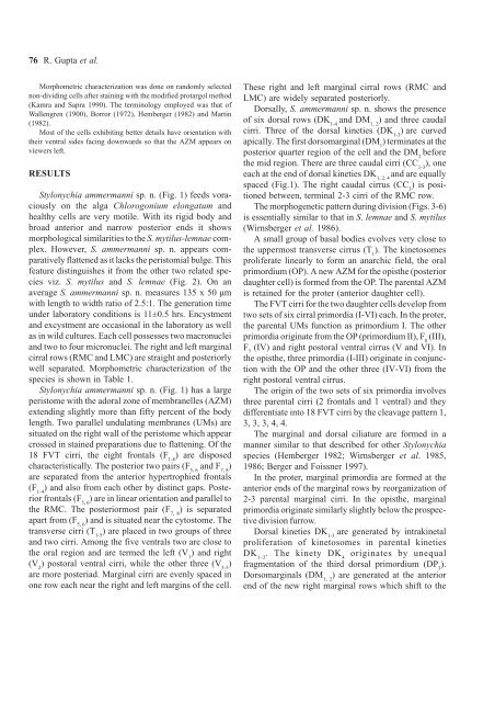
<strong>Stylonychia</strong> ammermanni <strong>sp</strong>. n. (Fig. 1) feeds voraciously<br />
on the alga Chlorogonium elongatum and<br />
healthy cells are very motile. With its rigid body and<br />
broad anterior and narrow posterior ends it shows<br />
morphological similarities to the S. mytilus-lemnae complex.<br />
However, S. ammermanni <strong>sp</strong>. n. appears comparatively<br />
flattened as it lacks the peristomial bulge. This<br />
feature distinguishes it from the other two related <strong>sp</strong>ecies<br />
viz. S. mytilus and S. lemnae (Fig. 2). On an<br />
average S. ammermanni <strong>sp</strong>. n. measures 135 x 50 µm<br />
with length to width ratio of 2.5:1. The generation time<br />
under laboratory conditions is 11±0.5 hrs. Encystment<br />
and excystment are occasional in the laboratory as well<br />
as in wild cultures. Each cell possesses two macronuclei<br />
and two to four micronuclei. The right and left marginal<br />
cirral rows (RMC and LMC) are straight and posteriorly<br />
well separated. Morphometric characterization of the<br />
<strong>sp</strong>ecies is shown in Table 1.<br />
<strong>Stylonychia</strong> ammermanni <strong>sp</strong>. n. (Fig. 1) has a large<br />
peristome with the adoral zone of membranelles (AZM)<br />
extending slightly more than fifty percent of the body<br />
length. Two parallel undulating membranes (UMs) are<br />
situated on the right wall of the peristome which appear<br />
crossed in stained preparations due to flattening. Of the<br />
18 FVT cirri, the eight frontals (F 1-8<br />
) are di<strong>sp</strong>osed<br />
characteristically. The posterior two pairs (F 5, 6<br />
and F 7, 8<br />
)<br />
are separated from the anterior hypertrophied frontals<br />
(F 1-4<br />
) and also from each other by distinct gaps. Posterior<br />
frontals (F 5, 6<br />
) are in linear orientation and parallel to<br />
the RMC. The posteriormost pair (F 7, 8<br />
) is separated<br />
apart from (F 5, 6<br />
) and is situated near the cytostome. The<br />
transverse cirri (T 1-5<br />
) are placed in two groups of three<br />
and two cirri. Among the five ventrals two are close to<br />
the oral region and are termed the left (V 1<br />
) and right<br />
(V 2<br />
) postoral ventral cirri, while the other three (V 3-5<br />
)<br />
are more posteriad. Marginal cirri are evenly <strong>sp</strong>aced in<br />
one row each near the right and left margins of the cell.<br />
These right and left marginal cirral rows (RMC and<br />
LMC) are widely separated posteriorly.<br />
Dorsally, S. ammermanni <strong>sp</strong>. n. shows the presence<br />
of six dorsal rows (DK 1-4<br />
and DM 1, 2<br />
) and three caudal<br />
cirri. Three of the dorsal kineties (DK 1-3<br />
) are curved<br />
apically. The first dorsomarginal (DM 1<br />
) terminates at the<br />
posterior quarter region of the cell and the DM 2<br />
before<br />
the mid region. There are three caudal cirri (CC 1-3<br />
), one<br />
each at the end of dorsal kineties DK 1, 2, 4<br />
and are equally<br />
<strong>sp</strong>aced (Fig.1). The right caudal cirrus (CC 3<br />
) is positioned<br />
between, terminal 2-3 cirri of the RMC row.<br />
The morphogenetic pattern during division (Figs. 3-6)<br />
is essentially similar to that in S. lemnae and S. mytilus<br />
(Wirnsberger et al. 1986).<br />
A small group of basal bodies evolves very close to<br />
the uppermost transverse cirrus (T 1<br />
). The kinetosomes<br />
proliferate linearly to form an anarchic field, the oral<br />
primordium (OP). A new AZM for the opisthe (posterior<br />
daughter cell) is formed from the OP. The parental AZM<br />
is retained for the proter (anterior daughter cell).<br />
The FVT cirri for the two daughter cells develop from<br />
two sets of six cirral primordia (I-VI) each. In the proter,<br />
the parental UMs function as primordium I. The other<br />
primordia originate from the OP (primordium II), F 8<br />
(III),<br />
F 7<br />
(IV) and right postoral ventral cirrus (V and VI). In<br />
the opisthe, three primordia (I-III) originate in conjunction<br />
with the OP and the other three (IV-VI) from the<br />
right postoral ventral cirrus.<br />
The origin of the two sets of six primordia involves<br />
three parental cirri (2 frontals and 1 ventral) and they<br />
differentiate into 18 FVT cirri by the cleavage pattern 1,<br />
3, 3, 3, 4, 4.<br />
The marginal and dorsal ciliature are formed in a<br />
manner similar to that described for other <strong>Stylonychia</strong><br />
<strong>sp</strong>ecies (Hemberger 1982; Wirnsberger et al. 1985,<br />
1986; Berger and Foissner 1997).<br />
In the proter, marginal primordia are formed at the<br />
anterior ends of the marginal rows by reorganization of<br />
2-3 parental marginal cirri. In the opisthe, marginal<br />
primordia originate similarly slightly below the pro<strong>sp</strong>ective<br />
division furrow.<br />
Dorsal kineties DK 1-3<br />
are generated by intrakinetal<br />
proliferation of kinetosomes in parental kineties<br />
DK 1-3<br />
. The kinety DK 4<br />
originates by unequal<br />
fragmentation of the third dorsal primordium (DP 3<br />
).<br />
Dorsomarginals (DM 1, 2<br />
) are generated at the anterior<br />
end of the new right marginal rows which shift to the