treatment of perforated infectious corneal ulcers - Journal of IMAB
treatment of perforated infectious corneal ulcers - Journal of IMAB
treatment of perforated infectious corneal ulcers - Journal of IMAB

You also want an ePaper? Increase the reach of your titles
YUMPU automatically turns print PDFs into web optimized ePapers that Google loves.
Intraocular pressure greater than 21 mm Hg on two<br />
separate occasions was taken as secondary glaucoma.<br />
All cases were photo documented.<br />
Surgical procedures<br />
All PK had performed under general anesthesia.<br />
A suitable donor cornea was received from the<br />
International Eye Bank <strong>of</strong> S<strong>of</strong>ia.<br />
Trephination was done per endothelium. The size <strong>of</strong><br />
the graft should be the smallest capable <strong>of</strong> incorporating<br />
the perforation site and any infected or ulcerated border.<br />
Donor button was oversized by 0.5 mm in all cases and<br />
secured with 16 to 20 interrupted 10-0 nylon sutures.<br />
Anterior and posterior synechiolysis,<br />
membranectomy (all 5 cases), and pupilloplasty (case 2 and<br />
4) were performed. The anterior chamber should be irrigated<br />
to remove the necrotic or inflammatory debris. Cataract<br />
removal with intraocular lens implantation was performed in<br />
one eye (case 3) despite the risk <strong>of</strong> expulsive hemorrhage<br />
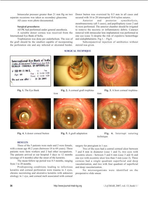
and endophthalmitis. Fig.1. – Fig.6.<br />
Subconjunctival injection <strong>of</strong> antibiotics without<br />
steroid was given.<br />
SURGICAL TECHNIQUE<br />
Fig. 1. The Eye Bank<br />
Fig. 2. A <strong>corneal</strong> graft trephination<br />
Fig. 3. A host <strong>corneal</strong> trephination<br />
Fig. 4. A donor <strong>corneal</strong> button Fig. 5. A graft adaptation Fig. 6. Interrupt suturing<br />
technique<br />
RESULTS<br />
Three <strong>of</strong> the 5 patients were male and 2 were female,<br />
with a mean age 48.2 years (between 18 to 69 years). Three<br />
patients were farm workers and 2 had other occupations.<br />
The patients arrived at our hospital 5 days to 12 months<br />
(average <strong>of</strong> 4 months) after the onset <strong>of</strong> the keratitis.<br />
The mean follow-up period was 8.5 months, ranging<br />
from 1 to 20 months.<br />
Predisposing conditions leading to <strong>infectious</strong><br />
keratitis and <strong>corneal</strong> perforation were trauma in 3 eyes,<br />
chronic necrotizing and ulcerative keratitis with unknown<br />
etiology in 1 eye, and <strong>corneal</strong> melt associated with <strong>corneal</strong><br />
surgery for pterygium in 1 eye.<br />
Two <strong>of</strong> the eyes had a central <strong>corneal</strong> ulcer between<br />
7 and 8 mm in diameter (case 1 and 5), two eyes with<br />
eccentric <strong>ulcers</strong> - between 5 and 6 mm (case 3 and 4) and<br />
one eye with eccentric ulcer less than 5 mm (case 2). Three<br />
corneas had a single quadrant superficial and deep<br />
vascularization, and two with four quadrant <strong>of</strong> superficial<br />
and deep vascularization.<br />
No microorganisms were identified on the<br />
preoperative slide smear.<br />
36 http://www.journal-imab-bg.org / J <strong>of</strong> <strong>IMAB</strong>, 2007, vol. 13, book 1 /