treatment of perforated infectious corneal ulcers - Journal of IMAB
treatment of perforated infectious corneal ulcers - Journal of IMAB
treatment of perforated infectious corneal ulcers - Journal of IMAB
Create successful ePaper yourself
Turn your PDF publications into a flip-book with our unique Google optimized e-Paper software.
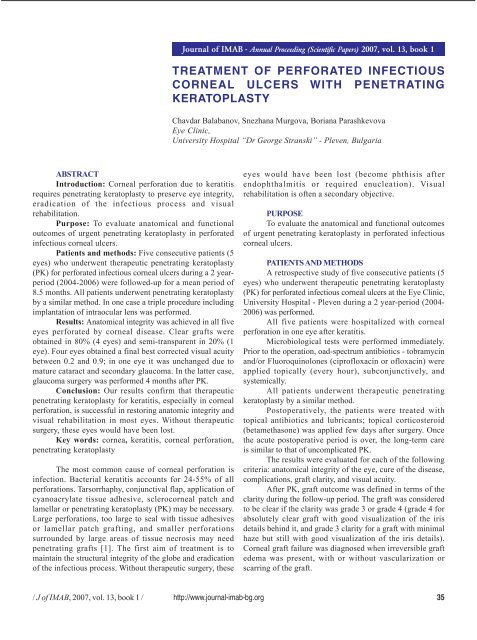
<strong>Journal</strong> <strong>of</strong> <strong>IMAB</strong> - Annual Proceeding (Scientific Papers) 2007, vol. 13, book 1<br />
TREATMENT OF PERFORATED INFECTIOUS<br />
CORNEAL ULCERS WITH PENETRATING<br />
KERATOPLASTY<br />
Chavdar Balabanov, Snezhana Murgova, Boriana Parashkevova<br />
Eye Clinic,<br />
University Hospital “Dr George Stranski” - Pleven, Bulgaria<br />
ABSTRACT<br />
Introduction: Corneal perforation due to keratitis<br />
requires penetrating keratoplasty to preserve eye integrity,<br />
eradication <strong>of</strong> the <strong>infectious</strong> process and visual<br />
rehabilitation.<br />
Purpose: To evaluate anatomical and functional<br />
outcomes <strong>of</strong> urgent penetrating keratoplasty in <strong>perforated</strong><br />
<strong>infectious</strong> <strong>corneal</strong> <strong>ulcers</strong>.<br />
Patients and methods: Five consecutive patients (5<br />
eyes) who underwent therapeutic penetrating keratoplasty<br />
(PK) for <strong>perforated</strong> <strong>infectious</strong> <strong>corneal</strong> <strong>ulcers</strong> during a 2 yearperiod<br />
(2004-2006) were followed-up for a mean period <strong>of</strong><br />
8.5 months. All patients underwent penetrating keratoplasty<br />
by a similar method. In one case a triple procedure including<br />
implantation <strong>of</strong> intraocular lens was performed.<br />
Results: Anatomical integrity was achieved in all five<br />
eyes <strong>perforated</strong> by <strong>corneal</strong> disease. Clear grafts were<br />
obtained in 80% (4 eyes) and semi-transparent in 20% (1<br />
eye). Four eyes obtained a final best corrected visual acuity<br />
between 0.2 and 0.9; in one eye it was unchanged due to<br />
mature cataract and secondary glaucoma. In the latter case,<br />
glaucoma surgery was performed 4 months after PK.<br />
Conclusion: Our results confirm that therapeutic<br />
penetrating keratoplasty for keratitis, especially in <strong>corneal</strong><br />
perforation, is successful in restoring anatomic integrity and<br />
visual rehabilitation in most eyes. Without therapeutic<br />
surgery, these eyes would have been lost.<br />
Key words: cornea, keratitis, <strong>corneal</strong> perforation,<br />
penetrating keratoplasty<br />
The most common cause <strong>of</strong> <strong>corneal</strong> perforation is<br />
infection. Bacterial keratitis accounts for 24-55% <strong>of</strong> all<br />
perforations. Tarsorrhaphy, conjunctival flap, application <strong>of</strong><br />
cyanoacrylate tissue adhesive, sclero<strong>corneal</strong> patch and<br />
lamellar or penetrating keratoplasty (PK) may be necessary.<br />
Large perforations, too large to seal with tissue adhesives<br />
or lamellar patch grafting, and smaller perforations<br />
surrounded by large areas <strong>of</strong> tissue necrosis may need<br />
penetrating grafts [1]. The first aim <strong>of</strong> <strong>treatment</strong> is to<br />
maintain the structural integrity <strong>of</strong> the globe and eradication<br />
<strong>of</strong> the <strong>infectious</strong> process. Without therapeutic surgery, these<br />
eyes would have been lost (become phthisis after<br />
endophthalmitis or required enucleation). Visual<br />
rehabilitation is <strong>of</strong>ten a secondary objective.<br />
PURPOSE<br />
To evaluate the anatomical and functional outcomes<br />
<strong>of</strong> urgent penetrating keratoplasty in <strong>perforated</strong> <strong>infectious</strong><br />
<strong>corneal</strong> <strong>ulcers</strong>.<br />
PATIENTS AND METHODS<br />
A retrospective study <strong>of</strong> five consecutive patients (5<br />
eyes) who underwent therapeutic penetrating keratoplasty<br />
(PK) for <strong>perforated</strong> <strong>infectious</strong> <strong>corneal</strong> <strong>ulcers</strong> at the Eye Clinic,<br />
University Hospital - Pleven during a 2 year-period (2004-<br />
2006) was performed.<br />
All five patients were hospitalized with <strong>corneal</strong><br />
perforation in one eye after keratitis.<br />
Microbiological tests were performed immediately.<br />
Prior to the operation, oad-spectrum antibiotics - tobramycin<br />
and/or Fluoroquinolones (cipr<strong>of</strong>loxacin or <strong>of</strong>loxacin) were<br />
applied topically (every hour), subconjunctively, and<br />
systemically.<br />
All patients underwent therapeutic penetrating<br />
keratoplasty by a similar method.<br />
Postoperatively, the patients were treated with<br />
topical antibiotics and lubricants; topical corticosteroid<br />
(betamethasone) was applied few days after surgery. Once<br />
the acute postoperative period is over, the long-term care<br />
is similar to that <strong>of</strong> uncomplicated PK.<br />
The results were evaluated for each <strong>of</strong> the following<br />
criteria: anatomical integrity <strong>of</strong> the eye, cure <strong>of</strong> the disease,<br />
complications, graft clarity, and visual acuity.<br />
After PK, graft outcome was defined in terms <strong>of</strong> the<br />
clarity during the follow-up period. The graft was considered<br />
to be clear if the clarity was grade 3 or grade 4 (grade 4 for<br />
absolutely clear graft with good visualization <strong>of</strong> the iris<br />
details behind it, and grade 3 clarity for a graft with minimal<br />
haze but still with good visualization <strong>of</strong> the iris details).<br />
Corneal graft failure was diagnosed when irreversible graft<br />
edema was present, with or without vascularization or<br />
scarring <strong>of</strong> the graft.<br />
/ J <strong>of</strong> <strong>IMAB</strong>, 2007, vol. 13, book 1 / http://www.journal-imab-bg.org 35
Intraocular pressure greater than 21 mm Hg on two<br />
separate occasions was taken as secondary glaucoma.<br />
All cases were photo documented.<br />
Surgical procedures<br />
All PK had performed under general anesthesia.<br />
A suitable donor cornea was received from the<br />
International Eye Bank <strong>of</strong> S<strong>of</strong>ia.<br />
Trephination was done per endothelium. The size <strong>of</strong><br />
the graft should be the smallest capable <strong>of</strong> incorporating<br />
the perforation site and any infected or ulcerated border.<br />
Donor button was oversized by 0.5 mm in all cases and<br />
secured with 16 to 20 interrupted 10-0 nylon sutures.<br />
Anterior and posterior synechiolysis,<br />
membranectomy (all 5 cases), and pupilloplasty (case 2 and<br />
4) were performed. The anterior chamber should be irrigated<br />
to remove the necrotic or inflammatory debris. Cataract<br />
removal with intraocular lens implantation was performed in<br />
one eye (case 3) despite the risk <strong>of</strong> expulsive hemorrhage<br />
and endophthalmitis. Fig.1. – Fig.6.<br />
Subconjunctival injection <strong>of</strong> antibiotics without<br />
steroid was given.<br />
SURGICAL TECHNIQUE<br />
Fig. 1. The Eye Bank<br />
Fig. 2. A <strong>corneal</strong> graft trephination<br />
Fig. 3. A host <strong>corneal</strong> trephination<br />
Fig. 4. A donor <strong>corneal</strong> button Fig. 5. A graft adaptation Fig. 6. Interrupt suturing<br />
technique<br />
RESULTS<br />
Three <strong>of</strong> the 5 patients were male and 2 were female,<br />
with a mean age 48.2 years (between 18 to 69 years). Three<br />
patients were farm workers and 2 had other occupations.<br />
The patients arrived at our hospital 5 days to 12 months<br />
(average <strong>of</strong> 4 months) after the onset <strong>of</strong> the keratitis.<br />
The mean follow-up period was 8.5 months, ranging<br />
from 1 to 20 months.<br />
Predisposing conditions leading to <strong>infectious</strong><br />
keratitis and <strong>corneal</strong> perforation were trauma in 3 eyes,<br />
chronic necrotizing and ulcerative keratitis with unknown<br />
etiology in 1 eye, and <strong>corneal</strong> melt associated with <strong>corneal</strong><br />
surgery for pterygium in 1 eye.<br />
Two <strong>of</strong> the eyes had a central <strong>corneal</strong> ulcer between<br />
7 and 8 mm in diameter (case 1 and 5), two eyes with<br />
eccentric <strong>ulcers</strong> - between 5 and 6 mm (case 3 and 4) and<br />
one eye with eccentric ulcer less than 5 mm (case 2). Three<br />
corneas had a single quadrant superficial and deep<br />
vascularization, and two with four quadrant <strong>of</strong> superficial<br />
and deep vascularization.<br />
No microorganisms were identified on the<br />
preoperative slide smear.<br />
36 http://www.journal-imab-bg.org / J <strong>of</strong> <strong>IMAB</strong>, 2007, vol. 13, book 1 /
Anatomical integrity was achieved in all the five eyes<br />
<strong>perforated</strong> from <strong>corneal</strong> disease.<br />
Therapeutic PK cured the disease in all keratitis<br />
cases.<br />
Postoperatively, three <strong>of</strong> the eyes (case 1, 3 and 4)<br />
had a grade 4 clarity <strong>of</strong> the corneas during the 1 to 8,5<br />
months follow-up period, one achieved a grade 3 clarity after<br />
trabeculectomy at five months, and one eye – a grade 2<br />
clarity (case 5). Fig.7. – Fig.21.<br />
Fig. 7. Case 1 (BSA, 49y): before<br />
PK; VOS=PPLC<br />
Fig. 8. Case 1 (BSA, 49y):<br />
surgery<br />
Fig. 9. Case 1 (BSA, 49y): 30<br />
days after PK; VOS=0,2<br />
Fig. 10. Case 2 (EIV, 45y): before<br />
PK; VOS=PPLC<br />
Fig. 11. Case 2 (EIV, 45y): 23<br />
days after PK;<br />
Fig. 12. Case 2 (EIV, 45y): 20<br />
months after PK; VOS=0.8<br />
Fig. 13. Case 3 (LPB, 45y):<br />
before PK; VOS=PPLC<br />
Fig. 14. Case 3 (LPB, 45y):<br />
surgery (PK+IOL)<br />
Fig. 15. Case 3 (LPB, 45y): 8,5<br />
months after PK; VOS=0,6<br />
/ J <strong>of</strong> <strong>IMAB</strong>, 2007, vol. 13, book 1 / http://www.journal-imab-bg.org 37
Fig. 16. Case 4 (ZMZ, 18y):<br />
before PK; VOS=PPLC<br />
Fig. 17. Case 4 (ZMZ, 18y): 3<br />
months after PK;<br />
Fig. 18. Case 4 (ZMZ, 18y): 8<br />
months after PK; VOS=0,9<br />
Fig. 19. Case 5 (BSA, 49y):<br />
before PK; VOS=PPLC<br />
Fig. 20. Case 5 (BSA, 49y):<br />
trabeculectomy – 4 th month<br />
Fig. 21. Case 5 (BSA, 49y): 5<br />
months after PK; VOS=PPLC<br />
The visual acuity results are shown in Table 1.<br />
Tab. 1. Visual acuity <strong>of</strong> the patients<br />
VISUS PL, PPL, HM HM - 0,04 0,05 - 0,1 0,2 - 0,4 > 0,5<br />
No(%) No(%) No (%) No (%) No(%)<br />
Before PKP 4 (80) 0 (0) 1 (20) 0 (0) 0 (0)<br />
After PKP 1 (20) 0 (0) 0 (0) 1 (20) 3 (60)<br />
The preoperative visual acuity ranged from light<br />
perception to 0.05. Postoperatively, the best corrected visual<br />
acuity among the 4 clear grafts ranged from 0.2 to 0.9. Only<br />
one eye with a grade 3 clarity cornea didn’t change visual<br />
acuity because <strong>of</strong> a mature cataract and compensate<br />
glaucoma (case 5). In case 2, despite a grade 2 clarity, the<br />
visual acuity was 0,8 because <strong>of</strong> peripheral localization <strong>of</strong><br />
the graft.<br />
No recurrent infection and <strong>corneal</strong> allograft rejection<br />
occured during the follow-up period <strong>of</strong> 1-8 months after<br />
PKP. Secondary glaucoma was noted in one <strong>of</strong> the operated<br />
eyes (case 5) and glaucoma surgery was performed 4 months<br />
after PK. The intraocular pressure was controlled and<br />
<strong>corneal</strong> edema disappeared.<br />
DISCUSSION<br />
Urgent penetrating keratoplasty can preserve eye<br />
integrity and eradicate the <strong>infectious</strong> process in a large part<br />
<strong>of</strong> <strong>perforated</strong> bacterial <strong>corneal</strong> <strong>ulcers</strong> [1, 4]. Visual<br />
rehabilitation is <strong>of</strong>ten a secondary objective. After restoring<br />
<strong>of</strong> structural integrity <strong>of</strong> the globe, a subsequent smaller<br />
optical penetrating keratoplasty is an option in some <strong>of</strong> the<br />
eyes with graft rejection [1, 2, 3, 6].<br />
Adapted antimicrobial <strong>treatment</strong> reduces graft<br />
reinfection and steroid <strong>treatment</strong> reduces the frequency <strong>of</strong><br />
some complications, especially graft rejection [2, 5].<br />
If PK were performed for a traumatic <strong>corneal</strong><br />
perforation, grafts had a better chance to remain clear if<br />
surgery could be delayed. Early intervention is<br />
recommended in case <strong>of</strong> <strong>infectious</strong> <strong>corneal</strong> perforation since<br />
38 http://www.journal-imab-bg.org / J <strong>of</strong> <strong>IMAB</strong>, 2007, vol. 13, book 1 /
the risk <strong>of</strong> endophthalmitis and the need for a larger diameter<br />
graft may be avoided [1, 10].<br />
We found that anatomical integrity was achieved in<br />
all eyes <strong>perforated</strong> from <strong>corneal</strong> disease. Therapeutic PK<br />
cured the disease in all keratitis cases. The clear grafts were<br />
obtained in 80% (4 eyes) and semi-transparent in 20% (1<br />
eye). These results are consistent with the literature.<br />
Nurozler found clear grafts in 23 eyes (60.9%) <strong>perforated</strong><br />
from <strong>corneal</strong> disease and 40.5% <strong>of</strong> them obtained a final<br />
visual acuity <strong>of</strong> 0.2 or better [11]. Gong reported 40%<br />
transparent grafts after PK in cases with suppurative <strong>corneal</strong><br />
ulcer and vision restored to 0.05 or better in 15 cases<br />
(37.5%) [7].<br />
In our study preoperative visual acuity in all patients<br />
was low – light perception only. Four eyes obtained a final<br />
visual acuity between 0.2 and 0.9, and it was unchanged<br />
compared to the preoperative status in 1 eye due to mature<br />
cataract and secondary glaucoma (case 5). Nobe obtained<br />
the same results, as 80% <strong>of</strong> the PK that were delayed 3<br />
months following primary repair <strong>of</strong> <strong>corneal</strong> laceration,<br />
remained clear, and 50% <strong>of</strong> these patients had a visual acuity<br />
<strong>of</strong> 0,3 or better [8]. In contrast, some recent studies report a<br />
visual acuity <strong>of</strong> 0,5 or better in only 15% to 41% <strong>of</strong> clear<br />
regrafts [10]. Visual outcome depends on various factors<br />
such as the causative agent, timing <strong>of</strong> surgery, degree <strong>of</strong><br />
inflammation, type <strong>of</strong> donor material used, and size <strong>of</strong> the<br />
graft used [6, 7, 8].<br />
The main causes for failure <strong>of</strong> grafts including<br />
recurrence <strong>of</strong> previous infection and secondary glaucoma<br />
are also the leading causes <strong>of</strong> failure <strong>of</strong> repeat grafts [3,5].<br />
Corneal neovascularisation is an independent risk factor<br />
that can jeopardize the outcome <strong>of</strong> a successfully performed<br />
keratoplasty by causing episodes <strong>of</strong> graft rejection.<br />
Ocular surface problem was another cause <strong>of</strong> failure<br />
<strong>of</strong> primary graft. However, some ocular surface problems<br />
persisted in some eyes resulting in mild haze and hence<br />
remained the leading cause for suboptimal best corrected<br />
visual acuity in these regrafts in spite <strong>of</strong> reasonably good<br />
graft clarity.<br />
It is very important to identify and remove any<br />
potential risk factors that would have made cornea<br />
susceptible to development <strong>of</strong> keratitis, including wearing<br />
contact lenses, trauma, aqueous tear deficiencies, recent<br />
<strong>corneal</strong> disease, structural alteration or malposition <strong>of</strong> the<br />
eyelids, immunologic diseases and other [1,2].<br />
CONCLUSIONS<br />
Our results confirm that therapeutic penetrating<br />
keratoplasty for keratitis, especially in <strong>corneal</strong> perforation,<br />
is successful in restoring anatomic integrity and visual<br />
rehabilitation in most eyes. Without therapeutic surgery,<br />
these eyes would have been lost [1].<br />
REFERANCE:<br />
1. Murillo-Lopez F.: Keratitis,<br />
Bacterial. Emedicine, April 18, 2006<br />
2. Bower K. S., Kowalski R. P.,<br />
Gordon YJ: Fluoroquinolones in the<br />
<strong>treatment</strong> <strong>of</strong> bacterial keratitis. Am J<br />
Ophthalmol 1996; 121(6): 712-5<br />
3. Sukhija J., Jain A. K.: Outcome <strong>of</strong><br />
therapeutic penetrating keratoplasty in<br />
<strong>infectious</strong> keratitis. Ophthalmic Surg<br />
Lasers Imaging. 2005;36(4):303-9.<br />
4. Hirst L. W., Smiddy W. E.,<br />
Stark W. J.: Corneal<br />
perforations.Changing methods <strong>of</strong><br />
<strong>treatment</strong>, 1960-1980. Ophthalmology<br />
1982; 89(6): 630-5<br />
5. Boujemaa C., Souissi K.,<br />
Daghfous F., Marrakchi S., Jeddi A.,<br />
Ayed S.: Urgent penetrating<br />
keratoplasty in <strong>perforated</strong> <strong>infectious</strong><br />
<strong>corneal</strong> <strong>ulcers</strong>. J Fr Ophtalmol. 2005;<br />
28(3):267-72.<br />
6. Sony P., Sharma N., Vajpayee R.<br />
B., Ray M.:Therapeutic keratoplasty for<br />
<strong>infectious</strong> keratitis: a review <strong>of</strong> the<br />
literature. CLAO J. 2002; 28(3): 111-8.<br />
7. Gong X.M., Chen J. Q., Feng C.<br />
M.: Penetrating keratoplasty in the<br />
<strong>treatment</strong> <strong>of</strong> <strong>corneal</strong> perforation.<br />
Zhonghua Yan Ke Za Zhi. 1994;30(1):8-<br />
10.<br />
8. Nobe JR, Moura BT, Robin JB,<br />
Smith RE. Results <strong>of</strong> penetrating<br />
keratoplasty for the <strong>treatment</strong> <strong>of</strong><br />
<strong>corneal</strong> perforations. Doheny Eye<br />
Institute, Los Angeles, CA 90033.<br />
1990;108 (7):<br />
9. Jones D. B. Decision-making in<br />
the management <strong>of</strong> microbial keratitis.<br />
Ophthalmology. 1981;88:814-820<br />
10. Cristol S. M., Alfonso E. C.,<br />
Guildford J. H., Roussel T. J.,<br />
Culbertson WW. Results <strong>of</strong> large<br />
penetrating keratoplasty in microbial<br />
keratitis. Cornea. 1996;15(6):571-6.<br />
11. Nurozler A. B., Salvarli S., Budak<br />
K., Onat M., Duman S.. Results <strong>of</strong><br />
therapeutic penetrating keratoplasty.<br />
Jpn J Ophthalmol. 2004;48(4):368-71.<br />
Address for correspondence:<br />
Chavdar Balabanov, MD, PhD<br />
Eye clinic, UMBAL Dr George Stranski - II-nd Clinical Base,<br />
91, Gen. Vladimir Vazov Str., 5800 Pleven, Bulgaria<br />
E-mail: <strong>of</strong>tal@mail.bg;<br />
/ J <strong>of</strong> <strong>IMAB</strong>, 2007, vol. 13, book 1 / http://www.journal-imab-bg.org 39


