View Complete Issue PDF
View Complete Issue PDF
View Complete Issue PDF

You also want an ePaper? Increase the reach of your titles
YUMPU automatically turns print PDFs into web optimized ePapers that Google loves.
ONE<br />
SOURCE!<br />
It Sounds Like A Who's Who Of The<br />
Industry - But You Deserve The Best<br />
Becker Orthopedics<br />
Campbell-Childs<br />
Dow Corning<br />
Gebhardt Leather<br />
Hosmer Dorrance<br />
Johnson & Johnson<br />
Ohio Willow Wood<br />
Pope Brace<br />
Sutton Shoe Machinery<br />
United States Mfg.<br />
Waterhouse Leather<br />
Velcro U.S.A.<br />
Bell-Horn<br />
Otto Bock<br />
Burlington<br />
Durr-Fillauer<br />
Kingsley Mfg.<br />
Knit-Rite<br />
Mauch Lab<br />
Orthomedics<br />
Sand-Rite<br />
Ralph Storr's<br />
USS Chemicals<br />
Vulcan Hart<br />
West Haven Buckle<br />
Ali med<br />
Apex<br />
Camp<br />
Foster Mfg.<br />
Feiner Bros.<br />
Grieve Corp.<br />
IPOS<br />
Jaeco<br />
Truform<br />
H. Weniger<br />
TOLL FREE<br />
(800) 241-1892<br />
Atlanta • Chicago • Dallas • Orlando<br />
Orlando<br />
Is,<br />
IS<br />
SOUTHERN PROSTHETIC SUPPüM<br />
fp
Oinical<br />
v<br />
-iï>rosthetics<br />
^Orthotics<br />
Editor<br />
Charles H. Pritham, CPO<br />
Managing Editor<br />
Sharada Gilkey<br />
Editorial Board<br />
Michael J. Quigley, CPO—Chairperson<br />
H. Richard Lehneis, Ph.D., CPO—Vice<br />
Chairperson<br />
C h a r l e s H<br />
- p r i t h a m » CPO-Editor<br />
Sharada Gilkey—Managing Editor<br />
J o h n N<br />
Assistant Editor<br />
-<br />
B i l l o c C P O<br />
k'<br />
John H. Bowker, M.D.<br />
Stephen Kemp<br />
M a r t y<br />
Carlson, CPO<br />
Clinical Prosthetics and Orthotics (ISSN 0735-0090) is published quarterly by the American Academy of Orthotists and Prosthetists,<br />
717 Pendleton St., Alexandria, VA 22314. Subscriptions: $20.00 domestic, $25.00 foreign, $35.00 air mail. Second-class<br />
postage paid at Alexandria, Virginia and additional mailing offices. POSTMASTER: Send address changes to Clinical<br />
Prosthetics and Orthotics, 717 Pendleton St., Alexandria, VA 22314.<br />
Requests for reproduction of articles from Clinical Prosthetics and Orthotics in other publications should be addressed to the<br />
Managing Editor, c/o the Academy National Headquarters. Requests will be considered only from scientific, educational,<br />
or scholarly periodicals.<br />
® 1987 by the American Academy of Orthotists and Prosthetists. Printed in the United States of America. All rights reserved.
We Don't Count Them<br />
Even After They've Hatched!<br />
As soft as they look from the outside, our<br />
customers are tough. It wasn't easy to earn their<br />
trust. We never take them for granted.<br />
At Kingsley Mfg. Co., we reinforce this trust on<br />
each order by providing the finest products, the<br />
most attentive service and the fastest possible<br />
delivery. Kingsley pursues an active design and<br />
development program which directly benefits<br />
everyone involved in the prosthetics field —<br />
especially our customers!<br />
Admittedly, we bend over backward for our<br />
customers. They deserve no less. Our performance<br />
is directly related to the service they<br />
provide their patients.<br />
We at Kingsley Mfg. Co. understand the reasons<br />
which make our customers so tough, and we're<br />
pleased they count on us to maintain the<br />
excellence they have grown to expect.<br />
KINGSLEY MFG. CO.<br />
POST omcE BOX 5010<br />
COSTA MESA, CALIFORNIA 92628-5010<br />
(7U) 645-4401 • (800) 854-3479<br />
A<br />
Cable Address; KiπorCET<br />
World's leading manufacturer of prosthetic feet with Natural Toes
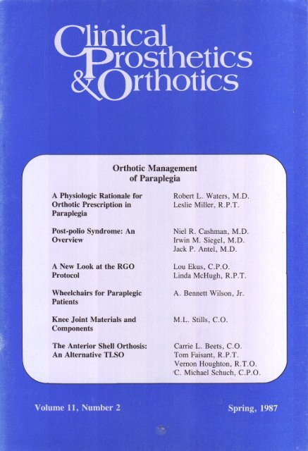
CONTENTS<br />
Volume 11, Number 2 Spring, 1987<br />
Orthotic Management<br />
of Paraplegia<br />
A Physiologic Rationale for Orthotic<br />
Prescription in Paraplegia 66<br />
Robert L. Waters, M.D.<br />
Leslie Miller, R.P.T.<br />
The criteria used at Rancho Los Amigos<br />
Medical Center to determine whether or<br />
not a paraplegic is a candidate for<br />
ambulation, based on the energy<br />
expenditure measurements of 150<br />
patients.<br />
Post-polio Syndrome: An Overview ... 73<br />
Niel R. Cashman, M.D.<br />
Irwin M. Siegel, M.D.<br />
Jack P. Antel, M.D.<br />
Patients who once had paralytic<br />
poliomyelitis may develop new<br />
complaints after decades of stable<br />
function. Over 50,000 persons in the<br />
U.S. are at risk of developing PPS.<br />
A New Look at the RGO Protocol 79<br />
Lou Ekus, C.P.O.<br />
Linda McHugh, R.P.T.<br />
A simplified approach to the selection,<br />
fitting, and training of patients for<br />
fitting with the Reciprocal Gait<br />
Orthosis.<br />
Wheelchairs for Paraplegic Patients ... 82<br />
A. Bennett Wilson, Jr.<br />
An overview of wheelchair options.<br />
The Anterior Shell Orthosis: An<br />
Alternative TLSO 95<br />
Carrie L. Beets, CO.<br />
Tom Faisant, R.P.T.<br />
Vernon Houghton, R.T.O.<br />
C. Michael Schuch, C.P.O.<br />
The authors present a TLSO which<br />
allows early mobilization of patients<br />
with spinal cord injury and other<br />
diagnoses, as well as independent<br />
donning and doffing.<br />
The Cast Off Valve: An Improved<br />
Method for Removing and Retaining<br />
Above Knee Casts and Prosthetic<br />
Sockets 101<br />
Albeit F. Rappoport, M.A., CP.<br />
Successfully tested at UCLA's<br />
Prosthetic Education Program and<br />
Prosthetic-Orthotic Laboratory, the<br />
Cast Off Valve allows a cast to remain<br />
undamaged for further reference.<br />
Early Mobility Aid for Non-walking<br />
Children 106<br />
Virgil Faulkner, C.P.O.<br />
N. Walsh, M.D.<br />
D. Currie, M.D.<br />
A low cost electrical mobility aid for<br />
children with multiple limb deficiencies.<br />
Knee Joint Materials and<br />
Components 91<br />
M.L. Stills, CO.<br />
An analysis of appropriate prescription,<br />
components, fabrication, fitting, and<br />
training for successful orthotic<br />
management of the paraplegic patient.
FEATURES<br />
Letters to the Editor<br />
Calendar<br />
vii<br />
xii<br />
ADVERTISER'S INDEX<br />
R.G. Abernethy<br />
The Hood Company<br />
Durr-Fillauer Medical Co<br />
Kingsley Manufacturing<br />
Knit-Rite<br />
Model and Instrument<br />
Development<br />
Motion Control<br />
PEL Supply, Inc<br />
Remploy<br />
Southern Prosthetic Supply<br />
iv<br />
xvi<br />
vii<br />
ii<br />
C3<br />
v<br />
xi<br />
vi<br />
vii<br />
C2<br />
Are yon caring for all of your patients'<br />
26 bones and 214 ligaments?<br />
You are if you prescribe Dr. Abernethy's custom<br />
shoes. Dr. Abernethy has brought a new design and<br />
principle to proper footcare. His custom shoes can<br />
eliminate most footproblems, fatigue and hard to fit<br />
feet. From arthritic towork, golf, tennis, jogging,<br />
hunting, walking and fashionable dress shoes. All<br />
handcrafted with the finest water-repellent leather. Calf,<br />
kid, kangaroo, suede and reptile. Twenty stylish<br />
patterns. The latest colors. Shoes as handsome as<br />
they are functional.<br />
Abernethy shoes have walked on the moon,<br />
in all 50 states, five foreigncountries, in Congress<br />
and hospitals.<br />
Have your patient step into a new world of comfort.<br />
Call 1-800-334-0128 or write forcasting information,<br />
prices and color brochure. Normal delivery is three<br />
weeks or less. Dr. Abernethy guarantees his shoes are<br />
correct. If not, hell re-do them or make them correct.<br />
R.G. Abernethy Industries, Inc.<br />
Route 1, Box 21 • Cæston, NC28615<br />
Sound shoes for a sound mind and body.<br />
From the Editor:<br />
Opinions expressed in Clinical Prosthetics and Orthotics are solely those of the authors. No<br />
endorsement implied or explicit is made by the editoral staff, or by the American Academy of<br />
Orthotists and Prosthetists.
THE SEATTLE FOOT...<br />
the foot with a natural<br />
spring in its step!<br />
A desire to improve<br />
the quality of life<br />
enjoyed by amputees, combined<br />
with a recognition<br />
of the<br />
limitations imposed by conventional<br />
prosthetic feet, brought a team of<br />
aerospace engineers, prosthetists,<br />
industrial designers and physicians<br />
together in Seattle. The result?<br />
THE SEATTLE FOOT"<br />
literally a giant step forward<br />
lower extremity<br />
amputees.<br />
Quite<br />
THE SEATTLE FOOT" has the features<br />
that amputees and prosthetists deserve.<br />
Dynamic... a Dupont Delrin keel stores<br />
and releases energy with each step to<br />
supply natural lift and thrust.<br />
Specific... available with a range of keel<br />
spring-rates to provide optimum energy<br />
storage for each amputee's body weight<br />
and activity level.<br />
Cosmetic... made from life-cast molds to<br />
achieve a new level of foot cosmesis<br />
Includes split between great and second<br />
toe.<br />
Versatile... beneficial for amputees of all<br />
' ages, activity levels, and types including<br />
-BK, AK, and Bilateral.<br />
Compatible... can be fit to new or existing<br />
endoskeletal or exoskeletal prostheses<br />
using conventional techniques.<br />
Tested... developed with input from over<br />
900 evaluation amputees.<br />
Supported... covered by a full year of<br />
warranty and an optional trial exchange<br />
program.<br />
for<br />
THE SEATLEFOOT" hasben<br />
enthusiasticalyacceptedby aw«Je<br />
majoπtyo>famputeswhoevaluated it.<br />
One oftheseindividuals isJimClark,<br />
aUniversityo(Washingtonprofessor.<br />
"THE SEATTLE FOOT" has<br />
vastly improved my options as an<br />
amputee. When I had a standard<br />
'SACH foot I became tired and sore<br />
after walking (or more than half<br />
an hour. With the natural absorption<br />
and spring of THE SEATTLE<br />
FOOT" I can now comfortably<br />
warn and jog for much longer. I<br />
recently even spent time in the<br />
Swiss Alps hiking up to ten miles<br />
a day "<br />
j m i:<br />
For the full story of THE SEATTLE FOOT 7 ', along with specifications,<br />
currently available sizes, and ordering information, call or write Or,<br />
contact one of our distributors<br />
Model and Instrument Development<br />
861 Poplar Place South<br />
Seattle, Washington 98U4<br />
(206)325-0715<br />
Distributed by Southern Prosthetic Supply
Orthotic and Prosthetic<br />
Parts and Supplies<br />
NEWS<br />
4666 Manufacturing Rd., Cleve., OH 44135<br />
HelpsRelieve<br />
LowBack ADJUSTABLEDOUBLE<br />
ACTIONANKLEJOINT<br />
NOWAVAILABLEFOR<br />
FRACTUREBRACING<br />
Discomfort<br />
The Orthopedic<br />
SACRO-EASE<br />
(posture correction<br />
'seat is designed to<br />
be custom fit by you for your patient.<br />
It helps relieve low back discomfort<br />
and can be moved from one seat to<br />
another since it is fitted to the<br />
individual and not a particular seat.<br />
The BRS model is 15" wide and<br />
most versatile. It fits office chairs, car<br />
and truck seats, even adult size (18")<br />
wheelchairs. It can be used on any<br />
existing seat bottom and backrest<br />
which is too soft to provide sufficient<br />
support under the hips and behind<br />
the lumbar spine.<br />
The Sacro-Ease units are easy to<br />
fit, involving manually bending the<br />
backrest over the edge of a counter<br />
top (a vice is not recommended).<br />
Homecare Sacro-Ease<br />
Also available is<br />
a Homecare Model<br />
of the Sacro-Ease.<br />
The lumbar pad<br />
can be adjusted by<br />
moving the side<br />
I straps up or down.<br />
The patient could<br />
buy this model and adjust the lumbar<br />
pad himself. This model would also be<br />
beneficial in an office, hospital,<br />
nursing home etc.; wherever more<br />
than one person may be using the<br />
device.<br />
4666 Manufacturing Rd.<br />
Cleveland, Ohio 44135<br />
(216)<br />
InOhio Cont.U.S. (800)321-1264<br />
267-5775<br />
(800)345-5105<br />
Orthotic & Prosthetic Parts & Supplies<br />
Here's the latest in functional fracture<br />
bracing from USMC. The Controlled Ankle<br />
Motion Walker (C.A.M.) is a new, unique way<br />
to provide range of motion at the ankle in the<br />
rehabilitation of injuries to the ankle or foot.<br />
The adjustable double action ankle joints<br />
provide a fixed position or range of motion.<br />
You can lock in dorsiflexion or plantar<br />
flexion or allow up to 45 degrees total<br />
range of motion. The C.A.M. restricts<br />
inversion/eversion. It is very lightweightj{<br />
made of high-tech plastic and has strong<br />
malleable uprights. A one piece stirrup is<br />
molded into the footplate and there are adjustable straps with<br />
velcro closures.<br />
The rocker has the roll point located on a continuous<br />
radius to give optimum walking assistance. Flat spots are<br />
eliminated for a smooth patient gait. A slip resistant sole has<br />
been bonded to the rocker.<br />
The C.A.M. is very popular for achilles tendonitis and<br />
non-displaced fractures of distal tibia/fibula following initial<br />
casting.<br />
Four foam pads are included with the C.A.M. to fill extra<br />
space (if needed) at either side of the ankle, at the heel and<br />
over the instep. It is available in one size only which will fit 90%<br />
of adults. Your stocking requirements are low as it can be<br />
used on a left or right foot.<br />
Central Location and<br />
Inventory<br />
<strong>Complete</strong><br />
PEL Supply is the Cadillac of Orthotic and Prosthetic<br />
product distributors. PEL has a central and very large,<br />
complete inventory in one location. It is easy to manage and<br />
control by PEL's excellent staff.<br />
No order is too small or too large at PEL Supply.<br />
Order orthotic and prosthetic parts and supplies from PEL.<br />
Receive friendly, fast service.<br />
PEL has today, what you need tomorrow.
Letters to the Editor<br />
Dear Editor:<br />
I think that the Winter, 1987 issue of Clinical<br />
Prosthetics and Orthotics turned out<br />
nicely, but am dismayed to see that editorial<br />
policy has dictated that "residual limb" and<br />
"limb" be used instead of "stump." You will<br />
recall the editorial you published some years<br />
ago in which I gave what I believe are very<br />
valid reasons for not using "residual limb" in<br />
scientific and technical publications. I haven't<br />
changed my mind on this and I wouldn't want<br />
the readers of C.P.O. to think that I have.<br />
I have no quarrel with clinicians avoiding the<br />
use of "stumps" in dealing with patients, but<br />
"residual limb" and simply "limb" are<br />
usually thought of, quite correctly, as the unaffected<br />
one and at best is ambiguous if not outright<br />
misleading, two things that should be<br />
avoided at all costs in scientific and technical<br />
writing.<br />
In my opinion the use of "residual limb" is<br />
an affectation that should be avoided by those<br />
who should know better.<br />
Sincerely,<br />
A. Bennett Wilson, Jr.<br />
!?I=MMI=DI<br />
Stump Socks<br />
Remploy garments for use with<br />
artificial limbs are among the<br />
best available anywhere. They<br />
are made to strictly functional<br />
specification, beautifully<br />
fashioned in the most<br />
comfortable gauge of pure<br />
wool yarn. After knitting, each<br />
garment is proofed against<br />
shrinkage, to a rigid approved<br />
specification. They are specially<br />
designed to withstand repeated<br />
washing without lessening the<br />
comfort to the wearer.<br />
For further information contact:<br />
Remploy °?I=MMI=DI<br />
PUBLIC RELATIONS DEPARTMENT,<br />
Russ Street, Broad Plain, BRISTOL<br />
BS2 OHJ. U.K. TEL: 011 44 272 277512<br />
TELEX: 23178 REMPLOY G<br />
FAX: 011 44 1 452 6898
AUTHOR INFORMATION<br />
OinicaL<br />
^prosthetics<br />
^Orthotics<br />
As professionals, we are obligated to do what we can to advance the state-of-the-art and<br />
share new developments with our colleagues. The most efficient way to transfer information,<br />
and the way that has the greatest impact, is through the written word. But, for many<br />
professionals, writing is a task that often becomes monumental to the point that we succumb<br />
to inertia. Writing, however, is not such a monumental task if we break it down into<br />
smaller, simpler tasks which we can complete one at a time.<br />
The initial and most difficult problem every writer faces is how to organize the material.<br />
The quickest way to organize material is through the use of an outline. In its most basic<br />
form, an article is divided into three parts—introduction, body, and conclusion. The introduction<br />
states the subject and gives pertinent background information that is necessary in<br />
order to understand the topic. The main body of the article is the intent to inform and<br />
answer a variety of questions. The body can include subheads, such as review of literature,<br />
method, clinical materials, discussion, and results. The conclusion restates the main points<br />
presented in the article.<br />
Clinical Prosthetics and Orthotics addresses broad, philosophical issues, and as such<br />
invites a more subjective style. Each issue of C.P.O. centers on a main topic. Usually, an<br />
issue will contain a lead article, an editorial, and one or more technical articles pertaining<br />
to the topic. Authors are solicited by the Academy editorial board; however, C.P.O. also<br />
accepts unsolicited articles. Unsolicited articles need not cover the topic at hand and may<br />
be of a more technical and objective nature. All articles are submitted to the editor, a<br />
professional in the field, who checks every article for accuracy, terminology, format, and<br />
references. The articles are then forwarded to the publications staff at the Academy National<br />
Office for production and printing.<br />
The chosen topics for Clinical Prosthetics and Orthotics, Volume 11, Number 4 through<br />
Volume 12, Number 3, and deadlines for submission are as follows:<br />
Volume 11, Number 4<br />
Volume 12, Number 1<br />
Volume 12, Number 2<br />
Volume 12, Number 3<br />
"Quadraplegia"<br />
Deadline: July 1, 1987<br />
"Prosthetic Management of the Partial Foot and Symes<br />
Amputations"<br />
Deadline: September 1, 1987<br />
"Orthotic Management of the Foot"<br />
Deadline: December 1, 1987<br />
"Disarticulation Amputations"<br />
Deadline: March 1, 1988<br />
Please remember that although these are the chosen topics for these particular issues,<br />
we gladly welcome submissions on other topics. Please feel free to contact the National<br />
Office if you have any questions on whether your article would be appropriate for C.P.O.<br />
If you have an article that has been previously published in another scientific journal and<br />
think it may be appropriate for C.P.O., please let us know.<br />
Submit articles to: Charles Pritham, CPO, Editor, c/o Durr-Fillauer Medical, Inc., Orthopedic<br />
Division, P.O. Box 5189, Chattanooga, Tennessee 37406.<br />
Questions should be submitted to: Sharada Gilkey, Managing Editor, Academy<br />
National Office, 717 Pendleton Street, Alexandria, Virginia 22314; or call (703) 836-7118.<br />
P.S. Author's kits are made available upon request, free of charge. Kits include manuscript<br />
guidelines, a patient permission form, reprint information, a reprint permission form, and<br />
information on prizes for published articles.
C.P.O. MANUSCRIPT GUIDELINES<br />
1. Manuscripts must be typewritten, double-spaced with wide margins.<br />
2. Indicate bibliographical references by means of Arabic numerals in parentheses (6).<br />
3. Write out numbers less than ten.<br />
4. Bibliography should follow the correct forms as shown below.<br />
Appli<br />
a. Book<br />
Murphy, Eugene F., "Lower-Extremity Component," Orthopedic<br />
ances Atlas, Vol. 2, J.W. Edwards, 1960, pp. 217-224.<br />
b. Journal Article<br />
Panton, Hugh J., "Considerations for Joints and Corset," Newsletter ...<br />
Amputee Clinics, 8:3: June, 1975, pp. 1-3, 6-7.<br />
c. Lecture or Verbal Presentation<br />
1. Holmgren, Gunnar, "The PTB Suction Prosthesis" from the written material of a<br />
lecture delivered at the third of the "Strathclyde Bioengineering Seminars," 8-11<br />
August, 1978.<br />
2. Wagner, F.W., Jr.: "Classification and treatment for diabetic foot lesions"; Instructional<br />
Course, American Academy of Orthopedic Surgeons, New Orleans, Louisiana,<br />
February, 1976.<br />
d. Personal Communication<br />
Irons, George, C.P.O., Personal communication, June 1977. Presently, Director of<br />
Research, United States Mfg., Glendale, California. Formerly, Research Prosthetist,<br />
Patient Engineering Service, Rancho Los Amigos Hospital, Downey, California.<br />
Arrange all references alphabetically.<br />
5. Illustrations<br />
a. Write authors name on the back of each illustration.<br />
b. Number all illustrations to correspond to references from the text.<br />
c. Submit either an original or a photographic black & white reproduction of illustrations.<br />
6. Photographs<br />
a. DO NOT WRITE ON THE BACK OF PRINTS OR USE PAPERCLIPS OR STAPLES<br />
ON PRINTS.<br />
b. Identify by number to correspond to references from the text on a piece of paper taped<br />
to the back of each print.<br />
7. List all captions in order on a separate sheet of paper, typed.<br />
8. Must include with the manuscript signed copies of releases from patients who are photographed<br />
and referred to in the text. For pictures of patients for which permission cannot be<br />
obtained, the eyes must be masked out.<br />
9. Mail the original manuscript with accompanying photographs/illustrations and one<br />
xerox to:<br />
Charles H. Pritham, C.P.O.<br />
c/o Durr-Fillauer Medical, Inc.<br />
Orthopedic Division<br />
P.O. Box 5189<br />
Chattanooga, TN 37406
Reprints<br />
of<br />
(^lİnİCal<br />
^prosthetics<br />
^Orthotics<br />
v v y<br />
Articles<br />
High quality reprints of articles appearing in Clinical Prosthetics and<br />
Orthotics are available for very reasonable rates listed below. All prices<br />
include your printed pages in black ink on 60 lb. white enamel (glossy)<br />
paper, stapled, carton packed, and shipped.<br />
Add'l 100's Add'l 1,000's<br />
No. Pages 100 200 300 400 500 (up to 1,000) (up to 5,000)<br />
2 $ 23 $ 26 $ 28 $ 30 $ 33 $ 2.40 $ 24<br />
4 44 47 51 54 58 3.70 37<br />
8 82 93 102 112 123 9.40 94<br />
10 105 117 129 141 153 11.70 117<br />
12 120 134 148 161 176 13.10 131<br />
16 152 168 184 201 218 16.00 160<br />
20 188 208 228 249 269 20.10 201<br />
24 224 248 272 296 320 23.50 235<br />
COVERS<br />
Additional for self cover title page with publication title, volume, number, date, article title, name<br />
of authors, and reprint line.<br />
Per article $ 7.50<br />
Additional for 67 lb. Vellum Bristol cover, printed one color on one side only with publication<br />
title, volume, number, date, article title, name of authors and reprint line.<br />
100 covers $ 44.00<br />
Additional 100 covers $ 4.90<br />
SHIPPING<br />
All prices are F.O.B. Hanover, Pennsylvania. Please furnish complete street address so shipment<br />
may be made by United Parcel Service. If other shipping is required, please indicate.<br />
INVOICE<br />
Will include shipping, and any preparation, composition or non-standard production costs not<br />
included in the above price schedules.<br />
Terms, net 30 days.<br />
Prices subject to change without notice.<br />
To order your reprint(s), write or call:<br />
Sharada Gilkey<br />
Managing Editor<br />
Clinical Prosthetics and Orthotics<br />
717 Pendleton Street<br />
Alexandria, VA 22314<br />
(703)836-7118
The HJ5IilElliiiiHmisfii<br />
f†ttαble, æmfor†αble, reliable<br />
Motion Control, Inc<br />
1290 West 2320 South. Suite A<br />
Salt Lake City, Utah 84119<br />
Toll Free 1-800-621-3347
A Physiologic Rationale<br />
for Orthotic Prescription<br />
in Paraplegia<br />
by Robert L. Waters, M.D.<br />
Leslie Miller, R.P.T.<br />
INTRODUCTION<br />
A difficult clinical decision to be made when<br />
treating a paraplegic patient is deciding if<br />
walking is a realistic goal, if orthoses should be<br />
prescribed, and what the functional outcome<br />
will be. It has been demonstrated that the energy<br />
expenditure for paraplegics, utilizing a<br />
crutch assisted swing-through gait pattern, is<br />
markedly elevated. Many patients have learned<br />
to walk with crutches and orthoses, but discontinued<br />
their use after discharge from a rehabilitation<br />
center. 2 , 3 , 4<br />
Studies of other forms of<br />
bracing also reveal elevated energy expenditure.<br />
12<br />
In this review, we will describe the indications<br />
for prescribing ankle-foot orthoses and<br />
knee-ankle-foot orthoses. We will then outline<br />
the criteria used at Rancho Los Amigos Medical<br />
Center to determine whether or not a paraplegic<br />
is a candidate for ambulation. These criteria<br />
are based on the results of energy expenditure<br />
measurements of 150 patients with<br />
traumatic paraplegia. 10<br />
Further investigation of<br />
the data collected revealed that the proprioception<br />
level or pattern seemed a reliable indicator<br />
of which patients would achieve ambulation,<br />
while muscle function seemed to determine the<br />
quality of their ambulation. These results have<br />
helped us to develop guidelines for projecting<br />
the functional outcome of ambulation of paraplegics.<br />
ORTHOTIC PRESCRIPTION<br />
The goal of orthotic management in paraplegia<br />
is to provide the external support necessary<br />
to compensate for the motor and sensory<br />
deficits. Joint range of motion, muscle<br />
strength, proprioception, sensation, and spasticity<br />
are evaluated when determining the orthotic<br />
prescription.<br />
Knee-Ankle-Foot Orthosis (KAFO)<br />
Quadriceps strength less than "Fair+" on<br />
manual muscle testing is the most common indication<br />
for a KAFO. The KAFO is locked at<br />
the knee while walking. Although some patients<br />
with less than "Fair+ " strength are able<br />
to ambulate a short distance without a locked<br />
knee (knee stabilization), knee instability<br />
usually occurs after a few steps. The exception<br />
is the patient with severe quadriceps spasticity<br />
which maintains the knee in extension, eliminating<br />
the need for external support.<br />
Another indication for a KAFO is impaired<br />
or absent knee proprioception. The lack of proprioception<br />
can result in knee instability even<br />
when the quadriceps strength is "Fair+" or<br />
greater, as the patient is unable to monitor joint<br />
position. If light touch sensation is present on<br />
the front of the thigh, a KAFO which allows<br />
knee flexion is usually sufficient to control the<br />
knee. The anterior stop of the knee mechanism<br />
limits extension at 180 degrees and the patient<br />
feels pressure from the anterior thigh cuff. In<br />
this regard, the brace serves as a transducer that<br />
converts proprioception (which is not perceived)<br />
into pressure (which is perceived).<br />
The final indication for extending bracing<br />
above the knee is a severe hyperextension<br />
thrust during stance. Paraplegics whose gait is
characterized by a hyperextension thrust may<br />
develop ligamentous instability, due to attenuation<br />
of the posterior cruciate ligament and<br />
posterior knee capsule resulting in hyperextension<br />
deformity.<br />
Range of motion at the hip from 0 degrees of<br />
extension to 110 degrees of flexion should be<br />
present. In the absence of hip extensor<br />
muscles, full hip extension range is necessary<br />
to allow the patient to lean backwards and<br />
move the center of gravity of the trunk posterior<br />
to the hip joint (Figure 1). Hip flexion to<br />
110 degrees, with the knee extended, enables<br />
the patient to come to standing with locked<br />
KAFO's and rise from the ground after a fall.<br />
Full knee extension is required for optimal fit.<br />
Ten degrees of dorsiflexion at the ankle is<br />
the minimum necessary for unassisted upright<br />
balance (Figure 1). Normal proprioception in at<br />
least one hip also facilitates unassisted<br />
standing.<br />
Inability to meet the joint range requirements<br />
described above commonly occurs and is most<br />
often due to spasticity or contracture. If satisfactory<br />
orthotic fit and posture cannot be<br />
achieved, a physical therapy regime that includes<br />
stretching exercises or serial casting is<br />
often successful when spasticity is mild and the<br />
deformity is not longstanding. When severe<br />
spasticity or deformity is present, or the deformity<br />
has been present for an extended time,<br />
the patient should be referred to an orthopedic<br />
surgeon.<br />
Good trunk strength is necessary to maintain<br />
an erect posture in the standing position<br />
without excessive weight bearing in the arms.<br />
High level paraplegics without adequate trunk<br />
strength must exert a strong upwards force by<br />
the arms throughout the entire gait cycle to prevent<br />
forward collapse and accomplish forward<br />
progression. This contributes to the high energy<br />
demand. (All swing-through gait candidates are<br />
required to perform 50 consecutive dips on parallel<br />
bars to insure they have sufficient upper<br />
extremity strength and endurance.)<br />
Ankle-Foot Orthosis (AFO)<br />
Quadriceps strength greater than "Fair"<br />
should be present to stabilize the knee if an<br />
AFO is prescribed. The patient must also have<br />
adequate hip flexion strength to swing the leg<br />
Figure 1. Standing posture.<br />
forward to achieve a reciprocal gait pattern.<br />
The indications for AFO are numerous and include<br />
any or all of the following: plantarflexion<br />
strength less than "Good," dorsiflexion<br />
strength less than "Fair," impaired ankle proprioception,<br />
and moderate to severe plantarflexion<br />
spasticity.<br />
During normal walking, the plantarflexors<br />
are active during the stance phase of gait to<br />
prevent excessive forward advancement of the<br />
tibia. As a result of forward momentum, the<br />
knee passively extends as the body advances<br />
forward over the stabilized tibia, and the demand<br />
placed on the quadriceps is minimal.<br />
Customary manual muscle testing methods fail<br />
to place a sufficient load on the plantarflexors<br />
to evaluate the force required during gait. The<br />
strength required to provide ankle and knee stability<br />
is present in patients who can perform 15<br />
to 20 toe raises on one leg. Failure to provide<br />
adequate orthotic stabilization of the ankle in<br />
patients with inadequate plantarflexion strength<br />
may result in ankle instability and knee instability,<br />
if the quadriceps and/or hip extension<br />
strength is also inadequate.
Knee wobble can be a sign of impaired ankle<br />
proprioception and/or weakness. This can be<br />
eliminated by an AFO with a rigid ankle or anterior<br />
ankle stop, which provides distal stability<br />
and kinesthetic information via the calf cuff.<br />
An AFO may be utilized to hold the ankle in<br />
the neutral position when dorsiflexion strength<br />
is impaired or there is excessive plantarflexion<br />
spasticity. When spasticity is severe, it may not<br />
be possible to maintain the foot in neutral, and<br />
the patient should be referred to an orthopedic<br />
surgeon if non-operative measures prove inadequate.<br />
When the ankle is held in a rigid orthosis,<br />
ankle stability is gained during midstance.<br />
However, a forward thrust is imposed, forcing<br />
the knee into flexion at the moment of heel<br />
contact. (This knee flexion torque is generated<br />
because the rigidly immobilized ankle rotates<br />
forward about the point of heel contact.)<br />
During normal gait, this torque is avoided by<br />
ankle plantarflexion, minimizing the effect of<br />
the heel lever.<br />
There are two courses of action available to<br />
provide ankle stability during stance, while<br />
still maintaining knee stability at heel strike. If<br />
the patient has "Fair+ " or better ankle dorsiflexion<br />
strength and intact proprioception, we<br />
fit a metal AFO with a double adjustable ankle<br />
joint. A set screw in the anterior channel provides<br />
an adjustable stop that prevents excessive<br />
dorsiflexion. The posterior stop is left open to<br />
allow free ankle plantarflexion. Springs can be<br />
added posteriorly if dorsiflexion strength is less<br />
than "Fair+." The advantage gained is that<br />
restriction of motion during terminal stance is<br />
maintained while the normal plantarflexion<br />
motion during heel contact is preserved,<br />
avoiding the undesired knee flexion torque. If<br />
the patient has less than "Fair" dorsiflexors or<br />
absent proprioception at the ankle, then the<br />
ankle is locked and either metal or plastic is<br />
used. To avoid the excessive knee flexion<br />
torque when the AFO is locked, the heel of the<br />
shoe is undercut. This decreases the heel lever<br />
and, thus, the knee flexion torque.<br />
ORTHOSIS WEIGHT<br />
Weight is an important factor to some patients,<br />
as is the availability of joint motion of<br />
the orthotic system. Plastic, because of its potential<br />
to be lighter than metal, is sometimes<br />
preferable. For the patient with weak hip<br />
flexors, efforts to minimize weight are warranted<br />
since any extra weight at the end of the<br />
limb will make it more difficult to lift the foot<br />
and advance the leg. Lehneis, et al. 8<br />
found that<br />
improving orthotic stability at the ankle reduces<br />
energy costs. It follows, then, that in any orthotic<br />
design, stability (control about a joint)<br />
should not be sacrificed merely to achieve<br />
lighter weight.<br />
EXERCISE PHYSIOLOGY<br />
It is necessary to understand several fundamental<br />
principles of exercise physiology to interpret<br />
the results of energy expenditure measurements<br />
in paraplegic patients. 1<br />
The use of<br />
oxygen consumption is based on the fact that<br />
during sustained exercise, most of the ATP for<br />
muscle contraction is generated by aerobic metabolic<br />
pathways. After several minutes of exercising<br />
at a constant submaximal workload, the<br />
rate of oxygen consumption rises until it<br />
reaches a level sufficient to meet the metabolic<br />
demands of the exercising muscle. Measurement<br />
of the rate of oxygen consumption at this<br />
time reflects the energy cost of the activity and<br />
indicates the exercise intensity. The oxygen<br />
cost per meter walked determines the efficiency<br />
of ambulation.<br />
The principle fuels for aerobic metabolism<br />
are carbohydrates and fats. The oxidation of<br />
glucose can be summarized by the following<br />
equation:<br />
GLUCOSE + 36 ADP + 6 O 2<br />
-><br />
6 CO 2<br />
+ 44 H 2<br />
O + 36 ATP<br />
During exercise, the extent to which anaerobic<br />
pathways contribute to the production of energy<br />
depends upon the intensity of the effort. In mild<br />
to moderate exercise (approximately 50 percent<br />
of the maximal aerobic capacity for untrained<br />
individuals), the oxygen supplied to the tissue<br />
for the aerobic energy producing reactions is<br />
usually sufficient to meet energy requirements.<br />
During more strenuous exercise, anaerobic oxidation<br />
processes also occurs.<br />
The amount of energy that can be produced<br />
by anaerobic means is limited. Nineteen times<br />
more energy is produced by the aerobic oxidation<br />
than by anaerobic oxidation. Also, accumulation<br />
of lactate in muscle and blood leads to
acidosis, limiting the extent to which intense<br />
exercise can be performed. From a practical<br />
standpoint, anaerobic oxidation provides an<br />
extra supply of energy for sudden bursts of<br />
strenuous effort, but these pathways cannot be<br />
routinely utilized for a prolonged time. In contrast,<br />
when exercise is performed below anaerobic<br />
threshold, an individual can sustain exercise<br />
for many hours without exhaustion.<br />
MAXIMAL<br />
AEROBIC CAPACITY<br />
The maximal aerobic capacity (VO 2<br />
max) is<br />
the single best indicator of physical work capacity<br />
and fitness. It measures the individual's<br />
maximum energy production capability. Generally,<br />
an individual is able to reach the VO 2<br />
maximum within two to three minutes of instituting<br />
strenuous exercise. Any disorder of the<br />
respiratory-cardiovascular muscle or metabolic<br />
systems that restricts the supply of oxygen to<br />
the muscle decreases the VO 2<br />
max. A physical<br />
conditioning program can increase aerobic capacity<br />
by several processes which include improving<br />
cardiac output, increasing the capacity<br />
of the muscle to extract oxygen from the blood,<br />
increasing the level of hemoglobin, and increasing<br />
the muscle mass. On the other hand,<br />
the maximal aerobic capacity can be reduced<br />
due to blood loss, paralysis, surgery, negative<br />
nitrogen balance, or bed rest. 1<br />
The important<br />
clinical implication is that the paraplegic patient<br />
is usually severely deconditioned as a consequence<br />
of the injury. The prescription of<br />
orthoses and a walking program should not be<br />
initiated until the patient has sufficient strength<br />
and maximal aerobic capacity to meet the required<br />
energy demand. The deconditioned<br />
paraplegic patient will respond to a physical<br />
conditioning program just as an able bodied<br />
subject with respect to increased strength, endurance,<br />
and maximal aerobic capacity. 5<br />
In able bodied subjects, the VO 2<br />
max also<br />
depends on the type of exercise. During lower<br />
limb exercise, the VO 2<br />
max is greater than the<br />
VO 2<br />
max for the upper limbs. Since paraplegic<br />
patients rely on the upper extremities to walk<br />
with the aid of crutches, their energy production<br />
capability is inherently limited. The<br />
problem in paraplegics is further compounded<br />
by the effects of the spinal injury. The upper<br />
extremity VO 2<br />
max for paraplegics is lower<br />
than for able bodied subjects, presumably due<br />
to the effects of paralysis and interruption of<br />
the autonomic neurological pathways which<br />
regulate blood flow and cause venous pooling<br />
in the lower extremities. 6,11<br />
For the typical<br />
adult male paraplegic, we establish a VO 2<br />
max<br />
of 20 ml/kg-min during upper arm cranking as<br />
the minimal criteria acceptable for entering gait<br />
training if a swing-through crutch assisted gait<br />
pattern will be required.<br />
ENERGY EXPENDITURE<br />
Wheeling Versus Normal Walking<br />
On a hard, level surface paraplegic wheelchair<br />
use is as efficient as normal walking. A<br />
comparison of the data in Figure 2 indicates<br />
that when propelling a chair around a 60.5<br />
meter circular track, the speed was almost as<br />
fast as normal walking (72 versus 80 m/min). 10<br />
The oxygen rate was approximately the same<br />
(11.5 versus 11.9 ml/kg/min) (Figure 3), as<br />
was the oxygen cost (.16 versus .15 ml/kg/<br />
min). The heart rate was higher in paraplegics<br />
using the wheelchair than in normal walking<br />
(123 versus 100 BPM) (Figure 4). As previously<br />
mentioned, this relates to the lower<br />
upper maximal aerobic capacity in paraplegics<br />
during arm exercise. From a clinical standpoint,<br />
it may be concluded that the wheelchair<br />
is a highly efficient means of transportation<br />
whose speed and energy requirements are comparable<br />
to that of normal walking.<br />
Swing Through<br />
Gait<br />
Crutch walking with a swing-through gait requires<br />
the arms and shoulder girdle to lift the<br />
entire weight of the body and swing it forward<br />
with each step. The average speed in paraplegics<br />
trained to use a swing-through crutch<br />
assisted gait was 64 percent lower than normal<br />
walking (20 versus 80 m/min) (Figure 2); the<br />
rate of oxygen consumption was 38 percent<br />
greater (16.5 versus 11.9 ml/kg/min) (Figure<br />
3); the oxygen cost was 560 percent greater<br />
(.84 versus .15 ml/kg/min); and the heart rate<br />
was increased 46 percent (145 versus 99 BPM)<br />
(Figure 4). 10<br />
This rate of energy expenditure<br />
requires most of the aerobic capacity of the<br />
typical adult male paraplegic with a complete<br />
T12 lesion and is well above the anaerobic
Figure 2. Average velocity in normal subjects and in patients using wheelchairs or orthoses.<br />
Figure 3. Rate of oxygen consumption in normal subjects and in patients using wheelchairs<br />
or orthoses.
Figure 4. Heart rate in normal subjects and in patients using wheelchairs or orthoses.<br />
threshold. The extreme exertion required for a<br />
swing-through gait demands a greater intensity<br />
of physical effort than a normal individual customarily<br />
expends on sports activity such as recreational<br />
jogging. Consequently, it is not surprising<br />
that while the athletic paraplegic may<br />
be willing to expend this level of exertion for<br />
recreational purposes, he is unwilling to sustain<br />
these efforts for normal activities of daily<br />
living. Even those patients, who are physiologically<br />
capable of sustaining the intense physical<br />
effort of a swing-through gait for a sustained<br />
time period to travel longer distances, find<br />
tachypnea (rapid breathing), tachycardia (rapid<br />
heart rate), and hidrosis (sweating), unacceptable<br />
for routine activities of daily living.<br />
We believe that the highly motivated paraplegic<br />
who is willing to exercise strenuously<br />
should not be discouraged from walking, but a<br />
more realistic approach should be taken for the<br />
average patient. The average patient should be<br />
given walking training and bilateral kneeankle-foot<br />
orthoses only if walking is necessary<br />
for psychological reasons, for purposes of exercise,<br />
or because of architectural barriers in<br />
the living environment. It should be clearly explained<br />
that the wheelchair should be considered<br />
as the primary means of mobility.<br />
We have tested three patients with "Fair+"<br />
hip flexors who used bilateral KAFO's and preferred<br />
a reciprocal gait pattern. 10<br />
Interestingly,<br />
the effort expended by these patients was just<br />
as great as in swing-through gait (Figures 2, 3,<br />
and 4).
Figure 5. Average distances necessary to perform customary activities of daily<br />
living.<br />
Energy Expenditure: Reciprocal Gait<br />
In a review of spinal cord injured patients,<br />
Hussey and Stauffer found that those patients<br />
who were able to walk in the community had<br />
pelvic control with at least "Fair" hip flexor<br />
strength and at least "Fair" extensor strength<br />
in one knee so that a maximum of one KAFO<br />
was required, enabling the patient to achieve a<br />
reciprocal gait pattern. 6<br />
Having "Fair+" or<br />
greater quadriceps strength sufficient to stabilize<br />
one knee eliminates the need for one<br />
KAFO and enables the patient to walk with a<br />
crutch assisted reciprocal gait pattern at a rate<br />
of energy expenditure and heart rate that are<br />
significantly below that required for a swingthrough<br />
gait pattern (Figures 3 and 4). Surprisingly,<br />
we found no difference in the speed and<br />
rate of energy expenditure in patients with one<br />
free knee or two free knees and requiring<br />
bracing only below the knee (Figures 2 and 3).<br />
Nevertheless, paraplegics who have intact<br />
hip flexion and knee extension bilaterally require<br />
orthoses only below the knees, and those<br />
who use a reciprocal crutch assisted gait pattern<br />
are still severely impaired (Figures 2, 3, and 4).<br />
Compared to normal walking, the rate of oxygen<br />
expenditure is 20 percent greater (16.3<br />
versus 11.9 ml/kg/min) (Figure 3), the heart<br />
rate 31 percent greater (131 versus 100 BPM)<br />
(Figure 4), and the speed 67 percent slower (80<br />
versus 20 m/min) (Figure 2). 10<br />
The typical paraplegic<br />
who uses crutches and a reciprocal gait<br />
still exerts a force of 25 to 50 percent of total<br />
body weight on the crutches with each step, accounting<br />
for the increased rate of energy expenditure.<br />
The only spinal cord injured patients<br />
we have tested whose energy expenditure<br />
during walking does not exceed normal values<br />
are those patients with minimal involvement<br />
who have intact sacral function (in addition to<br />
lumbar function) and a sufficient hip abductor<br />
and extensor strength to maintain an erect posture<br />
without crutches.<br />
The average distances necessary to perform<br />
different daily living activities are listed in<br />
Figure 5 and were obtained from numerous<br />
measurements made in different types of urban<br />
areas in Los Angeles. 8<br />
Since the average speed<br />
of walking in low lumbar paraplegics who
used bilateral ankle-foot orthoses and a reciprocal<br />
crutch assisted gait pattern was only 26<br />
m/min, it would take more than five minutes to<br />
travel 150 meters. Because five minutes of<br />
walking will require a strenuous effort, it is apparent<br />
why even the typical low lumbar paraplegic<br />
is a limited walker outside the home and<br />
is not able to routinely ambulate comfortably<br />
for activities which require walking a longer<br />
distance. In this regard, clinicians are justified<br />
in prescribing a wheelchair to any spinal injury<br />
patient who requires crutch assistance. The patients<br />
should be encouraged to use the wheelchair<br />
as necessary and be reassured that reliance<br />
on the wheelchair, when necessary,<br />
should not be considered a failure.<br />
AUTHORS<br />
Robert L. Waters, M.D. is Chairman of the Department<br />
of Surgery at Rancho Los Amigos Medical Center,<br />
HB-121, 7601 E. Imperial Highway, Downey, California<br />
90242.<br />
Leslie Miller, R.P.T. is a spinal cord injury clinical specialist<br />
at Rancho Los Amigos Medical Center.<br />
REFERENCES<br />
1<br />
Aastrand, P.O. and K. Rodahl, Textbook Work Physiology,<br />
Ed. 2, McGraw-Hill, Inc., New York, 1977.<br />
2<br />
Cerny, K., R. Waters, H. Hislop and J. Perry,<br />
"Walking and Wheelchair Energetics in Persons with Paraplegia,"<br />
Physical Therapy, 60:1133-1139, 1980.<br />
3<br />
Clinkingbeard, J.R., J.W. Gersten, and D. Hoehn,<br />
"Energy Cost of Ambulation in the Traumatic Paraplegic,"<br />
American Journal of Physical Medicine, 43:157-<br />
165, 1964.<br />
4<br />
Gordon, E.E., "Physiological Approach to Ambulation<br />
in Paraplegia," JAMA, 161:686-688, 1956.<br />
5<br />
Huang, C.T., A.B. McEachran, K.V. Kuhlemeier,<br />
M.J. DeVivo, and P.R. Fine, "Prescriptive Arm Ergometry<br />
to Optimize Muscular Endurance in Acutely Injured<br />
Paraplegic Patients," Arch. Phys. Med. Rehab., 64:578-<br />
582, 1983.<br />
6<br />
Hussey, R.W. and E.S. Stauffer, "Spinal Cord Injury:<br />
Requirements for Ambulation," Arch. Phys. Med. Rehab.,<br />
54:544-547, 1973.<br />
7<br />
Lehneis, H.R., E. Bergofsky, and W. Fresina, "Energy<br />
Expenditure with Advanced Lower Limb Orthoses and<br />
with Conventional Braces," Arch. Phys. Med. Rehab.,<br />
57:20, 1976.<br />
8<br />
Lerner-Frankiel, M.B., S. Vargas, M. Brown, L. Krusell,<br />
and W. Schoneberger, "Functional Community Ambulation:<br />
What Are Your Criteria?" Clin. Man. in Phys.<br />
Ther., 6:12-15, 1986.<br />
9<br />
Waters, R.L., H.J. Hislop, J. Perry, and D. Antonelli,<br />
"Energetics: Application to the Study and Management of<br />
Locomotor Disabilities," Orthop. Clin. North America,<br />
9:351-377, 1978.<br />
10<br />
Waters, R.L. and B.R. Lunsford, "The Energy Cost<br />
of Paraplegic Locomotion," Journal of Bone Joint Surgery,<br />
67A: 1245-1250, 1985.<br />
11<br />
Wolf, E. and A. Magora, "Orthostatic and Ergometric<br />
Evaluation of Cord-injured Patients," Scandinavian<br />
Journal of Rehabilitation Medicine, 8:93-96, 1976.
Post-polio Syndrome: An Overview<br />
by Neil R. Cashman, M.D.<br />
Irwin M. Siegel, M.D.<br />
Jack P. Antel, M.D.<br />
Poliomyelitis was a dreaded disease of developed<br />
countries, affecting tens of thousands of<br />
children and adults during each of the epidemic<br />
years up to the mid-1950s. The polio virus is a<br />
small RNA virus whose only natural host appears<br />
to be man. The vast majority of exposed<br />
persons develop either an inapparent infection<br />
or a non-specific flu-like illness (non-paralytic<br />
poliomyelitis). Secondary invasion of brain and<br />
spinal cord is associated with infection and<br />
death of motor neurons, with loss of innervation<br />
to muscle fibers, and consequent muscle<br />
weakness and atrophy. Postmortem studies<br />
show that muscle weakness in poliomyelitis is<br />
clinically apparent only when more than half of<br />
corresponding motor neurons are destroyed. 1<br />
Frequently, muscles can be reinnervated by<br />
healthy neighboring motor neurons by a process<br />
of axonal sprouting. Thus, partial or complete<br />
recovery of muscle bulk and strength can<br />
occur, in which subnormal numbers of motor<br />
neurons support increased (up to 8-fold)<br />
numbers of muscle fibers. 2<br />
It is estimated that<br />
about 250,000 people in the United States have<br />
survived paralytic poliomyelitis and are alive<br />
today. 3<br />
Recently, it has become clear that some patients<br />
who had paralytic poliomyelitis may develop<br />
new complaints after decades of stable<br />
function. 3-11<br />
These new symptoms have been<br />
designated the "post-polio syndrome" (PPS)<br />
or "late sequella of poliomyelitis." Although<br />
some reports of new weakness following polio<br />
can be found in the medical literature since<br />
1875, 5 recent epidemiologic studies indicate<br />
that new symptoms are common, occurring in<br />
approximately 25 percent of patients with antecedent<br />
paralytic poliomyelitis. 4<br />
If this estimate<br />
is correct, over 50,000 persons in the U.S. are<br />
at risk of developing PPS. From published reports,<br />
the mean latency of onset has been calculated<br />
to be 36 years. 5<br />
Thus, an increasing incidence<br />
of new cases will probably continue<br />
into the early 1990s, reflecting the last epidemics<br />
of the mid-1950s.<br />
The risk of developing PPS appears to correlate<br />
with severity of the original poliomyelitis.<br />
Thus, a patient with four-limb involvement and<br />
a history of respiratory dependence during his<br />
polio is more likely to develop new symptoms<br />
than a patient with one-limb involvement. 6<br />
The<br />
severity of the original onset of polio also<br />
seems to predict the latency of developing the<br />
syndrome; severely affected patients may develop<br />
new symptoms after only 10-20 years,<br />
whereas mildly affected patients are more<br />
likely to exhibit extended delays in time of<br />
onset of PPS. 6<br />
What causes PPS? Why should a patient who<br />
has had stable function for decades develop<br />
new symptoms? At the present time, there is<br />
little definitive data on this subject. Early conjecture<br />
focused on a possible reactivation of the<br />
polio virus which had remained latent in the<br />
nervous system since the original infection.<br />
However, there appears to be little or no evidence<br />
for inflammation in post-polio patients;<br />
spinal fluid is without the cells, protein, and<br />
immunoglobulin which characterize other<br />
nervous system viral infections. Some investigators<br />
have suggested that the normal attrition<br />
of neurons with aging may trigger the postpolio<br />
syndrome when superimposed on previous<br />
static damage of polio. 7<br />
However, agingrelated<br />
loss of neurons in the spinal cord normally<br />
begins about age 60 12 ; the onset of PPS<br />
most commonly occurs 30 years after polio and<br />
does not correlate with chronological age of the
Table 1.<br />
patient. 7 Weichers and Hubbel 8 and Dalakis, et<br />
al. 9<br />
have suggested that motor units grossly enlarged<br />
by reinnervation in recovery from poliomyelitis<br />
may begin to experience peripheral<br />
disintegration with the passage of time. Our<br />
own data support this hypothesis in part; late<br />
denervation is most common in muscles with<br />
the greatest degree of reinnervation. However,<br />
we find that group atrophy, a putative indicator<br />
of motor neuronal disease (and not terminal axonal<br />
degeneration), is also common in patients<br />
with prior poliomyelitis. 10<br />
Although a bewildering variety of new<br />
symptoms are recognized as occurring in PPS , 4<br />
most new complaints appear to be subsumed<br />
under the three major problems of new pain,<br />
new weakness, and fatigue (Table 1). Some investigators<br />
have theorized that new muscle atrophy<br />
and weakness constitutes a separate syndrome,<br />
"postpoliomyelitis progressive muscular<br />
atrophy" or PPMA. 9<br />
In this scheme,<br />
other symptoms of PPS, such as pain and fatigue,<br />
are thought to be manifestations of a separate<br />
"musculoskeletal" syndrome due to<br />
chronic strain of muscles and joints that have<br />
been forced to bear weight in an unnatural<br />
fashion. 11<br />
Common orthopedic deformities in<br />
patients with poliomyelitis include knee valgus,<br />
varus, and recurvatum, as well as ankle<br />
equinus. 13<br />
However, new weakness can result<br />
in new joint instability, and new joint problems<br />
may interfere with efficiency of movement. Although<br />
a symptomatic approach to separate<br />
complaints of PPS patients is warranted, there<br />
is little scientific data which supports a division<br />
of sub-syndromes of PPS at present. We have<br />
found that even patients without new symptoms<br />
have evidence of an ongoing neuromuscular<br />
disorder. 10<br />
New pain is the most common symptom in<br />
PPS based on our experience (Table 1), and is a<br />
frequent complaint in other series as well. 4 - 6<br />
We have evaluated patients experiencing pain<br />
in conjunction with an orthopod experienced in<br />
neuromuscular disease. Several causes of pain<br />
are commonly identified in PPS patients.<br />
Perhaps the most common cause is insertional<br />
tendonitis and/or bursitis from chronic overuse<br />
and strain of muscle groups with subnormal<br />
strength. Palpation of tendons and bursae at<br />
common sites of involvement, such as the pes<br />
tendon at the medial knee and the trochanteric<br />
bursa, will often reveal profound point tenderness<br />
consistent with this syndrome. A trial of<br />
rest and non-steroidal anti-inflammatory agents<br />
may induce remission in this remitting/relapsing<br />
syndrome. For certain local sites, a steroid<br />
injection may be useful; weight reduction<br />
and readjustment of weight-bearing (through<br />
retraining and/or orthotic devices) may also<br />
produce long-range benefits. Degenerative arthritis,<br />
found most often in weight-bearing<br />
joints (where walking aids are used, the joints<br />
of the upper extremities may indeed become<br />
weight bearing), may also respond to the same<br />
regimen. Nerve compression syndromes characterized<br />
by pain and paraesthesias, secondary<br />
to positional or repetitive stress, should also be<br />
considered in the differential diagnosis of pain<br />
in PPS patients.<br />
Another type of pain, unrelated to joint<br />
"wear and tear" is muscle pain. This occurs<br />
frequently during or after exercise, and may be<br />
associated with cramps, fasciculations, or intense<br />
local fatigability. This may be related to<br />
muscle substitution and/or overwork in denervated<br />
muscle, and may ultimately be associated<br />
with permanently increased weakness. 14<br />
Treatment<br />
of this muscle pain includes avoiding the<br />
circumstances which induce it. Rest, orthoses,<br />
or even intermittent wheelchair use should also<br />
be considered to reduce load on overworked<br />
muscle. Medications which reduce muscle<br />
cramps (quinine, diphenylhydantoin) may increase<br />
weakness, and should be avoided.<br />
Fatigue is also a common complaint in PPS<br />
patients, occurring in over 60 percent of our<br />
series (Table 1). Two types of fatigue are reported<br />
by patients: generalized fatigue re-
quiring rest or sleep, and local muscle fatigue.<br />
Local muscle fatigue is most common in<br />
muscles previously severely affected by polio,<br />
and is often associated with cramps and fasciculations.<br />
Local fatigue may be a manifestation<br />
of ongoing muscle denervation, and is also<br />
reported by patients with classic denervating<br />
diseases such as amyotrophic lateral sclerosis. 15<br />
Generalized (systemic) fatigue is common in<br />
PPS, but may also be a symptom of a variety of<br />
other states, including medical conditions such<br />
as diabetes mellitus, cardiopulmonary dysfunction,<br />
and thyroid disease. Depression ("low<br />
energy") may also lead to systemic fatigue.<br />
Once medical and psychiatric diseases have<br />
been ruled out, systemic fatigue in PPS may be<br />
a symptom of widespread neuromuscular junction<br />
transmission defects. We have found that<br />
patients with fatigue and marked increased<br />
jitter in single-fiber electromyography (an indicator<br />
of defective neuromuscular transmission)<br />
respond to agents which enhance neuromuscular<br />
transmission, such as the anticholinesterase<br />
pyridostigmine (Mestinon). Rest, ambulatory<br />
aids, and activity planning may also alleviate<br />
generalized fatigue.<br />
New weakness is the third major component<br />
of the "post-polio triad" (Table 1). When new<br />
weakness occurs with new muscle atrophy,<br />
PPS patients are thought by some investigators<br />
to suffer from a specific syndrome of postpoliomyelitis<br />
progressive muscular atrophy<br />
(PPMA). 9<br />
It has been suggested that that evidence<br />
of ongoing denervation (fibrillations and<br />
positive waves on EMG, increased jitter on<br />
single-fiber EMG, and atrophic muscle fibers<br />
on muscle biopsy) are diagnostic for this syndrome.<br />
9<br />
However, we have found that electrophysiologic<br />
and muscle biopsy evidence of denervation<br />
is as common in polio patients who<br />
are not having new symptoms, as in patients<br />
who have clinically defined PPMA. 10<br />
Moreover,<br />
evidence of denervation is most severe in<br />
muscles which show the most signs of old<br />
polio. 10<br />
Thus, late denervation appears to be a<br />
concomitant of massive monophasic antecedent<br />
denervation, and not a sign of new disease. In<br />
addition, we found that although 14 out of 15<br />
patients who complained of new atrophy also<br />
reported new weakness, only about one-half of<br />
patients who reported new weakness noted new<br />
atrophy. 5<br />
Thus, the relationship of atrophy to<br />
weakness is not clear.<br />
New muscle weakness may put extra stress<br />
on a previously borderline compensated<br />
muscle, producing pain, cramping, and an<br />
"overwork myopathy," with accelerated<br />
weakness as an end result. 14<br />
It has been estimated<br />
that a partially denervated muscle graded<br />
"good" must work two and a half times as<br />
hard as normal muscle to accomplish the same<br />
task. 1<br />
We caution patients with new weakness<br />
to reduce activity. Exercise programs must be<br />
undertaken with extreme caution, and exercise<br />
should never be performed to the point of pain<br />
or muscle cramps. We advice patients to exercise<br />
limbs not previously affected by polio or,<br />
if this is impossible, participate in a carefully<br />
graded program in a therapeutically heated<br />
pool. One should exercise enough to prevent<br />
atrophy of disuse, but not enough to cause<br />
damage from overuse. High repetition, low resistance<br />
exercises are favored, as well as<br />
stretching and isometric drills. Orthotic devices,<br />
including the ankle-foot orthosis and<br />
knee-ankle-foot orthosis, may provide support<br />
for certain critically weakened muscle groups,<br />
although adequate function of other muscle<br />
groups (e.g., knee and hip extensor function<br />
for an ankle-foot orthosis) is a prerequisite for<br />
effective use. Wheelchair use should also be<br />
considered, sometimes only intermittently, as<br />
prolonged activity may predispose the patient<br />
to osteoporosis or venous thrombosis. Training<br />
in effective transfers, efficient movements, etc.<br />
by the physical and occupational therapist may<br />
also be useful, as can home help aids such as a<br />
shower chair and raised toilet seat.<br />
Limb weakness may result in new joint instability,<br />
which in turn may be associated with<br />
new pain and increasing deformity. It has been<br />
noted, for example, that floor reaction with<br />
knee hyperextension serves a knee-locking<br />
function when the quadriceps is weak. 16<br />
However,<br />
profound degrees of weakness can provide<br />
a "positive feedback" situation where<br />
posterior knee ligaments are subjected to more<br />
torque stress, leading to further stretching. 16<br />
A<br />
knee-ankle-foot orthosis (fit with a posterior<br />
offset knee hinge) may prevent progressive<br />
joint damage in this situation.<br />
Pulmonary complaints may occur in patients<br />
with previously weakened diaphragm, intercostals,<br />
abdominal, or accessory muscles. Frequently,<br />
a patient with previous paralytic poliomyelitis,<br />
involving muscles of respiration, will
have borderline respiratory compensation for<br />
decades, and will undergo precipitous respiratory<br />
failure later in life. 17<br />
Increasing scoliosis,<br />
aspiration pneumonia, gradual loss of motor<br />
units with aging, and other factors may contribute<br />
to respiratory insufficiency. Respiratory<br />
symptoms (daytime somnolence, snoring,<br />
dyspnea, etc.) must be sought in all patients,<br />
particularly those with a history of respiratory<br />
involvement with polio. Baseline spirometry is<br />
also obtained in patients attending clinics.<br />
Muscle relaxants and medications which suppress<br />
respiratory drive should be avoided. Vaccines<br />
(pneumonia and flu vaccines) and cessation<br />
of smoking are also important in patient<br />
management. New respiratory muscle weakness<br />
may also present as sleep apnea, which<br />
may respond to medication (e.g., protriptyline),<br />
or may require night time oxygen or mechanical<br />
ventilation. Pulmonary complaints<br />
should always be evaluated and treated in conjunction<br />
with a pulmonary physician versed in<br />
neuromuscular diseases.<br />
Prognosis of the post-polio syndrome depends<br />
upon the symptoms experienced by the<br />
patient and upon individual (as yet uncharacterized)<br />
differences in disease progression. General<br />
health care measures (proper rest, nutrition,<br />
weight management, etc.) as well as psychosocial<br />
support are important. Inflammation<br />
in joints and muscles may be managed well<br />
with the treatments cited above. At least some<br />
patients with PPS fatigue respond to anticholinesterase<br />
medications. Progressive weakness,<br />
with or without atrophy, is the least responsive<br />
symptom of PPS. Respiratory complaints, particularly,<br />
should be considered seriously. Fortunately,<br />
weakness progresses slowly (about<br />
one percent per year according to a recent<br />
study), 9<br />
and plateaus in function are observed.<br />
Although rapid progression of weakness does<br />
occur in some PPS patients, other diagnoses<br />
such as medical illnesses or superimposed neurologic<br />
and orthopedic problems must be considered.<br />
A common complaint of post-polio patients<br />
is that health professionals do not understand or<br />
even believe their new symptoms. Although a<br />
breakthrough of understanding on PPS may not<br />
occur in the immediate future, it is the responsibility<br />
of all health personnel to listen carefully<br />
to patients with new problems and provide the<br />
best care possible. Specific symptomatic treatment<br />
should be made available where appropriate.<br />
The patient who has rehabilitated from<br />
the effects of acute polio must now be helped to<br />
accept the activity aids and lifestyle modifications<br />
necessary to ameliorate his "second disability."<br />
ACKNOWLEDGMENTS<br />
This paper was based on our experience at the University<br />
of Chicago Post-Polio Clinic, which was made possible by<br />
the fruitful collaboration of R. Maselli, R. Wollmann, R.<br />
Roos, E. Salazar, F. Brown, E. Nichols, R. Simon, P.<br />
Heidkamp, and R. Martia. We thank C. René de Cotret for<br />
preparing the manuscript.<br />
AUTHORS<br />
Neil R. Cashman, M.D. is an Assistant Professor of<br />
Neurology at the Montreal Neurological Institute, 3801<br />
University Street, Montreal, Quebec H3A 2B4, CANADA.<br />
He developed and directed the University of Chicago Post-<br />
Polio Clinic 1985-86.<br />
Irwin Siegel, M.D. is an Associate Professor in the departments<br />
of Orthopaedic Surgery and Neurological Services,<br />
Rush-Presbyterian-St. Luke's Medical Centre, Chicago,<br />
IL; Chairman of the Department of Orthopaedic Surgery<br />
at Louis A. Weiss Memorial Hospital in Chicago,<br />
Illinois; and Director of Muscular Dystrophy Clinics of the<br />
above facilities.<br />
Jack P. Antel, M.D. is Professor of Neurology and Neurologist-in-Chief<br />
at the Montreal Neurological Institute.<br />
REFERENCES<br />
1<br />
Sharrad, W.J.M., "Correlation Between Changes in<br />
the Spinal Cord and Muscle Paralysis in Poliomyelitis",<br />
Proceedings of the Royal Society of Medicine, 40, 1953, p.<br />
346.<br />
2<br />
Cöers, C, and Woolf, A.L. The Innervation of<br />
Muscle: A Biopsy Study, Blackwell Scientific Publications<br />
(Oxford), 1959.<br />
3<br />
Halstead, L.S., and D.O. Weichers, "Introduction,"<br />
Late Effects of Poliomyelitis, L.S. Halstead and D.O.<br />
Weichers, Symposia Foundation (Miami, FL), 1985, pp.<br />
xv-xx.<br />
4<br />
Codd, M.B., D.W. Mulder, L.T. Kurland, CM.<br />
Beard, and W.M. O'Fallon, "Poliomyelitis in Rochester,<br />
Minnesota, 1935-1955: Epidemiology and Long-term Sequelae:<br />
A Preliminary Report," Late Effects of Poliomyelitis,<br />
L.S. Halstead and D.O. Weichers, Symposia Foundation<br />
(Miami, FL), 1985, pp. 121-134.<br />
5<br />
Jubelt, B. and N.R. Cashman, "Neurologic Manifestations<br />
of the Post-Polio Syndrome," Critical Reviews in<br />
Clinical Neurobiology, in press.<br />
6<br />
Halstead, L.S., D.O. Weichers, and CD. Rossi,<br />
"Late Effects of Poliomyelitis: A National Survey," Late<br />
Effects of Poliomyelitis, L.S. Halstead and D.O. Weichers,<br />
Symposia Foundation (Miami, FL), 1985, pp. 11-31.<br />
7<br />
Tomlinson, B.E. and D. Irving, "Changes in Spinal<br />
Cord Motor Neurons of Possible Relevance to the Late Ef-
fects of Poliomyelitis," Late Effects of Poliomyelitis, L.S.<br />
Halstead and D.O. Weichers, Symposia Foundation<br />
(Miami, FL), 1985, pp. 57-70.<br />
8<br />
Weichers, D.O. and S.L. Hubbell, "Late Changes in<br />
the Motor Unit After Acute Poliomyelitis," Muscle and<br />
Nerve, 4, 1981, pp. 524-528.<br />
9<br />
Dalakis, M.C., G. Elder, M. Hallett, et al., "A Longterm<br />
Follow-up Study of Patients with Post-poliomyelitis<br />
Neuromuscular Symptoms," New England Journal of<br />
Medicine, 314, 1986, pp. 959-963.<br />
10<br />
Cashman, N.R., R. Maselli, R.L. Wollmann, R.<br />
Roos, R. Simon, and J.P. Antel, "Postpoliomyelitis Syndrome:<br />
Evidence of Ongoing Denervation in Symptomatic<br />
and Asymptomatic Patients," Proceedings of the Second<br />
Annual Symposium on the Late Effects of Poliomyelitis,<br />
Symposia Foundation (Miami, FL), in press.<br />
11<br />
Maynard, F.M., "Differential Diagnosis of Pain and<br />
Weakness in Post-polio Patients," Late Effects of Poliomyelitis,<br />
L.S. Halstead and D.O. Weichers, Symposia<br />
Foundation (Miami, FL), 1985, pp. 33-41.<br />
12<br />
Tomlinson, B.E. and D. Irving, "The Number of<br />
Limb Motor Neurons in the Human Lumbosacral Cord<br />
Throughout Life," Journal of Neurological Sciences, 34,<br />
1977, pp. 213-219.<br />
13<br />
Clark, D.R., J. Perry, and T.R. Lunsford, "Case<br />
Studies—Orthotic Management of the Adult Post Polio Patient,"<br />
Orthotics and Prosthetics, Vol. 40, No. 1, 1986,<br />
pp. 43-50.<br />
14<br />
Bennett, R.L. and G.C. Knowlton, "Overwork<br />
Weakness in Partially Denervated Skeletal Muscle," Clinical<br />
Orthopedaedics, 12, 1968, pp. 22-29.<br />
15<br />
Mulder, P.W., E.H. Lambert, and L.M. Eaton,<br />
"Myasthenic Syndrome in Patients with Amyotrophic Lateral<br />
Sclerosis", Neurology, 9, 1959, p. 627.<br />
16<br />
Perry, J., "Orthopedic Management of Post-polio Sequellae,"<br />
Late Effects of Poliomyelitis, L.S. Halstead and<br />
D.O. Weichers, Symposia Foundation (Miami, FL), 1985,<br />
pp. 193-206.<br />
17<br />
Ringel, S.P. and R.J. Martin, "Respiratory Complications<br />
and Their Management in Neuromuscular Disorders,"<br />
Interdisciplinary Rehabilitation of Multiple Sclerosis<br />
and Neuromuscular Disorders, F.P. Malone, J.S.<br />
Burks, and S.P. Ringel, J.B. Lippincott Co. (Philadelphia),<br />
1985, pp. 211-227.
A New Look at the RGO Protocol<br />
by Lou Ekus, C.P.O.<br />
Linda McHugh, R.P.T.<br />
INTRODUCTION<br />
The L.S.U. Reciprocal Gait Orthosis (RGO)<br />
is an orthotic device that gives structural stability<br />
to the patient with lower trunk and lower<br />
limb paralysis while allowing, through a cable<br />
coupling system, reciprocal hip joint motion for<br />
ambulation. The device has been used at the<br />
Shriners Hospital in Springfield, Massachusetts<br />
since December, 1980. Our experience with the<br />
Reciprocal Gait Orthosis has led us to a simplified<br />
approach in the selection, fitting, and<br />
training of patients suitable for fitting with this<br />
device.<br />
PATIENT DISTRIBUTION<br />
Sixteen fittings with the Reciprocal Gait<br />
Orthosis have been reviewed for this article.<br />
Seven of these children were under the age of<br />
four years at the time of their first fitting, with<br />
a total of 12 children under the age of eight at<br />
the time of first fitting. All 12 children were<br />
discharged from the hospital using the orthosis<br />
effectively. One child in this group later rejected<br />
the orthosis because he was able to ambulate<br />
with bilateral knee-ankle-foot orthoses<br />
and felt the Reciprocal Gait Orthosis was too<br />
much bracing. Out of this group, the remaining<br />
11 children are currently community ambulators<br />
and wear the orthosis for most of the day.<br />
In addition to these 12 children, we have<br />
four young adults who are fit with the Reciprocal<br />
Gait Orthosis. Three of them were 13<br />
years old at the initial fitting. Two of these<br />
children were discharged from the hospital<br />
using the orthosis effectively and are currently<br />
household ambulators. The last of the 13 year<br />
olds rejected the brace due to an extreme fear of<br />
the upright position. Our last fitting was done<br />
on a 21 year old male with severe hip and knee<br />
flexion contractures. This patient had a tremendous<br />
desire to ambulate and so the fitting<br />
was attempted. However, after numerous fittings<br />
and adjustments, the attempt was abandoned<br />
as a result of the severity of his contractures.<br />
PROTOCOL<br />
Our first patient was fit with a reciprocator in<br />
December, 1980. Subsequently, 12 children<br />
were fit following the general guidelines established<br />
by Louisiana State University. In November,<br />
1985, we developed our own written<br />
protocol. The protocol was extremely specific,<br />
outlining prerequisites before fitting with the<br />
Reciprocal Gait Orthosis. The protocol included<br />
such criteria as, 1) hip and knees free of<br />
flexion contractures greater than 20 degrees, 2)<br />
patient required to demonstrate independent<br />
mobility in a parapodium, and 3) parents required<br />
to admit children for training.<br />
After a review of our series up to that point,<br />
we realized that few of the patients actually met<br />
100 percent of the criteria in our existing protocol,<br />
and yet our success rate was quite high.<br />
After a further review of the fittings was done,<br />
a revised protocol was written and instituted in<br />
June, 1986. Our new protocol for fitting with<br />
the Reciprocal Gait Orthosis is outlined below:<br />
1) Parents and child will watch a video prepared<br />
by the hospital showing the fitting<br />
and training process for the Reciprocal<br />
Gait Orthosis.<br />
2) A team meeting will be held prior to admission<br />
with parents and child, physical<br />
therapist, orthotist, nurse, social service<br />
representative, and physician. At this<br />
meeting, goals are set for admission and<br />
parents are given the opportunity to ask
Figure 2. Lateral view.<br />
Figure 1. Front view of patient showing extensive<br />
pre-existing contractures and shoe wedging to accommodate<br />
the contractures.<br />
any member of the team questions that<br />
they might have. The child's abilities will<br />
be discussed, including a) ability to stand<br />
and move in the parapodium, b) emotional<br />
and cognitive ability to tolerate<br />
training, c) upper extremity strength, and<br />
d) any existing joint contractures and<br />
their influence on fitting and training.<br />
3) Goals will be set, regarding a) cooperation<br />
for training, b) balance and confidence<br />
with movement, c) quality of mobility,<br />
d) independency in transfers, and<br />
e) donning and doffing of the orthosis.<br />
Following fitting and dynamic alignment of<br />
the Reciprocal Gait Orthosis, gait training<br />
begins. It includes 1) momentary standing balance,<br />
2) training on the parallel bars (patient<br />
instructed to "shift weight" and "push<br />
back"), 3) progression to a rollator walker<br />
when consistent orthotic control, good balance,<br />
and even stride length are demonstrated in parallel<br />
bars, and 4) progression to Loftstrand<br />
crutches when improved independence in balance<br />
is achieved and the patient is cognitively<br />
able to use them.<br />
Three weeks into training, a second team<br />
meeting is held. Each goal is addressed and the<br />
team determines the best way to continue<br />
training based on the reassessed goals.<br />
At discharge, the patient will 1) ambulate<br />
with the walker, 2) exhibit consistent control in<br />
step length, balance and stability, 3) exhibit<br />
good standing balance, and 4) be able to negotiate<br />
a ramp.<br />
FITTING PROBLEMS<br />
Without a doubt, the most consistent<br />
problem we've seen in fitting the Reciprocal<br />
Gait Orthosis is existing hip, knee, and ankle<br />
contractures. We have fit patients with significant<br />
contractures of these joints and have accommodated<br />
for the contractures in alignment<br />
by wedging the shoes (Figures 1 and 2). Our<br />
intention is to enable the child to exhibit effec-
tive ambulation and then to consider joint releases<br />
when possible.<br />
We have seen, in a few cases, where it is<br />
difficult for the patient to comprehend that<br />
pushing back will advance the leg. To make<br />
this concept more easily understood in the early<br />
stages of training, the hip joints are flexed<br />
slightly more than usual to allow the patient to<br />
grasp this concept easily. This usually makes<br />
standing balance impossible. However, after a<br />
day or two, the orthosis can be extended and<br />
standing balance can be addressed. We found<br />
this to be an extremely useful tool in expediting<br />
the initial stages of training.<br />
EARLY INTERVENTION<br />
Taking into consideration the importance of<br />
upper limb strength, preservation of range of<br />
motion, and weight control before fitting a patient<br />
with the reciprocating orthosis, it is easy<br />
to see the importance of early intervention in<br />
cases of congenital deficiency. Through our<br />
myelodysplasia clinic, we are able to follow the<br />
patients on an ongoing basis from birth to insure<br />
continuing follow up in these areas. It is<br />
also possible to insure the delivery of an infant<br />
Stander at the appropriate time. The clinic also<br />
gives us the opportunity to observe the child in<br />
the Stander or parapodium. Mobility in these<br />
devices is a good indication of motivation, balance,<br />
and the child's awareness of his body<br />
moving through space. The myelodysplasia<br />
clinic gives us an invaluable opportunity to insure<br />
that all of the prerequisites are being nurtured<br />
and that we can initiate a fitting with the<br />
Reciprocating Gait Orthosis at the appropriate<br />
time.<br />
RESULTS<br />
Included in our series of 16 patients are 12<br />
children who are community ambulators. In<br />
addition, two children are ambulatory in their<br />
household or for short distances, and two rejected<br />
the Reciprocal Gait Orthosis as their<br />
means of mobility. The age of initial fitting for<br />
these children spanned two years to 21 years,<br />
with children under the age of eight all being<br />
community ambulators.<br />
CONCLUSION<br />
Clearly, the results demonstrate the importance<br />
of both early intervention and early fitting<br />
with the Reciprocal Gait Orthosis. We hope<br />
that children with congenital paraplegia who<br />
initiate ambulation with a Reciprocal Gait<br />
Orthosis at an early age will continue to be ambulatory<br />
further into adult life than those who<br />
have used knee-ankle-foot orthoses in the past.<br />
In conclusion, we would like to propose the<br />
idea that, based on experience with our protocol,<br />
the fitting and training of a child using<br />
the Reciprocal Gait Orthosis is no more difficult<br />
than other bracing modalities and can be<br />
approached with the same ease.<br />
AUTHORS<br />
Lou Ekus, C.P.O. is Director of Orthotics and Prosthetics<br />
at Shriners Hospital for Crippled Children, 516<br />
Carew Street, Springfield, Massachusetts 01104.<br />
Linda McHugh, R.P.T. is also with Shriners Hospital for<br />
Crippled Children.<br />
BIBLIOGRAPHY<br />
Douglas, R., P. Larson, R. Ambrosia, and R. McCall,<br />
"The LSU Reciprocation-Gait Orthosis," Orthopedics,<br />
Vol. 6, No. 7, July, 1983, pp. 834-838.<br />
McCall, R., R. Douglas, and N. Rightor, "Surgical<br />
Treatment in Patients with Myelodysplasia Before Using<br />
the LSU Reciprocation Gait System," Orthopedics, Vol.<br />
6, No. 7, July, 1983, pp. 843-848.<br />
Shanks, K., "LSU Reciprocating Gait Orthosis," Durr-<br />
Fillauer Medical, Inc.<br />
Ekus, L., "Reciprocator Orthosis: A Protocol,"<br />
ACPOC, April, 1985.
Wheelchairs for Paraplegic Patients<br />
by A. Bennett Wilson, Jr.<br />
The best current estimates of the incidence<br />
and prevalence of spinal cord injury in the U.S.<br />
is 30-32 and 900 cases per million of population<br />
respectively. 5<br />
About half of these cases are<br />
paraplegic. Added to this are paraplegics due to<br />
spina bifida, a few polio cases, etc. By definition,<br />
paraplegics have to rely on one or more<br />
assistive devices if mobility is to be achieved.<br />
Only a small segment of the paraplegic population<br />
make use of lower-limb orthoses, and<br />
even those who do have orthoses, and use<br />
them, need a wheelchair as well, in order to<br />
make the most of their available energy. For<br />
the very few who can "walk" enough not to<br />
feel the need for a wheelchair in work and activities<br />
of daily living, wheelchairs permit participation<br />
in athletic activities that would otherwise<br />
be impossible.<br />
Wheelchairs can be classified as either<br />
"manual" or "powered". The manual wheelchair<br />
is designed to be propelled by the occupant<br />
or by an attendant. Tests have shown that<br />
the energy cost of using a manual wheelchair<br />
for mobility on a smooth, level surface can be<br />
appreciably less than that of unimpaired<br />
persons walking on the same type of surface. 4<br />
The conditions, of course, are reversed when<br />
uneven surfaces or ascending surfaces are encountered.<br />
The "powered" wheelchair is designed<br />
to be propelled by a battery-powered<br />
electric motor or motors. Originally conceived<br />
to be used by patients unable to propel themselves,<br />
powered chairs are sometimes indicated<br />
so that a paraplegic can make more effective<br />
use of his own energy.<br />
The basic manual wheelchair has two sideframes<br />
connected by a cross-bar that is pivoted<br />
about its intersection and a flexible seat and<br />
back to allow folding, two large driving wheels<br />
at the rear, and two caster wheels at the front<br />
(Figure 1). 6<br />
This is a configuration that has<br />
evolved over the years since the original patented<br />
design of Everest and Jennings 2 in 1936<br />
for the folding mechanism, and represents a<br />
rather elegant compromise between maneuverability,<br />
stability, and portability. Many concerted<br />
attempts, especially in recent years, to<br />
develop better designs have not been very successful.<br />
The use of new materials has made it<br />
possible to produce significantly lighter wheelchairs,<br />
but the original configuration is basically<br />
the same.<br />
It must be remembered that a change in the<br />
design to emphasize one feature generally affects<br />
adversely one or more of the other features.<br />
An example is when the wheelbase of the<br />
basic chair is lengthened to provide more stability<br />
for the bilateral leg amputee; maneuverability<br />
is sacrificed. Designers of some of the<br />
"sports" chairs, in order to reduce weight,<br />
have eliminated the folding mechanism. Portability<br />
is achieved by connecting and disconnecting<br />
driving wheels for transport in an automobile.<br />
PRESCRIPTION<br />
CONSIDERATIONS<br />
Variations of the basic chair are available for<br />
amputees, hemiplegics, and others, but the<br />
basic chair of proper dimensions is generally<br />
the most suitable for paraplegic patients. The<br />
range of dimensions of the basic wheelchairs<br />
available in the United States are shown in<br />
Figure 2.<br />
Even when sensation is present, the hammock<br />
type seat is seldom used without<br />
cushions, which are needed to provide a better<br />
distribution of pressure over the thighs and buttocks<br />
for comfort, if for no other reason.<br />
Cushions and other seating systems affect the<br />
relationship between the user and the chair and,<br />
therefore, must be selected and taken into ac-
Figure 1. The basic wheelchair—folding frame, 24-inch diameter wheels in the rear, 8-inch diameter<br />
casters in the front, flexible seat and back.<br />
count before the final dimensions of the chair<br />
are determined.<br />
The importance of selecting the most appropriate<br />
chair and seat cushion cannot be over<br />
emphasized. The dimensions of the chair must<br />
distribute the forces of the body properly while<br />
also placing the user in a position, with respect<br />
to the driving wheels, to provide maximum efficiency<br />
during propulsion.<br />
SEAT WIDTH AND DEPTH<br />
Selection of the proper seat width is important<br />
to comfort and stability. A seat that is too<br />
narrow is not only uncomfortable, but access to<br />
the chair is made difficult. Furthermore, the<br />
chances of pressure sores developing is increased.<br />
A seat that is too wide encourages the<br />
user to lean toward one side, thus promoting<br />
scoliosis and increased pressure over the buttocks<br />
on one side. In addition, a seat wider than<br />
is necessary makes propulsion more difficult.<br />
A seat that is too shallow reduces the area in<br />
contact with the buttocks and thighs and causes<br />
more pressure on the soft tissues in contact with<br />
the seat than is necessary or safe. Furthermore,<br />
the location of the footrests is changed so that<br />
the feet and legs are not supported properly,<br />
and the balance of the user can be affected.<br />
A seat that is too long can restrict circulation<br />
in the legs.
Figure 2. Dimension ranges for the basic adult wheelchairs from major U.S. manufacturers.<br />
SEAT HEIGHT<br />
The height of the seat above the ground of<br />
the basic adult chair is 19 1/2 - 20 1/2 inches. Tall<br />
persons require a seat that is higher and deeper;<br />
short persons require a seat that is lower.<br />
Usually these requirements can be met by a<br />
stock chair; if not, properly dimensioned units<br />
can be had on special order. Obviously, the<br />
cushion or seating system to be used will affect<br />
the end result.<br />
BACKREST<br />
The backrest of the basic chair is made of a<br />
flexible material stretched between the two side<br />
SEAT TYPE<br />
Seats available from wheelchair manufacturers<br />
are sling or hammock types, made of a<br />
flexible material, and solid seats which are generally<br />
removable (Figure 3).<br />
The sling seats are, by far, the type most<br />
used. A solid seat installed to permit folding is<br />
available, or a removable solid wooden seat<br />
may be purchased or made.<br />
Figure 3. Seat types—a. hammock or sling; b.<br />
solid.
Figure 4. The basic wheelchair with the most popular types of arms—removable full-length, removable<br />
desk-type, and removable, adjustable desk-type.<br />
frames which are fixed with respect to the seat.<br />
The backrest should be high enough to provide<br />
support without inhibiting motion, yet not so<br />
low that the scapulae can hang over the back of<br />
the chair and cause discomfort.<br />
ARMS (Figure 4)<br />
The lightest chairs have fixed arms or none<br />
at all. But an overriding factor in wheelchair<br />
prescription is transfer into and from the wheelchair,<br />
especially when the patient is unable to<br />
stand for a brief period. For this reason, most<br />
patients require chairs with arms that can be removed<br />
easily.<br />
Chair arms not only provide support for the<br />
patient's arms in a resting attitude, but also<br />
provide lateral support and a reaction point for<br />
the hands when the asensitive patient elevates<br />
his body at regular intervals to prevent restriction<br />
of circulation and thus pressure sores.<br />
Both removable and fixed arms are available<br />
in full-length and desk models; both of these<br />
styles are available with the height fixed or adjustable.<br />
The desk models are foreshortened to permit<br />
the user to get closer to a desk or table top. The<br />
removable desk arm is by far the most popular<br />
type. The full length models are indicated when<br />
the forepart is needed to support the arms of the<br />
user in rising from the chair, or when lordosis,<br />
obesity, or some other physical factor makes it<br />
necessary to use the front part of the arm for<br />
support. The standard removable desk model<br />
can be reversed to provide this feature.<br />
WHEELS AND TIRES<br />
The basic chair has two 24 inch diameter rear<br />
wheels and two eight inch caster wheels in the<br />
front (Figure 5).
Figure 5. Basic wheelchair with standard 24-inch diameter wire-spoke wheel and two options: the cast<br />
magnesium wheel and a wheel with special built in hand rim.<br />
The standard rear wheel for many years has<br />
been a wire spoke wheel, but wheels of cast<br />
metal alloy and wheels of cast plastic have been<br />
made available recently to overcome the maintenance<br />
problems inherent in the wire wheel<br />
design without adding more weight.<br />
Three types of tires are available in several<br />
widths and tread types. Pneumatic, semi-pneumatic,<br />
and solid tires are available (Figure 6).<br />
The eight inch diameter wheel with solid<br />
rubber tires is standard on the basic chair, and<br />
is suitable for use on smooth surface and indoors.<br />
The semi-pneumatic and pneumatic tires<br />
provide shock absorption, and, thus, are more<br />
suitable for rough surfaces and outdoor use.<br />
Pneumatic tires provide a more cushioned<br />
ride and their shock absorber action tends to<br />
prolong the life of a wheelchair when kept inflated<br />
properly.<br />
HANDRIMS<br />
Handrims are attached to the driving wheels<br />
of wheelchairs to permit control without soiling<br />
the hands. The standard handrim is a circular<br />
steel tube. For users who have problems gripping<br />
the smooth surface of a metal ring, vinyl<br />
coated rings and a variety of knobs and projections<br />
can be added to the ring.
CASTERS<br />
Casters make steering possible and are available<br />
in two diameters: eight inches and five<br />
inches. The five inch model is available only<br />
with solid tires, and is used on children's chairs<br />
and in special circumstances on adult chairs<br />
and basketball chairs, when more maneuverability<br />
is desired.<br />
PARKING LOCKS<br />
Most users need some means of securing one<br />
or more wheels to keep the chair from rolling<br />
down inclines or to provide stability during<br />
transfer to and from the chair. Two types of<br />
parking locks are available from the large<br />
wheel (Figure 7): toggle and lever. Selection<br />
depends upon user preference.<br />
Pin type locks are also available. These retain<br />
a caster in the trail position and are used to<br />
prevent swiveling during lateral transfer.<br />
CUSHIONS<br />
The vast majority of paraplegics require, and<br />
can use successfully, seat cushions that are<br />
Figure 6. Basic wheelchair and optional casters<br />
available. Shown on the chair is the standard 8-<br />
inch diameter wheel with solid rubber tire. Next<br />
in order are: the 8-inch wheel with the semi-pneumatic<br />
tire; the 8-inch wheel with pneumatic tire; a<br />
5-inch diameter wheel with solid rubber tire.<br />
Figure 7. Two types of parking locks—left, toggle type; right, lever type. Variations of these two types of<br />
locks are available.
mass produced and are widely available at reasonable<br />
prices. A great many designs of seat<br />
cushions are available. Some have been developed<br />
by trial and error, the designs being based<br />
on what has proven to be acceptable to the inventor<br />
or his customers; other designs have a<br />
more scientific basis, but because the exact<br />
cause of decubitus ulcers is not known, precise<br />
criteria for design of wheelchair seating have<br />
not been established. 1<br />
Although each of the<br />
cushion designs available has advantages and<br />
disadvantages, most of which are not clearly<br />
defined, selection of seat cushions for individual<br />
cases is seldom simple or straightforward.<br />
Commercially available cushions may be divided<br />
roughly into five categories, including<br />
"miscellaneous" or "other", based on material<br />
and design (Figure 8).<br />
1. Foam<br />
2. Viscoelastic foam<br />
3. Gel<br />
4. Fluid<br />
5. Other<br />
Foam<br />
Cushions<br />
Foam cushions generally use polyurethane or<br />
polyether foam, and are available in various<br />
configurations. The simplest are homogeneous<br />
rectangular blocks 2-4 inches thick; some are<br />
contoured; and others are composed of two or<br />
more layers of material of different densities,<br />
some of which may contain hollow spaces or<br />
cores in an attempt to distribute the load to specific<br />
areas.<br />
Viscoelastic Foam Cushions<br />
Viscoelastic foam is less resilient than ordinary<br />
foam.<br />
Gel<br />
Cushions<br />
Gel cushions consist of rather firm emulsion<br />
enclosed in a "non breathing" plastic casing.<br />
Fluid Flotation<br />
Cushions<br />
Water, air, or water-and-foam particles are<br />
used in a flexible, tailored plastic bag to provide<br />
distribution of forces. The overall effect<br />
varies with the amount of fluid introduced.<br />
Figure 8. Various types of seat cushions that are<br />
available.<br />
Other<br />
Types<br />
Many other designs that combine several elements<br />
are available. Prominent among these are<br />
the ROHO, which uses a collection of air-filled<br />
tufts to distribute the loads and the VASIO<br />
(Veterans Administration Spinal Injury<br />
Orthosis), in which foams of two different densities<br />
are combined and contoured to meet the<br />
special needs of paraplegic patients.<br />
Each type and design has advantages and<br />
disadvantages, and, therefore, selection of the<br />
type most appropriate for individual patients is<br />
not easy. Until more is known, selection has to<br />
be made on a trial basis.
SPORTS CHAIRS<br />
Since the introduction of wheelchair basketball<br />
shortly after World War II, a constant<br />
stream of modifications and refinements has<br />
been made to the basic wheelchair to meet the<br />
needs of wheelchair athletes. Development of<br />
the lightweight, high-performance, sports chair<br />
has led to racing among wheelchair users and<br />
has made playing tennis from wheelchairs<br />
practical and enjoyable. These chairs have also<br />
been found useful in non-competitive recreation,<br />
such as camping and mountain climbing.<br />
Much that has been learned in developing and<br />
using sports chairs has resulted in improved<br />
performance and quality of prescription wheelchairs,<br />
just as automobile racing has led to improvements<br />
in the family car. At the same time,<br />
many of the people who have been using conventional<br />
wheelchairs are now using sports<br />
chairs full-time.<br />
Like the basic prescription wheelchair, the<br />
sports chair (Figure 9) has evolved through a<br />
series of refinements to where the general configurations<br />
of most chairs are strikingly similar.<br />
At least 20 manufacturers at this time offer one<br />
or more models. Most use 24 inch diameter<br />
wheels; some use 27 inch wheels. Weight<br />
varies from 16 to 38 pounds, due mainly to material<br />
selection and whether or not the chair can<br />
be folded. A number of designs incorporate<br />
provisions for folding; Others use wheels that<br />
can be disconnected (and connected) quickly<br />
without tools to make transportation easier.<br />
Nearly all use five inch diameter front<br />
castors, except one manufacturer that uses four<br />
inch wheels. Two make eight inch castors<br />
available as an option. Nearly all, if not all,<br />
have a feature that permits a choice of rear<br />
wheel axle position with respect to the frame<br />
(Figure 10). Only a very few offer arm rests.<br />
Many active wheelchair users prefer to use a<br />
sports type chair all the time, and in many instances<br />
options are offered that make regular<br />
use practical. Many models have adjustable<br />
features, and most manufacturers will provide a<br />
chair with dimensions to suit a given individual.<br />
A feature found on most sports chair, but not<br />
on other types, is the easy adjustability of<br />
wheelbase and seat height afforded by the positioning<br />
plate for the rear wheels. In many<br />
Figure 9. Three types of sports chairs. The one<br />
shown at the top is limited primarily for use in<br />
racing. The other two are more versatile.<br />
models, the position of the castor wheels can<br />
also be adjusted. Such adjustability, of course,<br />
permits the user to be seated in a position which<br />
puts the muscles in the upper limbs and
Figure 10. Schematic showing adjustability often found in sports chairs that<br />
permit an optimum relationship between position of the user and the wheels.<br />
Shoulders in the optimum arrangements for<br />
maximum biomechanical efficiency.<br />
Because refinements and advances are being<br />
introduced so frequently, the periodical<br />
SPORTS 'N' SPOKES, published by the Paralyzed<br />
Veterans of America, has been devoting<br />
one issue each year to sports chairs and their<br />
specifications. 3<br />
SUMMARY<br />
Because of increased competition and refinements<br />
brought about by the sports chair movement,<br />
paraplegics now have available high<br />
quality wheelchairs. No single chair design is<br />
apt to meet all the needs of each individual, but<br />
careful thought and attention to detail in prescription<br />
preparation can result in a chair that<br />
meets most of the needs of the paraplegic.<br />
AUTHOR<br />
A. Bennett Wilson, Jr. is an Associate Professor with the<br />
Department of Orthopedics and Rehabilitation at the University<br />
of Virginia, Charlotteville, Virginia 22908.<br />
LITERATURE CITED<br />
1<br />
Cochran, George Van B. and Vincent Palmieri, "Development<br />
of Test Methods for Evaluation of Wheelchair<br />
Cushions," Bulletin of Prosthetics Research, 10-22,<br />
17:1:9-30, Spring 1980.<br />
2<br />
Everest, H.A., et al., U.S. Patent No. 2,095.411, October<br />
12, 1937.<br />
3<br />
SPORTS 'N' SPOKES, 5201 N. 19th Avenue, Suite<br />
111, Phoenix, Arizona 85015.<br />
4<br />
Grimby, Gunnar, "On the Energy Cost of Achieving<br />
Mobility," Scand. J. Rehab. Med., Supplement 9, 1983,<br />
pp. 49-54.<br />
5<br />
University of Alabama at Birmingham, Spinal Cord Injury<br />
Project, "Spinal Cord Injury — The Facts and<br />
Figures," 1986.<br />
6<br />
Wilson, A. Bennett, Jr., Wheelchairs: A Prescription<br />
Aid, Rehabilitation Press, Charlottesville, VA, 1986.
Knee Joint Materials and Components<br />
by M.L. Stills, CO.<br />
The primary purpose of any orthotic knee<br />
joint, regardless of material or design, is to aid<br />
in providing stability to the patient's anatomical<br />
knee during loading of the extremity. In the<br />
paraplegic patient population, resistance to<br />
flexion of the knee is required during the periods<br />
of ground contact that occur during reciprocal<br />
gait. Orthotic knee joints can be used to<br />
provide medial-lateral control while permitting<br />
free flexion and extension, provide stance<br />
phase stability only during gait, or maintain<br />
locked knee extension during all phases of gait.<br />
Materials used in the fabrication of knee<br />
joints for management of paraplegics are generally<br />
a hybrid of various metals, or in some<br />
cases, high-strength, reinforced composite<br />
plastics. Aluminum, and/or stainless steel machined<br />
preformed components, are common<br />
and can be considered state-of-the-art.<br />
Mechanical knee joints are only a single<br />
component of a very complex system (Figures<br />
1 and 2). How that component is incorporated<br />
into the entire system has an effect on the outcome<br />
of successful orthotic management. The<br />
success or failure of the entire orthotic system<br />
is dependent on many variables, i.e., accuracy<br />
of the original prescription, fabrication procedures,<br />
placement and alignment of mechanical<br />
joints relative to anatomical joints, lever arms,<br />
overall fit, training in the use of the orthosis,<br />
and the motivation of the patient.<br />
FREE KNEE JOINTS<br />
Free knee joints, having only hyperextension<br />
stops, are used to provide medial-lateral stability<br />
to the knee, or in situations when the patient<br />
has adequate extension power, but due to<br />
knee ligament laxity or muscle imbalance, is<br />
unable to control hyperextension.<br />
Care must be taken when using free knee<br />
joints to check hyperextension. The orthotist<br />
must be assured that the patient has adequate<br />
voluntary muscle control to maintain knee extension.<br />
The orthosis may be required to permit<br />
a limited amount of hyperextension in order to<br />
provide stability during stance.<br />
OFFSET KNEE JOINT<br />
The purpose of the offset knee joint is to<br />
provide stance phase stability of the knee while<br />
permitting free knee flexion during swing<br />
phase. This should provide a more anatomical<br />
reciprocal type of gait and should reduce energy<br />
consumption.<br />
The patient must have adequate voluntary<br />
muscle control to place the mechanical joint in<br />
a fully extended position and to move the<br />
ground reaction force anterior to the axis of rotation.<br />
The combination of ground reaction<br />
force, posteriorly offset orthotic knee joint, and<br />
a mechanical extension stop can provide stance<br />
phase stability for the paraplegic.<br />
Many of the same factors that influence stability<br />
of the bilateral above-knee amputee also<br />
can be applied to the paraplegic patient using<br />
bilateral offset knee joints. Voluntary hip extension<br />
power is required. The use of crutches<br />
or assistive devices are almost always mandatory.<br />
Consideration must be given to the<br />
problems of uneven walking surfaces, changes<br />
in heel heights, and patient endurance. Dynamic<br />
extension assists are often added to this<br />
type joint, or an extension lock may be added<br />
and dropped into place when additional security<br />
is required.<br />
LOCKED KNEE JOINTS<br />
A locked knee joint (Figures 1 and 2) provides<br />
stability during the stance phase of gait<br />
and remains locked even during phases of nonground<br />
contact. A mechanism is generally pro-
Figure 1. Conventional metal and leather bilateral<br />
knee-ankle-foot orthosis with single axis drop<br />
lock knee and double adjustable ankle joint.<br />
vided to unlock the knee for cosmesis and comfort<br />
during sitting. Mechanisms for locking the<br />
knee joint in extension vary from simple<br />
gravity ring (drop) locks, spring-assisted drop<br />
locks, cams, pawls, and Swiss locks. Difficulty<br />
in unlocking the knee to permit sitting has led<br />
to the development of a variety of designs,<br />
again beginning with the simple ring lock, extensions<br />
added to drop locks, and bails (mechanical<br />
links between medial and lateral locks<br />
on a single extremity). To avoid accidentally<br />
unlocking a joint, designers have added ball retainers,<br />
springs, and elastic straps, all in an attempt<br />
to prevent accidental, unintentional<br />
flexion of the knee joint and subsequent falls<br />
and possible injury to the patient. There does<br />
not exist, however, a failsafe system that will<br />
Figure 2. Bilateral polypropylene knee-ankle-foot<br />
orthosis with single axis drop lock knee and semi<br />
rigid ankle. FES was used with KAFO to facilitate<br />
swing through during gait.<br />
completely eliminate the possibility of inadvertent<br />
knee flexion.<br />
Solid knee orthoses have been used with limited<br />
success because of functional difficulties.<br />
Granted, the knee is stable during gait, but the<br />
inability to flex the knee during sitting makes<br />
the use of public and private transportation difficult<br />
and many times impossible. Social and<br />
public functions are difficult to manage when<br />
the user of a solid knee type device tries to sit<br />
and avoid blocking aisles or passageways. Difficulties<br />
related to a stiff knee have greatly reduced<br />
the use of surgical knee arthrodesis.<br />
The use of medial and lateral components<br />
when fabricating knee-ankle-foot orthoses<br />
(KAFO) is commonplace. The use of such bilateral<br />
double upright construction certainly in-
creases the weight of an orthosis and requires<br />
that the fabricator use techniques that insure<br />
both medial and lateral joint surfaces are absolutely<br />
parallel and in alignment with each<br />
other.<br />
Nitschke in 1971 reported the results of<br />
using a single lateral upright in the fabrication<br />
of KAFOs. This technique reduced the weight<br />
of the KAFO and the problem of joint alignment.<br />
Incorporation of knee joints into a conventional<br />
metal and leather type KAFO provides<br />
the orthotist with the option of adjustability<br />
and limited skin contact (Figure 1). Incorporation<br />
of knee joints into laminated and thermoformed<br />
KAFOs (Figure 2) provides a means for<br />
more intimate fit, better control of the extremity,<br />
improved cosmesis, and lighter<br />
weight, but limited adjustability in alignment<br />
and fit of the orthosis.<br />
The Lower Extremity Telescoping Orthosis<br />
(LETOR) (Figure 3) incorporates a new concept<br />
in knee joints. It really does not have a<br />
knee joint, but a telescoping posterior rod that,<br />
when in its extended position, bridges the anatomical<br />
knee joint and does not permit knee<br />
flexion. By lowering the telescoping rod, knee<br />
flexion is permitted during sitting. This simple<br />
telescoping bar attachment and a solid ankle<br />
system provides knee stability in ambulation<br />
and becomes a valuable training system and<br />
may be used as a definitive orthosis for the limited<br />
household ambulator.<br />
Other methods of controlling the knee joint<br />
externally must include the use of Functional<br />
Electrical Stimulation (Figure 2). These externally<br />
applied electrodes provide a means of<br />
electrically stimulating the muscles controlling<br />
the knee. Work has been done using electrical<br />
stimulation with and without forms of external<br />
knee support with mixed results. This work is<br />
still considered experimental, but there is every<br />
indication that it may become a means of providing<br />
control of the knee in the paraplegic<br />
population.<br />
CONCLUSION<br />
A number of knee joint designs exist. Those<br />
developed from metal, i.e., stainless steel and/<br />
or aluminum, are best used when orthotically<br />
Figure 3. LETOR—Posterior telescoping rod<br />
bridges knee and prevents knee flexion.<br />
managing the paraplegic patient. Thermoplastic<br />
knee joint designs can be used in the unilaterally<br />
involved patient or when the problem is related<br />
to structural instability and not voluntary<br />
muscle control.<br />
Knee joints are made stable by including mechanical<br />
locks or stops, by alignment techniques<br />
to insure that the ground reaction force<br />
is anterior to the axis of rotation, or by the addition<br />
of springs, elastic straps, or cords that<br />
dynamically extend the knee.<br />
Ground reaction forces can be combined<br />
with the paraplegic's own intact anatomical<br />
knee joint to provide knee extension without<br />
orthotic extension above the knee joint (Figure<br />
4). This has been used with limited success in<br />
selected pediatric paraplegic patients.<br />
Present and future research may drastically<br />
alter components and materials used in the future.<br />
At present, however, the combination of<br />
appropriate prescription, components, fabrica-
tion and fitting skills, along with skilled<br />
training in the use of an orthosis, will result in<br />
the potential for successful orthotic management<br />
of the paraplegic patient.<br />
AUTHOR<br />
M.L. Stills is an Instructor of Orthopedic Surgery in the<br />
Division of Orthopedics at the University of Texas Health<br />
Science Center in Dallas, Texas and Assistant Professor at<br />
the University of Texas School of Allied Health Sciences.<br />
Figure 4. Floor reaction orthosis—posterior directed<br />
force on knee producing knee extension.<br />
Note hyperlordosis due to hip flexion contracture.<br />
REFERENCES<br />
1<br />
Anderson, E.G., and J.T. Henshaw, "The Design and<br />
Prescription of Above Knee Orthosis." Orthotics and<br />
Prosthetics, Vol. 31, No. 3. September, 1977, pp. 31-40.<br />
2<br />
Bajd, T., B.J. Andrews, A. Krulj, and J. Katakis,<br />
"Restoration of Walking in Patients with Incomplete<br />
Spinal Cord Injuries by Use of Surface Electrical Stimulation—Primary<br />
Results." Clinical Prosthetics and Orthotics,<br />
Vol. 10, No. 3, Summer, 1986, pp. 111-114.<br />
3<br />
Clark, D.R., J. Perry, and T.R. Lunsford, "Case<br />
Studies—Orthotic Management of Adult Post Polio Patients."<br />
Orthotics and Prosthetics, Vol. 40, No. 1, Spring,<br />
1986, pp. 43-50.<br />
4<br />
Condie, D., C. Pritham, A.B. Wilson, III, and M.<br />
Stills, Lower-Limb Orthotics, A Manual, First Edition, Rehabilitation<br />
Engineering Center, Moss Rehabilitation Hospital,<br />
Philadelphia, PA.<br />
5<br />
Foster, R., and J. Milani, "The Genucentric Knee<br />
Orthosis—A New Concept." Orthotics and Prosthetics,<br />
Vol. 33, No. 2, June, 1979, pp. 31-44.<br />
6<br />
Glancy, J., and R.E. Lindseth, "The Polypropylene<br />
Solid Ankle Orthosis." Orthotics and Prosthetics, Vol. 26,<br />
No. 1, March, 1972, pp. 14-26.<br />
7<br />
Pokora, M.B., J. Ober, and P.T. Milewski, "Lower<br />
Extremity Telescoping Orthosis LETOR." Prosthetics and<br />
Orthotics International, Vol. 8, No. 2, August, 1984, pp.<br />
114-116.<br />
8<br />
Pritham, C, and M. Stills, "Knee Cylinder," Orthotics<br />
and Prosthetics, Vol. 33, No. 4, December, 1979,<br />
pp. 11-18.<br />
9<br />
Rubin, G., M. Dixon, and M. Danisi, "VAPC Prescription<br />
Procedures for Knee Orthosis and Knee Ankle<br />
Foot Orthoses," Orthotics and Prosthetics, Vol. 31, No. 3,<br />
September, 1977, pp. 9-25.<br />
10<br />
Stills, M., "Lower Limb Orthotics." Report: The<br />
Current Status of Prosthetics and Orthotics and Trends for<br />
Future Research and Development, University of Miami,<br />
April 1-3, 1977.
The Anterior Shell Orthosis: An<br />
Alternative TLSO<br />
by Carrie L. Beets, CO.<br />
Tom Faisant, R.P.T.<br />
Vernon Houghton, R.T.O.<br />
C. Michael Schlich, C.P.O.<br />
INTRODUCTION<br />
Postoperative spinal management has undergone<br />
progressive changes in recent years. The<br />
merits of early mobilization following spinal<br />
surgery are well documented 13<br />
and it is now<br />
generally agreed that earlier mobilization leads<br />
to quicker and more successful patient recovery.<br />
The recent advent of DRGs and predetermined<br />
payment to hospitals, regardless of<br />
length of hospitalization, adds even more incentive<br />
to the concept of earliest possible mobilization.<br />
Traditional approaches to postoperative<br />
spinal immobilization have been plaster<br />
body casts, 2 , 4,8 , 13<br />
Jewett hyperextension<br />
orthoses, 3,5 , 6,7 , 8 and Knight-Taylor orthoses. 1 , 6,7<br />
More recent approaches include the use of total<br />
contact TLSO's (body jackets), either with an<br />
anterior or posterior opening, or a bivalved,<br />
clamshell design. 4,5,6,7,8,9,11,12<br />
Each of the<br />
above orthoses has inherent deficiencies with<br />
respect to very early patient mobilization attempts.<br />
Briefly, plaster casts lack total contact,<br />
lack volume adjustability, and do not promote<br />
or allow acceptable skin hygiene. Metal frame<br />
type orthoses such as a Jewett or Knight-Taylor<br />
do not control motion in all three planes, which<br />
is necessary for immediate postoperative mobilization.<br />
The ability of these orthoses to control<br />
lateral trunk flexion and/or rotary motion of the<br />
trunk is questionable. On the other hand, total<br />
contact TLSO's provide excellent control, but<br />
are very difficult to independently don and doff<br />
and, more important, they require rolling the<br />
patient into a prone position, or use of a Stryker<br />
frame, for molding. An additional deficiency of<br />
total contact TLSO's is they are too restrictive<br />
or confining, and actually slow the rehabilitation/recovery<br />
process by limiting range of motion<br />
necessary for independence.<br />
DEVELOPMENT<br />
AND DESCRIPTION<br />
In late 1977, Richard Rosenberger, CP.<br />
(deceased March, 1985) and physicians with<br />
the Department of Orthopaedics and Rehabilitation<br />
at the University of Virginia Medical<br />
Center developed the "anterior shell" orthosis<br />
as an alternative TLSO, designed to address all<br />
of the above mentioned deficiencies found in<br />
these other orthotic approaches. As its name<br />
implies, the anterior shell orthosis is a TLSO<br />
that provides total contact coverage to the anterior<br />
three quarters of the trunk, with the anterior<br />
trimlines the same as those of any standard<br />
body jacket type TLSO, and the lateral trimlines<br />
just posterior to the lateral midline of the<br />
trunk (Figure 1). Suspension and immobilization<br />
are afforded by this total contact anterior<br />
section coupled with a Jewett type posterior<br />
pad with adjustable straps and a two inch wide<br />
Velcro® posterior strap across the sacral-coccygeal<br />
junction of the pelvis (Figure 2). Although<br />
quite flexible upon first impression, this TLSO<br />
becomes sufficiently rigid when properly tightened<br />
on a patient (Figures 3 and 4), deriving its<br />
strength and rigidity from the tubular principle.<br />
This orthotic design provides a three point<br />
pressure system which is effective from T5 to
Figure 1. Anterior view of Orthoplast® anterior<br />
shell orthosis.<br />
Figure 2. Posterior view of Orthoplast® anterior<br />
shell orthosis.<br />
Figure 4. Lateral view of patient wearing<br />
orthosis. Note Jewett type posterior pad and strap<br />
arrangement.<br />
Figure 3. Anterior view of patient wearing<br />
orthosis.
Figure 5. Patient in supine position donning<br />
orthosis.<br />
Figure 6. Patient, lying down, rolls to side and<br />
fastens the posterior pad and strap. Allowing for<br />
the posterior pad and strap to fasten on the same<br />
side facilitates donning and doffing in the lying<br />
position.<br />
L5; however, a cervical extension can be added<br />
to the orthosis to extend its support to the upper<br />
thoracic region. Originally designed for postoperative<br />
spinal management following Harrington<br />
rod instrumentation secondary to traumatic<br />
injury, the anterior shell orthosis permits<br />
the cast impression to be taken with the patient<br />
comfortably supine without the need for<br />
proning or other patient movement.<br />
ADVANTAGES<br />
In addition to the advantage of not having to<br />
move the patient while casting, the anterior<br />
shell orthosis is felt to be superior to the bivalved<br />
and circumferential TLSO designs for<br />
postoperative management in other respects.<br />
Additional .advantages offered by the anterior<br />
shell orthosis include ease of donning and<br />
doffing the orthosis initially for the nursing<br />
staff and later, the ability to independently don<br />
and doff the orthosis by the patient while in the<br />
supine position (Figures 5 and 6), ease of inspection<br />
of the surgical wound site without<br />
having to doff the orthosis, increased air circulation<br />
to the surgical wound site, and more efficient<br />
cooling due to less body containment<br />
within the orthosis. The anterior shell orthosis<br />
provides anterior, posterior, lateral, and rotary<br />
control, however, because there is no posterior<br />
section, the lateral aspects are slightly more<br />
flexible than in a circumferential design. This<br />
quality of slight flexibility facilitates maneuverability<br />
during transfers and activities of<br />
daily living, yet the orthosis provides sufficient<br />
external stabilization to protect the Harrington<br />
rod instrumentation.<br />
INDICATIONS<br />
As the advantages of the anterior shell design<br />
were proven with experience with postoperative<br />
patients, opportunities were sought for its<br />
use with other spinal diagnoses (Table 1). Indications<br />
for use of the anterior shell orthosis<br />
now include various vertebral fractures, treated<br />
surgically or non-surgically; vertebral degeneration<br />
and pain due to diffused malignancy; progressive<br />
kyphosis due to osteoporosis, ankylosing<br />
spondylitis, and neurological conditions;<br />
degenerative joint disease; and postoperative<br />
management of spinal stenosis.<br />
EXPERIENCE<br />
Over a period spanning 1979-1985, 232 patients<br />
were treated orthotically with the anterior<br />
shell; 137 of these patients were treated postoperatively<br />
(Tables 2 and 3). Over this seven year<br />
period, no postoperative patients experienced<br />
failure of surgical instrumentation while in the<br />
orthosis. During the initial development phase<br />
in 1978, only one postoperative patient experienced<br />
failure of his surgical instrumentation<br />
while in the orthosis.
TECHNICAL INFORMATION<br />
Material<br />
Selection<br />
At the University of Virginia Medical<br />
Center, the anterior shell orthosis is normally<br />
fabricated utilizing Orthoplast®. This thermoplastic<br />
material offers quick and easy fabrication<br />
that permits removal from the mold immediately<br />
after cooling without risk of shrinkage<br />
or other distortion. This allows for quick fabrication<br />
and delivery of the orthosis. Other noteworthy<br />
advantages of Orthoplast® include preventilation<br />
for air circulation, light weight, and<br />
due to its low temperature thermomolding<br />
properties, it is easily adjusted or modified in<br />
hospital and clinical settings. In cases where<br />
the orthosis is going to be used definitively,<br />
thermoplastics such as polyethylene or Vitrathene<br />
are used in lieu of Orthoplast®.<br />
Table 1.<br />
TREATMENT REGIME<br />
Current treatment of thoracic and lumbar<br />
spinal cord injuries at the University of Virginia<br />
Medical Center includes molding and<br />
subsequent fit and delivery of an anterior shell<br />
orthosis within a few days post-surgery. Patients<br />
are usually maintained supine in bed until<br />
the orthosis is fit and delivered, with rehabilitation<br />
beginning immediately after fitting and delivery.<br />
At two weeks post-surgery, patients are<br />
allowed unlimited forward leaning in the<br />
orthosis for level and uneven surface transfers<br />
(wheelchair to bed, wheelchair to mat, etc.).<br />
Once the basic transfers are mastered, appropriately<br />
supervised advanced wheelchair transfers<br />
are permitted, including wheelchair to floor,<br />
floor to wheelchair, ascending and descending<br />
stairs in a sitting position, and in and out of a<br />
bathtub. At three to four weeks post-surgery,<br />
patients are taught independent donning and<br />
doffing of the orthosis in the supine position.<br />
Patient<br />
Molding<br />
To cast a patient for an anterior shell<br />
orthosis, a piece of 12 inch wide stockinette is<br />
split lengthwise and placed over the patient<br />
with the edges of the stockinette tucked under<br />
the patient to prevent shifting during casting. A<br />
piece of narrow stockinette is passed carefully<br />
under the patient in the lumbosacral region of<br />
the back and through to the other side. The two<br />
ends are pulled tight over the iliac crests, tied<br />
off, and placed under tension as for pelvic traction<br />
(Figure 7). Indelible anatomical markings<br />
are made and include the xiphoid process,<br />
sternal notch, costal margins, anterior superior<br />
iliac spines, and the superior border of the<br />
symphysis pubis. Plaster splints ase then applied<br />
making sure to cover from the symphysis<br />
pubis to the sternal notch anteriorally and down<br />
to the surface of the table on the sides, being<br />
sure to follow the patient's contours. When<br />
hardened, the plaster cast impression is removed<br />
and sealed and the positive model is<br />
poured.<br />
Model<br />
Modification<br />
The positive model is modified in a normal<br />
TLSO modification fashion, including flattening<br />
the anterior lower thoracic and abdominal<br />
area for increased intraabdominal pressure<br />
and defining the area above the iliac crests for
Table 2.<br />
Figure 7. Patient, in supine position, is ready to<br />
be casted. Patient does not have to be rolled or<br />
turned to complete casting.<br />
good suspension on the pelvis. Plaster buildups<br />
are added over the anterior superior iliac<br />
spines if the patient is thin. The lateral posterior<br />
border is extended two inches in the posterior<br />
direction from the iliac crests inferiorally, to<br />
cover the gluteals laterally and increase lateral<br />
stability.<br />
Because the anterior trimline of the orthosis<br />
extends to within an inch of the sternal notch,<br />
female patients require design variations in the<br />
model modification and the subsequent<br />
orthosis. For large busted female patients, an<br />
opening is frequently designed in the breast<br />
area to free the breasts. For smaller busted female<br />
patients, the breast area is built up on the<br />
plaster model to permit room for the breasts in<br />
the orthosis with the patient upright. In both situations,<br />
the area superior to the breast area is<br />
reduced on the plaster model to ensure good<br />
contact within the orthosis; also, the area superior<br />
to the breasts is reinforced in the fabrication<br />
process to ensure rigidity. When total contact<br />
for support and/or dispersement of pressure<br />
over a greater area is needed, as in cases of degenerative<br />
disease, such as osteoporosis, arthritis,<br />
and diffused cancer, the breast area is<br />
Table 3.<br />
built up slightly on the plaster model and incorporated<br />
into a solid design in the orthosis.<br />
Fabrication Techniques<br />
When molded with Orthoplast(tm), reinforcement<br />
is provided by a double thickness of Orthoplast(tm)<br />
in appropriate areas: the anterior superior<br />
and the lateral posterior edges. The<br />
metal anchor plates for attachment of the posterior<br />
pad straps are sandwiched in between<br />
layers of Orthoplast(tm) and later drilled and<br />
tapped for 8-32 screws.<br />
If vacuum formed using a more durable thermoplastic,<br />
reinforcement can be provided with<br />
hybrid carbon composite inserts (available from<br />
Durr Fillauer). In this fabrication technique, the
metal anchor plates for the posterior pad straps<br />
can be mounted on the plaster model for incorporation<br />
into the vacuum formed shell.<br />
In either case, the posterior pad is patterned<br />
after the Jewett orthosis posterior pad and has<br />
two sets of 1/2inch dacron straps with 3/16inch<br />
diameter holes, 1/2 inch apart in both ends for<br />
connection to the anterior shell. The posterior<br />
pad floats freely on the dacron straps, which<br />
are permanently attached to the metal anchorplate<br />
on the left side of the orthosis with 8-32<br />
screws and have roller buckles on the right<br />
hand ends of the straps. The right side straps,<br />
which are attached under 8-32 screw studs,<br />
pass through the roller buckles and double back<br />
on themselves for adjustable tension control<br />
and attachment to the stud-heads of the 8-32<br />
screw studs. The roller buckle system acts as a<br />
pulley system, thereby reducing the mechanical<br />
force needed to properly tighten the posterior<br />
pad.<br />
The final component in the system is the two<br />
inch wide Velcro® sacral-coccygeal strap,<br />
which is permanently attached on the left side<br />
of the anterior shell, passes through a two inch<br />
stainless steel loop on the right, and doubles<br />
back on itself for a secure closure.<br />
This adjustable closure system is described<br />
as was originally designed by Rosenberger, et<br />
al. It is not necessarily deemed to be the simplest.<br />
Any of the adjustable closure systems<br />
utilized in the available prefabricated spinal extension<br />
orthoses should provide a suitable alternative<br />
to the above closure system.<br />
SUMMARY<br />
The anterior shell orthosis provides quickly<br />
accessible orthotic support for early mobilization<br />
of patients with spinal cord injury and<br />
other diagnoses, allowing for independent donning<br />
and doffing with relative ease. Though<br />
sufficiently rigid to protect surgical instrumentation<br />
while boney fusion takes place, the anterior<br />
shell orthosis allows maximum maneuverability<br />
possible for a patient in a TLSO.<br />
ACKNOWLEDGMENTS<br />
The authors would like to acknowledge Michael Smith<br />
for his efforts in the chart reviews.<br />
AUTHORS<br />
Carrie Beets, CO. was formerly with the Division of<br />
Prosthetics and Orthotics at the University of Virginia Medical<br />
Center in Charlottesville, Virginia.<br />
Tom Faisant, R.P.T. is a Supervisor of Physical Therapy<br />
in the Adult Rehabilitation Unit at the University of Virginia<br />
Medical Center in Charlottesville, Virginia.<br />
Vernon Houghton, R.T.O. is an Orthotic Assistant in the<br />
Division of Prosthetics and Orthotics at the University of<br />
Virginia Medical Center in Charlottesville, Virginia.<br />
C. Michael Schuch, C.P.O. is Assistant Professor in the<br />
Department of Orthopaedics and Rehabilitation and Associate<br />
Director in the Division of Prosthetics, Orthotics, and<br />
Rehabilitation Engineering Services at the University of<br />
Virginia Medical Center in Charlottesville, Virginia.<br />
REFERENCES<br />
1<br />
Albee, F.H., E.J. Powers, and H.C. McDowell, Surgery<br />
of the Spinal Column, F.A. Davis Co., 1945, pp.<br />
213-215.<br />
2<br />
Bauer, R., "Preoperative Correction and Post-operative<br />
Fixation Using Harrington Instrumentation," Operative<br />
Treatment of Scoliosis, George Chapchal, editor, 1973,<br />
pp. 82-85.<br />
3<br />
Bradford, D.S. and R.C. Thompson, "Fractures of the<br />
Spine," Minnesota Medicine, 59:1976, pp. 711-720.<br />
4<br />
Dickson, J.H., P.R. Harrington and W.D. Erwin,<br />
"Results of Reduction and Stabilization of the Severely<br />
Fractured Thoracic and Lumbar Spine," Journal of Bone<br />
and Joint Surgery, 60A:1978, pp. 799-805.<br />
5<br />
The Unstable Spine, edited by S.B. Dunsker, H.H.<br />
Schmidek, J. Frymoyer and A. Kaan, pp. 12-15.<br />
6<br />
Edmonson, A.S. et al., "Report: Panel on Spinal Orthotics,"<br />
Orthotics and Prosthetics, Vol. 31, No. 4, December,<br />
1977, pp. 67-71.<br />
7<br />
Edmonson, A.S., "Spinal Orthotics," Orthotics and<br />
Prosthetics, Vol. 31, No. 4, December, 1977, pp. 31-42.<br />
8<br />
Flesch, J.R., et al., "Harrington Instrumentation and<br />
Spine Fusion for Unstable Fractures and Fracture-Dislocations<br />
of Thoracic and Lumbar Spine," Journal of Bone and<br />
Joint Surgery, 59A:1977, pp. 143-153.<br />
9<br />
Friddle, W.D. and L.P. Brown, "Greenville Spinal<br />
Orthosis, Polypropylene," Inter-Clinic Information Bulletin,<br />
15(9&10):Sept.-Oct. 1976, pp. 7-12.<br />
10<br />
Norton, P.L. and T. Brown, "The Immobilization Efficiency<br />
of Back Braces," Journal of Bone and Joint Surgery,<br />
39A:1957, pp. 111-139.<br />
11<br />
Van Hanswyk, E.P., H.A. Yuan, and W.A. Eckhardt,<br />
"Orthotic Management of Thoraco-Lumbar Spine<br />
Fractures With A Total-Contact TLSO," Orthotics and<br />
Prosthetics, Vol. 33, No. 3, September, 1979, pp. 10-19.<br />
12<br />
Wallace, S.L. and K. Fillauer, "Thermoplastic Body<br />
Jackets for Control of the Spine After Fusion in Patients<br />
With Scoliosis," Orthotics and Prosthetics, Vol. 33, No.<br />
3, September, 1978, pp. 20-24.<br />
13<br />
Wharton, G.W., "Stabilization of Spinal Injuries For<br />
Early Mobilization," Orthopedic Clinics of North America,<br />
9(2): April, 1976, pp. 271-276.
The Cast Off Valve: An Improved<br />
Method for Removing and Retaining<br />
Above Knee Casts and<br />
Prosthetic Sockets<br />
by Albert F. Rappoport, M.A., CP.<br />
INTRODUCTION<br />
The fabrication of a prosthesis continues to<br />
be a labor intensive process. The advent of prefabricated<br />
components, together with the use of<br />
central fabrication, has allowed many prosthetists<br />
to utilize their time more effectively. Time<br />
saving devices have always been welcomed by<br />
the prosthetic practitioner, especially when the<br />
quality of work is not compromised.<br />
Removal of an above-knee socket from a<br />
plaster model is a common procedure in most<br />
prosthetic facilities. There are several methods<br />
for removing the socket from the cast. These<br />
methods will be addressed later in the text and<br />
the problems of each discussed. The most improved<br />
method is the Cast Off Valve (Figure<br />
1). The Cast Off Valve uses compressed air,<br />
linking it directly to the above-knee socket<br />
(Figure 2). The female coupling of the air hose<br />
is attached to the male connector of the Cast<br />
Off Valve (Figure 3). The Cast Off Valve is<br />
then threaded into the suction valve housing of<br />
the above-knee socket. This method saves<br />
manpower, time, and energy by allowing removal<br />
of the socket from the cast in a matter of<br />
seconds. It is also effective in the duplication of<br />
any definitive above-knee suction socket. The<br />
concept is credited in its design to Judd Lundt,<br />
B.S.A.E., Assistant Director at UCLA's Prosthetic<br />
Education Program.<br />
Figure 1. The Cast Off Valve.<br />
METHODS OF REMOVING<br />
SOCKET FROM CAST<br />
Several methods have been used, with<br />
varying degrees of success, in removing an<br />
above-knee socket from a plaster model. The<br />
oldest method involves breaking the plaster out<br />
of the socket with a cold chisel and hammer, or<br />
air chisel. This is a labor intensive process<br />
which is still practiced by many prosthetists<br />
(Figure 4). This process is not always necessary<br />
to facilitate the removal of a definitive<br />
socket.
Figure 2. Female couple of air hose to male connector on Cast Off Valve.<br />
Figure 3. Cast Off Valve attached to female air hose coupling.<br />
BIVALVING<br />
Many times, the prosthetist would like to<br />
save the plaster model for further modification<br />
or reference. One approach to saving the model<br />
is to bivalve the socket with a cast saw (Figure<br />
5). Once the socket has been bivalved, the cast<br />
can be touched up with minor plastic additions<br />
and used again. After the socket is bivalved, it<br />
cannot be reused. This process is not only time<br />
consuming, but can be eliminated in many circumstances.<br />
COMPRESSED AIR<br />
The use of compressed air is by far the most<br />
popular method. It saves labor, time, sockets,<br />
and casts. A newly formed check socket or
Figure 4. Age old method of removing socket by breaking out plaster by hand.<br />
Figure 5. Bivalving socket to retain cast.
Figure 6. Removing socket from cast using compressed air. This two-person operation requires one person<br />
to use air gun to direct air through hole in bottom of socket and second person to tap proximal socket.<br />
laminated socket may be easily blown off using<br />
an air gun. The newly fabricated socket must<br />
be trimmed just proximal to the desired trim<br />
line. A hole must then be drilled at the distal<br />
end of the socket to correspond in size to the tip<br />
of the air gun (Figure 6). One person holds the<br />
air gun with compressed air in the hole at the<br />
distal end of the socket, while the other person<br />
gently taps, trying not to fracture the socket, at<br />
the proximal brim. This is continued until the<br />
air is forced through the socket and assists in<br />
forcing the socket off the cast. Some radical<br />
socket shapes may prevent the ease of this technique,<br />
in which case it may be helpful to attempt<br />
this procedure while the socket is still warm or<br />
to refer back to the previously mentioned<br />
methods. The compressed air technique is an<br />
effective way to remove the socket from the<br />
cast without damaging either one. Two drawbacks<br />
to this method are: 1) it requires two<br />
persons to remove the socket, and 2) it is possible<br />
for air to leak through the hole where the<br />
air gun is held at the socket's distal end.<br />
CAST OFF VALVE<br />
The use of the Cast Off Valve can improve<br />
the effectiveness of the compressed air method<br />
(Figure 7). This improved technique can be<br />
employed whenever a valve housing is used in<br />
either a laminated socket or clear check socket.<br />
The Cast Off Valve is designed to fit the valve<br />
housing and link the air hose coupling directly<br />
to the socket. This approach allows a stronger<br />
air pressure to be obtained and little chance for<br />
air leakage. The use of this method requires<br />
only one person, freeing the hands of a second<br />
person who holds the air gun in the hole (Figure<br />
6). First, the proximal brim of the socket<br />
should be trimmed with a cast saw. Once the<br />
Cast Off Valve is installed, the air hose can<br />
then be connected and the socket will blow off<br />
without any further effort. One may need to<br />
gently tap the proximal brim with a piece of<br />
wood dowling and hammer to assist the removal.<br />
(Note: certain radical socket shapes<br />
may prevent the use of this method.) In summary,<br />
the Cast Off Valve requires only one<br />
person to remove a socket from the cast with a<br />
minimum amount of effort, reduction of time<br />
and improved results over methods previously<br />
discussed.<br />
SOCKET DUPLICATION<br />
The Cast Off Valve also is excellent when an<br />
above-knee suction socket is to be duplicated
Figure 7. The Cast Off Valve is threaded into valve housing and air hose is connected to blow socket off<br />
with minimum effort and maximum results.<br />
from a definitive limb. No longer is an alginate<br />
impression or use of duplicating foam necessary.<br />
The patient's socket should be filled with<br />
plaster and a holding pipe inserted once the<br />
plaster has set. The valve housing must be<br />
cleared of any material so the Cast Off Valve<br />
can be inserted. The air hose coupling can then<br />
be hooked up and the socket is blown off in a<br />
matter of seconds. The socket is duplicated exactly<br />
in plaster and ready for lamination or<br />
check socket fabrication.<br />
SUMMARY<br />
The Cast Off Valve has been well accepted<br />
and tested clinically with great success for the<br />
past two years by the staff at UCLA's Prosthetic<br />
Education Program and Prosthetic-Orthotic<br />
Laboratory. The UCLA prosthetic staff<br />
has found this device to be valuable, in many<br />
cases, in removing an above-knee socket in<br />
both quadrilateral and CAT-CAM designs.<br />
This method allows the cast to remain undamaged<br />
for further reference and can be useful<br />
when duplicating a definitive socket. When<br />
working with an appropriately shaped cast, the<br />
Cast Off Valve allows the removal of the<br />
socket from the cast with improved results from<br />
the previously aforementioned methods.<br />
ACKNOWLEDGMENTS:<br />
This work is supported by VA Contract #633P-1667 Rehabilitation<br />
Research & Developmental Funds. Special<br />
thanks to Shirley M. Forsgren for the photography on this<br />
article and to Diane I. Lyons for preparation of the manuscript.<br />
Special thanks also to Dr. Ernest M. Burgress for his<br />
continued support in the research and development of advancing<br />
prosthetics. Thanks to Christopher Hoyt, CP,<br />
David Litig, CP, Mark Yamaka, CP, Richard Boryk, CPO,<br />
and Kenneth Neal, O/P Technician for their clinical evaluation<br />
of the cast-off valve at UCLA's Prosthetic-Orthotic<br />
Laboratory.<br />
AUTHOR<br />
Albert F. Rappoport, M.A., CP., is Chief of Research<br />
Prosthetics with the Prosthetics Research Study, 1102 Columbia<br />
Street, Room 409, Seattle, Washington 98104,<br />
(206) 622-7717. He is formerly Senior Prosthetist-Orthotist<br />
at UCLA's Prosthetic-Orthotic Laboratory in Los Angeles,<br />
California.
Early Mobility Aid for<br />
Non-walking Children<br />
by Virgil Faulkner, C.P.O.<br />
N. Walsh, M.D.<br />
D. Currie, M.D.<br />
INTRODUCTION<br />
The early establishment of mobility in children<br />
born with multiple congenital amputations<br />
and limb deficiencies is of paramount importance.<br />
The child's inability to manipulate objects,<br />
explore the environment, and function<br />
independently in an upright position can result<br />
in limited cognitive development. Prompt intervention<br />
is necessary to prevent this degenerative<br />
cycle from compounding the child's disabilities.<br />
A dominant concept in the field of<br />
cognitive development is Piaget's theory which<br />
emphasizes the importance of object manipulation<br />
for successful, early concept formation. 3<br />
Although children with multiple congenital amputations<br />
cannot manipulate objects or move<br />
about using their own limbs, they can develop<br />
early concept formation by observing the actions<br />
and words of people around them. However,<br />
because the child's exposure to these activities<br />
is often limited by the amount of time<br />
that an adult is willing to donate, it is not surprising<br />
that some of these children have not developed<br />
normal cognitive functions.<br />
Currently, devices used to provide mobility<br />
to most children with multiple limb deficiencies<br />
fall into three categories:<br />
1. A socket fitted to a base with small<br />
wheels or casters (Figure 1). 2<br />
2. A pylon-type swivel walker (Figure 2). 4<br />
3. An electrically powered cart (Figure 3). 5<br />
All three of these options present problems<br />
that often cannot be overcome. The socket on<br />
casters 2<br />
requires upper limbs or prostheses for<br />
propulsion. The swivel walker 4<br />
(which was<br />
popular in Australia and Canada during the late<br />
Figure 1. Socket mounted on caster base.<br />
60's and early 70's, but was not widely used in<br />
the United States) requires great energy expenditure.<br />
Also, as the child grows and the center<br />
of gravity is raised, the walker may become<br />
dangerous. The electric cart 5<br />
has provided the<br />
best opportunity for mobility to this group, but<br />
it is not widely used because each cart must be<br />
custom fabricated at a specialized center with<br />
most adjustments and repairs made at that<br />
center. Initial costs, as well as the costs of returning<br />
it to the center, often are prohibitive.<br />
EVALUATION<br />
The Rehabilitation Engineering Lab (REL) at<br />
The University of Texas Health Science Center<br />
at San Antonio recently was asked to help evaluate<br />
a two-year-old congenital amputee with bilateral<br />
upper limb amelia, hip disarticulation on<br />
the right, and proximal femoral focal deficiency<br />
(PFFD) with hemimelia on the left.
Figure 2. Swivel Walker.<br />
The child could move by log rolling. Toys<br />
could be manipulated to some extent by head,<br />
neck and trunk movements, but most toys had<br />
to be placed in position. The child could pick<br />
up objects with the mouth and with the toes.<br />
The child seemed to have appropriate cognitive<br />
development skills and was very curious about<br />
the environment. The referring orthopedist was<br />
not in favor of prosthetic fittings at this time.<br />
After the initial interview with the child and<br />
family, the rehabilitation team considered the<br />
available mobility devices. The limitations discussed<br />
above and the 120 mile distance of the<br />
family's home from the center made it necessary<br />
to examine alternative options. To solve<br />
the immediate mobility problems, we considered<br />
relatively inexpensive, commercially<br />
available electrically powered toy cars.<br />
DESIGN PROCEDURES<br />
The rehabilitation team wanted a commercially<br />
available system that could be delivered<br />
to the family the first visit with minimal customizing<br />
required. It was decided that a "joystick"<br />
system for total operation of the vehicle<br />
was the most appropriate way to proceed.<br />
We decided that a vehicle produced by Hedstrom,<br />
Inc, 1<br />
The Probe VI, had the most features<br />
suitable to our needs. These included<br />
Figure 3. Electrically powered cart, custom fabricated.<br />
size, weight, simple drive mechanism, and an<br />
electrical system. Also, it appeared that this vehicle<br />
would require minimal modification. This<br />
toy vehicle is a battery powered six wheel vehicle<br />
with a molded body. The vehicle comes<br />
complete with two six volt gel batteries, which<br />
power two six volt electric motors. The six<br />
wheel design provides good stability (Figure<br />
4).<br />
The joystick allows forward, reverse, right,<br />
and left turns. An additional advantage of the<br />
Hedstrom vehicle is the small turning radius<br />
made possible by the steering system. This<br />
system cuts power to the drive wheel on the<br />
side of the direction of the turn. When the joystick<br />
is in the forward or reverse position, equal<br />
power is applied to drive wheels on each side of<br />
the vehicle.<br />
DISCUSSION<br />
The child came to the REL for an introduction<br />
to the modified vehicle. The joystick was<br />
adapted so that the vehicle could be moved<br />
without the operator sitting in it. A safety strap<br />
and foam pad were added to aid in sitting balance.<br />
The child was able to operate the cart by<br />
remote control while sitting in the mother's lap.<br />
After a few minutes, the child was placed in the<br />
vehicle and soon was operating it (Figure 5).
Figure 4. Probe VI.<br />
The family was instructed in the vehicle's<br />
operation and sent home to practice. The total<br />
retail cost of the cart was $170.00. This vehicle<br />
should be sufficient as a mobility aid for several<br />
years with proper training and supervision.<br />
A vehicle of this type can be used as a mobility<br />
aid for children aged 18 months to seven<br />
years. 6<br />
Early mobility has been provided to a child<br />
with multiple limb deficiencies by minimal<br />
modification of a relatively inexpensive toy, an<br />
electrically-powered vehicle. The device was<br />
readily acceptable to both the patient and<br />
family. Such devices can give small children<br />
with similar disabilities a new dimension of<br />
mobility and represent an easily affordable adjunct<br />
to their rehabilitation.<br />
ACKNOWLEDGMENTS<br />
Photography done by Cono Farias, Photographic Technician<br />
II, Radiology Department, University of Texas Health<br />
Science Center at San Antonio.<br />
N.I.H. Biomed, Res. Sup. Grant, #S07 RR 05654 "A<br />
Breath Activated Switching Mechanism for the Electric<br />
Powered Prehension Orthosis."<br />
Figure 5. Child in modified Probe VI.<br />
AUTHORS<br />
Virgil Faulkner, C.P.O., N. Walsh, M.D., and D.<br />
Currie, M.D. are with the Department of Physical Medicine<br />
and Rehabilitation at the University of Texas Health<br />
Science Center at San Antonio, 7703 Floyd Curl Drive, San<br />
Antonio, Texas 78284-7799.<br />
REFERENCES<br />
1<br />
Hedstrom Co., Bedford, PA 15522; Dothan, AL,<br />
36302.<br />
2<br />
Management of the Juvenile Amputee, A Manual,<br />
Northwestern University, Section 13,613-BB.<br />
3<br />
Piaget, J., The Origins of Intelligence in Children, International<br />
University Press, New York, 1952.<br />
4<br />
Sauter, W.F., "Prostheses for Child Amputee," Orthopedic<br />
Clinic North America, 3:483-494, July, 1972.<br />
5<br />
Sumida, C, Y. Setoguchi, and J. Shaperman, "CAPP<br />
Electric Cart: Recent Developments," Artificial Limbs, 15,<br />
Autumn, 1971.<br />
6<br />
Zazula, J. L., M.S., O.T.R. and R. A. Foulds, M.S.,<br />
"Mobility Device for a Child with Phocomelia," Rehab<br />
Engineering Center, TUFTS, New England Medical<br />
Center, Boston, MA 02111: Arch. Physical Medicine and<br />
Rehabilitation, 64:137-139, 1983.
Calendar<br />
1987<br />
April 24-25, Spring Meeting, New York State<br />
Chapter of the American Academy of Orthotists<br />
and Prosthetists, Ramada Inn, Binghamton, New<br />
York. Contact: Bryan Finley, CP, Program<br />
Chairman, Binghamton Limb and Brace Co.,<br />
Inc., 142 Harry L. Drive, Johnson City, New<br />
York 13790; tel. (607) 797-1246.<br />
May 4-13, UCLA CAT-CAM/Narrow ML Above<br />
Knee Prosthetics Course, Los Angeles, California.<br />
Contact: Timothy B. Staats, MA, CP,<br />
UCLA POEP, Room 22-46, 1000 Veteran Avenue,<br />
Los Angeles, California 90024.<br />
May 13-15, Hosmer Electric Systems Workshop<br />
and Seminar, Memphis, Tennessee. Contact:<br />
Catherine Wooten, Hosmer Dorrance Corporation,<br />
561 Division Street, Campbell, California<br />
95008; tel. (800) 538-7748 or (408) 379-5151.<br />
May 21-23, NYU course, the ISNY Below-Knee<br />
Flexible Socket, New York, New York. Contact:<br />
Registrar, Prosthetics and Orthotics, 317 E. 34th<br />
Street, New York, New York 10016; tel. (212)<br />
340-6686.<br />
May 27-29, NYU course, The Narrow ML Above-<br />
Knee Socket, New York, New York. Contact:<br />
Registrar, Prosthetics and Orthotics, 317 E. 34th<br />
Street, New York, New York 10016; tel. (212)<br />
340-6686.<br />
May 27-30, UCLA Total Surface Bearing Suction<br />
Below Knee Prosthetics Course, Los Angeles,<br />
California. Contact: Timothy B. Staats, MA, CP,<br />
UCLA POEP, Room 22-46, 1000 Veteran Avenue,<br />
Los Angeles, California 90024.<br />
June 3-5, NYU course, The ISNY Below-Knee<br />
Flexible Socket, New York, New York. Contact:<br />
Registrar, Prosthetics and Orthotics, 317 E. 34th<br />
Street, New York, New York 10016; tel. (212)<br />
340-6686.<br />
June 4-6, Annual Meeting of the Association of<br />
Children's Prosthetic-Orthotic Clinics, Vancouver,<br />
British Columbia. Paper submissions:<br />
Francis J. Trost, M.D., Program Chairman, 2545<br />
Chicago Avenue, South, Minneapolis, Minnesota<br />
55404. Registration: Sidney Fishman, Ph.D., c/o<br />
NYU PGMS, 317 E. 34th Street, New York, New<br />
York 10016. Information: Yoshio Setoguchi,<br />
M.D., Child Amputee Prosthetics Project, UCLA<br />
Rehabilitation Center, 1000 Veteran Avenue,<br />
Room 25-26, Los Angeles, California 90024.<br />
June 5-7, AOPA Region IX, COPA, and the California<br />
Chapters of the Academy Combined Annual<br />
Meeting, Doubletree Inn, Monterey,<br />
fornia.<br />
Cali<br />
June 8-17, UCLA CAT-CAM/Narrow ML Above<br />
Knee Prosthetics Course, Los Angeles, California.<br />
Contact: Timothy B. Staats, MA, CP,<br />
UCLA POEP, Room 22-46, 1000 Veteran Avenue,<br />
Los Angeles, California 90024.<br />
June 12-14, Association of Cytogenetic Technologists<br />
Annual Meeting, Estes Park, Colorado.<br />
June 18-21, AOPA Region VI and the Midwest<br />
Chapter of the Academy Combined Annual<br />
Meeting, Embassy Suite, Indianapolis, Indiana.<br />
June 19-23, RESNA '87, the 10th Annual Conference<br />
on Rehabilitation Technology, San Jose,<br />
California. Contact: RESNA, Suite 700, 1101<br />
Connecticut Avenue, NW, Washington, D.C.<br />
20036; tel. (202) 857-1199.<br />
September 11-12, Ohio Orthotics and Prosthetics<br />
Association/Ohio Chapter, American Academy of<br />
Orthotists and Prosthetists combined meeting,<br />
"Bridging the Profession," Dayton, Ohio. Contact:<br />
Norma Jean Finissi, Executive Director,<br />
O.O.P.A./Ohio A.A.O.P., 4355 North High<br />
Street, #208, Columbus, Ohio 43214; tel. (614)<br />
267-1121.<br />
September 28-30, Hosmer Electric Systems Workshop<br />
and Seminar, Hosmer Dorrance Corporation,<br />
Campbell, California. Contact: Catherine<br />
Wooten, Hosmer Dorrance Corporation, 561 Division<br />
Street, Campbell, California 95008; tel.<br />
(800) 538-7748 or (408) 379-5151.<br />
November 11-13, Hosmer Electric Systems Workshop<br />
and Seminar, Hosmer Dorrance Corporation,<br />
Campbell, California. Contact: Catherine<br />
Wooten, Hosmer Dorrance Corporation, 561 Division<br />
Street, Campbell, California 95008; tel.<br />
(800) 538-7748 or (408) 379-5151.<br />
1988<br />
January 25-31, Academy Annual Meeting and Scientific<br />
Symposium, Newport Beach Marriott<br />
Hotel and Tennis Club, Newport Beach, California.<br />
Contact: Academy National Office, (703)<br />
836-7118.<br />
February 4-9, American Academy of Orthopaedic<br />
Surgeons Annual Meeting, Atlanta, Georgia.<br />
May 13-15, AOPA Region IX, COPA, and the California<br />
Chapters of the Academy Combined Annual<br />
Meeting.<br />
xii
1989 1990<br />
January 31-February 5, Academy Annual Meeting<br />
and Scientific Symposium, Wyndham Hotel, Orlando,<br />
Florida. Contact: Academy National Office,<br />
(703) 836-7118.<br />
February 9-19, American Academy of Orthopaedic<br />
Surgeons Annual Meeting, Las Vegas, Nevada.<br />
May 12-14, AOPA Region K, COPA, and the California<br />
Chapters of the Academy Combined Annual<br />
Meeting.<br />
January 22-28, Academy Annual Meeting and Scientific<br />
Symposium, Hyatt Regency Hotel,<br />
Phoenix, Arizona. Contact: Academy National<br />
Office, (703) 836-7118.<br />
February 8-13, American Academy of Orthopaedic<br />
Surgeons Annual Meeting, New Orleans, Louisiana.<br />
May 11-13, AOPA Region DC, COPA, and the California<br />
Chapters of the Academy Combined Annual<br />
Meeting.<br />
xiii
Hood Co. Announces Winners of<br />
Clinical Prosthetics and Orthotics<br />
Article Competition<br />
The Academy Editorial Board, through the sponsorship of the Hood Company, Inc.,<br />
a New York State Corporation, is proud to announce the winners of the 1986-87<br />
Clinical Prosthetics and Orthotics Published Article Competition.<br />
The prize for best article co-authored by an orthotics/prosthetics practitioner goes to<br />
Cindi Ford, P.T., Robert C. Grotz, M.D., and Joanne K. Shamp, CPO for their article<br />
"Neurophysical Above-Knee Orthosis," which appeared in Clinical Prosthetics and<br />
Orthotics Volume 10, Number 1.<br />
The prize for best article by an orthotics/prosthetics student goes to Greg Moore,<br />
RTO for his article "An Alternative Technique for Fabricating Flexor Hinge Hand<br />
Orthoses Using Total Contact," which appeared in Volume 10, Number 3.<br />
Both winners have received a $250 award made possible through the generosity of<br />
The Hood Company, Inc., 2225 Kenmore Avenue, P.O. Box 240, Buffalo, New York<br />
14207.<br />
The Academy is also pleased to announce that the Hood Company will be sponsoring<br />
an honorarium for this year's competition. The Published Article Competition<br />
winner will be invited to present the winning paper at the Academy Annual Meeting.<br />
Submit articles to: Charles H. Pritham, CPO, Editor, Clinical Prosthetics and Orthotics,<br />
% Durr-Fillauer Medical, Inc., Orthopedic Division, P.O. Box 5189, Chattanooga,<br />
Tennessee 37406.<br />
xiv
"Resource for the Rehabilitation<br />
Professional"<br />
Oinical<br />
^^prosthetics<br />
^Orthotics<br />
BULK GIFT SUBSCRIPTION RATES*<br />
* Minimum of five (5) orders to qualify for bulk rates<br />
Domestic Bulk Subscription (U.S., Canada, and Mexico)<br />
Foreign Bulk Subscription, via surface mail<br />
Foreign Bulk Subscription, via air mail<br />
$12.50 each<br />
$17.50 each<br />
$22.50 each<br />
Shipping and Handling Charge (optional) $5.00<br />
Add to total order.<br />
C.P.O. Bulk Subscription Order Form<br />
Please complete this order form and mail to:<br />
American Academy of Orthotists and Prosthetists<br />
717 Pendleton Street<br />
Alexandria, Virginia 22314<br />
Upon the receipt of your order and payment, we will begin your bulk subscription order with the next<br />
published quarterly issue of Clinical Prosthetics and Orthotics. Copies will be mailed to your<br />
address, unless otherwise instructed. Include the names and addresses of the recipients with this<br />
form so that we may send introductory letters, explaining that you have ordered a subscription<br />
on their behalf. If we are shipping the copies to the recipients for you, add $5.00 for this service<br />
to your total order.<br />
SENDER'S NAME/FIRM:<br />
ADDRESS:<br />
CITY: STATE: ZIP: .<br />
Total Orders: (Minimum of five total orders)<br />
Domestic (U.S., Canada, and Mexico) x $12.50 =$<br />
Foreign (surface) x $17.50 = $<br />
Foreign (air mail) x $22.50 =$<br />
TOTAL = $<br />
optional SHIPPING = $<br />
TOTAL ENCLOSED = $<br />
All checks must be payable in U.S. currency. Do not send cash or UNESCO coupons.<br />
Make checks payable to: The American Academy of Orthotists and Prosthetists.<br />
xv
5tumpshrinhers<br />
a much higher reach in the<br />
crotch area to ensure all is<br />
held firmly.<br />
more material at the top to<br />
hold in a superior manner.<br />
a better taper to ensure the<br />
stump has the best shape.<br />
a sewn in firm belt to help obtain<br />
the most comfortable<br />
result.<br />
Experts agree<br />
this new cat<br />
cam stump<br />
shrinker is the<br />
only one to use.<br />
1-800-547-4027
cJunn-FiLiauER mEDical, iπc<br />
4R52<br />
Tube Clamp<br />
Adaptor<br />
7E7<br />
Hip Joint with<br />
Extension Assist<br />
2R38<br />
Tube-Adaptor<br />
4R56<br />
Angled Tube Clamp Adaptor<br />
2R31<br />
SACH Foot Adaptor<br />
3R35<br />
Single Axis Knee Joint<br />
with stance phase control<br />
ill<br />
cJurr-fİll3uer mEoical, inc.<br />
Orthopedic Division<br />
P.O. BOX 5189<br />
CHATTANOOGA, TIM 37406<br />
2710 AMNICOLA HIGHWAY<br />
CHATTANOOGA, TN 37406<br />
PHONE 615-624-0946<br />
1-800-251-6398<br />
(TN) 1-800-572-7650<br />
TELEX: 558422<br />
CABLE DFORTHO 7/86<br />
Orthopedic<br />
Industry, Inc.<br />
Durr-Fillauer is proud to be a distributor for the many fine products<br />
of Otto Bock, both prosthetic and orthotic. We are particularly<br />
excited to have available the Titanium endoskeletal components<br />
that have gained such widespread popularity so fast.<br />
So, whether your plans call for Otto Bock Titanium components<br />
in particular or prosthetic and orthotic components in general, Durr-<br />
Fillauer is The Source.
Prosthetic Socks Since 1923 By KNIT-RIT<br />
The PP//L<br />
May Be Worn As A • Liner • Spacer • Filler<br />
CUDDLY<br />
SOFT<br />
Accommodates Volume Change—<br />
Worn alone or in combination with<br />
other socks<br />
Polypropylene fiber wicks moisture away from<br />
skin through Soft-Sock to Super-Sock, cotton<br />
sock, Orion/Lycra Sock, sheath, or any combination<br />
of these. The skin remains drier and more<br />
comfortable. Soft-Sock feels soft and cuddly worn<br />
next to the skin.<br />
Lightweight, with just enough Lycra® to provide<br />
the stretch needed for excellent fitting qualities.<br />
Eight sizes are all that are required to fit with<br />
most regular sock sizes. (#A through #4 or #5).<br />
Your Patient Benefits When You Specify the<br />
KNIT-RITE SOFT-SOCK® and Companion<br />
Products.<br />
• SUPER-SOCK® 100% fine virgin wool easy care prosthetic<br />
sock resists shrinkage and felting. Consistent<br />
thickness through its life.<br />
• The KNIT-RITE PROSTHETIC SHEATH—Stretches<br />
for the best fit.<br />
• PROSTHETIC and ORTHOTIC SOCKS in other fibers<br />
include Super-Sock®, "Old-Style" wool, Orion/Lycra®,<br />
Polypropylene/Lycra®, Cotton, Silkolene.<br />
• THE NEW KNIT-RITE STUMP SHRINKER —Taper knit<br />
full fashion seamless construction insures better compression<br />
at the very toe end and proportioned compression,<br />
more distal and less proximal.<br />
Available from Prosthetic Facilities<br />
Nationwide<br />
2020 GRAND AVENUE • RO. BOX 208 • KANSAS CITY, MISSOURI 64141 I N<br />
(816) 221-5200 • TOLL FREE (800) 821-3094 • TWX#9107710513 • CABLE CODE: KNIT-RITE
The American Academy<br />
of Orthotists and Prosthetists<br />
OFFICERS<br />
President:<br />
Wm. C. Neumann, CPO<br />
Methuen, Massachusetts<br />
President Elect:<br />
Alvin C. Pike, CP<br />
Minneapolis, Minnesota<br />
Vice President:<br />
John W. Michael, CPO<br />
Durham, North Carolina<br />
Secretary-Treasurer:<br />
Terry J. Supan, CPO<br />
Springfield, Illinois<br />
Immediate-Past<br />
President:<br />
David C. Schultz, CPO<br />
Milwaukee, Wisconsin<br />
DIRECTORS<br />
Joseph M. Leal, CP<br />
Tucson, Arizona<br />
Donald W. Holmes, CPO<br />
Alexandria Bay, New York<br />
Dennis E. Clark, CPO<br />
Waterloo, Iowa<br />
Charles H. Pritham, CPO<br />
Chattanooga, Tennessee<br />
Douglas S. Potter, CPO<br />
Coeur D'Alene, Idaho<br />
Joan C. Weintrob, CPO<br />
Potomac, Maryland<br />
Executive Director<br />
William L. McCulloch<br />
Alexandria, Virginia<br />
Director of<br />
Academy Affairs<br />
Norman E. McKonly<br />
Alexandria, Virginia<br />
American Academy<br />
of Orthotists and Prosthetists<br />
717 Pendleton Street<br />
Alexandria, Virginia 22314