LASER ION SOURCE DEVELOPMENT FOR THE - raraf
LASER ION SOURCE DEVELOPMENT FOR THE - raraf
LASER ION SOURCE DEVELOPMENT FOR THE - raraf

Create successful ePaper yourself
Turn your PDF publications into a flip-book with our unique Google optimized e-Paper software.
CENTER <strong>FOR</strong> RADIOLOGICAL RESEARCH • ANNUAL REPORT 2004<br />
Accelerator maintenance and repair time increased by<br />
50% over last year, returning to the level of 2001−2002, and<br />
was also about 50% higher than the long-term average due<br />
to continued problems in the power supply in the terminal<br />
used to spray negative charge on the charging belt. Despite<br />
several modifications to the supply to reduce sparking, one<br />
of two strings of high voltage diodes in the supply would<br />
short out. We believe we now have located the cause of this<br />
problem and have repaired the power supply. The vacuum<br />
leak in one of the sections of the acceleration tube is a problem<br />
that has troubled us for several years, but at the moment<br />
is only an annoyance since a procedure has been developed<br />
to reseal the leak each time we open the accelerator for repair.<br />
No replacement of the section is planned because the<br />
accelerator will be dismantled in about 2 months to make<br />
room for the new one. No major repairs or modifications to<br />
the accelerator were performed. Once the new accelerator is<br />
installed, we anticipate much less accelerator maintenance,<br />
not only because the Singletron will be new, but also because<br />
it will be charged electronically (similar to a Cockroft-<br />
Walton) and will have few moving parts (no belt or chains).<br />
It has an RF ion source that also should require less maintenance<br />
than the Duoplasmatron source we are presently using.<br />
During the 5−6 months for the removal of the Van de<br />
Graaff and installation of the Singletron there will be no<br />
“on-line” development or accelerator-based experiments. All<br />
biology will be performed using the stand-alone microbeam.<br />
However, considerable development will continue since<br />
much of it is concerned with optical or other imaging issues<br />
and these don’t require an accelerator.<br />
Training<br />
This year we have had several students train at RARAF.<br />
During the summer of 2004, five students from Stuyvesant<br />
High School in Manhattan (Lusana Ahsan, Ross Kelly, Perry<br />
Leung, Deep Parikh, and Chun-Che Peng) spent at least two<br />
half days each week for 6 weeks working on projects in biology<br />
or physics that they selected. At the end of their projects,<br />
the students gave very professional presentations of<br />
their work. Their knowledge and commitment to their projects<br />
was impressive. This summer program for high school<br />
students now will be offered every year.<br />
Ravash Eliassi, an undergraduate student from UCLA,<br />
spent ten weeks during the summer measuring the yields and<br />
neutron spectra produced by protons on a very thin beryllium<br />
target (Exp. 82). This type of target might be used to<br />
produce neutrons for the detection of explosives by resonant<br />
neutron scattering.<br />
David Ross, an undergraduate student from the University<br />
of North Texas spent 4 weeks starting in December<br />
2004 studying whether the phase of the cell cycle could be<br />
determined by the microbeam image analysis system using<br />
quantitative analysis of the Hoechst stain.<br />
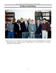
Personnel<br />
The Director of RARAF is Dr. David Brenner. The Van<br />
de Graaff accelerator facility is operated by Mr. Stephen<br />
Marino and Dr. Gerhard Randers-Pehrson. Our ranks have<br />
now swelled to a total of seven physicists, an increase of<br />
74<br />
two.<br />
Dr. Alan Bigelow, now an Associate Research Scientist,<br />
is continuing the development of the laser ion source and an<br />
optical system for 3-dimensional viewing of cells.<br />
Dr. Guy Garty, a Staff Associate, is working on the development<br />
of a stand alone microbeam, the secondary emission<br />
ion microscope (SEIM) and an inductive detector (LD 2 )<br />
for single ions.<br />
Mr. Greg Ross is a Programmer/Analyst, assisting with<br />
various programming tasks and working on the development<br />
of a stand alone microbeam and new methods of imaging<br />
cells.<br />
Dr. Giuseppe Schettino, a Post-Doctoral Fellow, arrived<br />
in November from the Gray Lab in England. He will work<br />
primarily on the development of the x-ray microbeam.<br />
Dr. Furu Zhan, a Post-Doctoral Fellow, returned to China<br />
in May, 2004.<br />
Biologists from the Center for Radiological Research are<br />
stationed at the facility in order to perform experiments:<br />
• Dr. Charles Geard, the Associate Director of the CRR,<br />
continues to spend most of each working day at RARAF.<br />
In addition to his own research, he is collaborates with<br />
some of the outside users on experiments using the single-particle<br />
microbeam facility.<br />
• Dr. Brian Ponnaiya is an Associate Research Scientist<br />
performing experiments using the track segment and microbeam<br />
irradiation facilities.<br />
• Ms. Gloria Jenkins, a Biology Technician, performs experiments<br />
on the microbeam facility for Dr. Geard.<br />
• Dr. Stephen Mitchell, a Post-Doctoral Fellow, continues<br />
to perform research involving neoplastic transformation<br />
of cells.<br />
Recent Publications of Work Performed at Raraf<br />
(2003−2004)<br />
1. Amundson SA, Do KT, Vinikoor L, Koch-Paiz CA,<br />
Bittner ML, Trent JM, Meltzer P and Fornace AJ Jr.<br />
Stress-specific signatures: Expression profiling of p53<br />
wild-type and null human cells. Oncogene Apr 11, 2005<br />
[Epub ahead of print].<br />
2. Balajee AS, Geard CR. Replication protein A and<br />
gamma-H2AX foci assembly is triggered by cellular response<br />
to DNA double-strand breaks. Exp Cell Res<br />
300:320-34, 2004.<br />
3. Balajee AS, Ponnaiya B, Baskar R and Geard CR. Induction<br />
of replication protein a in bystander cells. Radiat<br />
Res 162:677-86, 2004.<br />
4. Balajee AS and Geard CR. Replication protein A relocates<br />
into distinct nuclear foci and co-localizes with γ-<br />
H2AX in response to DNA damaging agents. (Submitted<br />
to Radiat Res, 2004.)<br />
5. Bigelow AW, Randers-Pehrson G, Kelly RP and Brenner<br />
DJ. Laser Ion Source for Columbia University’s Microbeam.<br />
Nucl Instr Meth B (in press 2005).<br />
6. Bigelow AW, Ross GJ, Randers-Pehrson G and Brenner<br />
DJ. The Columbia University Microbeam II endstation<br />
for cell imaging and irradiation. Nucl Instr Meth B (in<br />
press 2005).<br />
7. Garty G, Randers-Pehrson G and Brenner DJ. Develop-