

PDF Download - Glidewell Dental Labs
PDF Download - Glidewell Dental Labs
PDF Download - Glidewell Dental Labs

Create successful ePaper yourself
Turn your PDF publications into a flip-book with our unique Google optimized e-Paper software.
After most extractions, a loss of facial bone occurs in the<br />
vertical and horizontal dimensions. This bone loss can<br />
be regained surgically or prosthetically; however, adding<br />
facial bone around an implant requires the use of grafts,<br />
barriers, and techniques that are often more advanced<br />
than the average practitioner is prepared for. Therefore,<br />
preserving the facial and lingual surfaces with bone at the<br />
time of extraction is a more predictable procedure. If the<br />
surgeon is not familiar with these techniques, however,<br />
and their value is not communicated to the patient, the<br />
patient will likely experience a site collapse (Figs. 1a–1c).<br />
The most ideal time to augment the soft tissue profile is at<br />
the time of conventional implant placement.<br />
Figure 1c: This can give the final crown the appearance of being “stuck” on<br />
the gingiva.<br />
■ Understanding the Limitations of<br />
Stock Components<br />
Stock or standard abutments are designed in the shape<br />
of a perfect circle at the shoulder, or prosthetic base, of<br />
the abutment. The problem with this standard design is<br />
that no naturally occurring tooth is perfectly round at<br />
the alveolar crest or the cementoenamel junction (CEJ).<br />
While flared healing abutments are available, these<br />
stock components still provide only a generic solution,<br />
forming gingival contours that may be less than ideal for a<br />
specific patient.<br />
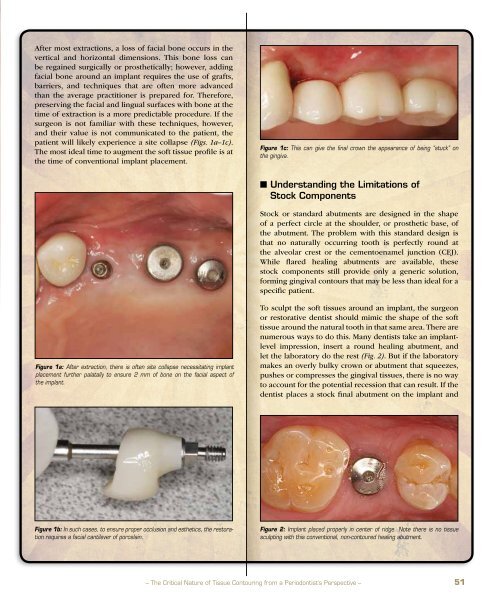
Figure 1a: After extraction, there is often site collapse necessitating implant<br />
placement further palatally to ensure 2 mm of bone on the facial aspect of<br />
the implant.<br />
To sculpt the soft tissues around an implant, the surgeon<br />
or restorative dentist should mimic the shape of the soft<br />
tissue around the natural tooth in that same area. There are<br />
numerous ways to do this. Many dentists take an implantlevel<br />
impression, insert a round healing abutment, and<br />
let the laboratory do the rest (Fig. 2). But if the laboratory<br />
makes an overly bulky crown or abutment that squeezes,<br />
pushes or compresses the gingival tissues, there is no way<br />
to account for the potential recession that can result. If the<br />
dentist places a stock final abutment on the implant and<br />
Figure 1b: In such cases, to ensure proper occlusion and esthetics, the restoration<br />
requires a facial cantilever of porcelain.<br />
Figure 2: Implant placed properly in center of ridge. Note there is no tissue<br />
sculpting with this conventional, non-contoured healing abutment.<br />
– The Critical Nature of Tissue Contouring from a Periodontist’s Perspective – 51


