The neurotoxic cascade in the ischemic penumbra
The neurotoxic cascade in the ischemic penumbra
The neurotoxic cascade in the ischemic penumbra
Create successful ePaper yourself
Turn your PDF publications into a flip-book with our unique Google optimized e-Paper software.
Radiopharmacie, Service de Médec<strong>in</strong>e Nucléaire,<br />
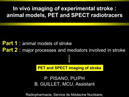
In vivo imag<strong>in</strong>g of experimental stroke :<br />
animal models, PET and SPECT radiotracers<br />
Part 1 : animal models of stroke<br />
Part 2 : major processes and mediators <strong>in</strong>volved <strong>in</strong> stroke<br />
PET and SPECT imag<strong>in</strong>g of stroke<br />
P. PISANO, PU/PH<br />
B. GUILLET, MCU, Assistant
valence and classification of acute strokes<br />
Third lead<strong>in</strong>g cause of death<br />
Lead<strong>in</strong>g cause of permanent disability<br />
No <strong>in</strong> vitro methods<br />
Acute stroke<br />
Hemorrhagic stroke<br />
Ischemic stroke 85%<br />
arachnoïd<br />
morrhage<br />
(SAH)<br />
Intra-cerebral<br />
Hemorrhage<br />
(ICH)<br />
Extra-cranial<br />
embolism<br />
Intra-cran<br />
thrombos
w to reproduce human thromboembolic stroke?
al of experimental <strong>ischemic</strong> stroke<br />
reduction of cerebral blood flow (CBF)<br />
reduction of oxygen and glucose supply<br />
to bra<strong>in</strong> tissue<br />
Size <strong>in</strong>farction predicted by<br />
<strong>in</strong>tensity<br />
duration<br />
of <strong>the</strong> CBF reduction<br />
100 %<br />
CBF Flow<br />
50 %<br />
25 %<br />
probability for <strong>in</strong>farction < 5%<br />
probability for <strong>in</strong>farction > 95%
BF reduction <strong>in</strong> focal <strong>ischemic</strong> strokes<br />
cal ischemia = reduction <strong>in</strong> blood flow to a very dist<strong>in</strong>ct specific bra<strong>in</strong> regio<br />
No blood flow<br />
<strong>the</strong> very central core of<br />
<strong>the</strong> ischemia,<br />
ut collateral circulation)<br />
from D<strong>in</strong>argl, 1999,<br />
Gradient of blood flow from <strong>the</strong> <strong>in</strong>ner core<br />
reach<strong>in</strong>g out to <strong>the</strong> limits of <strong>the</strong> <strong>ischemic</strong> area :
cal <strong>ischemic</strong> stroke by MCAO<br />
Willis circle :<br />
carotid and vertebrobasilar<br />
arterial system connected<br />
Occlusion of one of <strong>the</strong> major cerebral<br />
blood vessel middle cerebral artery (MCA<br />
X<br />
Proximal<br />
Distal<br />
X X
nes<strong>the</strong>sia Protocol and Monitor<strong>in</strong>g<br />
Anes<strong>the</strong>sia:<br />
<strong>in</strong>tubation and mechanical ventilation<br />
sevoflurane 1.2 MAC<br />
blood arterial pressure<br />
Monitor<strong>in</strong>g:<br />
temperature<br />
sevoflurane, O 2 et CO 2<br />
blood gases<br />
glycemiae<br />
Measurement and control of physiological parameters so that<br />
<strong>in</strong>farction size or cell <strong>in</strong>jury can be reproducible and consistent.
urgical protocol without craniotomy<br />
1h MCAO<br />
reperfusio<br />
A. Cérébrale<br />
Moyenne<br />
A. Ptérygo-Pallat<strong>in</strong>e<br />
A. Carotide Externe<br />
A. Carotide <strong>in</strong>terne<br />
A. Carotide Commune
utcome measurement of <strong>in</strong>jury<br />
eurological score (Longa et al., 1989).<br />
Neurobehavioral assessment<br />
S1<br />
otarod<br />
S2<br />
0<br />
1<br />
2<br />
3<br />
4<br />
no neurolocal deficit<br />
forelimb flexion<br />
unilateral circl<strong>in</strong>g<br />
sw<strong>in</strong>g<strong>in</strong>g of <strong>the</strong> body on <strong>the</strong> left side<br />
motionless<br />
Bra<strong>in</strong> <strong>in</strong>farct volume calculation<br />
mild deficit<br />
moderate deficit<br />
severe deficit<br />
unconciousness<br />
Swanson et al. JCBFM 1990<br />
h= <strong>in</strong>ter-slice<br />
distance
markers <strong>in</strong>itially developed to explore : - tumor hypoxia<br />
- myocardial<br />
ischemia<br />
Part 2 : <strong>neurotoxic</strong> <strong>cascade</strong> of<br />
ischemia<br />
PET and SPECT imag<strong>in</strong>g of <strong>penumbra</strong>
<strong>neurotoxic</strong> <strong>cascade</strong> <strong>in</strong> <strong>the</strong> <strong>ischemic</strong> <strong>penumbra</strong><br />
De Keyser et al., 1999<br />
15<br />
-O, 18 FDG, 18 F-MISO,<br />
11<br />
C-FMZ, 18F-CPFPX<br />
PLA2<br />
BBB disruption, endo<strong>the</strong>lial disturbances<br />
Cell <strong>in</strong>filtration<br />
Cell necrosis<br />
Tissue remodel<strong>in</strong>g, angiogenesis,
T imag<strong>in</strong>g of reduced CBF Phelps, Mol. Imag<strong>in</strong>g, 2004<br />
Initially, limited exclusively to <strong>the</strong> determ<strong>in</strong>ation of :<br />
Cerebral blood flow (CBF, 15O-H2O)<br />
Oxygen extraction fraction (15O-O2)<br />
Glucose metabolism (18FDG)<br />
In <strong>penumbra</strong>, oxygen extraction fraction (OEF) is <strong>in</strong>creased and neurons<br />
are functionally disturbed but still viable
ET imag<strong>in</strong>g of hypoxic cells with nitroimidazoles<br />
18F-fluoromisonidazole (18FMISO)<br />
FMISO<br />
Nitroréductase<br />
Radical anion<br />
Normoxia<br />
Diffuses<br />
Hypoxia<br />
Irreversibly bound to
ET imag<strong>in</strong>g of hypoxic cells<br />
Read et al., Neurology. 19<br />
18<br />
F-fluoromisonidazole (18FMISO) <strong>in</strong> humans<br />
22h post-<strong>in</strong>farct<br />
10d post-<strong>in</strong>farct<br />
MISO b<strong>in</strong>d<strong>in</strong>g is <strong>in</strong>dicative of :<br />
f<strong>in</strong>al CT <strong>in</strong>farct area<br />
- ongo<strong>in</strong>g tissue hypoxia<br />
- not merely recent tissue <strong>in</strong>ju
ET imag<strong>in</strong>g of hypoxic cells<br />
Ito et al., Acad Radiol 200<br />
68<br />
Ga-labeled metronidazole <strong>in</strong> a breast tumor-bear<strong>in</strong>g r
PECT imag<strong>in</strong>g of hypoxic cells<br />
Song et al., (Stroke 2003)<br />
99m<br />
Tc-metronidazole <strong>in</strong> patients with <strong>ischemic</strong> stroke<br />
EC-MN<br />
99m<br />
Tc-Neurolite®<br />
99m<br />
Tc-EC-MN
T imag<strong>in</strong>g of hypoxic cells by down-regulated receptors<br />
11<br />
C-flumazenil <strong>in</strong> cats<br />
Heiss et al., Stroke 19<br />
C I 1h 4h<br />
Defect <strong>in</strong> FMZ b<strong>in</strong>d<strong>in</strong>g predicts <strong>the</strong> size of <strong>the</strong> f<strong>in</strong>al <strong>in</strong>farct whereas
T imag<strong>in</strong>g of hypoxic cells by down-regulated receptors<br />
adenos<strong>in</strong>e A(1) receptors<br />
Tadashi et al., J Nucl Med 20<br />
• [1-methyl-( 11 )C]8-dicyclopropylmethyl-1-methyl-3-propylxanth<strong>in</strong>e (MPD<br />
•<br />
18<br />
F-CPFPX (8-cyclopentyl-3-(3 fluoropropyl)-1-propylxanth<strong>in</strong>e)<br />
-CPFPX<br />
Degree of decrease<br />
<strong>in</strong> A1R ligand b<strong>in</strong>d<strong>in</strong>g is a sensitive<br />
predictor of severe <strong>ischemic</strong> <strong>in</strong>sult<br />
Transient MCAO <strong>in</strong> cats
<strong>neurotoxic</strong> <strong>cascade</strong> <strong>in</strong> <strong>the</strong> <strong>ischemic</strong> <strong>penumbra</strong><br />
De Keyser et al., 1999<br />
-Annex<strong>in</strong><br />
PLA2<br />
BBB disruption, endo<strong>the</strong>lial disturbances<br />
Cell <strong>in</strong>filtration<br />
Cell necrosis<br />
Tissue remodel<strong>in</strong>g, angiogenesis,
apoptotic cells/necrotic cells = 1:1<br />
ag<strong>in</strong>g of apoptotic cell death<br />
ll death 4h after 2h MCAO<br />
apoptotic cells/necrotic cells = 9:1<br />
Charriaut-Marlangue C. et al., JCBFM, 19<br />
Apoptosis<br />
Necrosis
ag<strong>in</strong>g of apoptotic cell death<br />
Annex<strong>in</strong> V<br />
- 36-kDa prote<strong>in</strong><br />
- high aff<strong>in</strong>ity to phosphatidylser<strong>in</strong>e at <strong>the</strong><br />
surface of apoptotic cel<br />
- detection of <strong>the</strong> earliest stages of apoptosis<br />
SPECT<br />
– 99mTc-HYNIC-annex<strong>in</strong> V<br />
(hydraz<strong>in</strong>onicot<strong>in</strong>amide<br />
derivatized annex<strong>in</strong> V)<br />
– novel form of annex<strong>in</strong> V :<br />
PET<br />
– 18F-Annex<strong>in</strong> V<br />
– 124I-annex<strong>in</strong> V
ECT imag<strong>in</strong>g <strong>in</strong> rats submitted to<br />
2 h left MCAO followed by<br />
SPECT imag<strong>in</strong>g <strong>in</strong> rabbits suffer<strong>in</strong><br />
from neonatal hypoxic bra<strong>in</strong> <strong>in</strong>jur<br />
ECT imag<strong>in</strong>g of apoptosis<br />
99mTc-HYNIC-annex<strong>in</strong> V<br />
Mari et al., Eur J Nucl Med Mol Imag<strong>in</strong>g 2004<br />
D’Arceuil et al., Stroke 2000
T imag<strong>in</strong>g of apoptosis<br />
PET annex<strong>in</strong> V<br />
Radiolabeled annex<strong>in</strong> V PET imag<strong>in</strong>g of liver apoptosis<br />
Control<br />
Cycloheximide<br />
treated rats<br />
Untreated mice<br />
anti-Fas mAb-treated<br />
mice<br />
124<br />
I- Annex<strong>in</strong> V<br />
18<br />
F- Annex<strong>in</strong> V
<strong>neurotoxic</strong> <strong>cascade</strong> <strong>in</strong> <strong>the</strong> <strong>ischemic</strong> <strong>penumbra</strong><br />
De Keyser et al., 1999<br />
11<br />
C-PJ34<br />
PLA2<br />
BBB disruption, endo<strong>the</strong>lial disturbances<br />
Cell <strong>in</strong>filtration<br />
Cell necrosis<br />
Tissue remodel<strong>in</strong>g, angiogenesis,
T imag<strong>in</strong>g of free radical-<strong>in</strong>duced necrosis<br />
11<br />
C-PJ34 : phenanthrid<strong>in</strong>one derivative which<br />
b<strong>in</strong>ds to PARP<br />
PARP-1 is up-regulated dur<strong>in</strong>g cell necrosis<br />
streptozotoc<strong>in</strong>-<strong>in</strong>duced pancreas necrosis<br />
Control<br />
No sta<strong>in</strong><strong>in</strong>g was observed <strong>in</strong> control rats.<br />
STZ treated<br />
Accumulation of 11 C-PJ34 is shown <strong>in</strong> STZtreated<br />
rats as dark brown sta<strong>in</strong><strong>in</strong>g localized<br />
<strong>in</strong> <strong>the</strong> nuclei of islets <strong>in</strong> <strong>the</strong> rat pancreas
<strong>neurotoxic</strong> <strong>cascade</strong> <strong>in</strong> <strong>the</strong> <strong>ischemic</strong> <strong>penumbra</strong><br />
De Keyser et al., 1999<br />
11<br />
C-octanoate<br />
11<br />
C-PK11195<br />
PLA2<br />
BBB disruption, endo<strong>the</strong>lial disturbances<br />
Cell <strong>in</strong>filtration<br />
Cell necrosis<br />
Tissue remodel<strong>in</strong>g, angiogenesis,
ag<strong>in</strong>g of microglia <strong>in</strong>filtration after ischemia–reperfusion<br />
RICHARD B. BANATI GLIA (2002)<br />
peripheral<br />
benzodiazep<strong>in</strong>e<br />
receptors
T Imag<strong>in</strong>g of activated microglia <strong>in</strong>filtration<br />
Sette et al. Stroke,199<br />
11<br />
C-PK11195<br />
11C-PK11195 b<strong>in</strong>d<strong>in</strong>g after focal ischemia (MCAO) <strong>in</strong> <strong>the</strong> anes<strong>the</strong>tized baboon<br />
11<br />
C-flumazenil
T imag<strong>in</strong>g of dur<strong>in</strong>g MCAO-<strong>in</strong>duced astrocytes <strong>in</strong>filtration<br />
Kuge et al., Ann.Nucl.Med, 2<br />
11<br />
C-octanoate uptake <strong>in</strong> a dog 24h after MCAO
<strong>neurotoxic</strong> <strong>cascade</strong> <strong>in</strong> <strong>the</strong> <strong>ischemic</strong> <strong>penumbra</strong><br />
Dimeric RGD peptide<br />
Monomeric RGD peptide<br />
a 5 ß 3 <strong>in</strong>tegr<strong>in</strong> receptors<br />
VEGF receptors<br />
PLA2<br />
BBB disruption, endo<strong>the</strong>lial disturbances<br />
Cell <strong>in</strong>filtration<br />
Cell necrosis<br />
Tissue remodel<strong>in</strong>g, angiogenesis,
T imag<strong>in</strong>g of <strong>in</strong>tegr<strong>in</strong> receptors<br />
18<br />
F-FB-E[c(RGDyK)]2 dimeric ligand of avß3 <strong>in</strong>tegr<strong>in</strong> receptor<br />
<strong>in</strong>oculation of U87MG human glioblastoma cells <strong>in</strong>to mouse forebrai<br />
Tumor to contralateral background ratio = 9.5 +/- 0.8<br />
Chen et al., Mol Imag<strong>in</strong>g Biol, 2006
ECT imag<strong>in</strong>g of <strong>in</strong>tegr<strong>in</strong> receptors<br />
Meoli et al., J Cl<strong>in</strong> Invest. 200<br />
111In-RP748 : qu<strong>in</strong>olone targeted at avß3 <strong>in</strong>tegr<strong>in</strong> receptor<br />
111In-RP748, a<br />
gs with chronic myocardial <strong>in</strong>farction<br />
<strong>in</strong>creased myocardial uptake (red) <strong>in</strong> <strong>the</strong><br />
region of <strong>in</strong>jury-<strong>in</strong>duced angiogenes<br />
99m<br />
Tc-MIBI<br />
111<br />
In-RP748
ag<strong>in</strong>g of VEGF receptors<br />
VEGF is an angiogenic prote<strong>in</strong> secreted <strong>in</strong> response to<br />
hypoxia that b<strong>in</strong>ds to VEGF receptors Flt-1 and KDR<br />
overexpressed by <strong>ischemic</strong> microvasculature.
ag<strong>in</strong>g of VEGF receptors<br />
111In-labeled VEGF<br />
Lu et al., Circulation 2003<br />
Ischemic h<strong>in</strong>dlimb (rabbit)<br />
111<br />
In-labeled VEGF accumulation <strong>in</strong><br />
<strong>ischemic</strong> h<strong>in</strong>dlimb<br />
124I radiolabeled mAb aga<strong>in</strong>st VEGF Coll<strong>in</strong>gridge et al., Cancer Res. 200<br />
B<strong>in</strong>ds to angiogenic areas <strong>in</strong> tumor-bear<strong>in</strong>g mice
ture directions of stroke PET and SPECT imag<strong>in</strong>g<br />
Imag<strong>in</strong>g hypoxic cells with reporter genes<br />
Acycloguanos<strong>in</strong>e Pyrimid<strong>in</strong>e derivatives<br />
HSV-tk1<br />
HIF-1
Conclusion<br />
Most promis<strong>in</strong>g PET and SPECT radiotracers for<br />
stroke : angiogenesis radiotracers developed for<br />
tumor hypoxia<br />
Transferability from tumor to bra<strong>in</strong> hypoxia :<br />
pharmacok<strong>in</strong>etic considerations<br />
PET and SPECT track<strong>in</strong>g of stem cell <strong>the</strong>rapy of<br />
stroke by reporter genes transfected <strong>in</strong>side <strong>the</strong> cells.<br />
– Neural stem cells<br />
124I<br />
– Endo<strong>the</strong>lial progenitors
Leucocytes <strong>in</strong>filtration<br />
after ischemia–reperfusion
perimental models and Animal species<br />
enetically homogeneous<br />
ophisticated neurosensory and motor behavior measurements<br />
as outcome measures of <strong>in</strong>jury from ischemia<br />
mall animals : lissencephalic bra<strong>in</strong>s<br />
Focal cerebral ischemia (transient or permanent) :<br />
non human primate, dog, cat,<br />
rabbit, gu<strong>in</strong>ea pig, rat, mouse.<br />
Global cerebral ischemia<br />
- complete (transient) :<br />
- <strong>in</strong>complete (transient or premanent) :<br />
non human primate, dog, cat, rat,<br />
mouse, gerbil, sheep<br />
dog, cat, pig, rat, mouse, gerbil,<br />
sheep.<br />
Multifocalcerebral ischemia :<br />
Rabbit, rat.<br />
Intracerebelar hemorrhage :<br />
Subarachnoid hemorrhage and vasospasm :<br />
non human primate, dog, cat, rat,<br />
pig, rabbit.<br />
non human primate, dog, cat, rat,<br />
mouse, pig, rabbit.<br />
(from Traytsman, ILAR J. 2003; Andaluz et al., Neurosurg Cl<strong>in</strong> N Am 2


