Dermoscopy of pigmented skin lesions - Dermatology
Dermoscopy of pigmented skin lesions - Dermatology
Dermoscopy of pigmented skin lesions - Dermatology
Create successful ePaper yourself
Turn your PDF publications into a flip-book with our unique Google optimized e-Paper software.
CLINICAL REVIEW<br />
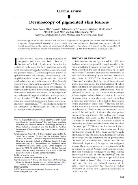
<strong>Dermoscopy</strong> <strong>of</strong> <strong>pigmented</strong> <strong>skin</strong> <strong>lesions</strong><br />
Ralph Peter Braun, MD, a Harold S. Rabinovitz, MD, b Margaret Oliviero, ARNP, MSN, b<br />
Alfred W. Kopf, MD, c and Jean-Hilaire Saurat, MD a<br />
Geneva,Switzerland;Miami,Florida;andNewYork,NewYork<br />
<strong>Dermoscopy</strong> is an in vivo method for the early diagnosis <strong>of</strong> malignant melanoma and the differential<br />
diagnosis <strong>of</strong> <strong>pigmented</strong> <strong>lesions</strong> <strong>of</strong> the <strong>skin</strong>. It has been shown to increase diagnostic accuracy over clinical<br />
visual inspection in the hands <strong>of</strong> experienced physicians. This article is a review <strong>of</strong> the principles <strong>of</strong><br />
dermoscopy as well as recent technological developments. ( J Am Acad Dermatol 2005;52:109-21.)<br />
In the last two decades a rising incidence <strong>of</strong><br />
malignant melanoma has been observed. 1-7<br />
Because <strong>of</strong> a lack <strong>of</strong> adequate therapies for<br />
metastatic melanoma, the best treatment currently<br />
is still early diagnosis and prompt surgical excision <strong>of</strong><br />
the primary cancer. 5,7 <strong>Dermoscopy</strong> (also known as<br />
epiluminescence microscopy, dermatoscopy, and<br />
amplified surface microscopy) is an in vivo method,<br />
that has been reported to be a useful tool for the early<br />
recognition <strong>of</strong> malignant melanoma. 8-11 The performance<br />
<strong>of</strong> dermoscopy has been investigated by<br />
many authors. Its use increases diagnostic accuracy<br />
between 5% and 30% over clinical visual inspection,<br />
depending on the type <strong>of</strong> <strong>skin</strong> lesion and experience<br />
<strong>of</strong> the physician. 12-16 This was confirmed by recent<br />
evidence-based publications and based on a metaanalysis<br />
<strong>of</strong> the literature. 17,18 This article is a review <strong>of</strong><br />
the principles <strong>of</strong> dermoscopy as well as recent<br />
technological developments.<br />
From the Pigmented Skin Lesion Unit, Department <strong>of</strong> <strong>Dermatology</strong>,<br />
University Hospital Geneva a ; Skin and Cancer Associates,<br />
Plantation, Fla, and Department <strong>of</strong> <strong>Dermatology</strong>, University <strong>of</strong><br />
Miami School <strong>of</strong> Medicine b ; and The Ronald O. Perelman<br />
Department <strong>of</strong> <strong>Dermatology</strong>, New York University School <strong>of</strong><br />
Medicine. c<br />
Funding sources: The work <strong>of</strong> Dr Braun has been supported by the<br />
Swiss Cancer League. Dr Kopf has the following funding<br />
sources: The Ronald O. Perelman Department <strong>of</strong> <strong>Dermatology</strong>,<br />
New York University School <strong>of</strong> Medicine, Joseph H. Hazen<br />
Foundation, Mary and Emanuel Rosenthal Foundation, Kaplan<br />
Comprehensive Cancer Center (Cancer Center Support Core<br />
Grant No. 5P30-CA-16087), Blair O. Rogers Medical Research<br />
Fund, The Rahr Family Foundation, and Stravros S. Niarchos<br />
Foundation Fund <strong>of</strong> the Skin Cancer Foundation.<br />
Conflict <strong>of</strong> interest: None.<br />
Accepted for publication November 9, 2001.<br />
Reprint requests: R. P. Braun, MD, Department <strong>of</strong> <strong>Dermatology</strong>,<br />
University Hospital Geneva, 24, rue Micheli-du-Crest, CH—<br />
1211Geneva 14, Switzerland. E-mail: braun@melanoma.ch.<br />
0190-9622/$30.00<br />
ª 2005 by the American Academy <strong>of</strong> <strong>Dermatology</strong>, Inc.<br />
doi:10.1016/j.jaad.2001.11.001<br />
HISTORY OF DERMOSCOPY<br />
Skin surface microscopy started in 1663 with<br />
Kolhaus who investigated the small vessels in the<br />
nailfold with the help <strong>of</strong> a microscope. 11,19 In 1878,<br />
Abbe described the use <strong>of</strong> immersion oil in light<br />
microscopy 20 and this principle was transferred to<br />
<strong>skin</strong> surface microscopy by the German dermatologist,<br />
Unna, in 1893. 21 He introduced the term<br />
‘‘diascopy’’ and described the use <strong>of</strong> immersion oil<br />
and a glass spatula for the interpretation <strong>of</strong> lichen<br />
planus and for the evaluation <strong>of</strong> the infiltrate in lupus<br />
erythematosus. The term ‘‘dermatoscopy’’ was introduced<br />
in 1920 by the German dermatologist<br />
Johann Saphier who published a series <strong>of</strong> communications<br />
using a new diagnostic tool resembling<br />
a binocular microscope with a built-in light source<br />
for the examination <strong>of</strong> the <strong>skin</strong>. 22-25 He used this new<br />
tool in various indications and did some interesting<br />
morphological observations on anatomical structures<br />
<strong>of</strong> the <strong>skin</strong> which indicated the high performance<br />
<strong>of</strong> his equipment. Skin surface microscopy<br />
was further developed in the United States by<br />
Goldman in the 1950s. He published a series <strong>of</strong><br />
interesting articles on new devices on what he called<br />
‘‘<strong>Dermoscopy</strong>.’’ 26-29 He was the first dermatologist to<br />
use this new technique for the evaluation <strong>of</strong> <strong>pigmented</strong><br />
<strong>skin</strong> <strong>lesions</strong>. In 1971, Rona MacKie 30 clearly<br />
identified, for the first time, the advantage <strong>of</strong> surface<br />
microscopy for the improvement <strong>of</strong> preoperative<br />
diagnosis <strong>of</strong> <strong>pigmented</strong> <strong>skin</strong> <strong>lesions</strong> and for the<br />
differential diagnosis <strong>of</strong> benign versus malignant<br />
<strong>lesions</strong>. These investigations were continued mainly<br />
in Europe by several Austrian and German groups.<br />
The first Consensus Conference on Skin Surface<br />
Microscopy was held in 1989 in Hamburg 31 and the<br />
Consensus Netmeeting on <strong>Dermoscopy</strong>, which was<br />
held in 2001 in Rome 32 (http://www.dermoscopy.<br />
org), was the first international meeting <strong>of</strong> its<br />
kind. Today dermoscopy has become a routine<br />
technique in Europe and is gaining acceptance in<br />
other countries.<br />
109
110 Braun et al<br />
Table I. Vascular architecture <strong>of</strong> <strong>pigmented</strong> <strong>skin</strong> <strong>lesions</strong> according to Kreusch and Koch 57<br />
PHYSICAL ASPECTS<br />
Light is either reflected, dispersed, or absorbed by<br />
the stratum corneum because <strong>of</strong> its refraction index<br />
and its optical density, which is different from air. 33<br />
Thus, deeper underlying structures cannot be adequately<br />
visualized. However, when various immersion<br />
liquids are used, they render the <strong>skin</strong> surface<br />
translucent and reduce the reflection, so that underlying<br />
structures are readily visible. The application<br />
<strong>of</strong> a glass plate flattens the <strong>skin</strong> surface and<br />
provides an even surface. Optical magnification is<br />
used for examination. Taken together, these optical<br />
means allow the visualization <strong>of</strong> certain epidermal,<br />
dermo-epidermal, and dermal structures.<br />
MATERIAL FOR DERMOSCOPY<br />
<strong>Dermoscopy</strong> requires optical magnification and<br />
liquid immersion. This can be performed with very<br />
simple, inexpensive equipment. 34,35 Specially designed<br />
handheld devices with 10 to 20 times magnification<br />
are commercially available (Dermatoscope<br />
[Heine AG]; DermoGenius Basic [Rodenstock<br />
Präzisionsoptik]; Episcope [Welch-Allyn]; DermLite<br />
[3Gen, LLC]). Photographic documentation can be<br />
performed with a dermoscopic attachment to a standard<br />
camera (Dermaphot, Heine, AG) which can be<br />
used also with some digital cameras. Most recently,<br />
digital cameras have been designed that are attached<br />
to computers. This allows easy storage, retrieval, and<br />
follow-up <strong>of</strong> <strong>pigmented</strong> <strong>skin</strong> <strong>lesions</strong>. For dermatologists<br />
with less experience in dermoscopy, some <strong>of</strong><br />
the systems may <strong>of</strong>fer the possibility <strong>of</strong> computerassisted<br />
diagnosis for malignant melanoma or for<br />
consulting an expert through telemedicine.<br />
Morphological aspect Type <strong>of</strong> pathology<br />
Tree-like vessels Thick, arborizing vessels, superficial Pigmented BCC <strong>of</strong> any type (discrete in<br />
superficial BCCs)<br />
Corona vessels ‘‘Surround’’ the tumor<br />
Thinner than tree-like vessels<br />
Less curved than tree-like vessels<br />
Sebaceous gland hyperplasia<br />
Comma-shaped vessels Short, strong, curved,<br />
Located on the tumor<br />
Short distance, parallel to <strong>skin</strong> surface<br />
Dermal nevi<br />
Point vessels Short capillary loops Thin malignant melanomas<br />
Dense packed red points Epithelial tumors such as actinic keratosis,<br />
Not in the holes <strong>of</strong> the pigment network Bowen’s disease, etc (short vertical height)<br />
Hairpin vessels Long capillary loops <strong>of</strong> thicker tumors at the<br />
border<br />
Thick melanomas<br />
Whitish halo in tumors with keratin SCC keratoacanthoma, seborrheic keratosis<br />
BCC, Basal cell carcinoma; SCC, squamous cell carcinoma.<br />
JAM ACAD DERMATOL<br />
JANUARY 2005<br />
DERMOSCOPIC CRITERIA<br />
Colors<br />
The use <strong>of</strong> dermoscopy allows the identification<br />
<strong>of</strong> many different structures and colors, not seen by<br />
the naked eye.<br />
Colors play an important role in dermoscopy.<br />
Common colors are light brown, dark brown, black,<br />
blue, blue-gray, red, yellow, and white. The most<br />
important chromophore in melanocytic neoplasms is<br />
melanin. 11,13,36 The color <strong>of</strong> melanin essentially<br />
depends on its localization in the <strong>skin</strong>. The color<br />
black is due to melanin located in the stratum<br />
corneum and the upper epidermis, light to dark<br />
brown in the epidermis, gray to gray-blue in the<br />
papillary dermis and steel-blue in the reticular<br />
dermis. 11,13 The color blue occurs when there is<br />
melanin localized within the deeper parts <strong>of</strong> the <strong>skin</strong><br />
because the portions <strong>of</strong> visible light with shorter<br />
wavelengths (blue-violet end <strong>of</strong> spectrum) are more<br />
dispersed than portions with longer wavelengths<br />
(red end <strong>of</strong> visible spectrum). 37,38 The color red is<br />
associated with an increased number or dilatation <strong>of</strong><br />
blood vessels, trauma, or neovascularization. The<br />
color white is <strong>of</strong>ten caused by regression and/or<br />
scarring. 11<br />
Dermoscopic structures<br />
In this context we will use the nomenclature as<br />
proposed by the recent Consensus Netmeeting (held<br />
in Rome in 2001) with some revisions 32 :<br />
Pigment network. The pigment network is<br />
a grid-like (honeycomb-like) network consisting<br />
<strong>of</strong> <strong>pigmented</strong> ‘‘lines’’ and hypo<strong>pigmented</strong><br />
‘‘holes.’’ 11,13,36,37,39 The anatomic basis <strong>of</strong> the
JAM ACAD DERMATOL<br />
VOLUME 52, NUMBER 1<br />
Fig 1. Two-step procedure for the classification <strong>of</strong> <strong>pigmented</strong><br />
<strong>skin</strong> <strong>lesions</strong>. Adapted from Argenziano G et al.<br />
J Am Acad Dermatol 2003;48:679-93. 32<br />
pigment network is either melanin pigment in<br />
keratinocytes, or in melanocytes along the dermoepidermal<br />
junction. 40 The reticulation (network) represents<br />
the rete ridge pattern <strong>of</strong> the epidermis. 41-43<br />
The relatively hypomelanotic holes in the network<br />
correspond to tips <strong>of</strong> the dermal papillae and the<br />
overlying suprapapillary plates <strong>of</strong> the epidermis.<br />
13,36,37<br />
The pigment network can be either typical or<br />
atypical. A typical network is relatively uniform,<br />
regularly meshed, homogeneous in color, and usually<br />
thinning out at the periphery. 36,39,44 An atypical<br />
network is nonuniform, with darker and/or broadened<br />
lines and ‘‘holes’’ that are heterogeneous in<br />
area and shape. The lines are <strong>of</strong>ten hyper<strong>pigmented</strong><br />
and may end abruptly at the periphery. 36,39,44<br />
If the rete ridges are short or less <strong>pigmented</strong>, the<br />
pigment network may not be visible. Areas devoid <strong>of</strong><br />
any network (but without signs <strong>of</strong> regression) are<br />
called ‘‘structureless areas.’’ 11,45<br />
Dots. Dots are small, round structures less than<br />
0.1 mm in diameter, which may be black, brown,<br />
gray, or blue-gray. 11,13,32,36 Black dots are caused by<br />
pigment accumulation in the stratum corneum and in<br />
the upper part <strong>of</strong> the epidermis. 11,37,42,46 Brown dots<br />
represent focal melanin accumulations at the dermoepidermal<br />
junction. 47 Gray-blue granules (peppering)<br />
are caused by tiny melanin structures in the<br />
papillary dermis. Gray-blue or blue granules are due<br />
to loose melanin, fine melanin particles or melanin<br />
‘‘dust’’ in melanophages or free in the deep papillary<br />
or reticular dermis. 11,37,42,46<br />
Globules. Globules are symmetrical, round to<br />
oval, well-demarcated structures that may be brown,<br />
black, or red. 11,13,32,36 They have a diameter which is<br />
usually larger than 0.1 mm and correspond to nests<br />
<strong>of</strong> <strong>pigmented</strong> benign or malignant melanocytes,<br />
clumps <strong>of</strong> melanin, and/or melanophages situated<br />
Braun et al 111<br />
Fig 2. Algorithm for the determination <strong>of</strong> melanocytic<br />
versus nonmelanocytic <strong>lesions</strong> according to the proposition<br />
<strong>of</strong> the Board <strong>of</strong> the Consensus Netmeeting. Adapted<br />
from Argenziano G et al. J Am Acad Dermatol 2003;48:<br />
679-93. 32<br />
usually in the lower epidermis, at the dermoepidermal<br />
junction, or in the papillary dermis. 11,37,42,46<br />
Both dots and globules may occur in benign as<br />
well as in malignant melanocytic proliferations. In<br />
benign <strong>lesions</strong>, they are rather regular in size and<br />
shape and quite evenly distributed (frequently in the<br />
center <strong>of</strong> a lesion). 32,36 In melanomas they tend to<br />
vary in size and shape and are frequently found in<br />
the periphery <strong>of</strong> <strong>lesions</strong>. 32,36,48<br />
Branched streaks. Branched streaks are an<br />
expression <strong>of</strong> an altered <strong>pigmented</strong> network in which<br />
the network becomes disrupted or broken up. 11,32,45<br />
Their pathological correlations are remnants <strong>of</strong> <strong>pigmented</strong><br />
rete ridges and bridging nests <strong>of</strong> melanocytic<br />
cells within the epidermis and papillary dermis. 11<br />
Radial streaming. Radial streaming appears as<br />
radially and asymmetrically arranged, parallel linear<br />
extensions at the periphery <strong>of</strong> a lesion. 13,49 Histologically,<br />
they represent confluent <strong>pigmented</strong> junctional<br />
nests <strong>of</strong> <strong>pigmented</strong> melanocytes. 13,36<br />
Pseudopods. Pseudopods represent fingerlike<br />
projections <strong>of</strong> dark pigment (brown to black) at the<br />
periphery <strong>of</strong> the lesion. 13,49,50 They may have small<br />
knobs at their tips, and are either connected to the<br />
pigment network or directly connected to the tumor<br />
body. 13,50 They correspond as well to intraepidermal<br />
or junctional confluent radial nests <strong>of</strong> melanocytes.<br />
13,50 Menzies et al 49 found pseudopods to be<br />
one <strong>of</strong> the most specific features <strong>of</strong> superficial<br />
spreading melanoma.<br />
Streaks. ‘‘Streaks’’ is a term used by some<br />
authors interchangeably with radial streaming or
112 Braun et al<br />
Fig 3. A, Macroscopic picture <strong>of</strong> a superficial spreading malignant melanoma (Breslow<br />
thickness 0.52 mm; Clark level II). B, <strong>Dermoscopy</strong> <strong>of</strong> A shows (atypical) pigment network and<br />
branched streaks and can therefore be considered a melanocytic lesion.<br />
Fig 4. A, Macroscopic picture <strong>of</strong> a blue nevus. B, <strong>Dermoscopy</strong> <strong>of</strong> A shows steel-blue areas (no<br />
pigment network, no aggregated globules, no branched streaks).<br />
Fig 5. A, Macroscopic picture <strong>of</strong> a seborrheic keratosis. B, <strong>Dermoscopy</strong> <strong>of</strong> A shows comedolike<br />
openings (a), multiple milia-like cysts (b), and fissures (c).<br />
Fig 6. A, Macroscopic picture <strong>of</strong> a seborrheic keratosis. B, <strong>Dermoscopy</strong> <strong>of</strong> A shows comedolike<br />
openings and multiple milia-like cysts.<br />
JAM ACAD DERMATOL<br />
JANUARY 2005
JAM ACAD DERMATOL<br />
VOLUME 52, NUMBER 1<br />
Fig 7. A, Macroscopic picture <strong>of</strong> a basal cell carcinoma. B, <strong>Dermoscopy</strong> <strong>of</strong> A shows maple<br />
leafelike areas, ovoid nests, and arborized telangiectasia.<br />
Fig 8. A, Macroscopic picture <strong>of</strong> a basal cell carcinoma. B, <strong>Dermoscopy</strong> <strong>of</strong> A shows multiple<br />
spoke wheel areas.<br />
Fig 9. A, Macroscopic picture <strong>of</strong> an angioma. B, <strong>Dermoscopy</strong> <strong>of</strong> A shows red lagoons.<br />
pseudopods. This is because both these structures<br />
have the same histopathological correlation. 11,37,42,46<br />
Streaks can be irregular, when they are unevenly<br />
distributed (malignant melanoma), or regular (symmetrical<br />
radial arrangement over the entire lesion).<br />
The latter is particularly found in the <strong>pigmented</strong><br />
spindle cell nevi (Reed’s nevi). 51-53<br />
Structureless areas. Structureless areas represent<br />
areas devoid <strong>of</strong> any discernible structures (eg,<br />
globules, network). They tend to be hypo<strong>pigmented</strong>,<br />
which is due to the absence <strong>of</strong> pigment or<br />
diminution <strong>of</strong> pigment intensity within a <strong>pigmented</strong><br />
<strong>skin</strong> lesion. 11<br />
Blotches. A blotch (also called black lamella by<br />
some authors) is caused by a large concentration <strong>of</strong><br />
Braun et al 113<br />
melanin pigment localized throughout the epidermis<br />
and/or dermis visually obscuring the underlying<br />
structures. 41-43,46<br />
Regression pattern. Regression appears as<br />
white scar-like depigmentation (lighter than the surrounding<br />
<strong>skin</strong>) or ‘‘peppering’’ (speckled multiple<br />
blue-gray granules within a hypo<strong>pigmented</strong> area).<br />
Histologically, regression shows fibrosis, loss <strong>of</strong> pigmentation,<br />
epidermal thinning, effacement <strong>of</strong> the rete<br />
ridges, and melanin granules free in the dermis or in<br />
melanophages scattered in the papillary dermis. 43,46<br />
Blue-white veil. Blue-white veil is an irregular,<br />
indistinct, confluent blue pigmentation with<br />
an overlying white, ground-glass haze. 13,32 The<br />
pigmentation cannot occupy the entire lesion.
114 Braun et al<br />
Table II. Pattern analysis according to Pehamberger et al 39 (modified)<br />
Lentigo simplex Junctional nevus Compound nevus Dermal nevus Blue nevus<br />
Regular pigment<br />
network without<br />
interruptions<br />
Regular border, thins<br />
out at periphery<br />
Black dots over grids<br />
<strong>of</strong> pigment<br />
network<br />
Brown-black globules<br />
at center <strong>of</strong> the<br />
lesion<br />
Regular pigment<br />
network without<br />
interruptions<br />
Regular border, thins<br />
out at periphery<br />
Heterogeneous holes<br />
<strong>of</strong> pigment<br />
network<br />
Histopathologically this corresponds to an aggregation<br />
<strong>of</strong> heavily <strong>pigmented</strong> cells or melanin in the<br />
dermis (blue color) in combination with a compact<br />
orthokeratosis. 13,32,36,43,46,54<br />
Vascular pattern. Pigmented <strong>skin</strong> <strong>lesions</strong> may<br />
have dermoscopically visible vascular patterns,<br />
which include ‘‘comma vessels,’’ ‘‘point vessels,’’<br />
‘‘tree-like vessels,’’ ‘‘wreath-like vessels,’’ and ‘‘hairpin-like<br />
vessels’’ (Table I). 55-57 Atypical vascular<br />
patterns may include linear, dotted, or globular red<br />
structures irregularly distributed within the lesion.<br />
32,36,58,59 Some <strong>of</strong> the vascular patterns may be<br />
caused by neovascularization. For the evaluation <strong>of</strong><br />
vascular patterns, there has to be as little pressure as<br />
possible on the lesion during examination because<br />
otherwise the vessels are simply compressed and<br />
will not be visible. The use <strong>of</strong> ultrasound gel for<br />
immersion helps to reduce the pressure necessary<br />
for the best evaluation <strong>of</strong> the <strong>skin</strong> lesion. 57<br />
Milia-like cysts. Milia-like cysts are round whitish<br />
or yellowish structures which are mainly seen in<br />
seborrheic keratosis.* They correspond to intraepidermal<br />
keratin-filled cysts and may also be seen in<br />
congenital nevi as well as in some papillomatous<br />
melanocytic nevi. At times, milia-like cysts are<br />
<strong>pigmented</strong>, and thus, can resemble globules.<br />
*References 8, 11, 13, 32, 36, 37, 49, 55, 60-63.<br />
Comedo-like openings (crypts, pseud<strong>of</strong>ollicular<br />
openings). Comedo-like openings (with<br />
‘‘blackhead-like plugs’’) are mainly seen in seborrheic<br />
keratoses or in some rare cases in papillomatous<br />
melanocytic nevi. y The keratin-filled invaginations<br />
<strong>of</strong> the epidermis correspond to the comedo-like<br />
structures histopathologically.<br />
Regular pigment<br />
network without<br />
interruptions<br />
Regular border, thins<br />
out at periphery<br />
Heterogeneous holes<br />
<strong>of</strong> pigment<br />
network<br />
No criteria for<br />
melanocytic lesion<br />
Brown globules Brown globules Homogeneous<br />
colors<br />
Homogeneous colors Homogeneous colors Symmetric papular<br />
appearance<br />
All criteria for<br />
melanocytic lesion<br />
possible<br />
Color heterogeneity<br />
possible<br />
Steel-blue areas<br />
No pigment network No pigment network<br />
Brown globules lll-defined<br />
White veils are<br />
possible<br />
‘‘Pseudonetwork’’ No pseudonetwork<br />
‘‘Comma’’-shaped<br />
blood vessels<br />
JAM ACAD DERMATOL<br />
JANUARY 2005<br />
y References 8, 11, 13, 32, 36, 37, 49, 55, 60-64.<br />
Fissures and ridges (‘‘brain-like appearance’’).<br />
Fissures are irregular, linear keratin-filled<br />
depressions, commonly seen in seborrheic keratoses.<br />
63 They may also be seen in melanocytic nevi<br />
with congenital patterns and in some dermal melanocytic<br />
nevi. Multiple fissures might give a ‘‘brainlike<br />
appearance’’ to the lesion. 32,36,63,65 This pattern<br />
has also been named ‘‘gyri and sulci’’ or ‘‘mountain<br />
and valley pattern’’ by some authors. 11<br />
Fingerprint-like structures. Some flat seborrheic<br />
keratoses (also known as solar lentigines) can<br />
show tiny ridges running parallel and producing<br />
a pattern that resembles fingerprints. 11,32,65,66<br />
Moth-eaten border. Some flat seborrheic keratoses<br />
(mainly on the face) have a concave border<br />
so that the pigment ends with a curved structure,<br />
which has been compared to a moth-eaten garment.<br />
11,13,32,63,65,66<br />
Leaf-like areas. Leaf-like areas (maple leafelike<br />
areas) are seen as brown to gray-blue discrete<br />
bulbous blobs, sometimes forming a leaf-like pattern.*<br />
Their distribution reminds one <strong>of</strong> the shape<br />
<strong>of</strong> finger pads. In absence <strong>of</strong> a pigment network,<br />
they are suggestive <strong>of</strong> <strong>pigmented</strong> basal cell carcinoma.<br />
11,32,67<br />
*References 8, 9, 11, 13, 32, 36, 37, 39, 55, 65,<br />
67, 68.<br />
Spoke wheelelike structures. Spoke wheele<br />
like structures are well-circumscribed, brown to<br />
gray-blue-brown, radial projections meeting at<br />
a darker brown central hub. 11,32,67 In the absence<br />
<strong>of</strong> a pigment network, they are highly suggestive <strong>of</strong><br />
basal cell carcinoma.
JAM ACAD DERMATOL<br />
VOLUME 52, NUMBER 1<br />
Table II. Cont’d<br />
Malignant melanoma Atypical (Clark) nevus Angioma Seborrheic keratosis Pigmented BCC<br />
Heterogeneous<br />
(colors and<br />
structures)<br />
Asymmetry (colors<br />
Irregular pigment<br />
network with<br />
interruptions<br />
Table III. ABCD rule <strong>of</strong> dermoscopy according to Stoiz et al (modified) 11,45<br />
Large blue-gray ovoid nests. Ovoid nests are<br />
large, well-circumscribed, confluent or near-confluent,<br />
<strong>pigmented</strong> ovoid areas, larger than globules,<br />
and not intimately connected to a <strong>pigmented</strong><br />
tumor body. 11,32,67 When a network is absent,<br />
ovoid nests are highly suggestive <strong>of</strong> basal cell<br />
carcinoma.<br />
No features <strong>of</strong><br />
melanocytic lesion<br />
No features for<br />
melanocytic lesion<br />
Braun et al 115<br />
No features for<br />
melanocytic lesion<br />
Heterogeneous holes No pigment network Pigment network Maple-leafelike<br />
and structures)<br />
usually absent pigmentation<br />
Irregular pigment Irregular border Red, red-blue, or Milia-like cysts Telangiectasia<br />
network<br />
red-black lagoons,<br />
( globules, saccules)<br />
Irregular border with Heterogeneity <strong>of</strong> Abrupt border cut-<strong>of</strong>f Pseud<strong>of</strong>ollicular Tree-like blood<br />
abrupt peripheral colors<br />
openings,<br />
vessels<br />
margin<br />
comedo-like<br />
openings (plugs)<br />
Structureless areas Gray-white veil Rough surface ‘‘Dirty’’ gray-brown to<br />
gray-black colors<br />
Gray-blue or red-rose Absence <strong>of</strong> primary<br />
Abrupt border cut-<strong>of</strong>f<br />
veils<br />
criteria for<br />
malignant<br />
melanoma<br />
Red<br />
Pseudopods/radial<br />
streaming<br />
Point and hairpin<br />
vessels<br />
Opaque gray-brown<br />
colors<br />
Points Weight factor Subscore range<br />
Asymmetry Complete symmetry 0 1.3 0-2.6<br />
Asymmetry in 1 axis 1<br />
Asymmetry in 2 axis 2<br />
Border 8 segments, 1 point for abrupt cut-<strong>of</strong>f <strong>of</strong> pigment 0-8 0.1 0-0.8<br />
Color 1 point for each color: 1-6 0.5 0.5-3.0<br />
White<br />
Red<br />
Light brown<br />
Dark brown<br />
Black<br />
Blue-gray<br />
Differential structures 1 point for every structure: 1-5 0.5 0.5-2.5<br />
Pigment network<br />
Structureless areas<br />
Dots<br />
Globules<br />
Streaks<br />
Total score range: 1.0-8.9<br />
Multiple blue-gray globules. Multiple bluegray<br />
globules are round, well-circumscribed structures<br />
that are, in the absence <strong>of</strong> a pigment network,<br />
highly suggestive <strong>of</strong> basal cell carcinoma. 11,32,67<br />
They have to be differentiated from multiple bluegray<br />
dots (which correspond to melanophages and<br />
melanin dust).
116 Braun et al<br />
Table IV. The 7-point checklist according to<br />
Argenziano et al 44<br />
Criteria<br />
Major criteria<br />
7-point score<br />
Atypical pigment network 2<br />
Blue-white veil 2<br />
Atypical vascular pattern<br />
Minor criteria<br />
2<br />
Irregular streaks 1<br />
Irregular pigmentation 1<br />
Irregular dots/globules 1<br />
Regression structures 1<br />
DIFFERENTIAL DIAGNOSIS OF<br />
PIGMENTED LESIONS OF THE SKIN<br />
There are many publications on the subject <strong>of</strong> the<br />
differential diagnosis <strong>of</strong> <strong>pigmented</strong> <strong>lesions</strong> <strong>of</strong> the<br />
<strong>skin</strong>. The 5 algorithms most commonly used are<br />
pattern analysis 8,39,62 ; the ABCD rule <strong>of</strong> dermoscopy<br />
11,45,70 ; the 7-point checklist 32,36,44 ; the<br />
Menzies method 13,32,49 ; and the revised pattern<br />
analysis. 71<br />
The Board <strong>of</strong> the Consensus Netmeeting agreed<br />
on a two-step procedure for the classification <strong>of</strong><br />
<strong>pigmented</strong> <strong>lesions</strong> <strong>of</strong> the <strong>skin</strong> (Fig 1). A similar<br />
approach has been proposed by other authors in<br />
the past.<br />
The first step is the differentiation between<br />
a melanocytic and a nonmelanocytic lesion. For<br />
this decision, the algorithm in Fig 2 is used.<br />
Are aggregated globules, pigment network,<br />
branched streaks (Fig 3), homogeneous blue pigmentation<br />
(blue nevus: Fig 4), or a parallel pattern<br />
(palms, soles, and mucosa) visible? If this is the case,<br />
the lesion should be considered as a melanocytic<br />
lesion (Fig 3). If not, the lesion should be evaluated<br />
for the presence <strong>of</strong> comedo-like plugs, multiple<br />
milia-like cysts, and comedo-like openings, irregular<br />
crypts, light brown fingerprint-like structures, or<br />
‘‘fissures and ridges’’ (brain-like appearance) pattern.<br />
If so, the lesion is suggestive <strong>of</strong> a seborrheic<br />
keratosis (Figs 5 and 6). If not, the lesion has to be<br />
evaluated for the presence <strong>of</strong> arborizing blood<br />
vessels (telangiectasia), leaf-like areas, large bluegray<br />
ovoid nests, multiple blue-gray globules, spoke<br />
wheel areas, or ulceration. If present, the lesion is<br />
suggestive <strong>of</strong> basal cell carcinoma (Figs 7 and 8). If<br />
not, one has to look for red or red-blue (to black)<br />
lagoons. If these structures are present, the lesion<br />
should be considered a hemangioma (Fig 9) oran<br />
angiokeratoma. If all the preceding questions were<br />
answered with ‘‘no,’’ the lesion should still be<br />
considered to be a melanocytic lesion.<br />
Table V. ‘‘The Menzies Method’’ according to<br />
Menzies et al 13,49<br />
Negative features<br />
Point and axial symmetry <strong>of</strong> pigmentation<br />
Presence <strong>of</strong> a single color<br />
Positive features<br />
Blue-white veil<br />
Multiple brown dots<br />
Pseudopods<br />
Radial streaming<br />
Scar-like depigmentation<br />
Peripheral black dots-globules<br />
Multiple colors (5 or 6)<br />
Multiple blue/gray dots<br />
Broadened network<br />
JAM ACAD DERMATOL<br />
JANUARY 2005<br />
Once the lesion is identified to be <strong>of</strong> melanocytic<br />
origin, the decision has to be made if the melanocytic<br />
lesion is benign, suspect, or malignant. To accomplish<br />
this, 4 different approaches are the most<br />
commonly used.<br />
Pattern analysis (Pehamberger et al)<br />
Pattern recognition has historically been used by<br />
clinicians and histopathologists to differentiate benign<br />
<strong>lesions</strong> from malignant neoplasms. A similar<br />
process has been found useful with dermoscopy,<br />
and has been termed ‘‘pattern analysis.’’ It allows<br />
distinction between benign and malignant growth<br />
features. It was described by Pehamberger and<br />
colleagues based on the analysis <strong>of</strong> more than 7000<br />
<strong>pigmented</strong> <strong>skin</strong> <strong>lesions</strong>. 8,39,62 Table II shows the<br />
typical patterns <strong>of</strong> some common, <strong>pigmented</strong> <strong>skin</strong><br />
<strong>lesions</strong> using pattern analysis.<br />
ABCD rule <strong>of</strong> dermatoscopy (Stolz et al)<br />
The ABCD rule <strong>of</strong> dermatoscopy, described by<br />
Stolz et al in 1993 was based on an analysis <strong>of</strong> 157<br />
<strong>pigmented</strong> <strong>skin</strong> <strong>lesions</strong>. 11,70 The complete ABCD<br />
rule is explained in Table III.<br />
For the evaluation <strong>of</strong> asymmetry, the lesion is<br />
divided into 4 segments (2 perpendicular axes). The<br />
axes are oriented so that the lowest asymmetry is<br />
obtained. For asymmetry in both axes, a value <strong>of</strong> 2 is<br />
obtained. To calculate the subscore, the value <strong>of</strong> each<br />
ABCD category has to be multiplied by the corresponding<br />
weight factor. To obtain the total score<br />
value, the different ABCD subscores have to be<br />
added.<br />
The total score ranges from 1 to 8.9. A lesion with<br />
a total score greater than 5.45 should be considered<br />
as melanoma. A lesion with a total score <strong>of</strong> 4.75<br />
or less can be considered as benign. A lesion with<br />
a score value between 4.75 and 5.45 should be
JAM ACAD DERMATOL<br />
VOLUME 52, NUMBER 1<br />
Table VI. Pattern <strong>of</strong> benign and malignant <strong>lesions</strong><br />
Benign Malignant<br />
Dots Centrally located or situated right on the Unevenly distributed and scattered focally at<br />
network<br />
the periphery<br />
Globules Uniform in size, shape, and color, symmetrically Globules that are unevenly distributed and<br />
located at the periphery, centrally located, or when reddish are highly suggestive <strong>of</strong><br />
uniform throughout the lesion as in a<br />
cobblestone pattern<br />
melanoma<br />
Streaks Radial streaming or pseudopods tend to be Radial streaming or pseudopods tend to be<br />
symmetrical and uniform at the periphery focal and irregular at periphery<br />
Blue-white veil Tends to be centrally located Tends to be asymmetrically located or diffuse<br />
almost over entire lesion<br />
Blotch Centrally located or may be diffuse<br />
Asymmetrically located or there are <strong>of</strong>ten<br />
hyper<strong>pigmented</strong> area that extends almost to<br />
periphery <strong>of</strong> the lesion<br />
multiple asymmetrical blotches<br />
Network Typical network that consists <strong>of</strong> light to dark Atypical network that may be nonuniform with<br />
uniform <strong>pigmented</strong> lines and<br />
black/brown or gray thickened lines and<br />
hypo<strong>pigmented</strong> holes<br />
holes <strong>of</strong> different sizes and shapes<br />
Network borders Either fades into the periphery or is<br />
symmetrically sharp<br />
Focally sharp<br />
considered ‘‘suspicious’’ and should therefore be<br />
monitored closely or removed. 11,70<br />
7-point checklist<br />
In 1998 Argenziano and colleagues described a 7point<br />
checklist based on the analysis <strong>of</strong> 342 <strong>pigmented</strong><br />
<strong>skin</strong> <strong>lesions</strong>. 32,36,44 They distinguish 3 major<br />
criteria and 4 minor criteria (Table IV). Each major<br />
criterion has a score <strong>of</strong> 2 points while each minor<br />
criterion has a score <strong>of</strong> 1 point. A minimum total<br />
score <strong>of</strong> 3 is required for the diagnosis <strong>of</strong> malignant<br />
melanoma.<br />
Menzies method<br />
In the Menzies method 13,32,49 for diagnosing<br />
melanoma, both <strong>of</strong> the following negative features<br />
must not be found: a single color (tan, dark brown,<br />
gray, black, blue, and red, but white is not considered)<br />
and ‘‘point and axial symmetry <strong>of</strong> pigmentation’’<br />
(refers to pattern symmetry around any axis<br />
through the center <strong>of</strong> the lesion). This does not<br />
require the lesion to have symmetry <strong>of</strong> shape.<br />
Additionally, at least one positive feature must be<br />
found (Table V).<br />
Exceptions to the algorithms<br />
The ABCD rule is not applicable for <strong>pigmented</strong><br />
<strong>lesions</strong> on the palms, soles, or face. 11 Palms and soles<br />
have a particular anatomy which is characterized by<br />
marked orthokeratosis and the presence <strong>of</strong> sulci and<br />
gyri. The sweat ducts join the surface at the summits<br />
<strong>of</strong> the gyri. 11,32 A classification <strong>of</strong> 10 different<br />
dermoscopy patterns on the palms and soles has<br />
been proposed by Saida et al. 72<br />
Braun et al 117<br />
The face has a very particular anatomic architecture<br />
especially concerning the dermoepidermal<br />
junction where rete ridges are shorter. That is why<br />
facial <strong>lesions</strong> <strong>of</strong>ten do not exhibit a regular pigment<br />
network. <strong>Dermoscopy</strong> shows a broadened pigment<br />
reticulation which is called a ‘‘pseudonetwork.’’ This<br />
does not correspond to the projection <strong>of</strong> <strong>pigmented</strong><br />
rete ridges. It is due to a homogeneous pigmentation<br />
which is interrupted by the surface openings <strong>of</strong> the<br />
adnexal structures. 11,66,73<br />
The differential diagnosis <strong>of</strong> a pseudonetwork is<br />
solar lentigo, seborrheic keratosis, lentigo simplex,<br />
melanoma in situ, lichen planuselike keratosis, and<br />
<strong>pigmented</strong> actinic keratosis. 11,66,73 These <strong>lesions</strong><br />
are <strong>of</strong>ten difficult to distinguish dermoscopically.<br />
However, when there are multiple colors and<br />
a broadened, thickened, and irregular ‘‘pseudonetwork,’’<br />
melanoma is <strong>of</strong>ten the diagnosis suggested.<br />
Other, more specific characteristics include an ‘‘annular<br />
granular’’ or ‘‘rhomboidal pattern.’’ 11,66,73<br />
Revised pattern analysis<br />
The overall general appearance <strong>of</strong> color, architectural<br />
order, symmetry <strong>of</strong> pattern, and homogeneity<br />
(CASH) are important components in distinguishing<br />
these two groups. Benign melanocytic <strong>lesions</strong> tend<br />
to have few colors, architectural order, symmetry <strong>of</strong><br />
pattern, or homogeneity. Malignant melanoma <strong>of</strong>ten<br />
has many colors and much architectural disorder,<br />
asymmetry <strong>of</strong> pattern, and heterogeneity.<br />
The reticular pattern or network pattern is the<br />
most common global feature in melanocytic <strong>lesions</strong>.<br />
This pattern represents the junctional component
118 Braun et al<br />
<strong>of</strong> a melanocytic nevus (Clark nevus, dysplastic<br />
nevus). 32,36,71<br />
Another pattern is the so-called globular pattern.<br />
It is characterized by the presence <strong>of</strong> numerous<br />
‘‘aggregated globules.’’ This pattern is commonly<br />
seen in a congenital nevus, superficial type. 32,36,71<br />
The cobblestone pattern is very similar to the<br />
globular pattern but is composed <strong>of</strong> closer aggregated<br />
globules, which are somehow angulated, resembling<br />
cobblestones.<br />
The homogeneous pattern appears as diffuse<br />
pigmentation, which might be brown, gray-blue,<br />
gray-black, or reddish black. 32,36,71 No pigment<br />
network or any other distinctive dermoscopy structure<br />
is found. An example is the homogeneous steelblue<br />
color seen in blue nevi.<br />
The so-called starburst pattern is characterized by<br />
the presence <strong>of</strong> streaks in a radial arrangement,<br />
which is visible at the periphery <strong>of</strong> the lesion. 32,36,71<br />
This pattern is commonly seen in Reed nevi or Spitz<br />
nevi.<br />
The parallel pattern is exclusively found on the<br />
palms and soles due to the particular anatomy <strong>of</strong><br />
these areas. 32,36,71<br />
The combination <strong>of</strong> 3 or more distinctive dermoscopic<br />
structures (ie, network, dots, and globules as<br />
well diffuse areas <strong>of</strong> hyperpigmentation and hypopigmentation)<br />
within a given lesion is called multicomponent<br />
pattern. 32,36,71 This pattern is highly<br />
suggestive <strong>of</strong> melanoma, but might be observed in<br />
some cases in acquired melanocytic nevi and congenital<br />
nevi.<br />
The term ‘‘<strong>lesions</strong> with indeterminate patterns’’<br />
are dermoscopic patterns that can be seen in both<br />
benign and malignant <strong>pigmented</strong> <strong>lesions</strong>. Clinically<br />
and dermoscopically, one cannot make a distinction<br />
between whether they are melanomas or atypical<br />
nevi.<br />
In addition to the global features already mentioned,<br />
the local features (dermoscopic structures<br />
such as the pigment network, dots, and globules, etc)<br />
are important to evaluate melanocytic <strong>lesions</strong><br />
(Table VI).<br />
PERSPECTIVES<br />
Because computer hardware has become userfriendly<br />
and more affordable, digital dermoscopy<br />
will become more integrated into the clinical setting.<br />
The currently available digital dermoscopic systems<br />
already have an acceptable picture quality which<br />
comes close to a photograph. 74 Digital images <strong>of</strong>fer<br />
the possibility <strong>of</strong> computer storage and retrieval <strong>of</strong><br />
dermoscopic images and patient data. 48,75-78 Some<br />
systems even <strong>of</strong>fer the potential <strong>of</strong> ‘‘computerassisted<br />
diagnosis.’’ 79-94 Because diagnostic accuracy<br />
JAM ACAD DERMATOL<br />
JANUARY 2005<br />
with dermoscopy has been shown to depend on the<br />
experience <strong>of</strong> the dermatologist, such objective<br />
systems might help less-experienced dermatologists<br />
in the future.<br />
Another expanding field is teledermoscopy. At<br />
the beginning <strong>of</strong> the digital dermoscopic era, teledermoscopy<br />
was used between experts to exchange<br />
difficult or interesting images. The development <strong>of</strong><br />
new electronic media and the evolution <strong>of</strong> the<br />
Internet will have an important impact as the infrastructure<br />
becomes available to almost everyone,<br />
and the exchange is now easy to perform. Recent<br />
studies were able to show the feasibility and importance<br />
<strong>of</strong> teledermoscopy. 95-98 This was recently used<br />
in a Consensus Netmeeting on <strong>Dermoscopy</strong> held in<br />
Rome during the first World 57,69 Congress on <strong>Dermoscopy</strong><br />
(http://www.dermoscopy.org). 32<br />
We thank Dr G. Argenziano, Dr J. Kreusch, Pr<strong>of</strong>essor S.<br />
Menzies, Pr<strong>of</strong>essor H. Pehamberger, and Pr<strong>of</strong>essor W.<br />
Stolz for their suggestions and their permission for the<br />
reproductions. We also thank Dr S. Rabinovitz for her help<br />
during the entire editing process and for her valuable<br />
suggestions.<br />
REFERENCES<br />
1. Rigel DS, Friedman RJ, Kopf AW. The incidence <strong>of</strong> malignant<br />
melanoma in the United States: Issues as we approach the<br />
21st century. J Am Acad Dermatol 1996;34:839-47.<br />
2. Rigel DS, Friedman RJ, Kopf AW. Lifetime risk for development<br />
<strong>of</strong> <strong>skin</strong> cancer in the U.S. population: current estimate is now 1<br />
in 5. J Am Acad Dermatol 1996;35:1012-3.<br />
3. Landis SH, Murray T, Bolden S, Wingo PA. Cancer statistics,<br />
1999. CA Cancer J Clin 1999;49:8-31.<br />
4. Landis SH, Murray T, Bolden S, Wingo PA. Cancer statistics,<br />
1998. CA Cancer J Clin 1998;48:6-29.<br />
5. Rigel DS, Carucci JA. Malignant melanoma: prevention, early<br />
detection, and treatment in the 21st century. CA Cancer J Clin<br />
2000;50:215-36.<br />
6. Burton RC. Malignant melanoma in the year 2000. CA Cancer<br />
J Clin 2000;50:209-13.<br />
7. Rigel DS. Malignant melanoma: perspectives on incidence and<br />
its effects on awareness, diagnosis, and treatment. CA Cancer<br />
J Clin 1996;46:195-8.<br />
8. Pehamberger H, Binder M, Steiner A, Wolff K. In vivo<br />
epiluminescence microscopy: improvement <strong>of</strong> early diagnosis<br />
<strong>of</strong> melanoma. J Invest Dermatol 1993;100:356S-62S.<br />
9. Soyer HP, Argenziano G, Chimenti S, Ruocco V. <strong>Dermoscopy</strong> <strong>of</strong><br />
<strong>pigmented</strong> <strong>skin</strong> <strong>lesions</strong>. Eur J Dermatol 2001;11:270-6.<br />
10. Soyer HP, Argenziano G, Talamini R, Chimenti S. Is dermoscopy<br />
useful for the diagnosis <strong>of</strong> melanoma? Arch Dermatol<br />
2001;137:1361-3.<br />
11. Stolz W, Braun-Falco O, Bilek P, Landthaler M, Burgdorf WHC,<br />
Cognetta AB. Color alas <strong>of</strong> drmatoscopy. 2nd ed. Berlin:<br />
Blackwell Wissenschafts-Verlag; 2002.<br />
12. Mayer J. Systematic review <strong>of</strong> the diagnostic accuracy <strong>of</strong><br />
dermatoscopy in detecting malignant melanoma. Med J Aust<br />
1997;167:206-10.<br />
13. Menzies SW, Crotty KA, Ingvar C, McCarthy WH. An atlas <strong>of</strong><br />
surface microscopy <strong>of</strong> <strong>pigmented</strong> <strong>skin</strong> <strong>lesions</strong>. Sydney:<br />
McGraw-Hill Book Company; 2003.
JAM ACAD DERMATOL<br />
VOLUME 52, NUMBER 1<br />
14. Binder M, Schwarz M, Winkler A, Steiner A, Kaider A, Wolff K,<br />
et al. Epiluminescence microscopy. A useful tool for the diagnosis<br />
<strong>of</strong> <strong>pigmented</strong> <strong>skin</strong> <strong>lesions</strong> for formally trained<br />
dermatologists. Arch Dermatol 1995;131:286-91.<br />
15. Binder M, Puespoeck-Schwarz M, Steiner A, Kittler H, Muellner<br />
M, Wolff K, et al. Epiluminescence microscopy <strong>of</strong> small<br />
<strong>pigmented</strong> <strong>skin</strong> <strong>lesions</strong>: short-term formal training improves<br />
the diagnostic performance <strong>of</strong> dermatologists. J Am Acad<br />
Dermatol 1997;36:197-202.<br />
16. Westerh<strong>of</strong>f K, McCarthy WH, Menzies SW. Increase in the<br />
sensitivity for melanoma diagnosis by primary care physicians<br />
using <strong>skin</strong> surface microscopy. Br J Dermatol 2000;143:<br />
1016-20.<br />
17. Bafounta ML, Beauchet A, Aegerter P, Saiag P. Is dermoscopy<br />
(epiluminescence microscopy) useful for the diagnosis <strong>of</strong><br />
melanoma? Results <strong>of</strong> a meta-analysis using techniques<br />
adapted to the evaluation <strong>of</strong> diagnostic tests. Arch Dermatol<br />
2001;137:1343-50.<br />
18. Kittler H, Pehamberger H, Wolff K, Binder M. Diagnostic<br />
accuracy <strong>of</strong> dermoscopy. Lancet Oncol 2002;3:159-65.<br />
19. Gilje O, O’Leary PA, Baldes EY. Capillary microscopic examination<br />
in <strong>skin</strong> siease. Arch Dermatol 1958;68:136-45.<br />
20. Diepgen P. Geschichte der Medizin. Berlin: De Gruyter; 1965.<br />
21. Unna P. Die Diaskopie der Hautkrankheiten. Berl Klin Wochen<br />
1885;42:1016-21.<br />
22. Saphier J. Die Dermatoskopie. I. Mitteilung. Arch Dermatol<br />
Syphiol 1920;128:1-19.<br />
23. Saphier J. Die Dermatoskopie. II. Mitteilung. Arch Dermatol<br />
Syphiol 1921;132:69-86.<br />
24. Saphier J. Die Dermatoskopie. IV. Mitteilung. Arch Dermatol<br />
Syphiol 1921;136:149-58.<br />
25. Saphier J. Die Dermatoskopie. III. Mitteilung. Arch Dermatol<br />
Syphiol 2002;134:314-22.<br />
26. Goldman L. Some investigative studies <strong>of</strong> <strong>pigmented</strong> nevi<br />
with cutaneous microscopy. J Invest Dermatol 1951;16:407-26.<br />
27. Goldman L. Clinical studies in microscopy <strong>of</strong> the <strong>skin</strong> at<br />
moderate magnification. Arch Dermatol 1957;75:345-60.<br />
28. Goldman L. A simple portable <strong>skin</strong> microscope for surface<br />
microscopy. Arch Dermatol 1958;78:246-7.<br />
29. Goldman L. Direct <strong>skin</strong> microscopy as an aid in the early<br />
diagnosis <strong>of</strong> precancer and cancer <strong>of</strong> the <strong>skin</strong> in the elderly.<br />
J Am Geriatr Soc 1980;28:337-40.<br />
30. MacKie RM. An aid to the preoperative assessment <strong>of</strong><br />
<strong>pigmented</strong> <strong>lesions</strong> <strong>of</strong> the <strong>skin</strong>. Br J Dermatol 1971;85:232-8.<br />
31. Bahmer FA, Fritsch P, Kreusch J, Pehamberger H, Rohrer C,<br />
Schindera I, et al. Diagnostische Kriterien in der Auflichtsmikroskopie.<br />
Konsensus-Treffen der Arbeitsgruppe Analytische<br />
Morphologie der Arbeitsgemeinschaft Dermatologische Forschung,<br />
17 November 1989 in Hamburg. Hautarzt 1990;41:513-4.<br />
32. Argenziano G, Soyer HP, Chimenti S, Talamini R, Corona R, Sera<br />
F, et al. <strong>Dermoscopy</strong> <strong>of</strong> <strong>pigmented</strong> <strong>skin</strong> <strong>lesions</strong>: results <strong>of</strong><br />
a consensus meeting via the Internet. J Am Acad Dermatol<br />
2003;48:679-93.<br />
33. Anderson RR, Parrish JA. The optics <strong>of</strong> human <strong>skin</strong>. J Invest<br />
Dermatol 1981;77:13-9.<br />
34. Braun RP, Saurat JH, Krischer J. Diagnostic pearl: unmagnified<br />
diascopy for large <strong>pigmented</strong> <strong>lesions</strong> reveals features similar<br />
to those <strong>of</strong> epiluminescence microscopy. J Am Acad Dermatol<br />
1999;41:765-6.<br />
35. Bahmer FA, Rohrer C. Rapid and simple macrophotography <strong>of</strong><br />
the <strong>skin</strong>. Br J Dermatol 1986;114:135-6.<br />
36. Argenziano G, Soyer HP, De Giorgi V, Piccolo D, Carli P, Delfino<br />
M, et al. <strong>Dermoscopy</strong>: a tutorial. 1st ed. Milano: EDRA; 2000.<br />
37. Kenet RO, Kang S, Kenet BJ, Fitzpatrick TB, Sober AJ, Barnhill<br />
RL. Clinical diagnosis <strong>of</strong> <strong>pigmented</strong> <strong>lesions</strong> using digital<br />
Braun et al 119<br />
epiluminescence microscopy. Grading protocol and atlas.<br />
Arch Dermatol 1993;129:157-74.<br />
38. Reisfeld PL. Blue in the <strong>skin</strong>. J Am Acad Dermatol 2000;42:<br />
597-605.<br />
39. Pehamberger H, Steiner A, Wolff K. In vivo epiluminescence<br />
microscopy <strong>of</strong> <strong>pigmented</strong> <strong>skin</strong> <strong>lesions</strong>. I. Pattern analysis <strong>of</strong><br />
<strong>pigmented</strong> <strong>skin</strong> <strong>lesions</strong>. J Am Acad Dermatol 1987;17:571-83.<br />
40. Krischer J, Skaria A, Guillod J, Lemonnier E, Salomon D, Braun<br />
R, et al. Epiluminescent light microscopy <strong>of</strong> melanocytic<br />
<strong>lesions</strong> after dermoepidermal split. <strong>Dermatology</strong> 1997;<br />
195:108-11.<br />
41. Massi D, De Giorgi V, Soyer HP. Histopathologic correlates <strong>of</strong><br />
dermoscopic criteria. Dermatol Clin 2001;19:259-68.<br />
42. Soyer HP, Kenet RO, Wolf IH, Kenet BJ, Cerroni L. Clinicopathological<br />
correlation <strong>of</strong> <strong>pigmented</strong> <strong>skin</strong> <strong>lesions</strong> using dermoscopy.<br />
Eur J Dermatol 2000;10:22-8.<br />
43. Yadav S, Vossaert KA, Kopf AW, Silverman M, Grin-Jorgensen<br />
C. Histopathologic correlates <strong>of</strong> structures seen on dermoscopy<br />
(epiluminescence microscopy). Am J Dermatopathol<br />
1993;15:297-305.<br />
44. Argenziano G, Fabbrocini G, Carli P, De Giorgi V, Sammarco E,<br />
Delfino M. Epiluminescence microscopy for the diagnosis <strong>of</strong><br />
doubtful melanocytic <strong>skin</strong> <strong>lesions</strong>. Comparison <strong>of</strong> the ABCD<br />
rule <strong>of</strong> dermatoscopy and a new 7-point checklist based on<br />
pattern analysis. Arch Dermatol 1998;134:1563-70.<br />
45. Nachbar F, Stolz W, Merkle T, Cognetta AB, Vogt T, Landthaler<br />
M, et al. The ABCD rule <strong>of</strong> dermatoscopy: high prospective<br />
value in the diagnosis <strong>of</strong> doubtful melanocytic <strong>skin</strong> <strong>lesions</strong>.<br />
J Am Acad Dermatol 1994;30:551-9.<br />
46. Soyer HP, Smolle J, Hödl S, Pachernegg H, Kerl H. Surface<br />
microscopy: a new approach to the diagnosis <strong>of</strong> cutaneous<br />
<strong>pigmented</strong> tumors. Am J Dermatopathol 1989;11:1-10.<br />
47. Guillod JF, Skaria AM, Salomon D, Saurat JH. Epiluminescence<br />
videomicroscopy: black dots and brown globules revisited by<br />
stripping the stratum corneum. J Am Acad Dermatol<br />
1997;36:371-7.<br />
48. Kittler H, Seltenheim M, Dawid M, Pehamberger H, Wolff K,<br />
Binder M. Frequency and characteristics <strong>of</strong> enlarging common<br />
melanocytic nevi. Arch Dermatol 2000;136:316-20.<br />
49. Menzies SW, Ingvar C, McCarthy WH. A sensitivity and<br />
specificity analysis <strong>of</strong> the surface microscopy features <strong>of</strong><br />
invasive melanoma. Melanoma Res 1996;6:55-62.<br />
50. Menzies SW, Crotty KA, McCarthy WH. The morphologic<br />
criteria <strong>of</strong> the pseudopod in surface microscopy. Arch<br />
Dermatol 1995;131:436-40.<br />
51. Argenziano G, Scalvenzi M, Staibano S, Brunetti B, Piccolo D,<br />
Delfino M, et al. Dermatoscopic pitfalls in differentiating<br />
<strong>pigmented</strong> Spitz naevi from cutaneous melanomas. Br J<br />
Dermatol 1999;141:788-93.<br />
52. Argenziano G, Soyer HP, Ferrara G, Piccolo D, H<strong>of</strong>mann-<br />
Wellenh<strong>of</strong> R, Peris K, et al. Superficial black network: an<br />
additional dermoscopic clue for the diagnosis <strong>of</strong> <strong>pigmented</strong><br />
spindle and/or epithelioid cell nevus. <strong>Dermatology</strong> 2001;<br />
203:333-5.<br />
53. Steiner A, Pehamberger H, Binder M, Wolff K. Pigmented Spitz<br />
nevi: improvement <strong>of</strong> the diagnostic accuracy by epiluminescence<br />
microscopy. J Am Acad Dermatol 1992;27:697-701.<br />
54. Massi D, De Giorgi V, Carli P, Santucci M. Diagnostic<br />
significance <strong>of</strong> the blue hue in dermoscopy <strong>of</strong> melanocytic<br />
<strong>lesions</strong>: a dermoscopic-pathologic study. Am J Dermatopathol<br />
2001;23:463-9.<br />
55. Kreusch J, Rassner G. Auflichtmikroskopie pigmentierter<br />
Hauttumoren. Stuttgart: Thieme; 1991.<br />
56. Kreusch J, Rassner G, Trahn C, Pietsch-Breitfeld B, Henke D,<br />
Selbmann HK. Epiluminescent microscopy: a score <strong>of</strong>
120 Braun et al<br />
morphological features to identify malignant melanoma.<br />
Pigment Cell Res 1992:295-8.<br />
57. Kreusch J, Koch F. Auflichtmikroskopische Charakterisierung<br />
von Gefassmustern in Hauttumoren. Hautarzt 1996;47:264-72.<br />
58. Argenziano G, Fabbrocini G, Carli P, De Giorgi V, Delfino M.<br />
Epiluminescence microscopy: criteria <strong>of</strong> cutaneous melanoma<br />
progression. J Am Acad Dermatol 1997;37:68-74.<br />
59. Argenziano G, Fabbrocini G, Carli P, De Giorgi V, Delfino M.<br />
Clinical and dermatoscopic criteria for the preoperative<br />
evaluation <strong>of</strong> cutaneous melanoma thickness. J Am Acad<br />
Dermatol 1999;40:61-8.<br />
60. Argenyi ZB. <strong>Dermoscopy</strong> (epiluminescence microscopy) <strong>of</strong><br />
<strong>pigmented</strong> <strong>skin</strong> <strong>lesions</strong>. Current status and evolving trends.<br />
Dermatol Clin 1997;15:79-95.<br />
61. Carli P, De Giorgi V, Soyer HP, Stante M, Mannone F, Giannotti<br />
B. Dermatoscopy in the diagnosis <strong>of</strong> <strong>pigmented</strong> <strong>skin</strong> <strong>lesions</strong>:<br />
a new semiology for the dermatologist. J Eur Acad Dermatol<br />
Venereol 2000;14:353-69.<br />
62. Steiner A, Pehamberger H, Wolff K. In vivo epiluminescence<br />
microscopy <strong>of</strong> <strong>pigmented</strong> <strong>skin</strong> <strong>lesions</strong>. II. Diagnosis <strong>of</strong> small<br />
<strong>pigmented</strong> <strong>skin</strong> <strong>lesions</strong> and early detection <strong>of</strong> malignant<br />
melanoma. J Am Acad Dermatol 1987;17:584-91.<br />
63. Braun RP, Rabinovitz H, Krischer J, Kreusch J, Oliviero M, Naldi<br />
L, et al. <strong>Dermoscopy</strong> <strong>of</strong> <strong>pigmented</strong> seborrheic keratosis. Arch<br />
Dermatol 2002;138:1556-60.<br />
64. Provost N, Kopf AW, Rabinovitz HS, Oliviero MC, Toussaint S,<br />
Kamino HH. Globulelike dermoscopic structures in <strong>pigmented</strong><br />
seborrheic keratosis. Arch Dermatol 1997;133:540-1.<br />
65. Braun RP, Rabinovitz H, Oliviero M, Kopf AW, Saurat JH,<br />
Thomas L. Dermatoscopy <strong>of</strong> <strong>pigmented</strong> <strong>lesions</strong>. Ann Dermatol<br />
Venereol 2002;129:187-202.<br />
66. Schiffner R, Schiffner-Rohe J, Vogt T, Landthaler M, Wlotzke U,<br />
Cognetta AB, et al. Improvement <strong>of</strong> early recognition <strong>of</strong><br />
lentigo maligna using dermatoscopy. J Am Acad Dermatol<br />
2000;42:25-32.<br />
67. Menzies SW, Westerh<strong>of</strong>f K, Rabinovitz H, Kopf AW, McCarthy<br />
WH, Katz B. Surface microscopy <strong>of</strong> <strong>pigmented</strong> basal cell<br />
carcinoma. Arch Dermatol 2000;136:1012-6.<br />
68. Soyer HP, Argenziano G, Ruocco V, Chimenti S. <strong>Dermoscopy</strong> <strong>of</strong><br />
<strong>pigmented</strong> <strong>skin</strong> <strong>lesions</strong> (Part II). Eur J Dermatol 2001;11:<br />
483-98.<br />
69. Wang SQ, Katz B, Rabinovitz H, Kopf AW, Oliviero M. Lessons<br />
on dermoscopy #4. Poorly defined <strong>pigmented</strong> lesion. Diagnosis:<br />
<strong>pigmented</strong> BCC. Dermatol Surg 2000;26:605-6.<br />
70. Stolz W, Riemann A, Cognetta AB, Pillet L, Abmayr W, Hölzel D,<br />
et al. ABCD rule <strong>of</strong> Dermatoscopy: a new practical method for<br />
early recognition <strong>of</strong> malignant melanoma. Eur J Dermatol<br />
1994;4:521-7.<br />
71. Braun RP, Rabinovitz H, Oliviero M, Kopf AW, Saurat JH.<br />
Pattern analysis: a two step procedure for the dermoscopic<br />
diagnosis <strong>of</strong> melanoma. Clin Dermatol 2002;20:236-9.<br />
72. Saida T, Oguchi S, Ishihara Y. In vivo observation <strong>of</strong> magnified<br />
features <strong>of</strong> <strong>pigmented</strong> <strong>lesions</strong> on volar <strong>skin</strong> using video<br />
macroscope. Usefulness <strong>of</strong> epiluminescence techniques in<br />
clinical diagnosis. Arch Dermatol 1995;131:298-304.<br />
73. Cognetta AB Jr, Stolz W, Katz B, Tullos J, Gossain S. Dermatoscopy<br />
<strong>of</strong> lentigo maligna. Dermatol Clin 2001;19:307-18.<br />
74. Kittler H, Seltenheim M, Pehamberger H, Wolff K, Binder M.<br />
Diagnostic informativeness <strong>of</strong> compressed digital epiluminescence<br />
microscopy images <strong>of</strong> <strong>pigmented</strong> <strong>skin</strong> <strong>lesions</strong> compared<br />
with photographs. Melanoma Res 1998;8:255-60.<br />
75. Braun RP, Lemonnier E, Guillod J, Skaria A, Salomon D, Saurat<br />
JH. Two types <strong>of</strong> pattern modification detected on the followup<br />
<strong>of</strong> benign melanocytic <strong>skin</strong> <strong>lesions</strong> by digitized epiluminescence<br />
microscopy. Melanoma Res 1998;8:431-7.<br />
JAM ACAD DERMATOL<br />
JANUARY 2005<br />
76. Kittler H, Pehamberger H, Wolff K, Binder M. Follow-up <strong>of</strong><br />
melanocytic <strong>skin</strong> <strong>lesions</strong> with digital epiluminescence microscopy:<br />
patterns <strong>of</strong> modifications observed in early melanoma,<br />
atypical nevi, and common nevi. J Am Acad Dermatol<br />
2000;43:467-76.<br />
77. Menzies SW, Gutenev A, Avramidis M, Batrac A, McCarthy WH.<br />
Short-term digital surface microscopic monitoring <strong>of</strong> atypical or<br />
changing melanocytic <strong>lesions</strong>. Arch Dermatol 2001;137:1583-9.<br />
78. Stolz W, Schiffner R, Pillet L, Vogt T, Harms H, Schindewolf T,<br />
et al. Improvement <strong>of</strong> monitoring <strong>of</strong> melanocytic <strong>skin</strong> <strong>lesions</strong><br />
with the use <strong>of</strong> a computerized acquisition and surveillance<br />
unit with a <strong>skin</strong> surface microscopic television camera. J Am<br />
Acad Dermatol 1996;35:202-7.<br />
79. Binder M, Steiner A, Schwarz M, Knollmayer S, Wolff K,<br />
Pehamberger H. Application <strong>of</strong> an artificial neural network in<br />
epiluminescence microscopy pattern analysis <strong>of</strong> <strong>pigmented</strong><br />
<strong>skin</strong> <strong>lesions</strong>: a pilot study. Br J Dermatol 1994;130:460-5.<br />
80. Binder M, Kittler H, Seeber A, Steiner A, Pehamberger H, Wolff<br />
K. Epiluminescence microscopy-based classification <strong>of</strong> <strong>pigmented</strong><br />
<strong>skin</strong> <strong>lesions</strong> using computerized image analysis and<br />
an artificial neural network. Melanoma Res 1998;8:261-6.<br />
81. Binder M, Kittler H, Dreiseitl S, Ganster H, Wolff K, Pehamberger<br />
H. Computer-aided epiluminescence microscopy <strong>of</strong><br />
<strong>pigmented</strong> <strong>skin</strong> <strong>lesions</strong>: the value <strong>of</strong> clinical data for the<br />
classification process. Melanoma Res 2000;10:556-61.<br />
82. Day GR, Barbour RH. Automated melanoma diagnosis: where<br />
are we at? Skin Res Technol 2000;6:1-5.<br />
83. Debeir O, Decaestecker C, Pasteels JL, Salmon I, Kiss R, Van<br />
Ham P. Computer-assisted analysis <strong>of</strong> epiluminescence microscopy<br />
images <strong>of</strong> <strong>pigmented</strong> <strong>skin</strong> <strong>lesions</strong>. Cytometry<br />
1999;37:255-66.<br />
84. Elbaum M, Kopf AW, Rabinovitz HS, Langley RG, Kamino H,<br />
Mihm MC Jr, et al. Automatic differentiation <strong>of</strong> melanoma<br />
from melanocytic nevi with multispectral digital dermoscopy:<br />
a feasibility study. J Am Acad Dermatol 2001;44:207-18.<br />
85. Fleming MG, Steger C, Zhang J, Gao J, Cognetta AB, Pollak I,<br />
et al. Techniques for a structural analysis <strong>of</strong> dermatoscopic<br />
imagery. Comput Med Imaging Graph 1998;22:375-89.<br />
86. Gutenev A, Skladnev VN, Varvel D. Acquisition-time image<br />
quality control in digital dermatoscopy <strong>of</strong> <strong>skin</strong> <strong>lesions</strong>. Comput<br />
Med Imaging Graph 2001;25:495-9.<br />
87. Gutkowicz-Krusin D, Elbaum M, Jacobs A, Keem S, Kopf AW,<br />
Kamino H, et al. Precision <strong>of</strong> automatic measurements <strong>of</strong><br />
<strong>pigmented</strong> <strong>skin</strong> lesion parameters with a MelaFind(TM) multispectral<br />
digital dermoscope. Melanoma Res 2000;10:563-70.<br />
88. Menzies SW. Automated epiluminescence microscopy: human<br />
vs machine in the diagnosis <strong>of</strong> melanoma. Arch Dermatol<br />
1999;135:1538-40.<br />
89. Moncrieff M, Cotton S, Claridge E, Hall P. Spectrophotometric<br />
intracutaneous analysis: a new technique for imaging <strong>pigmented</strong><br />
<strong>skin</strong> <strong>lesions</strong>. Br J Dermatol 2002;146:448-57.<br />
90. Murali A, Stoecker WV, Moss RH. Detection <strong>of</strong> solid pigment in<br />
dermatoscopy images using texture analysis. Skin Res Technol<br />
2000;6:193-8.<br />
91. Rubegni P, Ferrari A, Cevenini G, Piccolo D, Burroni M, Perotti<br />
R, et al. Differentiation between <strong>pigmented</strong> Spitz naevus and<br />
melanoma by digital dermoscopy and stepwise logistic<br />
discriminant analysis. Melanoma Res 2001;11:37-44.<br />
92. Schmid P. Segmentation <strong>of</strong> digitized dermatoscopic images<br />
by two-dimensional color clustering. IEEE Trans Med Imaging<br />
1999;18:164-71.<br />
93. Smolle J. Computer recognition <strong>of</strong> <strong>skin</strong> structures using<br />
discriminant and cluster analysis. Skin Res Technol 2000;6:58-63.<br />
94. Stoecker WV, Moss RH. Digital imaging in dermatology.<br />
Comput Med Imaging Graph 1992;16:145-50.
JAM ACAD DERMATOL<br />
VOLUME 52, NUMBER 1<br />
95. Braun RP, Meier M, Pelloni F, Ramelet AA, Schilling M,<br />
Tapernoux B, et al. Teledermatoscopy in Switzerland: a preliminary<br />
evaluation. J Am Acad Dermatol 2000;42:770-5.<br />
96. Piccolo D, Smolle J, Argenziano G, Wolf IH, Braun R, Cerroni L,<br />
et al. Teledermoscopy—results <strong>of</strong> a multicentre study on 43<br />
<strong>pigmented</strong> <strong>skin</strong> <strong>lesions</strong>. J Telemed Telecare 2000;6:132-7.<br />
97. Piccolo D, Smolle J, Wolf IH, Peris K, H<strong>of</strong>mann-Wellenh<strong>of</strong> R,<br />
Dell’Eva G, et al. Face-to-face diagnosis vs telediagnosis <strong>of</strong><br />
<strong>pigmented</strong> <strong>skin</strong> tumors: a teledermoscopic study. Arch Dermatol<br />
1999;135:1467-71.<br />
98. Provost N, Kopf AW, Rabinovitz HS, Stolz W, DeDavid M, Wasti<br />
Q, et al. Comparison <strong>of</strong> conventional photographs and telephonically<br />
transmitted compressed digitized images <strong>of</strong> melanomas<br />
and dysplastic nevi. <strong>Dermatology</strong> 1998;196:299-304.<br />
ADDITIONAL READING<br />
1. Argenziano G, Soyer HP, De Giorgi V, Piccolo D, Carli P, et al.<br />
<strong>Dermoscopy</strong>: A tutorial. 1st ed. Milan: EDRA; 2000.<br />
Braun et al 121<br />
2. Menzies SW, Crotty KA, Ingvar C, McCarthy WH. An atlas <strong>of</strong><br />
surface microscopy <strong>of</strong> <strong>pigmented</strong> <strong>lesions</strong>. Sydney: McGraw-Hill<br />
Company; 1996.<br />
3. Stolz W, Braun-Falco O, Bilek P, Landthaler M, Burgdorf WHC,<br />
Cognetta AB. Color atlas <strong>of</strong> dermatoscopy. 2nd ed. Berlin:<br />
Blackwell Wessenschafts-Verlag; 2002.<br />
4. Soyer HP, Argenziano G, Chimenti S, Rabinovitz HS, Stolz W,<br />
Kopf AW, et al. <strong>Dermoscopy</strong> <strong>of</strong> <strong>pigmented</strong> <strong>lesions</strong>. Milan: EDRA;<br />
2001.<br />
5. Rabinovitz HS, Cognetta AB. Dermatologic clinics. Philadelphia<br />
(PA): WB Saunders; 2001.<br />
6. Johr R, Soyer HP, Argenziano G, H<strong>of</strong>mann-Wellenh<strong>of</strong> R,<br />
Scalvenzi M. <strong>Dermoscopy</strong>: The essentials. London: Mosby; 2004.<br />
8. Marghoob AA, Braun RP, Kopf AW. Atlas <strong>of</strong> dermoscopy.<br />
London: Taylor & Francis; 2005.<br />
9. Malvehy J, Puig S. Principles <strong>of</strong> dermoscopy. Barcelona: CEGE<br />
Editors; 2002.


