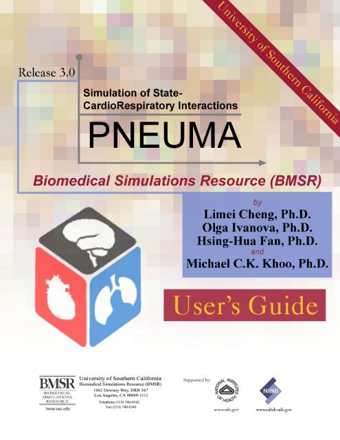
User Guide Release 3.0 - Biomedical Simulations Resource ...
User Guide Release 3.0 - Biomedical Simulations Resource ...
User Guide Release 3.0 - Biomedical Simulations Resource ...

You also want an ePaper? Increase the reach of your titles
YUMPU automatically turns print PDFs into web optimized ePapers that Google loves.
PNEUMA <strong>Release</strong> <strong>3.0</strong> software described in this document is furnished by the <strong>Biomedical</strong> <strong>Simulations</strong><br />
<strong>Resource</strong> under the terms of a release agreement.<br />
PNEUMA may be used only under the terms of the release agreement.<br />
PNEUMA <strong>User</strong>’s <strong>Guide</strong><br />
Contact: pneuma.bmsr@gmail.com<br />
<strong>Release</strong> <strong>3.0</strong> – January 2013<br />
<strong>Release</strong> 2.0 – January 2011<br />
<strong>Release</strong> 1.1 – March 2003<br />
<strong>Release</strong> 1.0 – August 2002<br />
Beta <strong>Release</strong> – September 2001<br />
Supported by: NIH Grant P41-EB001978<br />
© 2013 <strong>Biomedical</strong> <strong>Simulations</strong> <strong>Resource</strong> (BMSR)<br />
University of Southern California
PNEUMA <strong>Release</strong> Agreement<br />
Before you use PNEUMA, please read the following conditions for using our package.<br />
Thank you for your cooperation.<br />
<br />
<br />
<br />
<br />
PNEUMA is restricted to non-profit research and instructional purposes aimed at<br />
further knowledge in the area of cardiorespiratory system modeling and<br />
simulation. PNEUMA is supported by University of Southern California (USC)<br />
<strong>Biomedical</strong> <strong>Simulations</strong> <strong>Resource</strong> (BMSR) (NIH Grant P41-EB001978 ).<br />
Any publications of research results that were obtained in part by the use of<br />
PNEUMA will contain proper acknowledgement of the BMSR at USC. Reprints<br />
of such publications will be sent to the BMSR for the record.<br />
I will not distribute PNEUMA, in whole or part, to others without the expressed<br />
permission of the BMSR.<br />
I understand that neither USC nor the BMSR make any warranties, expressed or<br />
implied, that PNEUMA is free of errors or is consistent with any standard of<br />
merchantability, or that it will meet my requirements for any particular<br />
application. I understand that PNEUMA should not be relied on for solving a<br />
problem whose incorrect solution could result in injury to a person or loss of<br />
property, and that if I do use PNEUMA in such a manner it is at my own risk. I<br />
understand that USC, the BMSR and the authors disclaim any and all liability for<br />
direct or consequential damages resulting from my use of PNEUMA.
Contents<br />
Getting Started……………………………………………………………….............<br />
Individual Model……………………………………………………………..............<br />
Overall Pneuma Model………………………………………………………............<br />
Open Pneuma…………………………………………………………………...........<br />
Constant Parameters………………………………………………………….............<br />
Adjustable Inputs…………………………………………………………….............<br />
Interventions…………………………………………………………………............<br />
Block Description……………………………………………………………............<br />
Contact and Support………………………………………………………….............<br />
Blocks Reference…………………………………………………………….............<br />
Overall Pneuma………………….……………………………………………...........<br />
Reflexes (Reflex_Ursino.mdl)……………………………………………….............<br />
Carotid Baroreceptors……………………………………………….……...........<br />
Chemoreflex…...…………………………………………………………...........<br />
Lung Stretch Receptors Reflex………………………………………….............<br />
Offsets……………………………………………………………………............<br />
Autonomic Control…………………………...….…………………………..............<br />
SA Node (SA_Node_Ursino.mdl)…………………..……………………….............<br />
-Sympathetic Control…………………………...………………….…..............<br />
Parasympathetic Control…………………………….…………………..............<br />
-Sympathetic Control of Peripheral Resistance (TPR_Ursino.mdl)….……............<br />
Variable Breathing Period (PNEUMA.mdl)…...…………………………….............<br />
Variable Heart Period (PNEUMA.mdl)….………………………….……….............<br />
Cardiovascular System (PNEUMA.mdl)…………………………………….............<br />
Neuromuscular Drive (NeuroMuscular.mdl)…………………………………...........<br />
Respiratory Muscle Activity (Pmus_Flow_Younes.mdl)……………………............<br />
Pleural Pressure (Pleural_Schuessler.mdl)…………………………………..............<br />
Gas Exchange and Transport (Gas_Exchange.mdl)…………………………............<br />
Dead Space (Dead_Space_Khoo.mdl)………………..………………………...........<br />
Alveolar Gas Exchange (Lungs_Khoo.mdl)……..………………………….............<br />
Cardiovasuclar Mixing, Convection and Dissociation (Cardio_Mix_Lange.mdl,<br />
Dissociation_Spencer.mdl).………………..……..……..…...…….…….…….…….<br />
Brain Compartment (Brain_Khoo.mdl)…………………………………….…….….<br />
Body Tissues Compartment (Body_Khoo.mdl)……………………………....……..<br />
Ventilatory Response (Vent_Drive_Khoo.mdl)……………………………....……..<br />
Upper Airway / State Change (State_UA_Khoo_Borbely.mdl)……………....……..<br />
Upper Airway………………………………………………….……….….…….<br />
Sleep Mechanism……………………………………………………..…...…….<br />
Metabolic Control (PNEUMA.mdl)..……………………………………..…...…<br />
Autonomic and Metabolic Interactions………………………………….……….<br />
Appendix I: Software Package..……………………………………………….……..<br />
Appendix II: Saved Data Files…...…………………………………………....……..<br />
Appendix III: Save/Load Date for Advance <strong>User</strong>s………..…………………………<br />
Appendix IV: Overall Parameter Set and Initial Conditions……………...…………<br />
2<br />
2<br />
2<br />
2<br />
6<br />
8<br />
9<br />
15<br />
15<br />
16<br />
17<br />
19<br />
20<br />
22<br />
24<br />
25<br />
27<br />
31<br />
33<br />
34<br />
35<br />
37<br />
40<br />
42<br />
48<br />
50<br />
53<br />
55<br />
57<br />
60<br />
62<br />
66<br />
68<br />
70<br />
72<br />
73<br />
75<br />
79<br />
86<br />
88<br />
91<br />
92<br />
94
2<br />
Getting Started<br />
Thank you for trying out PNEUMA and its modularized component models. Before using<br />
PNEUMA, please take a moment to read the <strong>Release</strong> Agreement first. To download the<br />
associated files or their updates, please go to bmsr.usc.edu and click on Software. Before<br />
you begin to use PNEUMA or its individual model components, please take a moment to<br />
make sure that you have downloaded the most recent files that you will be using. In<br />
Appendix I, there is a full list of files that are included in zipped format on the BMSR-<br />
PNEUMA web site. After you unzip the downloaded file, please refer to the appendix<br />
and check that you have the correct files.<br />
Individual Sub-Models<br />
PNEUMA is implemented using Simulink and Matlab version R2007b or higher (© The<br />
Mathworks Inc., Natick, MA), which provides a graphical programming environment that<br />
promotes modularization of the overall model into hierarchically smaller subsystems.<br />
This allows the user to customize parts of the overall model in accordance to his/her<br />
simulations needs. Alternatively, the user may also choose to focus on a specific<br />
PNEUMA block and use it to study the corresponding mechanism of interest. Therefore,<br />
depending on the user’s interest and needs, individual component blocks may be<br />
downloaded and used.<br />
Please refer to the reference and the “.m” file for variable names and values of each<br />
compartment. Some of the components are difficult to decompose into smaller modules<br />
and therefore may not be suitable for your application. If you have suggestions or would<br />
like to request modifications to PNEUMA components that would better suit your<br />
simulation needs, please feel free to send us feedback. Contact information is provided in<br />
the Support and Contact section of this manual.<br />
PNEUMA V.<strong>3.0</strong>: What’s New<br />
In Pneuma <strong>Release</strong> <strong>3.0</strong>, we have incorporated a metabolic component with autonomicmetabolic<br />
interactions into the existing integrative comprehensive simulation model. This<br />
metabolic component of PNEUMA is based on prior models of glucose-insulin regulation<br />
by Bergman et al. (1979) and free fatty acid (FFA) regulation by Roy and Parker (2006).<br />
Changes in sympathetic activity from the autonomic portion of PNEUMA produce<br />
changes in epinephrine output, which in turn affects the metabolism of glucose, insulin<br />
and FFA. Inputs from the dietary intake of glucose and external interventions, such as<br />
insulin injections, have also been incorporated into the model. Also incorporated is<br />
autonomic “feedback” from the metabolic component to the rest of PNEUMA in the<br />
following way: changes in insulin level are assumed to lead to changes in sympathetic<br />
tone. The “Control Panel” along with other input panels have been improved to facilitate<br />
greater user interaction and control of the simulations
3<br />
References<br />
Bergman, R. N., Ider, Y. Z., Bowden, C.R.,and Cobelli,C.(1979).Quantitative estimation<br />
of insulin sensitivity. Am. J. Physiol. 236, E667–E677.<br />
Roy, A., and Parker, R. S. (2006). Dynamic modeling of free fatty acid, glucose, and<br />
insulin: an extended “Minimal Model”. Diabetes Technol. Ther. 8, 617–626.<br />
Using Pneuma<br />
To begin using Pneuma, unzip the “Pneuma<strong>Release</strong>3.zip” file and check that you have<br />
all the necessary files. For the list of files in “Pneuma<strong>Release</strong>3.zip” file, please refer to<br />
“Getting Started” section. After you have unzipped the file and you are ready to run the<br />
program in the MATLAB environment, make sure that you are in the directory where the<br />
unzipped files are located. To Open Pneuma, in the Matlab command prompt, type<br />
“PNEUMA_MAIN_CONTROL_PANEL”.<br />
If you are running Pneuma using a version of Matlab higher than Matlab75 (version<br />
2007b), a series of warnings may appear due to compatibility issues, but these warnings<br />
should disappear after the first time you open PNEUMA.
The Control Panel graphic user interface (GUI) will appear, as shown below.<br />
4
5<br />
Next, input the parameter values.<br />
Start Time: time to start the simulation. (default is zero seconds)<br />
End Time: end-time (in seconds) of the simulation (for example:<br />
3600*24*7 will end up with 7-day simulation.<br />
Max Step: the simulation is using variable integration time steps, and it<br />
requires that the user specify the maximum allowable time step. A large<br />
max time step is not recommended (the default is 0.01 second).<br />
Saved Sample Time: some of the parameter/variable values can be saved<br />
to data files after each simulation and the user has the option of specifying<br />
the sample time of the saved segment. If given value -1, it will be default<br />
sample time of the simulation which can be used for saving data with<br />
“Saved Segment Time” as 0.5 day. The suggested sample time for saving<br />
is 0.1 second for neural-cardio-respiratory system. The sample time for<br />
metabolic system is constant as 6 seconds.<br />
Saved Segment Time: each data file can be saved as long as the segment<br />
time. The suggested time is 7 days.<br />
The “Run” and “Stop” buttons allow the user to run and terminate the simulation.<br />
Currently, the Real Time Workshop allows the simulation to run using the accelerated<br />
mode even with standard Matlab Simulink package. So the “Run” operation will run in<br />
accelerated mode that allows the simulation to run faster. If the user prefers to execute<br />
the model in normal mode, it will have to be run under Simulink model itself rather than<br />
using that GUI button (see options under Simulation tab in Simulink model window). If<br />
you decide to stop the simulation before the End Time that you have specified, some<br />
data will be stored to files (Saved Sample Time option) and all the variables are in the<br />
workspace which can be saved later. The “Reset” button will reset all variable values to<br />
their defaults.<br />
Under “Open” menu, the user has three options. “Open Pneuma Model Ctrl+O” will<br />
show the Pneuma model in Simulink. <strong>User</strong> can explore the modules in Pneuma and<br />
incorporate other blocks if needed. “Open Display Panel Ctrl+D” allows the user to see<br />
the output from some of the more common measurements such as arterial blood pressure,<br />
heart rate and so on. Having achieved some familiarity with PNEUMA, the user may<br />
want to add more inputs to the display panel or create new displays. “Open Program<br />
Status Ctrl+P” will show the Pneuma Progress module in Simulink, that displays the<br />
total duration of simulation, current simulation time and percentage of simulation<br />
completed, based on total duration of simulation and current simulation time.<br />
If the user wants to load or save the simulation workspace, click under “File” menu and<br />
two selections will show up. “Load Data Ctrl+L” opens the standard Matlab open file<br />
window, which allows the user to specify the data file and load the data into workspace.<br />
“Save Data Ctrl+S” opens the standard Matlab save file window, which allows saving<br />
the workspace data to the directory of user's choice.
6<br />
Constant Parameters<br />
When the user clicks on “CONSTANT PARAMETERS” button, another graphical<br />
interface will appear, as shown below:<br />
These are the constant parameters used in the model. The values may be changed before<br />
the simulation, if desired. It is recommended that these parameters be left at their default<br />
values. Each model subsystem is listed along with the constant parameter in that<br />
compartment. Each title button gives user the opportunity to open the Simulink<br />
implementation of that particular subsystem.
If the mouse cursor is placed and held at a particular box with number, the help text for<br />
the corresponding parameter will appear so that the user will know what physiological<br />
entity that parameter represents, as shown below:<br />
7
8<br />
Adjustable Parameters<br />
This panel allows the user to vary parameters before or during the simulation. Click on<br />
“ADJUSTABLE PARAMETERS” button and the following panel will appear:<br />
The user can adjust the value either by using the slider bar or by typing directly into the<br />
box. Both the “min” and the “max” values are shown for each slider bar. These values<br />
can be changed as well. But the values that fall within the default spans indicated are<br />
recommended, since these are consistent with physiologically feasible ranges.
9<br />
The above panel shows the parameters that may be altered in value while the simulation<br />
is being executed. Since the model is continually being revised, the actual parameters that<br />
can be adjusted may be different in different versions of the program.<br />
External Interventions<br />
Here, the user is permitted to apply a variety of external interventions to the model.<br />
Click on “EXTERNAL INTERVENTIONS” and the graphical panel opens up, shown<br />
below:<br />
The panel shows the interventions that have been included in the model at the present<br />
time. Before you run each intervention, please click “Reset” button on “Control Panel” to<br />
reload the original parameter set, then enter your new start/stop time and other parameters<br />
on the Control Panel, then go back to the External Interventions. Again, as this software<br />
gets updated, other interventions will be added.<br />
The followings are some typical examples for the interventions.<br />
A. Hypoxia. To simulate hypoxia, simply enter values into “Start Time” and “Duration<br />
Time” such as start at 800 sec with duration 300 sec, then enter value into “Change in<br />
PIO2” such as “-90” by default, then go back to Control Panel and click on “Run”<br />
button, make sure the simulation “End Time” is equal or longer than the hypoxia end<br />
time, shown below:
10<br />
B. Normocapnic Hypoxia. To simulate normocapnia in hypoxia, first click on check box<br />
of “Normocapnia”, then define the hypoxia condition as in Hypoxia, then give the<br />
same “Start Time” and “Duration” in CO2 Inhalation part as O2 Inhalation part, then<br />
go back to Control Panel and click on “Run” button, shown below:<br />
C. Non-Normocapnic Hypoxia. To simulate non-normocapnic including hypercapnic<br />
hypoxia, first click on check box of “Non-Normocapnia”, then define the hypoxia<br />
condition as in Hypoxia, then give the same “Start Time” and “Duration” in CO2<br />
Inhalation part as O2 Inhalation part, then enter value into “Change in PICO2” such<br />
as “40” by default, then go back to Control Panel and click on “Run” button, make<br />
sure the simulation “End Time” is equal or longer than the hypercapnia end time,<br />
shown below:
11<br />
D. Normal Sleep. To simulate normal sleep, simply click on check box “Sleep Enable”,<br />
then go back to Control Panel and click on “Run” button. You can change the<br />
parameter set for sleep to simulation different interventions. For overnight sleep,<br />
make sure your “End Time” in Control Panel is longer than 3600*8+200 seconds (>8<br />
hrs) , shown below:<br />
E. OSA Sleep. To simulate obstructive sleep apnea (OSA) sleep, first click on check box<br />
“Sleep Enable”, then drag the slider button in “Upper Airway Mechanism” or directly<br />
enter value into “Pcrit” such as -2.66 which will simulate a moderate OSA, then go<br />
back to Control Panel and click on “Run” button. For overnight sleep, make sure your
12<br />
“End Time” in Control Panel is longer than 3600*9+200 seconds (>9 hrs) , shown<br />
below:<br />
F. CPAP with OSA Sleep. To simulate continuous positive airway pressure (CPAP),<br />
first set up OSA sleep as the above example in OSA Sleep. Then click on check box<br />
“CPAP”, enter values into “Start Time” and “Duration”, give values for the positive<br />
pressure such as 15 cmH2O, then go back to Control Panel and click on “Run”<br />
button. You can try 1 hour CPAP shown as below or overnight CPAP for OSA Sleep.<br />
In our model, the default mode is to repeat CPAP every night if the CPAP duration is<br />
longer than 1 day. For example, if the simulation runs for 30-day OSA sleep with 10-<br />
day CPAP, on in the middle of the 30-day run time simulation, then the CPAP “Start<br />
Time” could be 3600*24*10-1800 sec (which is 0.5 hour short than 10 days) and<br />
“Duration Time” could be 3600*24*10+3600*2 sec (which is 2 hours longer than 10<br />
days).
13<br />
G. Maneuvers. To simulate Mueller Maneuver, click on check box “Mueller Maneuver”,<br />
then use the default setup which can be entered with different values as you desire,<br />
then go back to Control Panel and click on “Run” button. To simulate Valsalva<br />
Maneuver, click on check box “Valsalva Maneuver”, then use the default setup which<br />
can be entered with different values as you desired, then go back to Control Panel and<br />
click on “Run” button, shown below:<br />
H. CSR-CHF Sleep. To simulate central sleep apnea (CSA characterized with Cheney-<br />
Stokes Respiration CSR) with congestive heart failure (CHF), first activate Sleep<br />
as in Normal Sleep. Then to change heart contractility, go to “Adjustable<br />
Parameters”, enter value such as “0.475*0.3” or drag the slider bar for
14<br />
“Gain_Emaxlv” and enter value such as “2392*0.3” or drag the slider bar for<br />
“Basal_Emaxlv” in “Heart Contractility” area, then increase chemoreflex gain such as<br />
increase “Peripheral Chemo-Gain” by directly entering value as “0.0063*6” (example<br />
value) or dragging the slider bar, then increase “Lung-Chemo Volume” by directly<br />
entering value as “0.588*1.5” (example value) or dragging the slider bar. Lastly, go<br />
back to Control Panel and click on the “Run” button, as shown below:<br />
These are brief descriptions to help the user get started using our package. Please feel free<br />
to explore the model. Since this is an open source environment, contribution of newer<br />
code or model will also help us to improve our implementation and to better suit the<br />
needs of other users as well.
15<br />
Block Description<br />
For the complete descriptions of all the individual Simulink model blocks, please refer to<br />
the “Blocks Reference” section.<br />
Contact and Support<br />
The whole model and its modularized components will be updated from time to time. So,<br />
please check the website for newer update or if you wish to join the mailing list,<br />
notification will be sent to you regarding our progress on the update.<br />
FAQ will be set up as we get more questions and comments. In the meantime, please<br />
send all your valuable comments and feedbacks to pneuma.bmsr@gmail.com. Once we<br />
have the solution, then we will post it in the forum so that other users can benefit from it.<br />
The PNEUMA project is supported by the USC <strong>Biomedical</strong> <strong>Simulations</strong> <strong>Resource</strong> (NIH<br />
Grant P41-EB001978). Comments and feedback on all aspects of this software are<br />
welcome.
Blocks Reference<br />
16
17<br />
PNEUMA V.<strong>3.0</strong><br />
Description<br />
PNEUMA is implemented using SIMULINK. The open architecture of PNEUMA allows<br />
to group models into hierarchies to create a simplified view of components or<br />
subsystems. High-level information is presented clearly and concisely, while detailed<br />
information is easily hidden in subsystems within the model hierarchy. Current<br />
PNEUMA implementation builds up on 557 model parameters and allows the tracing of<br />
93 model states. It is a hybrid model that simultaneously addresses fast and slow<br />
physiological processes (i.e. single heart beat and circadian rhythm) that are implemented<br />
in mixed discrete and continuous modes.<br />
The modular design of PNEUMA makes it possible to perform simulations in which<br />
specific physiological mechanisms are excluded or added in order to better determine<br />
their contribution to the closed-loop operation of the overall system of interconnected<br />
components. This allows the user to explore alternative models of physiologic function in<br />
silico, which could be very useful in circumventing the challenges of attempting to study<br />
the systems in question experimentally or clinically. As well, the modularity of<br />
PNEUMA enables users to replace one or more of the model blocks with their own<br />
modules of specific physiological components.<br />
General References:<br />
1. Cheng, L., and Khoo, M. C. K. Modeling the autonomic and metabolic effects of<br />
obstructive sleep apnea: a simulation study. Front Physiol 2:111, 2012. doi:<br />
10.3389/fphys.2011.00111.<br />
2. Cheng, L., Ivanova, O., Fan, H., and Khoo, M. C. K. An integrative model of<br />
respiratory and cardiovascular control in sleep-disordered breathing. Respiratory<br />
Physiology and Neurobiology 174, 4-28, 2010.
18<br />
Simulink Model. Overall Pneuma<br />
Maneuvers<br />
Double Click<br />
to load Initial Conditions<br />
200<br />
t_stop<br />
Progress<br />
Display<br />
Panel<br />
Variable Respiratory Rhythm<br />
Mech Vent Neural<br />
External Pressure<br />
Tidal Volume Vt<br />
Sleep/Awake<br />
PaO2<br />
PaCO2<br />
SAO2<br />
RespMus Drive<br />
Total Ventilatory Drive<br />
ftas<br />
Metabolic Control<br />
PbCO2<br />
ftas _v<br />
ftbs<br />
Central Neural Control<br />
ftp<br />
AI -Arousal Index<br />
PaCO2<br />
CaO2<br />
AI -Arousal Index<br />
fcs<br />
Cardiovascular System<br />
SI-Sleep Wake State Index<br />
Ppl<br />
Blood Flow<br />
deltaFtas<br />
REM<br />
Respiratory System
19<br />
Reflexes (Reflex_Ursino.mdl)<br />
Description<br />
The reflexes model includes the key cardiorespiratory reflexes: baroreflex, chemoreflex<br />
and lung stretch receptor influences on respiration and heart-rate control.<br />
Reflexes<br />
ABP<br />
D state<br />
Carotid<br />
Baroreceptors<br />
f cs<br />
P CO2<br />
P O2<br />
Chemoreflex<br />
f chemo<br />
V t<br />
Lung Stretch<br />
Receptors Reflex<br />
f ls
20<br />
Carotid Baroreceptors<br />
This block represents the pressor receptors that are located in the carotid sinus. In<br />
response to arterial blood pressure changes, it produces both parasympathetic and the<br />
sympathetic neural activity changes. During sleep, baro-sensitivity is assumed to<br />
increase slightly. The input for this compartment is the arterial blood pressure, ABP, and<br />
the output is the carotid sinus firing frequency, fcs.<br />
Carotid Baroreceptors Equation:<br />
<br />
P Pn<br />
<br />
fcs,<br />
min fcs,maxexp(<br />
<br />
kcs<br />
kcs<br />
fcs<br />
<br />
P P <br />
1<br />
exp(<br />
n Pn<br />
) <br />
kcs<br />
kcs<br />
<br />
Pn Pn _ sleep<br />
(1 AI ) SI<br />
kcs Kcs _ sleep<br />
(1 AI ) SI<br />
Pn<br />
<br />
) <br />
<br />
Reference: Ursino, M, Interaction between carotid baroregulation and the pulsating<br />
heart: a mathematical model. American Journal of Physiology, 275:H1733-H1747, 1998.<br />
Simulink Model: Baroreceptors
21<br />
Input: ABP Arterial Blood Pressure<br />
Output: f cs Carotid Sinus firing frequency<br />
Variables: P n Center pressure for sigmoidal function<br />
k cs Parameter for sigmoidal slope control<br />
f cs,min Lower threshold for sigmoidal function<br />
f cs,max Upper saturation for sigmoidal function<br />
Pn Pressure change in sleep<br />
Slope change in sleep<br />
kcs
22<br />
Chemoreflex<br />
Description<br />
The inputs to the chemoreflexes are Oxygen (O2) and Carbon Dioxide (CO2) levels in<br />
the arterial blood. This reflex affects both the heart rate and the peripheral vasculatures.<br />
Inputs for this block are the oxygen and carbon dioxide partial pressure, PaO2 and<br />
PaCO2. Output is the chemoreceptors firing, fac.<br />
Chemoreflex Equations:<br />
<br />
chemo<br />
PaO<br />
, Pa<br />
2 CO2<br />
where<br />
<br />
<br />
_____ <br />
Pa <br />
O Pa<br />
f f<br />
2 O2<br />
chemo,min<br />
chemo,max<br />
exp<br />
<br />
kchemo<br />
<br />
<br />
PaCO<br />
<br />
K<br />
ln 2<br />
f<br />
_____ <br />
______ <br />
<br />
<br />
<br />
Pa Pa<br />
<br />
O O<br />
PaCO2<br />
<br />
1<br />
exp<br />
2 2 <br />
kchemo<br />
<br />
<br />
<br />
K<br />
<br />
<br />
K K<br />
<br />
<br />
K<br />
H<br />
H<br />
H<br />
PaO<br />
80<br />
<br />
2<br />
1.2<br />
<br />
30 <br />
1.6<br />
if Pa<br />
if 40 Pa<br />
if Pa<br />
O2<br />
O2<br />
80<br />
O2<br />
40<br />
80<br />
df<br />
chemo<br />
dt<br />
1<br />
<br />
<br />
chemo<br />
<br />
<br />
f<br />
chemo<br />
<br />
chemo<br />
<br />
Reference: Ursino, M, A mathematical model of CO2 effect on cardiovascular<br />
regulation. American Journal of Physiology – Heart and Circulatory Physiology,<br />
281:H2036-H2052, 2001.
23<br />
Inputs: PaCO2 Arterial CO2 partial pressure<br />
PaO2 Arterial O2 partial pressure<br />
Output: fchemo Chemoreceptor firing<br />
Variables: fchemo,max Lower saturation for the sigmoidal function<br />
fchemo,min Upper saturation for the sigmoidal function<br />
_____<br />
Pa O2 Center point in the sigmoidal function<br />
kchemo Slope control parameter for the sigmoidal<br />
function<br />
_____<br />
Pa CO2 Normalizing PaCO2 value<br />
KH<br />
Constant value for the static response<br />
f<br />
Constant value for the static response<br />
τchemo Time constant for the chemoreflex<br />
Simulink Model: Chemoreflex
24<br />
Lung Stretch Receptor Reflex<br />
Lung inflation or deflation can produce changes in heart rate through the lung stretch<br />
receptors. The input for this block is the tidal volume, Vt. The output is the lung stretch<br />
receptor activity, fls.<br />
Lung Stretch Receptors Reflex Equations<br />
<br />
lung GlungVT<br />
df<br />
lung<br />
dt<br />
1<br />
<br />
<br />
lung<br />
<br />
<br />
f<br />
lung<br />
<br />
lung<br />
<br />
Reference: Ursino, M, A mathematical model of CO 2 effect on cardiovascular<br />
regulation. American Journal of Physiology – Heart and Circulatory Physiology,<br />
281:H2036-H2052, 2001.<br />
Simulink Model: Lung Stretch Receptors Reflex<br />
Inputs: Vt Tidal volume<br />
Output: fls Lung stretch receptors firing rate<br />
Variables: Gls Constant gain<br />
Τls Time constant
25<br />
Offsets (CNS Response in PNEUMA.mdl)<br />
Offsets for Autonomic Control are the central nervous system response to the partial<br />
blood pressure of carbon dioxide and oxygen in the cerebral circulation. The input for<br />
this block are partial arterial blood pressure PaCO2 and PaO2. The outputs are the<br />
offsets for autonomic control, Offset res,vein,heart , respectively.<br />
Offsets Equations<br />
Offset <br />
d<br />
d<br />
res, vein, heart san, spn, sbn O2 sa, O2 sp, O2sb CO2 sa, CO2 sp, CO2sb<br />
O2 sa, O2 sp, O2sb<br />
dt<br />
CO2 sa, CO2 sp, CO2sb<br />
dt<br />
1<br />
( O 2 sa, O2 sp, O2 sb<br />
Wsa , sp,<br />
sb )<br />
<br />
isc<br />
1<br />
[ CO 2 sa, CO2 sp, CO2 sb<br />
gccsa, sp, sb<br />
( PaCO 2<br />
PaCO 2n<br />
)]<br />
<br />
cc<br />
W X /(1 exp(( P - PO2 n ) / kisc ))<br />
sa, sp, sb sa, sp, sb aO2 sa, sp, sb sa, sp,<br />
sb<br />
Reference: Ursino, M, A mathematical model of CO 2 effect on cardiovascular<br />
regulation. American Journal of Physiology - Heart and Circulatory Physiology,<br />
281:H2036-H2052, 2001.<br />
Inputs: PaCO2 Arterial CO2 partial pressure<br />
PaO2<br />
Arterial O2 partial pressure<br />
Output: Offset res,vein,heart CNS Response as offsets of autonomic<br />
control<br />
Variables:<br />
X sa<br />
Saturation for the offset of α-sympathetic<br />
activity on peripheral resistance<br />
θ san<br />
Nominal level of offset of α-sympathetic<br />
activity on peripheral resistance<br />
PO2n sa Central point for the sigmoidal function<br />
kisc sa<br />
Parameter of α-sympathetic activity on<br />
peripheral resistance<br />
X sb<br />
Saturation for the offset of -sympathetic<br />
activity<br />
θ sbn<br />
Nominal level of offset of -sympathetic<br />
activity<br />
PO2n sb Central point for the sigmoidal function<br />
kisc sb<br />
Parameter of -sympathetic activity<br />
X sp<br />
Saturation for the offset of α-sympathetic<br />
activity on peripheral resistance<br />
Nominal level of offset of α-sympathetic<br />
θ spn
26<br />
PO2n sp<br />
kisc sp<br />
τ isc<br />
τ cc<br />
activity on peripheral resistance<br />
Central point for the sigmoidal function<br />
Parameter of α-sympathetic activity on<br />
unstressed volume of veins<br />
Time constant for oxygen response<br />
Time constant for carbon dioxide response<br />
Simulink Model: Offsets (CNS Response)<br />
-C-<br />
theta _sa_n<br />
1<br />
PaO 2<br />
f(u)<br />
Wsa_Fcn<br />
1/tao _isc<br />
1/tao _isc<br />
1<br />
s<br />
theta_O2_sa<br />
1<br />
Offset _Resistance<br />
2<br />
PaCO 2<br />
-C-<br />
PaCO 2_n<br />
-K-<br />
Gain 2<br />
1/tao _cc<br />
1/tao _cc<br />
1<br />
s<br />
theta_CO2_sa<br />
-C-<br />
theta _sp_n<br />
f(u)<br />
Wsp_Fcn1<br />
1/tao _isc<br />
1/tao _isc1<br />
1<br />
s<br />
theta_O2_sp<br />
2<br />
Offset _veins<br />
-K-<br />
Gain 1<br />
1/tao _cc<br />
1/tao _cc1<br />
1<br />
s<br />
theta_CO2_sp<br />
-C-<br />
theta _sb_n<br />
f(u)<br />
Wsb_Fcn2<br />
1/tao _isc<br />
1/tao _isc2<br />
1<br />
s<br />
theta_O2_sb<br />
3<br />
Offset _heart<br />
gcc_sb<br />
Gain 3<br />
1/tao _cc<br />
1/tao _cc2<br />
1<br />
s<br />
theta_CO2_sb
27<br />
Autonomic Control (submodels refer to Autonomic.mdl)<br />
Description<br />
Influences from the central respiratory control (RSA), baroreflexes, chemoreflexes and<br />
lung stretch receptors reflexes are integrated in this compartment and these inputs<br />
determine the total -sympathetic, -sympathetic and parasympathetic influences on<br />
heart rate and peripheral resistance. The inputs for this compartment are the central<br />
respiratory drive, N t , chemoreflex, f chemo , lung stretch receptors reflex, f ls , carotid<br />
baroreceptors firing, f cs, and CNS response, Offsets. The outputs are the -sympathetic<br />
response, f tas , -sympathetic response, f tbs and parasympathetic response, f tp . The models<br />
shown below is in PNEUMA.mdl, but the submodel is referred to Autonomic.mdl.<br />
Autonomic Control<br />
N t<br />
f chemo<br />
f ls<br />
f cs<br />
Offsets<br />
Autonomic Integration<br />
Central Respiratory Control<br />
Chemoreflex<br />
Lung Stretch Receptors<br />
Reflex<br />
Baroreflex<br />
CNS Response<br />
f tas<br />
f tbs<br />
f tp<br />
Autonomic Integration Equations:<br />
(a) Alpha-Sympathetic Activity<br />
f f ( f f ) <br />
tas _ res, vein s, s,0<br />
s,<br />
<br />
<br />
exp k G f G f G f G N Offset<br />
<br />
s baro, as cs chemo, as chemo lung,<br />
as lung RSA,<br />
as t res,<br />
vein<br />
(b) Beta-Sympathetic Activity<br />
f f ( f f ) <br />
tbs<br />
s, s,0<br />
s,<br />
<br />
<br />
exp k G f G f G f G N Offset<br />
<br />
s baro, bs cs chemo, bs chemo lung, bs lung RSA,<br />
bs t<br />
heart
28<br />
(c) Parasympathetic Activity<br />
<br />
fcs<br />
fcs,0<br />
f<br />
f exp<br />
para ,0 para , <br />
k<br />
<br />
p <br />
f <br />
<br />
<br />
G f G f G N Offset<br />
f f <br />
1 exp k<br />
<br />
p <br />
tp chemo, p chemo lung, p lung RSA, p t para _ n<br />
cs cs,0<br />
Reference: Ursino, M, A mathematical model of CO2 effect on cardiovascular<br />
regulation. American Journal of Physiology – Heart and Circulatory Physiology,<br />
281:H2036-H2052, 2001.<br />
Simulink Model: Autonomic Control<br />
Band-Limited<br />
White Noise<br />
4<br />
Gain2<br />
0<br />
Gain3<br />
noise<br />
1<br />
ftas_blocker<br />
1<br />
Total Alpha-Symp<br />
(ftas_res)<br />
4<br />
Nt Central Respiratory<br />
Neural Drive<br />
0.4<br />
G_CRSA<br />
0.34<br />
1<br />
lung feedback<br />
G_lung_asymp<br />
5<br />
Offset_Alpha-symp1<br />
G_offset_asymp1<br />
6<br />
Offset_Alpha-symp2<br />
G_offset_asymp2<br />
0.24<br />
1<br />
1<br />
4<br />
G_chemo_asymp<br />
fas<br />
Alpha-Symp<br />
Integration<br />
f(u)<br />
Alpha-Sympathetic<br />
Response<br />
f(u)<br />
Alpha-Sympathetic<br />
Response1<br />
fs<br />
(u60)*u<br />
f(u)<br />
1<br />
2<br />
Total Alpha-Symp<br />
ftas_blocker1<br />
(ftas_vein)<br />
2<br />
Chemoreflex<br />
1<br />
G_chemo<br />
2.8<br />
G_chemo_bsymp<br />
7<br />
Offset_Beta-symp<br />
G_lung_bsymp<br />
1<br />
G_offset_bsymp<br />
fbs<br />
Beta-Symp<br />
Integration<br />
f(u)<br />
Beta-Sympathetic<br />
Response<br />
fs<br />
(u60)*u<br />
1<br />
3<br />
Total Beta-Symp<br />
ftbs_blocker<br />
(ftbs)<br />
0.24<br />
3<br />
fcs<br />
Carotid Sinus<br />
1<br />
G_lung_para<br />
f(u)<br />
fp<br />
G_fcs<br />
-Ctheta_para_n<br />
Parasympathetic<br />
BaroResponse<br />
0.03<br />
G_chemo_para<br />
1<br />
G_offset_para<br />
Parasymp<br />
Integration<br />
(u>=0)*u<br />
1<br />
ftp_blocker<br />
4<br />
Total Parasymp<br />
(ftp)
29<br />
Simulink Model: Alpha-Sympathetic Response<br />
Simulink Model: Beta-Sympathetic Response<br />
Simulink Model: Parasympathetic Baroresponse
30<br />
Inputs: N t Respiratory Neural firings<br />
e f chemo Chemoreceptor firings<br />
f lung<br />
Lung stretch receptors firings<br />
f cs<br />
Baroreceptor firings<br />
CNS response<br />
Offset res,vein,heart<br />
Outputs: f tp Total parasympathetic response<br />
f tbs<br />
Total β-Sympathetic response<br />
Total α-Sympathetic response<br />
f tas_res,vein<br />
Variables: f para,0 Lower threshold of the parasympathetic baroreflex<br />
sigmoidal function<br />
f para,<br />
Upper saturation of the parasympathetic baroreflex<br />
sigmoidal function<br />
f cs,0<br />
Center point for the sigmoidal function<br />
k p<br />
Slope control parameter for the sigmoidal function<br />
G RSA,p<br />
Central RSA gain for parasympathetic response<br />
G chemo,p Chemoreflex gain for parasympathetic response<br />
G lung,p<br />
Lung stretch receptor reflex gain for<br />
parasympathetic response<br />
f s,0<br />
Lower limit of the sympathetic exponential decay<br />
function<br />
f s,<br />
Upper saturation of the sympathetic exponential<br />
decay function<br />
k s<br />
Constant for the exponential function<br />
G RSA,bs<br />
Central RSA gain for -sympathetic response<br />
G chemo,bs Chemoreflex gain for -sympathetic response<br />
G lung,bs<br />
Lung stretch receptor reflex gain for -sympathetic<br />
G baro,bs<br />
Baroreflex gain for -sympathetic<br />
G RSA,as<br />
Central RSA gain for -sympathetic response<br />
G chemo,as Chemoreflex gain for -sympathetic response<br />
G lung,as<br />
Lung stretch receptor reflex gain for -sympathetic<br />
Baroreflex gain for -sympathetic<br />
G baro,as
31<br />
SA Node (SA_Node_Ursino.mdl)<br />
Description<br />
This module translates changes in -sympathetic and parasympathetic efferent activity<br />
into changes in heart rate. In sleep, the model assumes that the parasympathetic response<br />
increases while there is small decrease in the sympathetic activity. The inputs for this<br />
subsystem are the total -sympathetic firing frequency, ftbs, and parasympathetic firing<br />
frequency, ftp, and the output is the heart period, HP (= reciprocal of instantaneous heart<br />
rate).<br />
SA Node<br />
f tbs<br />
SI<br />
-sympathetic<br />
Response<br />
HP bs<br />
f tp<br />
SI<br />
parasympathetic<br />
Response<br />
HP p<br />
HP<br />
Basal Heart<br />
Period HP basal<br />
SA Node Equation:<br />
HP<br />
<br />
HP<br />
bs<br />
HP<br />
p<br />
HP<br />
basal<br />
Reference: Ursino, M, Interaction between carotid baroregulation and the pulsating<br />
heart: a mathematical model. American Journal of Physiology, 275:H1733-H1747, 1998.
32<br />
Simulink Model: SA Node<br />
3<br />
Gbs(SI)<br />
1<br />
ftbs<br />
Transport<br />
Delay<br />
Saturation<br />
ln<br />
Math<br />
Function<br />
-0.13<br />
Gain _HPbs<br />
1<br />
toux (7).s+1<br />
Transfer Fcn<br />
ftbs_min<br />
Constant<br />
HP_basal<br />
Constant 1<br />
1<br />
HP<br />
2<br />
ftp<br />
Transport<br />
Delay<br />
4<br />
1<br />
Gps(SI)<br />
0.09<br />
Gain _HPpara<br />
1<br />
toux (8).s+1<br />
Transfer Fcn 1<br />
Inputs: f tbs Total beta-sympathetic firing frequency<br />
f tp<br />
Total parasympathetic firing frequency<br />
Output: HP Heart Period (equivalent to RR-interval)<br />
Variable: HP basal Basal value for HP for denervated heart<br />
HP bs Change in HP modulated by -sympathetic<br />
response<br />
HP p Change in HP modulated by parasympathetic<br />
response
33<br />
-Sympathetic Control<br />
Description<br />
This response is modeled assuming first-order dynamics. The time-constant and delay<br />
associated with the -sympathetic effect on the heart period is longer than that of the<br />
parasympathetic response. There is slight decrease in -sympathetic response in sleep.<br />
The input for this compartment is the -sympathetic firing frequency, ftbs and the output<br />
is the corresponding component of heart period change, HPbs.<br />
-Sympathetic Control Equations:<br />
G G ( SI ) ln[ f ( t D ) f 1],<br />
f f<br />
<br />
bs()<br />
t <br />
0,<br />
ftbs<br />
f<br />
G ( SI ) 1 SI (1 AI ) G<br />
bs bs tbs bs tbs min tbs tbs min<br />
bs bs _ sleep<br />
tbs min<br />
d<br />
dt<br />
HPbs<br />
<br />
<br />
1<br />
bs<br />
<br />
HPbs(<br />
t)<br />
<br />
bs<br />
( t)<br />
<br />
Reference: Ursino, M, Interaction between carotid baroregulation and the pulsating<br />
heart: a mathematical model. American Journal of Physiology, 275:H1733-H1747, 1998.<br />
Input: ftbs Total beta-sympathetic firing frequency<br />
SI<br />
Sleep Index for sleep wake state<br />
AI<br />
Arousal Index<br />
Output: ΔHPbs Heart Period change modulated by -symp.<br />
Variables: Dbs -sympathetic time delay<br />
ftbsIC -sympathetic initial output after time delay<br />
ftbs_min Lower limit for the natural log function<br />
Gbs<br />
-sympathetic Gain varied with sleep drive<br />
τbs<br />
-sympathetic time constant<br />
delta_HPbsIC Initial input to the -symp first order dynamic<br />
system<br />
Gbs_sleep -sympathetic Gain of sleep factor
34<br />
Parasympathetic Response<br />
Description<br />
The vagal effect on heart rate is modeled assuming first-order dynamics. During sleep,<br />
parasympathetic activity increases, and this is partially responsible for the decrease in the<br />
heart rate. The input for this compartment is the parasympathetic firing frequency, ftp<br />
and the output is the corresponding component of heart period change, HPp.<br />
Parasympathetic Response Equations:<br />
Gps<br />
ps( t) ftp( t Dps)<br />
G ( SI )<br />
<br />
d<br />
dt<br />
HP<br />
<br />
ps<br />
1<br />
p<br />
τ para<br />
G ( SI ) 1 SI (1 AI ) G<br />
<br />
ΔHPp(t)<br />
σp(t)<br />
ps para _ sleep<br />
<br />
Reference: Ursino, M, Interaction between carotid baroregulation and the pulsating<br />
heart: a mathematical model. American Journal of Physiology, 275:H1733-H1747, 1998.<br />
Input: ftp Total parasympathetic firing frequency<br />
SI<br />
Sleep Index for sleep wake state<br />
AI<br />
Arousal Index<br />
Outputs: ΔHPp Heart Period change modulated by<br />
parasympathetic<br />
Variables: Dpara Parasympathetic time delay<br />
ftpIC Parasympathetic initial output after time delay<br />
Gpara Parasympathetic Gain varied with sleep drive<br />
τpara Parasympathetic time constant<br />
delta_HPpIC Initial input to the parasympathetic first order<br />
dynamic system<br />
Gpara_sleep Parasympathetic Gain of sleep factor
35<br />
-Sympathetic Control of Peripheral Resistance<br />
(TPR_Ursino.mdl)<br />
Description<br />
This block models -sympathetic control of peripheral vascular resistance, using a firstorder<br />
dynamic system as in the case of the -sympathetic component. During sleep in<br />
normals, the accompanying decrease in -sympathetic activity contributes substantially<br />
to a decrease in blood pressure. The inputs are the total -sympathetic firing frequency,<br />
ftas and the state/sleep drive, Dstate. The output is the proportional change in the<br />
peripheral resistance, TPR.<br />
Total Peripheral Resistance<br />
ftas<br />
SI<br />
Vascular Resistance<br />
Changes (baro, lung<br />
stretch, central, chemo)<br />
TPR<br />
Equations for Total Peripheral Resistance Change:<br />
G G ( SI ) ln[ f ( t D ) f 1],<br />
f f<br />
Z<br />
j<br />
<br />
0,<br />
f f<br />
dTPR<br />
j 1<br />
( TPR<br />
j<br />
Z<br />
j )<br />
dt <br />
j as tas _ i j tas min tas _ i tas min<br />
TPR () t TPR TPR<br />
j<br />
j j j0<br />
G ( SI ) 1 SI (1 AI ) G<br />
as as _ sleep<br />
tas _ i tas min<br />
Reference: Ursino, M, Interaction between carotid baroregulation and the pulsating<br />
heart: a mathematical model. American Journal of Physiology, 275:H1733-H1747, 1998.
36<br />
Simulink Model: Alpha-Sympathetic Modulation on Peripheral Resistance<br />
3<br />
1-u<br />
AI (arousal Index)<br />
4<br />
SI (Sleep Index)<br />
Gas_sleep<br />
Gas_sleep<br />
2<br />
Alpha Symp<br />
(ftas_vein)<br />
1<br />
Alpha Symp<br />
(ftas)<br />
1<br />
Gas(SI)<br />
{Rsp}<br />
fes<br />
Rsp<br />
Gas(SI)<br />
Rsp<br />
Peri Circ Rsp<br />
fes<br />
Rep<br />
Rep<br />
Gas(SI)<br />
Peri Circ Rep<br />
fes<br />
Rmp_n<br />
Gas(SI)<br />
Rmp_n<br />
{Rep }<br />
{Rmpn }<br />
Peri Circ Rmp<br />
fes<br />
Vusv<br />
Gas(SI)<br />
Vusv<br />
{Vusv}<br />
Venous Circ Vusv<br />
fes<br />
Vuev<br />
Gas(SI)<br />
Vuev<br />
{Vuev }<br />
Venous Circ Vuev<br />
1<br />
u<br />
{Gbp }<br />
Gbp<br />
{Rmp }<br />
Rmp<br />
1<br />
u<br />
1<br />
u<br />
1<br />
{Rhp }<br />
Rhp u<br />
1<br />
u<br />
TPR _change<br />
Goto<br />
fes<br />
Vumv<br />
Gas(SI)<br />
Venous Circ Vumv<br />
{Vumv }<br />
Vumv<br />
Inputs: ftas Total alpha-sympathetic firing frequency<br />
SI<br />
Sleep Index for sleep wake state<br />
AI Arousal Index<br />
Outputs: TPR_change TPR change factor<br />
Variables: fasIC -sympathetic initial output after time delay<br />
fas_min Lower limit for the natural log function<br />
Gas_sleep -sympathetic Gain varied with sleep<br />
Gas_sp -sympathetic Gain for splanchnic peripheral resistance<br />
τas_sp -sympathetic time constant<br />
Das_sp Delay -sympathetic time constant<br />
Gas_ep -sympathetic Gain for extra-splanchnic peripheral resistance<br />
τas_ep -sympathetic time constant<br />
Das_ep Delay -sympathetic time constant<br />
Gas_mp -sympathetic Gain for skeletal muscle peripheral resistance<br />
τas_mp -sympathetic time constant<br />
Das_mp Delay -sympathetic time constant<br />
Vusv0 Basal level of unstressed volume of splanchnic venous<br />
circulation<br />
Gas_usv -sympathetic Gain for unstressed volume of splanchnic venous<br />
circulation<br />
τ as_usv -sympathetic time constant<br />
D as_usv Delay -sympathetic time constant
37<br />
Variable Breathing Period (PNEUMA.mdl)<br />
Description<br />
The variable breathing period is controlled from the central neural control system by the<br />
total chemoreflex drive [52]. The inspiratory and expiratory periods of a single breath are<br />
set to be of equal duration. The ventilatory drive is controlled by central and peripheral<br />
chemoreflexes. The combination of ventilatory drive and breathing period determines the<br />
neuromuscular drive.<br />
Reference: Duffin J., R.M. Mohan, P. Vasiliou, R. Stephenson, S. Mahamed, “A model<br />
of the chemoreflex control of breathing in humans: model parameter measurement,”<br />
Respiration Physiology, vol. 120, pp. 13-26, 2000.
38<br />
Simulink Model: Variable respiratory rhythm generator<br />
Breathing_Enable_Signal<br />
Ventilatory Drive (L/sec)<br />
1<br />
D_VENTILATORY<br />
Breathing_Enable_Signal<br />
2<br />
F_breathing<br />
Breathing Frequency<br />
(breaths/minute)<br />
Breathing_Frequency<br />
D_Vent<br />
Breathing Period (sec/breath)<br />
T_breathing<br />
Variable Breathing Period (sec)<br />
ENABLE SIGNAL<br />
Breathing Period<br />
Enable_Breathing_Period<br />
VENTILATORY<br />
DRIVE (chemical)1<br />
Breathing Period<br />
Enable Breathing Signal<br />
Enable Breathing Signal<br />
Breathing Period Basal<br />
Breathing_Period_Basal<br />
3.5<br />
Breathing Period Update<br />
Variable Breathing Period<br />
Variable Breathing Period Reset<br />
Variable Breathing Period (sec)<br />
VBP Variable Breathing Period<br />
Respiratory Rhythm<br />
RESPIRATORY RHYTHM<br />
1<br />
Respiratory Rhythm_Generator
39<br />
Simulink Model: Ventilatory drive breathing frequency/period<br />
1<br />
D_Vent<br />
7500<br />
-C-<br />
u<br />
para<br />
BF<br />
fcn<br />
count<br />
Control_Constant2<br />
Embedded<br />
MATLAB Function<br />
Breathing Frequency (breaths/minute)<br />
1<br />
u<br />
Math<br />
Function<br />
60<br />
Breathing Period (sec)<br />
2<br />
Breathing Period (sec)<br />
T_breathing<br />
Breathing Frequency (breaths/minute)<br />
1<br />
F_breathing<br />
BF_BP Scope1
40<br />
Variable Heart Period (PNEUMA.mdl)<br />
Description<br />
The variable heart period module is modulated by the major reflexes and<br />
cardiorespiratory interactions in a closed loop mode. The sinoatrial node is modeled as a<br />
simple pacemaker, regulated by the parasympathetic and the beta-sympathetic inputs. The<br />
variable heart period is generated from continuous SA output using an<br />
integration/saturation mechanism. The beta-sympathetic branch affects the heart rate<br />
contractility, thus modulating the systolic period. Greater beta-sympathetic tone increases<br />
myocardial elastance and shortens ventricular systole. Each active atria-ventricular<br />
compartment is characterized by a time-varying nonlinear elastance function, describing<br />
the changes in ventricular elastance due to the beta-sympathetic tone input. The diastolic<br />
filling time is the difference between the heart period and systolic period and is thus<br />
controlled indirectly. The activation of the right and left hearts is fully synchronized and<br />
occurs simultaneously.<br />
Reference: Dempsey, J.A., Smith, C.A., Eastwood, P.R., Wilson, C.R., Khoo, M.C.K.<br />
Sleep induced respiratory instabilities. In: Pack, A.I. (Ed.), Sleep Apnea Pathogenesis,<br />
Diagnosis and Treatment. Dekker M., New York. 2002.
41<br />
Simulink Model: Variable Heart Period<br />
1<br />
1<br />
HP_SAnode<br />
1<br />
u<br />
1<br />
s<br />
Threshold Integrated HP<br />
HPin<br />
u1 if(u1
42<br />
Cardiovascular System (PNEUMA.mdl)<br />
Description<br />
The cardiovascular subsystem is capable of simulating the pulsatile nature of the heart<br />
and blood flow through the pulmonary and systemic circulations. Included in the model<br />
are descriptions of atria-ventricular mechanics, hemodynamics of the systemic and<br />
pulmonary circulations, SA node, change of total peripheral resistance and baroreflex.<br />
The inputs for this combined subsystem are the α-sympathetic firing rates, ftas_res,vein,<br />
β-sympathetic firing rates, ftbs, parasympathetic firing rate, ftp, arterial PaCO2, CaO2,<br />
arousal index, AI, sleep-wake state index, SI, and the pleural pressure, Ppl. To<br />
incorporate the effects of pleural pressure changes on the circulatory system we modulate<br />
basal blood pressure values for systemic and pulmonary components in thoracic cavity<br />
and heart. The output is the arterial blood pressure, ABP, heart period, HP, cardiac<br />
output, CO, and blood flow to lung for gas exchange.<br />
Cardiovascular System<br />
Pulmonary<br />
Circulation<br />
R PA<br />
C PC<br />
R PC<br />
C PV<br />
R PV<br />
L PA<br />
p LA<br />
C LA<br />
R SV<br />
R SP<br />
C SV<br />
C SP<br />
R LA<br />
Q RV<br />
R RV<br />
C PA<br />
R EV<br />
R EP<br />
C EV<br />
C EP<br />
C LV<br />
R LV<br />
p LV<br />
C RV<br />
Q MP<br />
Q LV<br />
pRA<br />
R RA R MV R<br />
C MP<br />
MV<br />
C MP<br />
Q BP<br />
C RA<br />
R BV<br />
R<br />
C BP<br />
BV<br />
C BP<br />
QHP<br />
R VC<br />
C VC<br />
RHV<br />
RHP<br />
CHV<br />
C HP<br />
p SP<br />
C SA<br />
L SA<br />
R SA<br />
Systemic<br />
Circulation
43<br />
Reference:<br />
1. Ursino, M. Interaction between carotid baroregulation and the pulsating heart: a<br />
mathematical model. American Journal of Physiology, 275:H1733-H1747, 1998.<br />
2. Ursino, M., Magosso, E. Acute cardiovascular response to isocapnic hypoxia. I. A<br />
mathematical model. American Journal of Physiology – Heart and Circulatory<br />
Physiology, 279, H149-165, 2000.
44<br />
Simulink Model. Cardiovascular System<br />
2<br />
fcs<br />
2<br />
ftas_vein<br />
1<br />
ftas_res<br />
TPR Change<br />
Alpha Symp (ftas)<br />
Alpha Symp (ftas_vein)<br />
AI (arousal Index )<br />
{Ppa }<br />
{Vpa }<br />
{Pla }<br />
{Pra }<br />
{Vra } {Vla }<br />
{Vrv} {Vlv}<br />
Carotid<br />
Baroreceptor<br />
8<br />
SI Sleep -Wake<br />
State Index<br />
3<br />
ftbs<br />
4<br />
ftp<br />
SI (Sleep Index )<br />
SI Sleep Wake State Index<br />
Beta-Symp (ftbs)<br />
Parasymp (ftp)<br />
HP<br />
HP<br />
HP<br />
{Vpp }<br />
{gPpl }<br />
{Rmpn }<br />
{Gbp }<br />
{Wlv}<br />
{Wrv}<br />
{Vusv}<br />
{Rhp }<br />
{RHeart } {LHeart }<br />
{Vpv} {Vu}<br />
{Vuev } {Vumv }<br />
{Rmp } {Rsp}<br />
Arousal _CardIC<br />
{Rep }<br />
{Vsa}<br />
{Psp}<br />
{Pvc}<br />
SA-node and Autonomic Control<br />
ABP<br />
7<br />
AI<br />
Arousal Index<br />
1<br />
G_pleural<br />
{gPpl }<br />
9<br />
Ppl<br />
Qrv<br />
PULMONARY CIRCULATORY SYSTEM<br />
1<br />
Blood Flow to Lung<br />
for Gas Exchange<br />
Qla<br />
Local Flow Regulation<br />
PaCO2<br />
5<br />
CaO2<br />
6<br />
RIGHT HEART<br />
Qra<br />
Qmp<br />
LEFT HEART<br />
Qlv<br />
Qbp<br />
SYSTEMIC CIRULATORY SYSTEM<br />
Qhp
45<br />
Simulink Model. SA-Node and Autonomic Control<br />
Vusa<br />
Vusp<br />
2<br />
Beta-Symp (ftbs)<br />
f tbs<br />
Gbs(SI)<br />
Emaxlv<br />
{Emaxlv}<br />
{Emaxlv}<br />
{Emaxrv}<br />
Vuep<br />
[Vuev]<br />
[Vusv]<br />
Vupv<br />
Heart Emaxlv<br />
f tbs<br />
Emaxrv<br />
Gbs(SI)<br />
Heart Emaxrv<br />
{Emaxrv}<br />
phi<br />
Right Ventricle<br />
Pmaxrv<br />
Heart Beat<br />
Right Ventricle<br />
{RHeart}<br />
Vupa<br />
Vula<br />
Vupp<br />
{Vu}<br />
3<br />
Parasymp<br />
(ftp)<br />
f tbs<br />
f tp<br />
Gbs(SI)<br />
Gps(SI)<br />
HP<br />
HP phi<br />
phi<br />
phi<br />
Left Ventricle<br />
Pmaxlv<br />
Heart Beat<br />
Left Ventricle<br />
{LHeart}<br />
Vura<br />
SA Node<br />
Vump<br />
Vuhp<br />
Vuhv<br />
Vubp<br />
Vubv<br />
4<br />
Arousal_CardIC<br />
1-u<br />
1<br />
SI Sleep Wake State Index<br />
Gbs_sleep<br />
1<br />
Gpara_sleep<br />
HP_SAnode<br />
RR Interval<br />
Heart_Period<br />
1<br />
HP<br />
[Vumv]<br />
1<br />
Simulink Model. Left Heart<br />
1<br />
Qla<br />
Left Heart<br />
Qla<br />
Vula<br />
1<br />
s<br />
_<br />
Out<br />
+<br />
1/Cla<br />
{Vla}<br />
Pla<br />
[gPpl]<br />
{Pla}<br />
Vula (Left Atrium Unstressed Volume)<br />
Vulv (Left Ventricle Unstressed Volume)<br />
Vlv (Left Ventricle Volume)<br />
Vla (Left Atrium Volume)<br />
Pla (Left Atrium Pressure)<br />
Plv (Left Ventricle Pressure)<br />
Qla (Flow into Left Atrium)<br />
Qilv (Flow into Left Ventricle)<br />
Qolv (Flow out of Left Ventricle)<br />
LHeart (Left Ventricle Function)<br />
Qilv<br />
1/Rla<br />
mitral<br />
valve<br />
Plv<br />
[ABP]<br />
[LHeart]<br />
1<br />
s<br />
SV<br />
Qolv<br />
Vulv<br />
1<br />
s<br />
_<br />
Out<br />
+<br />
[ABP]<br />
{Vlv}<br />
{Wlv}<br />
Pmaxlv<br />
aortic<br />
valve<br />
Pmax<br />
P<br />
Flow Qolv<br />
Pmaxlv--Psa<br />
----------------------<br />
Rlv<br />
[HP]<br />
1<br />
Qlv<br />
-Kml/sec<br />
to l/min<br />
CO
46<br />
Simulink Model. Right Heart<br />
Right Heart<br />
1<br />
Qirv<br />
Qra<br />
Vura<br />
1<br />
s<br />
_<br />
Out<br />
+<br />
1/Cra<br />
{Vra}<br />
Pra<br />
{Pra}<br />
[gPpl]<br />
Vura (Right Atrium Unstressed Volume)<br />
Vurv (Right Ventricle Unstressed Volume)<br />
Vrv (Right Ventricle Volume)<br />
Vra (Right Atrium Volume)<br />
Pra (Right Atrium Pressure)<br />
Prv (Right Ventricle Pressure)<br />
Qra (Flow into Right Atrium)<br />
Qirv (Flow into Right Ventricle)<br />
Qrv (Flow out of Right Ventricle)<br />
LHeart (Left Ventricle Function)<br />
CHF Gain1<br />
Qirv<br />
1/Rra<br />
1<br />
tricuspid<br />
valve<br />
Prv<br />
Switch2<br />
[Ppa]<br />
Pmaxrv<br />
[RHeart]<br />
Qirv<br />
Vurv<br />
1<br />
s<br />
_<br />
Out<br />
+<br />
{Vrv}<br />
Pmaxrv<br />
[Ppa]<br />
{Wrv}<br />
pulmonary<br />
valve<br />
Pmax<br />
P<br />
Flow<br />
Pmaxrv--Ppa<br />
----------------------<br />
Rrv<br />
1<br />
Qrv<br />
Product<br />
Simulink Model. Systemic Circulation<br />
Systemic Circulation<br />
1<br />
Qlv<br />
Qlv<br />
Qsa<br />
Systemic<br />
Arteries<br />
Qsa<br />
Qvc<br />
Qmp<br />
Qbp<br />
Qhp<br />
Systemic<br />
Peripheral &<br />
Venous<br />
Circulation<br />
Qvc<br />
2<br />
Qmp<br />
3<br />
Qbp<br />
4<br />
Qhp<br />
Qra<br />
Vena Cava<br />
{Vvc}<br />
1<br />
Qra
47<br />
Simulink Model. Pulmonary Circulation<br />
Pulmonary Circulation<br />
1<br />
Qrv<br />
Qrv<br />
Qpa<br />
Vupa<br />
1<br />
s<br />
_<br />
Out<br />
+<br />
1/Cpa<br />
{Vpa}<br />
Ppa<br />
Ppp<br />
{Ppa}<br />
Ppa (Pulmonary Arteries Pressure)<br />
Ppp (Pulmonary Peripheral Pressure)<br />
Ppv (Pulmonary Veins Pressure)<br />
Pla (Left Atrium Pressure)<br />
Qor (Flow from Right Heart)<br />
Qpa (Flow to Pulmonary Aorta)<br />
Qla (Flow to Left Atrium)<br />
Vupa (Pulmonary Arteries Unstressed Volume)<br />
Vupp (Pulmonary Peripheral Unstressed Volume)<br />
Vupv (Pulmonary Veins Unstressed Volume)<br />
Qpa<br />
Rpa<br />
[gPpl]<br />
Qpa<br />
1/Lpa<br />
1<br />
s<br />
Qpp<br />
1<br />
Qpa<br />
Vupp<br />
1<br />
s<br />
_<br />
Out<br />
+<br />
Ppp<br />
{Vpp}<br />
1/Cpp<br />
Ppp<br />
-K-<br />
1/Rpp<br />
ml/liter<br />
Qpv<br />
Vupv<br />
1<br />
s<br />
_<br />
Out<br />
+<br />
[gPpl]<br />
1/Cpv<br />
Ppv<br />
{Vpv}<br />
2<br />
Qla<br />
Qpv<br />
Qpv<br />
1/Rpv<br />
Pla<br />
[Pla]<br />
Simulink Model. Local Flow Regulation<br />
4<br />
CaO2<br />
6<br />
Qbp<br />
3<br />
PaCO2<br />
5<br />
Qmp<br />
CaO2<br />
PaCO2<br />
Qbp<br />
Gsleep<br />
CaO2<br />
PaCO2<br />
Qmp<br />
Gsleep<br />
Gbp<br />
Cerebral Circulation<br />
Regulation<br />
Rmp<br />
Muscular Circulation<br />
Regulation<br />
{Gbp}<br />
Brain Peripheral<br />
Resistance<br />
{Rmp}<br />
Muscular Peripheral<br />
Resistance<br />
7<br />
Qhp<br />
1 1-u<br />
AI (arousal Index)<br />
2<br />
SI (Sleep Index)<br />
-K-<br />
Gas_sleep<br />
CaO2<br />
PaCO2<br />
Qhp<br />
Gsleep<br />
1<br />
Rhp<br />
Coronary Circulation<br />
Regulation<br />
Gas(SI)<br />
{Rhp}<br />
Coronary Peripheral<br />
Resistance
48<br />
Neuromuscular Drive (NeuroMuscular.mdl)<br />
Description<br />
Inspiratory muscular activity is produced by neural drive arising from the respiratory<br />
centers. The muscles have to overcome the resistive and elastic forces of the lungs and<br />
chestwall to generate the airflow. The muscular drive is modulated by the<br />
autorhythmicity, chemical and state drives. In the case of the mechanical assisted<br />
ventilation, the internal neural activity will diminish with a period of time. The inputs for<br />
this compartment are the chemical drive, Dchemo, external pressure, Dext and the staterelated<br />
drive, Dstate. The output is the neuromuscular drive, Nt.<br />
Neuromuscular Drive<br />
Chemical Drive, State Drive<br />
Respiratory Autorhythmicity<br />
External Assisted Pressure<br />
Neuromuscular<br />
<br />
Drive Profile<br />
Nt<br />
Neuromuscular Drive Equations:<br />
Respiratory<br />
Autorhythmicity SquareFuncTI ( , TT )<br />
<br />
<br />
TI<br />
N( t)<br />
0<br />
<br />
0<br />
Dtotaldt<br />
0 t TI<br />
TI<br />
t TT
49<br />
Simulink Model: Neuromuscular Drive<br />
Demux<br />
Resp_Rhythm_Generator<br />
on_off_rhythm_test<br />
Mux<br />
1<br />
RespMus Drive<br />
RespMus_blocker<br />
Chemical<br />
Drive<br />
3<br />
1<br />
G_RespMus<br />
20<br />
S_wake<br />
0.3<br />
Nt Save block<br />
Resp Neural Prof ile<br />
State<br />
drive<br />
2<br />
Resp_sig<br />
Mux<br />
2<br />
Total Drive<br />
1<br />
Mechanial<br />
Ventilation<br />
Mux<br />
f(u)<br />
Inputs: Dchemo Chemical Drive<br />
Dext External drive or pressure<br />
Dstate State related Drive<br />
Output: Nt Neural-Muscular Drive<br />
Variables: Gstate State Drive gain<br />
TTmean Breathing Period<br />
TImean Inspiration Period<br />
Inhale Boolean variable for inhalation
50<br />
Respiratory Muscle Activity (Pmus_Flow_Younes.mdl)<br />
Description<br />
During the breathing process, the respiratory muscles have to overcome the resistive and<br />
the elastic forces of the respiratory system. By equating the force generated from the<br />
respiratory muscles with the pressure from the respiratory system, the airflow pattern can<br />
be obtained using a simple mechanics model, and tidal volume can be computed from<br />
the flow. During normal breathing, expiratory muscular activity is minimum. The inputs<br />
are the neural signals, Nt, the upper airway conductance, Cond ua , the expiratory pressure,<br />
Pexp and the external pressure, Pao. The outputs are the airflow, Flow, tidal volume, Vt<br />
and the muscular pressure, Pmus.<br />
Respiratory Muscle Activity<br />
Pexp<br />
Pao<br />
Nt<br />
Cond ua<br />
Inspiratory<br />
Muscular<br />
Pressure<br />
Respiratory<br />
Resistive,<br />
Elastic Force<br />
Vt, Flow, Pmus<br />
Respiratory Mechanics Equations:<br />
P isom = G neuromusc D Total<br />
Y<br />
rs<br />
Yua<br />
<br />
1 ( R R R ) Y<br />
AW LT CW ua<br />
Note: when Yua=0, then Yrs = 0.<br />
.<br />
V<br />
t<br />
<br />
<br />
Vt<br />
/ 0.28VC<br />
isom t 2<br />
t t<br />
P ( t) e V V V / 0.28VC<br />
(0.25 Yrs b Vt ErsYrs Pao Yrs ) 4 b ( Pisom e Yrs VtErsY rs<br />
Pao Yrs)<br />
0.25<br />
Vt<br />
/ 0.28VC<br />
Y b V E Y P Y<br />
2<br />
Pisom<br />
() t e vt<br />
rs t rs rs ao rs<br />
2
51<br />
P<br />
P<br />
P<br />
mus<br />
PL<br />
alv<br />
.<br />
Vt<br />
<br />
<br />
<br />
<br />
P<br />
R<br />
R<br />
isom<br />
CW<br />
LT<br />
e<br />
V<br />
/ 0.28VC<br />
.<br />
t<br />
t<br />
V E<br />
.<br />
V E<br />
t<br />
.<br />
( b<br />
( V b<br />
t<br />
CW<br />
LT<br />
V<br />
V<br />
t<br />
t<br />
V<br />
V<br />
t<br />
t<br />
)<br />
0.25V<br />
)<br />
P<br />
P<br />
mus<br />
PL<br />
.<br />
t<br />
V (0.25 ( ) t / 0.28VC<br />
V<br />
2 V V / 0.28VC<br />
GP t e b GV ) 4 ( ( ) t<br />
tE<br />
GPE<br />
GPAO<br />
b GP t e GVt<br />
E GPE<br />
GPAO)<br />
2<br />
/ 0.28VC<br />
v<br />
0.25GP(<br />
t)<br />
e<br />
Vt b GVt<br />
E GPE<br />
GP<br />
<br />
AO<br />
2<br />
Reference: Younes, M. and Riddle W. A model for the relation between respiratory<br />
neural and mechanical outputs. II. Methods. Journal of Applied Physiology, 51(4): 979-<br />
989, 1981.<br />
Simulink Model: Respiratory Muscle Activity and Flow Generation
52<br />
Inputs: Nt Neural-Muscular Drive<br />
Condua Upper Airway conductance<br />
Pexp Expiratory Pressure<br />
Pao External Pressure<br />
Outputs: Vt Lung Volume<br />
Flow Air flow<br />
Pmus Muscle Pressure<br />
Variables: Flowo Initial air flow<br />
tau_resp Inspiratory muscle response time<br />
delta_t Integration step time<br />
VC Vital Capacity<br />
Vo Initial lung volume<br />
pt_frcIC1 Initial condition for respiratory muscle reaction<br />
pt_frcIC2 Initial condition for respiratory muscle reaction<br />
FlowIC Initial condition for airflow<br />
VtIC Initial condition for lung volume
53<br />
Pleural Pressure(Pleural_Schuessler.mdl)<br />
Description<br />
Pleural pressure influences the arterial blood pressure by increasing the venous return and<br />
decreasing the cardiac output. The combination of the respiratory muscle force and the<br />
static chest wall pressure yields pleural pressure. The inputs are airflow, Flow, muscular<br />
pressure, Pmus and external pressure, Pao. The output is the pleural pressure, Ppl.<br />
Pleural Pressure<br />
Flow<br />
Pmus<br />
Pao<br />
Pleural<br />
Pressure<br />
Mechanisms<br />
Ppl<br />
Pleural Pressure Equation:<br />
PPL<br />
<br />
. .<br />
PAO<br />
K AW K AW V <br />
t V<br />
1, 2,<br />
t<br />
<br />
<br />
.<br />
PE<br />
RCW<br />
Vt<br />
ECWVt<br />
P<br />
<br />
Vt<br />
.<br />
V<br />
( b t 0.25Vt<br />
)<br />
.<br />
( V<br />
Vt<br />
t b )<br />
Reference: Schuessler, T.F., Gottfried, S.B. and Bates, J.H.T. A model of the<br />
spontaneously breathing patient: applications to intrinsic PEEP and work of breathing.<br />
Journal of Applied Physiology, 82(5): 1694-1703, 1997.
54<br />
Simulink Model: Pleural Pressure<br />
Simulink Model: Chest Wall Mechanics<br />
Simulink Model: Airway Pressure<br />
Inputs: Flow Air flow<br />
Pmus Inspiratory muscle pressure<br />
Pao External Pressure<br />
Output: Ppl Pleural Pressure<br />
Variables: Rcw Chest Wall resistance<br />
Ecw Chest Wall elastance<br />
k1,aw Constant for upper airway pressure<br />
k2,aw Constant for upper airway pressure
55<br />
Gas Exchange and Transport (Gas_Exchange.mdl)<br />
Description<br />
This subsystem models gas transport through the dead space, CO 2 and O 2 exchange in the<br />
alveoli, the CO 2 and O 2 dissociation curves, and the transport of CO 2 and O 2 in the blood<br />
to the chemoreceptors along with vascular mixing. Also included in this module are CO2<br />
exchange in the brain compartment, gas exchange in the body tissues, conversion of<br />
blood gases into respiratory drive by the chemoreflexes, and chemoreflex effects on<br />
peripheral vascular resistance. The inputs are airflow, Flow, tidal volume, V t and cardiac<br />
output, CO (in this case, it is synonymous with blood flow, Q. The output is the<br />
chemoreflex-related ventilatory drive, D chem and chemoreflex modulation of total<br />
peripheral resistance, TPR chemo.<br />
Gas Exchange and Transport<br />
Flow<br />
V t<br />
Q<br />
Dead<br />
Space<br />
Brain Region<br />
P bCO2<br />
Central Chemoreceptors<br />
D c<br />
D chemo<br />
P dCO2<br />
P dO2<br />
P aCO2<br />
D p<br />
Gas Exchange at<br />
the Lungs<br />
P ACO2<br />
P AO2<br />
Cardiovascular<br />
Mixing,<br />
Convection and<br />
Dissociation<br />
P aCO2<br />
S aO2<br />
P aCO2<br />
P aO2<br />
Peripheral Chemoreceptors<br />
Flow<br />
Regulation<br />
TPR chemo<br />
C vCO2 C vO2<br />
C aCO2<br />
C aO2<br />
Body Tissues<br />
Part
56<br />
Simulink Model. Gas Exchange<br />
PACO2<br />
Pd5CO2<br />
PbCO2<br />
f(u)<br />
Qb<br />
3<br />
Air Flow<br />
PAO2<br />
Pd5O2<br />
Flow<br />
DEAD SPACE<br />
-C-<br />
Lung -Chemo Volume<br />
Lung-Chemoreceptor Delay Volume<br />
PACO2<br />
PaCO2<br />
1 VARIATION OF CEREBRAL<br />
BLOOD FLOW W / PaCO 2<br />
PaCO 2<br />
Fcn<br />
PaCO 2<br />
Qb<br />
PaCO2<br />
PbCO2<br />
4<br />
PbCO 2<br />
PAO2<br />
Q<br />
Cardiovascular<br />
Mixing and Convection<br />
PaO2<br />
2<br />
PaO 2<br />
SI Sleep Wake State Index<br />
BRAIN COMPARTMENT<br />
CaCO2<br />
2<br />
SI Sleep Wake State Index<br />
CVCO 2<br />
Pd5CO2<br />
PACO2<br />
PACO2<br />
Pd5O2<br />
1<br />
Vt - Tidal Volume<br />
Flow<br />
Vt - Tidal Volume<br />
Q<br />
CVO2<br />
CaO2<br />
PAO2<br />
GAS EXCHANGE<br />
IN THE LUNGS<br />
PAO2<br />
PACO2<br />
PAO2<br />
Dissociation<br />
CaCO2<br />
SAO 2<br />
CaO2<br />
SAO2<br />
3<br />
SAO2<br />
5<br />
CaO 2<br />
0.85<br />
SI Sleep Wake State Index<br />
CaCO2<br />
CaO2<br />
Qt<br />
BODY TISSUES<br />
COMPARTMENT<br />
Blood Flow to Tissues<br />
CvCO 2<br />
CvO 2<br />
4<br />
Blood Flow
57<br />
Dead Space (Dead_Space_Khoo.mdl)<br />
Description<br />
We assume that no gas exchange with blood occurs in the dead space. The inputs are<br />
airflow, Flow, tidal volume, V t and blood flow, Q. The outputs are the CO 2 , P dCO2 and<br />
the O 2 , P dO2 partial pressure for the dead space.<br />
Dead Space<br />
Flow<br />
V t<br />
CO<br />
Dead Space<br />
For CO 2 and O 2<br />
P dCO2<br />
P dO2<br />
Dead Space Equations:<br />
CO 2<br />
Inspiration<br />
.<br />
.<br />
Vd( 1) Pd(1)<br />
CO V[<br />
P<br />
(1) ]<br />
2 I CO<br />
P<br />
2<br />
d CO2<br />
.<br />
.<br />
Vd<br />
( i)<br />
Pd(<br />
i)<br />
CO V[<br />
P ( 1) ( ) ]<br />
2 5<br />
2 d i P<br />
i<br />
CO2<br />
d i CO2<br />
Expiration<br />
.<br />
.<br />
Vd<br />
( i)<br />
Pd(<br />
i)<br />
CO V[<br />
P ( 1) ( ) ] 1 4<br />
2 d i P<br />
i<br />
CO2<br />
d i CO2<br />
.<br />
.<br />
Vd( 5) Pd(5)<br />
CO V[<br />
P<br />
(5) ]<br />
2 A CO<br />
P<br />
2<br />
d CO2<br />
O 2<br />
Inspiration<br />
V<br />
V<br />
. .<br />
d( 1) Pd(1)<br />
O<br />
V[<br />
P<br />
]<br />
2 I P<br />
O d(1)<br />
2 O2<br />
. .<br />
d ( i)<br />
P d ( i)<br />
O<br />
V[<br />
P<br />
]<br />
2<br />
2 d(<br />
i1)<br />
P ( )<br />
i <br />
O d i<br />
2 O2<br />
Expiration<br />
. .<br />
Vd<br />
( i)<br />
P d(<br />
i)<br />
V[<br />
Pd<br />
( i1)<br />
Pd<br />
( i ]<br />
1<br />
i 4<br />
V<br />
O2 O )<br />
2 O2<br />
.<br />
.<br />
d( 5) P d(5)<br />
O<br />
V[<br />
P<br />
]<br />
2 A P<br />
O d(5)<br />
2 O2<br />
5<br />
Reference: Khoo, M.C.K., A model-based evaluation of the single-breath CO 2<br />
ventilatory response test. Journal of Applied Physiology, 68(1):393-399, 1990.
58<br />
Simulink Model: Entire Dead Space<br />
subsystem<br />
Dead Space Compartments for CO 2<br />
Individual Dead Space Compartment for CO 2<br />
Note: Dead Space Compartments for CO 2 and O 2 designs are the same. Only Part of<br />
CO 2 implementations are shown as examples.
59<br />
Inputs: Flow Air flow<br />
V t Lung Volume<br />
CO Cardiac Output<br />
Outputs: P dCO2 Dead Space CO 2 partial pressure<br />
P dO2 Dead Space O 2 partial pressure<br />
Variables:<br />
Dead (i),co2I<br />
C<br />
Dead (i),o2I<br />
C<br />
V d(i)<br />
P I,CO2<br />
P I,O2<br />
Initial condition for i th CO 2 dead space<br />
Initial condition for i th O 2 dead space<br />
i th dead space volume<br />
Inspiratory CO 2 partial pressure<br />
Inspiratory O 2 partial pressure
60<br />
Alveolar Gas Exchange (Lungs_Khoo.mdl)<br />
CO 2 and the O 2 exchange in the lungs are both modeled assuming first-order dynamics.<br />
The rate of exchange is affected by the gas concentration in the blood, the gas partial<br />
pressure and the blood flow rate. The CO 2 storage space is larger than that for O 2 to<br />
account for the larger capacity of lung tissue and lung water for CO 2 . The inputs are the<br />
CO 2 , P dCO2 and O 2 , P dO2 partial pressure for dead space, arterial CO 2 , C aCO2 and O 2 , C aO2<br />
concentration, venous CO 2 , C vCO2 and O 2 , C vO2 concentration, tidal volume, V t , airflow,<br />
Flow and blood flow, Q. The outputs are alveolar CO 2 , P ACO2 and O 2 , P AO2 partial<br />
pressure.<br />
Gas Exchange in the Lungs<br />
P dCO2 , C aCO2<br />
CO 2 exchange in the<br />
Lungs<br />
P ACO2<br />
V t , Flow<br />
C vCO2<br />
Q<br />
P dO2 , C aO2<br />
C vO2<br />
O 2 exchange in<br />
the Lungs<br />
P AO2<br />
Gas Exchange in the Lungs Equations:<br />
Inspiration<br />
V<br />
V<br />
.<br />
.<br />
co P Aco2<br />
[863 Q(<br />
C<br />
2 vco C<br />
2 aco2<br />
)<br />
o<br />
.<br />
Ao<br />
P<br />
[863 Q(<br />
C<br />
.<br />
C<br />
) V<br />
.<br />
) VA(<br />
Pd<br />
(5 co<br />
( P<br />
2 2<br />
vo2<br />
ao2<br />
A d(5)<br />
Expiration<br />
V<br />
V<br />
co2<br />
o<br />
2<br />
.<br />
Aco2<br />
P<br />
.<br />
Ao<br />
P<br />
2<br />
.<br />
[863 Q(<br />
Cvco<br />
[863 Q(<br />
C<br />
.<br />
vo<br />
2<br />
2<br />
C<br />
C<br />
ao<br />
2<br />
aco2<br />
)]<br />
.<br />
)]<br />
o<br />
2<br />
2<br />
P<br />
P<br />
Ao<br />
2<br />
Aco2<br />
)]<br />
)]<br />
Reference: Khoo, M.C.K., A model-based evaluation of the single-breath CO 2<br />
ventilatory response test. Journal of Applied Physiology, 68(1):393-399, 1990.
61<br />
Simulink Model: Gas<br />
Exchange in the Lungs<br />
Simulink Model: Gas Exchange in the Lungs for CO 2<br />
Note: O 2 compartment is the same implementation as CO 2 and it is not shown here.<br />
Inputs: P dCO2 Dead Space CO 2 partial pressure<br />
P dO2 Dead Space O 2 partial pressure<br />
C aCO2 Arterial CO 2 concentration<br />
C aO2 Arterial O 2 concentration<br />
C vCO2 Venous CO 2 concentration<br />
C vO2 Venous O 2 concentration<br />
V t Tidal volume<br />
Flow Airflow<br />
CO Cardiac output<br />
Outputs: P ACO2 Alveolar CO 2 partial pressure<br />
P AO2 Alveolar O 2 partial pressure<br />
Variables: V co2 Lungs storage volume for CO 2<br />
V o2 Lungs storage volume for O 2<br />
P Aco2IC Initial condition for Partial CO 2 pressure<br />
P Ao2IC Initial condition for Partial O 2 pressure<br />
s Pulmonary shunt<br />
lambda Concentration / Pressure conversion
62<br />
Cardiovascular Mixing, Convection and Dissociation<br />
(Cardio_Mix_Lange.mdl, Dissociation_Spencer.mdl)<br />
Description<br />
This module includes effects for pulmonary shunt, convection and mixing in the heart<br />
and vasculature, as well as the delay taken for arterial blood to travel from the gas<br />
exchange site to the chemoreceptors. The mixing and convection processes are affected<br />
by the blood flow rate and are modeled assuming a second order dynamic system. Partial<br />
pressures are converted to gas concentration in the blood, using the blood-gas<br />
dissociation equations. The inputs for this compartment are the alveolar CO 2 , P ACO2 and<br />
O 2 , P AO2 partial pressure. The outputs are the arterial CO 2 , P aO2 and O 2 , P aO2 partial<br />
pressure.<br />
Cardiovascular Mixing, Convection and Dissociation<br />
P ACO2<br />
Cardiovascular mixing<br />
and convection for<br />
CO 2<br />
Dissociation<br />
P aCO2<br />
C aCO2<br />
P AO2<br />
Cardiovascular mixing<br />
and convection for O 2<br />
P aO2<br />
Dissociation<br />
C aO2<br />
Cardiovascular Mixing Equations:<br />
..<br />
PaCO<br />
..<br />
Pao<br />
2<br />
2<br />
1<br />
[ P<br />
( T1*<br />
T2)<br />
1<br />
[ P<br />
( T1*<br />
T2)<br />
Ao<br />
ACO<br />
2<br />
2<br />
( t T<br />
( t T<br />
a<br />
a<br />
) ( T1<br />
T2)<br />
P<br />
) ( T1<br />
T2)<br />
P<br />
.<br />
ao<br />
.<br />
aCO<br />
Reference: Lange, R.L., Horgan, J.D., Botticelli, J.T., Tsagaris, T, Carlisle, R.P., and<br />
Kuida.H., Pulmonary to arterial circulatory transfer function: importance in respiratory<br />
control. Journal of Applied Physiology, 21(4):1281-1291, 1966.<br />
2<br />
P<br />
2<br />
ao<br />
P<br />
2<br />
]<br />
aCO<br />
2<br />
]
63<br />
Simulink Model: Cardiovascular Mixing<br />
PACO2<br />
2<br />
4<br />
Q<br />
1<br />
Lung -Chemoreceptor Delay Volume<br />
3<br />
PAO2<br />
CO2 Cardiovascular Mixing<br />
and Convection Effects<br />
PACO2<br />
Q<br />
Lung-Chemoreceptor Delay Volume<br />
Q<br />
PAO2<br />
Lung-Chemoreceptor Delay Volume<br />
O2 Cardiovascular<br />
Mixing and Convection<br />
Effects<br />
PaCO2<br />
PaO2<br />
1<br />
PaCO 2<br />
2<br />
PaO 2<br />
Simulink Model: Cardiovascular Mixing for CO 2<br />
PaCO 2secondIC<br />
T 1+T2<br />
1/(T1*T2)<br />
1<br />
s<br />
1<br />
s<br />
1<br />
PaCO 2<br />
PaCO 2firstIC<br />
PACO2<br />
3<br />
Lung -Chemoreceptor Delay Volume<br />
1<br />
2<br />
Q<br />
Product 1<br />
PACO2_delayIC<br />
PACO2 delay<br />
To<br />
Note: O 2 implementation is the same as CO 2 .<br />
Cardiovascular Convection Equation:<br />
K dp<br />
Ta Q<br />
Inputs: P ACO2 Alveolar CO 2 partial pressure<br />
P AO2 Alveolar O 2 partial pressure<br />
Outputs: P aCO2 Arterial CO 2 partial pressure<br />
P aO2 Arterial O 2 partial pressure<br />
Variables: K dp Peripheral Chemoreceptors delay time constant<br />
T 1 Time constant for cardiovascular mixing<br />
T 2 Time constant for cardiovascular mixing<br />
P aO2first IC Initial condition for first order P ao2 system<br />
P aO2second IC Initial condition for second order P ao2 system
64<br />
P aCO2first IC<br />
P aCO2second I<br />
C<br />
P aO2_delay IC<br />
P aco2_delay IC<br />
Initial condition for first order P aco2 system<br />
Initial condition for second order P aco2 system<br />
Initial condition for O 2 convection<br />
Initial condition for CO 2 convection<br />
Dissociation Equations:<br />
C<br />
C<br />
O<br />
F<br />
1/ a<br />
_<br />
1<br />
O2<br />
C<br />
2<br />
O2<br />
1/<br />
a<br />
1<br />
F<br />
1<br />
O2<br />
CO<br />
F<br />
1/ a<br />
_<br />
2<br />
CO2<br />
C<br />
2<br />
CO2<br />
1/<br />
a<br />
1<br />
F<br />
2<br />
CO2<br />
FO<br />
2<br />
<br />
PA<br />
O2<br />
(1 1PA<br />
K1(1<br />
1PA<br />
CO2<br />
CO2<br />
)<br />
)<br />
FCO<br />
2<br />
<br />
PA<br />
CO2<br />
(1 2PA<br />
K2<br />
(1 <br />
2PA<br />
O2<br />
O2<br />
)<br />
)<br />
Reference: Spencer, J.L., Firouztale, E., and Mellins, R.B. “Computational Expressions<br />
For Blood Oxygen and Carbon Dioxide Concentrations”, Annals of <strong>Biomedical</strong><br />
Engineering, Vol 7, pp. 59-66, 1979.<br />
Simulink Model: Dissociation
65<br />
Inputs: P ACO2 Alveolar CO 2 partial pressure<br />
P AO2 Alveolar O 2 partial pressure<br />
Outputs: P aCO2 Arterial CO 2 partial pressure<br />
P aO2 Arterial O 2 partial pressure<br />
Z Molar conversion factor<br />
C1 Maximum concentration of hemoglobin-bound<br />
oxygen<br />
C2 Maximum carbon dioxide concentration<br />
a1 Parameter in O 2 dissociation equation<br />
a2 Parameter in CO 2 dissociation equation<br />
alpha1 Parameter in O 2 dissociation equation<br />
alpha2 Parameter in CO 2 dissociation equation<br />
K1 Parameter in O 2 dissociation equation<br />
K2 Parameter in CO 2 dissociation equation<br />
beta1 Parameter in O 2 dissociation equation<br />
beta2 Parameter in CO 2 dissociation equation<br />
S aco2_delay I<br />
C<br />
Initial Condition for Oxygen Saturation Delay
66<br />
Brain Compartment (Brain_Khoo.mdl)<br />
Description<br />
Cerebral flow is highly sensitive to changes of the CO 2 tension in the brain. The brain<br />
CO 2 tension is controlled by the metabolic rate and the blood flow rate. The inputs for<br />
this compartment are the arterial CO 2 partial pressure, P aCO2 and blood flow in the brain<br />
region, Q b . The output is the brain arterial CO 2 partial pressure, P bCO2 .<br />
Brain Tissues and Cerebral Flow<br />
P aCO2<br />
Brain CO 2 interaction<br />
and cerebral flow<br />
P bCO2<br />
Cerebral Flow Equation:<br />
.<br />
SbCO<br />
PbCO<br />
[ MRbCO<br />
2<br />
2<br />
2<br />
.<br />
Q<br />
b<br />
S<br />
CO<br />
2<br />
( P<br />
aCO<br />
2<br />
P<br />
bCO<br />
2<br />
) h]<br />
Reference: Read, D.J.C. and Leigh, J. Blood-brain tissue Pco 2 relationships and<br />
ventilation during rebreathing. Journal of Applied Physiology, 23(1):53-70, 1967.<br />
Simulink Model: Brain Tissues<br />
Brain Tissues Equation:<br />
.<br />
.<br />
. .<br />
.<br />
2<br />
Q b [1<br />
0.03( Pbco<br />
40)] Q 0.03( ) / 0<br />
2 b0<br />
Qb<br />
MRbco<br />
h Q<br />
2 b0<br />
Sco<br />
<br />
2<br />
Reference: Khoo, M.C.K., A model-based evaluation of the single-breath CO 2<br />
ventilatory response test. Journal of Applied Physiology, 68(1):393-399, 1990.
67<br />
Simulink Model: Variation of Cerebral Blood Flow<br />
Input: P aCO2 arterial CO 2 partial pressure<br />
Output: P bCO2 brain arterial CO 2 partial pressure<br />
Variables: MR bco2 Metabolic production rate for CO 2 in the brain<br />
tissue<br />
S co2 Dissociation slope for CO 2 in the blood<br />
S bco2 Dissociation slope for CO 2 in the brain tissue<br />
P bco2IC Initial condition for partial CO 2 pressure from the<br />
brain
68<br />
Body Tissues Compartment (Body_Khoo.mdl)<br />
Description<br />
Gas exchange that occurs outside of the lungs and the brain is modeled as taking place in<br />
a single compartment. The rates of O 2 consumption and CO 2 production are dependent on<br />
the metabolic rate of the body tissues. The inputs are the arterial O 2 and CO 2<br />
concentrations, C aO2 and C aCO2 . The outputs are the venous O 2 and CO 2 concentrations,<br />
C vO2 and C vCO2 .<br />
Body Tissues<br />
C aCO2<br />
C aO2<br />
Body tissues exchange<br />
for O 2 and CO 2<br />
C vCO2<br />
C vO2<br />
Body Tissues Equations:<br />
Vt<br />
Vt<br />
CO<br />
O<br />
2<br />
2<br />
.<br />
VCO<br />
C<br />
.<br />
VO<br />
C<br />
2<br />
2<br />
[ MR<br />
[ MR<br />
O<br />
CO<br />
2<br />
2<br />
.<br />
Q(<br />
C<br />
.<br />
Q(<br />
C<br />
aO<br />
aCO<br />
2<br />
2<br />
C<br />
C<br />
VO<br />
VCO<br />
2<br />
)]<br />
2<br />
)]<br />
Reference: Khoo, M.C.K., A model-based evaluation of the single-breath CO 2<br />
ventilatory response test. Journal of Applied Physiology, 68(1):393-399, 1990.<br />
Simulink Model: Body Tissues Simulink Model: Body Tissues for CO 2
69<br />
Note: O 2 compartment is the same as CO 2 compartment<br />
Inputs: C AO2 Arterial O 2 concentration<br />
C ACO2 Alveolar CO 2 partial pressure<br />
Outputs: C vCO2 Arterial CO 2 partial pressure<br />
Cv O2 Oxygen Saturation<br />
Variables: V tco2 Body tissure storage volume for CO 2<br />
V to2 Body tissure storage volume for O 2<br />
MR co2 Metabolic production rate for CO 2<br />
MR o2 Metabolic consumption rate for O 2<br />
C vco2 IC Initial condition for mixed venous CO 2<br />
concentration<br />
C vo2 IC Initial condition for mixed venous O 2<br />
concentration
70<br />
Ventilatory Response (Vent_Drive_Khoo.mdl)<br />
Description<br />
The chemical driven ventilatory response is determined from the central and the<br />
peripheral chemoresponses during sleep-wake state. The central response is driven<br />
primarily by CO 2 while the peripheral response is modulated both by oxygen and carbon<br />
dioxide. The inputs for this compartment are the brain CO 2 partial pressure, P bCO2 ,<br />
arterial CO 2 partial pressure, P aCO2 and oxygen saturation, SA O2 . The output is the<br />
chemical drive for ventilation, D chem .<br />
Ventilatory Response<br />
P bCO2<br />
P aCO2<br />
SA O2<br />
SI<br />
Chemical drive<br />
for ventilation<br />
D Total<br />
Ventilatory Response Equations:<br />
(a) For normal breathing,<br />
D<br />
Total<br />
Y, TH<br />
L<br />
Y<br />
TH<br />
H<br />
<br />
0, OW<br />
zp0<br />
Y X /2 X /2 [ z e u( t)]<br />
p0<br />
<br />
<br />
Total _ O<br />
<br />
X <br />
0, DTotal<br />
_ O<br />
0<br />
(b) For sleep-disordered breathing,<br />
D<br />
t<br />
5D _<br />
2 /(1 e<br />
Total O<br />
) 1, D 0<br />
D<br />
Total Total _ O<br />
where<br />
DTotal _ O<br />
(1 0.4 SI ) ( Dvent Dstate<br />
)<br />
D SI<br />
S<br />
state<br />
wake<br />
Dvent<br />
Dc<br />
Dp<br />
D<br />
D<br />
C<br />
P<br />
G ( P I ), P I<br />
C bCO2 C bCO2<br />
C<br />
<br />
0,<br />
Otherwise<br />
P aCO2 pCO2 pO2 aCO2 pCO2 pO2<br />
G ( P I ) ( I SAO2), P I & I SAO2<br />
<br />
0,<br />
Otherwise
71<br />
Reference: Khoo, M.C.K., A model-based evaluation of the single-breath CO 2<br />
ventilatory response test. Journal of Applied Physiology, 68(1):393-399, 1990.<br />
Simulink Model: Ventilatory Drive<br />
SI<br />
2<br />
PbCO 2<br />
Ic<br />
0.075<br />
Gc<br />
1<br />
Gc_blocker<br />
(u>0)*u<br />
1<br />
0.3<br />
S_wake<br />
6<br />
PaCO 2<br />
IpCO 2<br />
0.0063<br />
1<br />
(u>0)*u<br />
4<br />
Sleep /Awake 5<br />
AI<br />
Mux<br />
f(u)<br />
Dchemo<br />
1<br />
1 for up<br />
0 for ground<br />
1<br />
D_Total<br />
IpO 2<br />
Gp<br />
Gp _blocker<br />
Switch<br />
3<br />
SAO2<br />
In1 Out1<br />
Dynamic Drive<br />
Inputs: P bCO2 Brain CO 2 partial pressure<br />
P aCO2 Arterial CO 2 partial pressure<br />
SA O2 Oxygen saturation<br />
SI Sleep-wake state index<br />
Output: D Total chemical drive for ventilation<br />
Variables: I c Central apneic threshold<br />
I pCO2 Peripheral apneic threshold for CO 2<br />
I pO2 Peripheral apneic threshold for O 2<br />
Gc Gain for central chemical drive<br />
Gp Gain for peripheral chemical drive<br />
Factor of wakefulness to sleep<br />
S wake
72<br />
Upper Airway / State Change<br />
(State_UA_Khoo_Borbely.mdl)<br />
Description<br />
Upper airway muscle tone decreases from wakefulness to sleep. This introduces the<br />
possibility of upper airway collapse under certain conditions. The simple model of upper<br />
airway mechanics employed here assumes that upper airway conductance (= reciprocal of<br />
resistance) is directly proportional to the "wakefulness" (or state-related ventilatory)<br />
drive.<br />
Upper Airway and State Change Interactions<br />
Awake / Sleep<br />
State Change<br />
Mechanism<br />
SI<br />
S SWA<br />
S aw/sleep<br />
SI<br />
AI<br />
P frc<br />
Insp<br />
P ao<br />
Upper Airway<br />
Mechanism<br />
Y ua
73<br />
Upper Airway<br />
Description<br />
Upper airway model is driven by pleural pressure, P pl , total respiratory flow, Q total in the<br />
airways, lower airway resistance, R la and sleep-wakefulness state drive, SD. During the<br />
obstruction, the upper airway is narrowed, therefore the upper airway resistance to the<br />
airflow increases. Because the upper airway is entirely blocked during full obstruction,<br />
and its resistance becomes infinitely large, for modeling purposes we prefer to use the<br />
upper airway conductance, Y ua which is the inverse of resistance. We model upper<br />
airway conductance as a function of upper airway opening surface area, A. It is a known<br />
fact that in patients with Obstructive Sleep Apnea the upper airway muscle tone is<br />
reduced and more prone to collapse. Therefore, the upper airway opening surface area<br />
depends on the airway pressure and upper airway compliance, C ua that is in turn a<br />
function of upper airway sensitivity, s ua and also depends on the sleep-wakefulness state<br />
drive SD. The upper airway muscle tone is represented by the upper airway sensitivity. In<br />
wakefulness, the sensitivity remains low, but with the sleep onset the sensitivity<br />
increases. The net effect is to impose an additional load on respiratory effort.<br />
All these mechanisms are inter-dependent on each other and connected to lower<br />
respiratory airways as well in a closed loop mode.<br />
Upper Airway Equations:<br />
<br />
P<br />
<br />
ao P <br />
ua Y <br />
ua V V<br />
ua<br />
where Y ua = 1 / R ua<br />
Pcrit<br />
( SI )<br />
<br />
<br />
Pua<br />
V ua dt V ua R<br />
b<br />
<br />
ua<br />
uaw<br />
0,<br />
Pua<br />
Pcrit<br />
<br />
Yua ( SI ) kua Aua , where Aua A0<br />
ua<br />
(1 Pua / Pcrit ( SI )), Pcrit Pua<br />
0<br />
<br />
A0<br />
ua, Pua<br />
0<br />
P<br />
, SI 0 (awake)<br />
crit _ awake<br />
<br />
2<br />
crit<br />
( ) <br />
crit _ awake<br />
/(1 <br />
ua<br />
( ) ) /(1 ),0 1,<br />
P SI P S SI Sleepawake SI<br />
<br />
Pcrit _ awake<br />
/(1 Sua), SI 1 (sleep)<br />
(A.40)<br />
where Sleepawake is 0 during sleep and 1 during wakefulness, Sua<br />
sensitivity and is directly related to P<br />
crit<br />
.<br />
is upper airway
74<br />
Simulink Model: Upper Airway Mechanism<br />
Enable<br />
C_ua Upper Airway Copmliance<br />
C_ua_Upper_Airway_Compliance<br />
P_ua_Upper Airway Pressure<br />
Y_ua_Upper Airway Conductance<br />
P_crit<br />
P_crit<br />
1<br />
Y_ua<br />
Upper Airway Conductance<br />
Upper Airway Conductance3<br />
C_ua Upper Airway Copmliance<br />
C_ua Upper Airway Compliance<br />
C_ua Upper Airway Copmliance<br />
C_ua Upper Airway Compliance<br />
P_ua Pressure in Upper Airways<br />
Upper_Airway_Compliance<br />
Flow in Upper Airway<br />
SI Sleep Wake State Index<br />
Sleep/awake<br />
Q_ua Upper Airway Flow<br />
Q_ua Upper Airway Flow<br />
SI Sleep Wake State Index<br />
1<br />
SI Sleep Wake State Index<br />
SI Sleep Wake State Index<br />
6<br />
7 Total Ventilatory Drive 2<br />
Sleep/awake<br />
Respiratory Rhythm<br />
P_ua<br />
5<br />
Tidal Volume<br />
SI Sleep Wake State Index<br />
Ventilatory Drive<br />
Respiratory Rhythm<br />
P_pl Pleural Pressure<br />
P_ua vs. P_cirt<br />
Tidal Volume<br />
Q_ua Upper Airway Flow<br />
Q_la Lower Airway Flow<br />
C_ua Upper Airway Copmliance<br />
Y_ua Upper Airway Conductance<br />
P_ua<br />
P_pl Pleural Pressure<br />
P_ua<br />
P_ua Upper Airway Pressure<br />
Q_la Lower Airway Flow<br />
P_pl Pleural Pressure<br />
Q_la Lower Airway Flow<br />
4<br />
Pleural Pressure<br />
Upper Airway Scope<br />
Pressure in Upper Airway<br />
Q_ua Upper Airway Flow<br />
Q_total<br />
Total Respiratory Flow Airways<br />
3<br />
Q_total<br />
1<br />
Q_la Lower Airway Flow<br />
Coefficient 1<br />
Q_total
75<br />
Inputs: SI Sleep-wake State Drive<br />
P pl<br />
Pleural Pressure<br />
Q total<br />
Total Respiratory Flow<br />
R la<br />
Lower Airway Resistance<br />
Sleep/Awake Sleep or Awake state<br />
D Total<br />
Total ventilatory drive<br />
Vt<br />
Tidal volume<br />
Resp_Rhm Respiratory rhythm<br />
Outputs: Y ua Upper Airway Conductance<br />
Variable: S ua Upper Airway sensitivity<br />
R uaw<br />
Upper airway wall resistance<br />
Maximum area of opening in upper airway<br />
A 0ua<br />
K ua<br />
P crit_awake<br />
C ua<br />
P ua<br />
V <br />
ua<br />
Proportionality coefficient between A ua and<br />
Yua;<br />
Critical upper airway pressure in wakefulness<br />
Upper airway compliance<br />
Upper airway pressure<br />
Upper airway flow<br />
V Total flow in airways<br />
Sleep Mechanism<br />
In the sleep mechanism model, the awake/sleep state is determined by a combination of<br />
the circadian rhythm and a sleep propensity index that is correlated with slow wave<br />
activity. The upper circadian threshold marks the point at which sleep onset occurs,<br />
while the lower limit triggers awakening. The circadian rhythm is modeled as a skewed<br />
sine function. The NREM and REM stages during sleep are determined by the slow<br />
wave activity with no activity in REM stage and an overall decaying throughout the night<br />
for the NREM stage. The input for this compartment is the total ventilatory drive, D vent .<br />
The outputs are state drive, SI, awake/sleep state change, S aw/sleep , sleep stage, S SWA and<br />
arousal, D arousal .<br />
Sleep Mechanism Equations:
76<br />
Process C:<br />
CH/ L A[0.97sin 0.22sin(2 ) 0.07sin(3 ) 0.03sin(4 ) 0.001sin(5 )]<br />
X<br />
where A 0.12, X X 0.9 for C , X X 0.15 for C .<br />
Process S:<br />
H H L L<br />
Awake State ( S C ): S( t) 1 [1 S( t t)]<br />
e <br />
L<br />
( 0.055 t /3600)<br />
dS<br />
Sleep State ( S CH<br />
): <br />
gc<br />
SWA<br />
dt<br />
dSWA<br />
SWA<br />
rc<br />
SWA(1 ) fc<br />
SWA REMT ( t) SWA<br />
n( t)<br />
dt<br />
S<br />
REMT ( t) REM 0.2 AI<br />
REM<br />
1 REM<br />
<br />
<br />
0 NREM<br />
1, D<br />
Total<br />
Thre , where Thre=I<br />
vent (0.7+0.3 )<br />
<br />
AI S SWA<br />
0, Otherwise<br />
<br />
<br />
1.2<br />
S<br />
SWA<br />
SWA<br />
<br />
S<br />
SSWA, sleep onset transition<br />
<br />
Dstate<br />
SI SSWAcombined<br />
, sleep<br />
0, awake<br />
1, 0<br />
S<br />
SWAcombined<br />
AI <br />
<br />
0, AI 1<br />
Reference: Khoo, M.C.K. A model-based evaluation of the single-breath CO 2 ventilatory<br />
response test. Journal of Applied Physiology, 68(1):393-399, 1990.<br />
Achermann, P., Borbely, A.A. Mathematical models of sleep regulation. Frontiers in<br />
Bioscience, 8, s683-693, 2003.
77<br />
Simulink Model: Sleep Mechanism<br />
0.9<br />
Circadian _High<br />
Sine Wave<br />
Process Circadian H<br />
Process Circadian H<br />
Circadian High Sine Wave<br />
Mux<br />
f(u)<br />
S between H and L ?<br />
XOR<br />
1-u<br />
Fcn8<br />
0<br />
Sleep Enable<br />
f(u)<br />
Process S<br />
0.15<br />
Circadian _Low<br />
Process Circadian L<br />
S<br />
Mux<br />
u1 if(u1 > 0)<br />
f(u)<br />
-0.0005<br />
1<br />
SI Sleep Wake State Index<br />
Sleep Index<br />
Saturation<br />
If<br />
u1<br />
f(u)<br />
Fcn2<br />
Mux<br />
u1<br />
u2<br />
f(u)<br />
Mux<br />
2.1<br />
Step _Size<br />
2<br />
1/200<br />
1<br />
s<br />
f(u)<br />
Mux<br />
x o<br />
Circadian _Process<br />
Mux<br />
Fcn4<br />
Process Circadian H<br />
Process Circadian L<br />
Process S<br />
REM<br />
REM<br />
SWA<br />
4<br />
REM<br />
3<br />
SWA/S<br />
Airway Cond<br />
Saturation 1<br />
Clock<br />
u2<br />
S<br />
if { }<br />
In1 Out1<br />
SWA_scaled<br />
SWA<br />
If Action<br />
Subsystem<br />
t0<br />
u2<br />
t<br />
t0<br />
u2<br />
t<br />
fcn<br />
fcn<br />
So<br />
Sleep/Awake<br />
AI - Arousal Index<br />
y<br />
y<br />
REM<br />
Diet<br />
In1<br />
Triggered<br />
Subsystem<br />
1-u<br />
Fcn1<br />
5<br />
Diet<br />
Sleep Awake<br />
2<br />
1<br />
AI - Arousal Index<br />
Out1<br />
In1<br />
Saving CircadianProcess<br />
Dstate<br />
S/SWA Combined<br />
SWA/Sleep State<br />
REM
78<br />
Simulink Model: SWA/Sleep State<br />
3<br />
SWA<br />
1<br />
s<br />
f(u)<br />
2<br />
SWA_scaled<br />
2<br />
Sleep /Awake<br />
1<br />
So<br />
-gc<br />
1<br />
s<br />
x o<br />
Product 1<br />
1<br />
S<br />
1.2<br />
Gain<br />
u=1<br />
1<br />
rc<br />
fc<br />
Product 8<br />
1-u<br />
u
79<br />
Metabolic Control (PNEUMA.mdl)<br />
Description<br />
The metabolic control include the circadian regulation of epinephrine secretion,<br />
epinephrine regulation on dynamic fluctuations in glucose and free-fatty acid in plasma,<br />
metabolic coupling among tissues and organs provided by insulin and epinephrine, as<br />
well as the effect of insulin on peripheral vascular sympathetic activity. The inputs for<br />
this compartment are the alpha-sympathetic activity, f tas,res , sleep state index, SI, REM<br />
sleep, REM, and diet uptake, DIET. The output is the change in the -sympathetic<br />
response, Δf tas .<br />
Metabolic Control<br />
Metabolic Control Equations:<br />
Glucose Insulin and Free-fatty Acid Dynamics:<br />
dG( t) k u ( t) u ( t)<br />
p G t p G p X t G t p X G p Z t G t p G Z <br />
dt<br />
EG 2int 2ext<br />
1<br />
( )<br />
1 b 4<br />
( ) ( )<br />
4 b b 6<br />
( ) ( )<br />
6 b b<br />
VolG
80<br />
dI()<br />
t<br />
( G( t TDi ) Gh ) t n( I( t) Ib) p5u1( t)<br />
dt<br />
dX () t<br />
p2( X ( t) X<br />
b) p3( I( t) Ib)<br />
dt<br />
dY () t<br />
pF 2( Y ( t) Yb ) pF 3( I( t) Ib)<br />
dt<br />
dF( t) G G kEFu3int ( t) u3ex<br />
t( t)<br />
p7F( t) p7Fb p8Y ( t) F( t) p8Y bFb p9 G( t) F( t)<br />
p9<br />
GbFb<br />
<br />
dt<br />
VolF<br />
dZ()<br />
t<br />
k2( Z( t) Zb) k1( F( t) Fb)<br />
dt<br />
G<br />
0.0055G<br />
where p9 0.00021e <br />
Epinephrine Regulation:<br />
V<br />
<br />
( E( t) E(0))<br />
2<br />
o<br />
E<br />
x, i<br />
Vx, i 1.0 <br />
<br />
x, i<br />
<br />
E<br />
2<br />
xi ,<br />
( E( t) E(0))<br />
<br />
<br />
<br />
<br />
where subscript x = “heart”, “muscle”, “gastrointestinal tract”, “adipose tissue” or “other<br />
tissues”; subscript i = “glucose” (assuming the metabolic pathway: GLC <br />
G6PGLY) or “FFA” (assuming the metabolic pathway: TGL FFA ACoA).<br />
u2int ( t) Vxi<br />
,<br />
( t)<br />
u ( t) V ( t)<br />
3int xi ,<br />
x<br />
x<br />
( t) (0) ( f ) [1.0 exp( t<br />
/ )]<br />
E E E b tas , meta E<br />
Autonomic and Metabolic Interactions (Forward Pathway):<br />
as sleep<br />
( f ) <br />
SIG<br />
tas, meta tas, meta tas, meta 0<br />
REM<br />
_<br />
[ f f 1] (1 b REM ) (1 SI a<br />
)<br />
f f (1 SI G<br />
)<br />
tas, meta tas as _ sleep<br />
f f (1 SI G<br />
)<br />
tas, meta 0 tas,0 as _ sleep<br />
(0) <br />
b<br />
<br />
Ce,0 tas, meta tas, meta 0<br />
E E K ( f f ) (1 SI)<br />
<br />
Autonomic and Metabolic Interactions (Feedback Pathway):<br />
exp[( I Ib) / kisc,<br />
I<br />
] 1<br />
W()<br />
I kas kas ftas, I 0<br />
exp[( I I ) / k ] 1<br />
f W( I) [1 exp( t<br />
/ )]<br />
tas<br />
I<br />
b<br />
isc,<br />
I
81<br />
f f f<br />
tas,<br />
FB tas tas<br />
where f f and f , respectively.<br />
tas tas, res tas,<br />
vein<br />
Reference:<br />
1. Kim, J., Saidel, G. M., and Cabrera, M. E. Multi-scale computational model of<br />
fuel homeostasis during exercise: effect of hormonal control. An Biomed Eng<br />
35(1): 69-90, 2006.<br />
2. Roy, A., and Parker, R. S. Dynamic modeling of free fatty acid, glucose, and<br />
insulin: an extended “Minimal Model”. Diabetes Tech Therapeu 8(6): 617-626,<br />
2006.<br />
Simulink Model: Metabolic Control<br />
Insulin Input<br />
Time Unit : Second<br />
1<br />
Ftas_r<br />
2<br />
SI<br />
3<br />
REM<br />
Ftas_r<br />
SI<br />
REM<br />
Epi_Heart Glucose<br />
Epi_Muscle Glucose<br />
Epi_Heart FFA<br />
Epi _Muscle FFA<br />
Epi_GI FFA<br />
Epinephrine Amount Ce (t)<br />
W_alphaSYMP<br />
Epinephrine Regulation<br />
on Heart and Muscle<br />
Glu _Epi<br />
FFA_Epi<br />
4<br />
DIET<br />
Gain<br />
-K-<br />
-K-<br />
-K-<br />
u1(t) Plasma Glucose Concn , G(t)<br />
u2(t)<br />
Glucose Input<br />
Plasma Insulin Concn , I(t)<br />
u3(t)<br />
Plasma FFA Concn , F(t)<br />
W_alphaSYMP<br />
DeltaFtas<br />
Glucose-Insulin-FFA Minimal Model<br />
1<br />
DeltaFtas<br />
Display<br />
Glu_in(t)<br />
Gclamp _in (t)<br />
G(t)<br />
Gclamp _in(t)<br />
InsPump _in(t)<br />
Glucose Insulin Interventions<br />
Simulink Model: Glucose-Insulin-FFA Dynamics<br />
2<br />
u2(t)<br />
u2(t)<br />
Z(t)<br />
G(t)<br />
X(t)<br />
Glucose Dynamics_LC<br />
1<br />
Plasma<br />
Glucose<br />
Concn ,<br />
G(t)<br />
f(u)<br />
1<br />
s<br />
Integrator 1<br />
1/tao _I<br />
Gain 1<br />
Sum 2<br />
4<br />
DeltaFtas<br />
I(t) X(t)<br />
Remote<br />
Insulin<br />
X(t)<br />
Remote<br />
Compartment<br />
p3 / (s + p2)1<br />
1<br />
u1(t)<br />
G(t)<br />
I(t)<br />
u1(t)<br />
Insulin Dynamics _ B&P<br />
2*Fcn+2<br />
2<br />
Plasma Insulin<br />
Concn , I(t)<br />
4<br />
W_alphaSYMP<br />
In1<br />
Saving deltaFtas_WalphaSYMP<br />
Remote FFA<br />
Z(t)<br />
F(t) Z(t)<br />
Remote<br />
Ins_FFA<br />
pF3 / (s + pF2)<br />
I(t)<br />
Y(t)<br />
G(t)<br />
F(t)<br />
Y (t)<br />
FFA Dynamics_LC<br />
u3(t)<br />
3<br />
u3(t)<br />
3<br />
Plasma FFA<br />
Concn , F(t)
82<br />
Simulink Model: Glucose Dynamics<br />
p1<br />
Gb<br />
1<br />
-K-<br />
u2(t)<br />
1/VolG<br />
p6*Gb *Zb<br />
p4*Xb *Gb<br />
1<br />
s<br />
G(t)<br />
1<br />
G(t)<br />
p 6<br />
2<br />
Z(t)<br />
p4<br />
3<br />
X(t)<br />
Simulink Model: Insulin Dynamics<br />
Gamma<br />
1<br />
G(t)<br />
Thresholding<br />
Operator 1<br />
Gamma<br />
Sum 2<br />
Gb<br />
Random<br />
Number<br />
Ti<br />
Variable<br />
Time Delay<br />
1<br />
s<br />
Int 2<br />
Product<br />
Thresholding<br />
Operator 2<br />
Sum 3<br />
n<br />
1<br />
s<br />
Int 1<br />
Threshold Glucose<br />
Concentration (h)<br />
Ib<br />
1<br />
I(t)<br />
1<br />
Constant<br />
Insulin Destruction<br />
Gain , Nu = 0.36<br />
Sum 1<br />
2<br />
u1(t)<br />
p5
83<br />
Simulink Model: Free-Fatty Acid Dynamics<br />
f(u )<br />
1<br />
G(t)<br />
p9<br />
Gb*Fb<br />
2<br />
Y(t)<br />
p 8<br />
Orinally Design<br />
Yb=0.01 *Xb<br />
p7<br />
-K-<br />
3<br />
u3(t)<br />
1/VolF<br />
p7*Fb<br />
1<br />
s<br />
F(t)<br />
1<br />
F(t)<br />
p8*0.1*Xb *Fb<br />
Simulink Model: Epinephrine Regulations<br />
1<br />
Ftas_r<br />
Ftas _r<br />
Ce(t)<br />
1microMol = 1e6 pMol<br />
2<br />
SI<br />
3<br />
REM<br />
SI<br />
W_alphaSYMP<br />
REM<br />
Epi Dynamics<br />
1e6<br />
7<br />
W_alphaSYMP<br />
6<br />
Epinephrine<br />
Amount Ce (t)<br />
Epi on Heart<br />
Epi on Muscle<br />
Epi on GI<br />
Ce(t)<br />
Epi on adipose<br />
4Fluxes of Epi in Heart<br />
5Fluxes of Epi in Muscle<br />
Epi Modulation<br />
FLux Scope<br />
(micronmol /min )<br />
1<br />
Epi _Heart<br />
Glucose<br />
(micronmol /min )<br />
EPI Modulation<br />
(micronmol /min )<br />
3<br />
Epi _Heart<br />
FFA<br />
2<br />
Epi _Muscle<br />
Glucose<br />
4<br />
Epi _Muscle<br />
FFA<br />
5<br />
Epi _GI<br />
FFA
84<br />
Simulink Model: Epinephrine Dynamics<br />
1<br />
s<br />
Integrator<br />
Gain<br />
1/tao _e<br />
1<br />
Ce (t)<br />
1<br />
Ftas_r<br />
f(u)<br />
Sum 1<br />
9<br />
2<br />
SI<br />
Ftas_REM _Sleeo ABS Fcn<br />
REM factor = 0.4<br />
Ftas,r0_metabolic<br />
Gas factor _M<br />
Terminator<br />
f(u )<br />
Ftas_Ce0*(1-SI*Gas_SleepM )<br />
f(u)<br />
Ftas_Ce0*(1-SI)<br />
f(u)<br />
Ftas_metabolic<br />
f(u)<br />
Ftas_Ce 0<br />
2<br />
W_alphaSYMP<br />
3<br />
REM<br />
f(u)<br />
Ftas_r0_metabolic<br />
f(u)<br />
Ftas_REM _Sleep Fcn<br />
REM factor = 0.4<br />
Ftas,r0_metabolic<br />
Gas factor _M<br />
Neg Power<br />
Saturation<br />
Ce_0<br />
Simulink Model: Epinephrine Regulations on Heart<br />
1<br />
Ce(t)<br />
Heart : GLC_to_G6P<br />
f(u)<br />
Fcn<br />
1<br />
Epi on Heart (Fluxes)<br />
Heart : GLY_to_G6P<br />
-C-<br />
-C-<br />
-C-<br />
-C-<br />
Heart : FFA_to_ACoA<br />
f(u)<br />
Fcn2<br />
Add<br />
2<br />
Epi on Heart (Sum )<br />
Heart : TGL _to_FFA<br />
f(u)<br />
Fcn3
85<br />
Simulink Model: Epinephrine Regulations on Muscle<br />
-C-<br />
1<br />
Ce(t)<br />
-C-<br />
Muscle : GLC_to_G6P<br />
-C-<br />
Muscle : GLY_to_G6P<br />
-C-<br />
Muscle : FFA_to_ACoA<br />
f(u)<br />
Fcn<br />
f(u)<br />
Fcn1<br />
f(u)<br />
Fcn2<br />
1<br />
Epi on Muscle (Fluxes)<br />
2<br />
Epi on Muscle (Sum )<br />
Add<br />
-C-<br />
Muscle : PYR_to_ALA<br />
f(u)<br />
Fcn4<br />
Muscle : TGL _to_FFA<br />
f(u)<br />
Fcn3<br />
Inputs: f tas,res Alpha-sympathetic Response<br />
SI Sleep State Index<br />
REM REM Sleep Signal<br />
DIET Diet Glucose Uptake<br />
Outputs: Δf tas l Change in Alpha-sympathetic Response<br />
Variables: G Plasma Glucose Concentration<br />
I Plasma Insulin Concentration<br />
X Remote Plasma Insulin Concentration<br />
Y Remote Plasma Insulin Concentration that<br />
Promotes FFA Production<br />
F Plasma FFA Concentration<br />
Z Remote Plasma FFA Concentration<br />
E Epinephrine Concentration In Plasma
86<br />
Autonomic and Metabolic Interactions<br />
Description<br />
PNEUMA is extended from previous Version 2.0, an existing integrative model of<br />
respiratory, cardiovascular and sleep-wake state control, to incorporate a sub-model of<br />
glucose-insulin-fatty acid regulation. This computational model is capable of simulating<br />
the complex dynamics of cardiorespiratory control, chemoreflex and state-related control<br />
of breath-to-breath ventilation, state-related and chemoreflex control of upper airway<br />
potency, respiratory and circulatory mechanics, as well as the metabolic control of<br />
glucose insulin dynamics and its interactions with the autonomic control.<br />
The interactions between autonomic and metabolic control include the circadian<br />
regulation of epinephrine secretion, epinephrine regulation on dynamic fluctuations in<br />
glucose and free-fatty acid in plasma, metabolic coupling among tissues and organs<br />
provided by insulin and epinephrine, as well as the effect of insulin on peripheral<br />
vascular sympathetic activity.<br />
These model simulations provide insight into the relative importance of the various<br />
mechanisms that determine the acute and chronic physiological effects of sleepdisordered<br />
breathing. The model can also be used to investigate the effects of a variety of<br />
interventions, such as different glucose clamps, the intravenous glucose tolerance test and<br />
the application of continuous positive airway pressure on obstructive sleep apnea<br />
subjects. incorporates several key cardiorespiratory reflexes and interactions. The<br />
schematic diagram below shows the overall scheme in which these interactions have been<br />
incorporated.
Scheme 1. Interactions of Autonomic Control and Metabolic Control<br />
87
88<br />
Appendix I: Software Package<br />
Here are all the files for either the whole Pneuma or its individual modules. Please check<br />
to make sure that you have downloaded all those files you need.<br />
Overall PNEUMA Package:<br />
Pneuma<strong>Release</strong>3.zip<br />
Individual Modules:<br />
Cardiovascular System:<br />
Cardiovascular<br />
FILES<br />
PNEUMA.mdl<br />
pneuma_acc.dll<br />
PNEUMA_MAIN_CONTROL_PANEL.fig<br />
PNEUMA_MAIN_CONTROL_PANEL.m<br />
pneuma_variables.m<br />
pneuma_gains.m<br />
constant_parameters_6.fig<br />
constant_parameters_6.m<br />
adjustable_inputs_6.fig<br />
adjustable_inputs_6.m<br />
About.fig<br />
About.m<br />
directory_list.fig<br />
directory_list.m<br />
directory_list_load.m<br />
directory_list_load.fig<br />
directory_list_save.fig<br />
directory_list_save.m<br />
acquire_data.m<br />
acquire_data_save.m<br />
interventions.fig<br />
interventions.m<br />
cond_check.m<br />
release_note.pdf<br />
modaldlg.fig<br />
modaldlg.m<br />
CNS.bmp<br />
cpap2.bmp<br />
CV_pic.bmp<br />
IC_pic.bmp<br />
Metabolic2.bmp<br />
Respiratory_System.bmp<br />
PNEUMA<strong>Release</strong>3_MANUAL.pdf<br />
Cardiovascular.mdl<br />
Cardiovascular_IC.m
89<br />
Autonomic Control:<br />
Autonomic<br />
SA Node:<br />
SA_Ursino<br />
Total Peripheral Resistance change:<br />
TPR_Ursino<br />
Autonomic.mdl<br />
Autonomic_IC.m<br />
SA_Node_Ursino.mdl<br />
SA_Node_Ursino_IC.m<br />
TPR_Ursino.mdl<br />
TPR_Ursino_IC.m<br />
Respiratory System:<br />
Respiratory<br />
NeuroMuscular Profile:<br />
NeuroMuscular<br />
Respiratory Mechanics (whole):<br />
Resp_Mech<br />
Respiratory Mechanics (Pmus):<br />
Pmus_Flow_Younes<br />
Respiratory.mdl<br />
Respiratory_IC.m<br />
NeuroMuscular.mdl<br />
NeuroMuscular_IC.m<br />
Resp_Mech.mdl<br />
Resp_Mech_IC.m<br />
Pmus_Flow_Younes.mdl<br />
Pmus_Flow_Younes_IC.m<br />
Respiratory Mechanics (Pleural Pressure):<br />
Pleural_Schuessler<br />
Pleural_Schuessler.mdl<br />
Pleural_Schuessler_IC.m<br />
State/Upper Airway Interaction:<br />
State_UA_Khoo<br />
Gas Exchange (Overall model):<br />
Gas_Exchange<br />
Gas Exchange (Individual):<br />
Dead_Space_Khoo<br />
Lungs_Khoo<br />
Cardio_Mix_Lange<br />
Dissociation_Spencer<br />
State_UA_Khoo.mdl<br />
State_UA_Khoo_IC.m<br />
Gas_Exchange.mdl<br />
Gas_Exchange_IC.m<br />
Dead_Space_Khoo.mdl<br />
Dead_Space_Khoo_IC.m<br />
Lungs_Khoo.mdl<br />
Lungs_Khoo_IC.m<br />
Cardio_Mix_Lange.mdl<br />
Cardio_Mix_Lange_IC.m<br />
Dissociation_Spencer.mdl<br />
Dissociation_Spencer_IC.m
90<br />
Brain_Khoo<br />
Body_Khoo<br />
Vent_Drive_Khoo<br />
Reflex_Ursino<br />
State_UA_Khoo_Borbely<br />
Brain_Khoo.mdl<br />
Brain_Khoo_IC.m<br />
Body_Khoo.mdl<br />
Body_Khoo_IC.m<br />
Vent_Drive_Khoo.mdl<br />
Vent_Drive_Khoo_IC.m<br />
Reflex_Ursino.mdl<br />
Reflex_Ursino.IC<br />
State_UA_Khoo_Borbely.mdl<br />
State_UA_Khoo_Borbely_IC
91<br />
Appendix II: Saved Data Files<br />
Here is the list of saved data files and the corresponding contents inside the files.<br />
Because of the limitations in Matlab ® that no data file bigger than 1 GB can be loaded,<br />
for the purpose of saving longer time simulation results, the saved data files for each<br />
group of data are segmented into 10 small data files naming from ***1.mat to ***10.mat<br />
where *** is the data file’s name. Each file must be smaller than 1 GB which is large<br />
enough for a 10-week simulation (3600*24*70 second run time) with sample period 0.1<br />
sec. However, if the simulation time is longer than 12-weeks (3600*24*84 second run<br />
time), then the sampling interval (step duration) must be longer than 0.1 second to ensure<br />
each data file is smaller than its limitation 1 GB.<br />
Data File Name<br />
Contents<br />
Autonomic#.mat<br />
BreathingPeriod#.mat<br />
CARDIO#.mat<br />
Autonomic Control Output Data:<br />
ftas_r, ftas_v, ftbs and ftp<br />
Variable Breathing Period Input/Output Data<br />
Cardiovascular System Outputs: Heart Period HP,<br />
Stroke Volume SV, Cardiac Output CO, TPR and<br />
ABP<br />
CARDIORESPIRATORY#.mat Overall Main Outputs of Cardiorepiratory<br />
Interactions: State Drive SI, HR, ABP, Ppl, PaCO2,<br />
SaO2, Breathing Frequency BF, Tidal Volume Vt,<br />
Total Ventilatory Drive D Total<br />
CircadianProcess#.mat<br />
deltaFtas#.mat<br />
GIMM_FFA_SEC#.mat<br />
Nt#.mat<br />
PVleft#.mat<br />
Resp_Rhythm#.mat<br />
stpres#.mat<br />
TPR#.mat<br />
varHeartPeriod#.mat<br />
where # represents number 1 to 10 for each data file.<br />
Circadian Process Data in Sleep Mechanism<br />
Insulin Effects on Peripheral Sympathetic Activity<br />
Metabolic Model’s Inputs and Outputs: G(t), I(t),<br />
F(t), E(t), Gin(t), I(t)_in<br />
Central Respiratory Neural Drive Nt<br />
Pressure and Volume of Left Atria<br />
Respiratory Rhythm Resp_Rhythm<br />
Dynamic Drives for Ventilatory Drive: X, Y and Z<br />
All the resistances and unstress volumes controlled<br />
by alpha-sympathetic activities<br />
Variable Heart Period Input/Output Data
92<br />
Appendix III: Saved/Load Data for Advanced <strong>User</strong>s<br />
For the advanced user, a potentially useful option that is available when PNEUMA V.<strong>3.0</strong><br />
is run using Matlab ® versions higher than R2009a is the ability to load initial states presimulation<br />
and to save final states post-simulation. This allows for a simulation to be<br />
continued starting at the time when the previous simulation run was terminated (assuming<br />
the final states have been saved prior to termination). For example, before clicking on<br />
“RUN” in the Control Panel for 1000 sec simulation, the advanced user can set up the<br />
final state as “yinitial1000” as shown below by opening Configuration in PNEUMA.mdl.<br />
Then, click “Run” to save all the “data” in the workspace when the simulation is stopped<br />
at 1000 sec by using “Save Data” in “File” menu. To load these data for the purpose of<br />
resuming the simulation run from t=1000 sec, modify the configuration as below by<br />
checking the box “Initial State” and change its name into “yinitial1000”; to save the final<br />
state, modify the configuration as below by checking the box “Final States” and change<br />
its name into “yFianl2000” shown as below:
To continuously save/load the states, be sure to provide different names for the initial and<br />
final states. Please note that this feature is only available when using PNEUMA V.<strong>3.0</strong> in<br />
Matlab ® versions higher than R2009a.<br />
93
94<br />
Appendix IV: Overall Parameter Set and Initial<br />
Conditions<br />
Here are the parameters and initial conditions for the complete PNEUMA model. During<br />
simulations and before simulations, some of these parameters and conditions can be<br />
modified. They are also the parameters/variables in the work space that will be saved into<br />
a data file when you select “Save data” under “File”. When you choose “Load data”,<br />
those parameters will be loaded into the workspace and some of these<br />
parameters/variables will be used as initial conditions in the subsequent simulation. All<br />
the saved simulation outputs can be extracted in Matlab ® by the user for further plotting<br />
or analysis.<br />
Parameter Definition Values Units<br />
Cardiovascular System<br />
Resistances<br />
R PA Pulmonary arterial flow resistance 0.023 mmHg*s/mL<br />
R PP Pulmonary peripheral flow resistance 0.0894 mmHg*s/mL<br />
R PV Pulmonary venous flow resistance 0.0056 mmHg*s/mL<br />
R SA Systemic arterial flow resistance 0.06 mmHg*s/mL<br />
R SP Splanchnic peripheral flow resistance 3.307 mmHg*s/mL<br />
R EP Extra-splanchnic peripheral resistance 3.52 mmHg*s/mL<br />
R MPN Skeletal muscle peripheral flow resistance 4.48 mmHg*s/mL<br />
R BPN Cerebral peripheral flow resistance 6.57 mmHg*s/mL<br />
R HPN Coronary peripheral flow resistance 19.71 mmHg*s/mL<br />
R SV Splanchnic venous flow resistance 0.038 mmHg*s/mL<br />
R EV Extra-splanchnic venous resistance 0.04 mmHg*s/mL<br />
R MV Skeletal muscle venous flow resistance 0.05 mmHg*s/mL<br />
R BV Cerebral venous flow resistance 0.075 mmHg*s/mL<br />
R HV Coronary venous flow resistance 0.224 mmHg*s/mL<br />
R VC_0 Nominal vena cava flow resistance 0.025 mmHg*s/mL<br />
R LA Left atrial flow resistance 0.0025 mmHg*s/mL<br />
R RA Right atrial flow resistance 0.0025 mmHg*s/mL<br />
Compliances<br />
C PA Pulmonary arterial compliances 0.76 mL/mmHg<br />
C PP Pulmonary peripheral compliances 5.8 mL/mmHg<br />
C PV Pulmonary venous compliances 25.37 mL/mmHg<br />
C SA Systemic arterial compliances 0.28 mL/mmHg<br />
C SP Splanchnic peripheral compliances 2.05 mL/mmHg<br />
C EP Extra-splanchnic peripheral compliances 0.668 mL/mmHg<br />
C MP Skeletal muscle peripheral compliances 0.525 mL/mmHg
95<br />
C BP Cerebral peripheral compliances 0.358 mL/mmHg<br />
C HP Coronary peripheral compliances 0.119 mL/mmHg<br />
C SV Systemic venous compliances 61.11 mL/mmHg<br />
C EV Extra-splanchnic venous compliances 20 mL/mmHg<br />
C MV Skeletal muscle venous compliances 15.71 mL/mmHg<br />
C BV Cerebral venous compliances 10.71 mL/mmHg<br />
C HV Coronary venous compliances 3.57 mL/mmHg<br />
C LA Left atrial compliances 19.23 mL/mmHg<br />
C RA Right atrial compliances 31.25 mL/mmHg<br />
Inertances<br />
L PA Pulmonary arterial inertance 0.00018 mmHg*s 2 /mL<br />
L SA Systemic arterial inertance 0.00022 mmHg*s 2 /mL<br />
Unstressed Volume<br />
V UPA Pulmonary arterial unstressed volume 0 mL<br />
V UPP Pulmonary peripheral unstressed volume 123 mL<br />
V UPV Pulmonary venous unstressed volume 120 mL<br />
V USA Systemic arterial unstressed volume 0 mL<br />
V USP Splanchnic peripheral unstressed volume 274.4 mL<br />
V UEP Extra-splanchnic peripheral unstressed volume 134.64 mL<br />
V UMP Skeletal muscle peripheral unstressed volume 105.8 mL<br />
V UBP Cerebral peripheral unstressed volume 72.13 mL<br />
V UHP Coronary peripheral unstressed volume 24 mL<br />
V USV Splanchnic venous unstressed volume 1121 mL<br />
V UEV Extra-splanchnic venous unstressed volume 550 mL<br />
V UMV Skeletal muscle venous unstressed volume 432.14 mL<br />
V UBV Cerebral venous unstressed volume 294.64 mL<br />
V UHV Coronary venous unstressed volume 98.21 mL<br />
V VC_0 Vena cava unstressed volume 130 mL<br />
V ULA Left atrial unstressed volume 25 mL<br />
V URA Right atrial unstressed volume 25 mL<br />
V ULV Left ventricular unstressed volume 16.77 mL<br />
V URV Right ventricular unstressed volume 40.88 mL<br />
Vena Cava<br />
Kr_vc Gain for vena cava flow resistance 0.001 mmHg*s/mL<br />
Vvc_max Maximum volume of vena cava 350 mL<br />
Vvc_min Minimum volume of vena cava 50 mL<br />
D 1 Parameter for P-V curve of vena cava 0.3855 mmHg<br />
D 2 Parameter for P-V curve of vena cava -5 mmHg<br />
K 1 _vc Parameter for P-V curve of vena cava 0.15 mmHg
96<br />
K 2 _vc Parameter for P-V curve of vena cava 0.4 mmHg<br />
Respiratory System<br />
Pleural Pressure and Alveolar Pressure<br />
Rcw Chest wall resistance 1.03 cmH 2 O*s/L<br />
R LT Lung transmural resistance 1.69 cmH 2 O *s/L<br />
Raw Airway wall resistance 1.016 cmH 2 O *s/L<br />
Ecw Chest wall elastance 5 cmH 2 O /L<br />
E LT Lung transmural elastance 5 cmH 2 O /L<br />
k 1,aw Constant for upper airway pressure 1.85 cmH 2 O *s 2 / L 2<br />
k 2,aw Constant for upper airway pressure 0.43 cmH 2 O *s 2 / L 2<br />
Gas Exchange and Transport<br />
Dead Space<br />
Dead (i),co2IC Initial condition for i th CO 2 dead space 39.562 L<br />
Dead (i),co2IC Initial condition for i th CO 2 dead space 39.674 L<br />
Dead (i),co2IC Initial condition for i th CO 2 dead space 39.813 L<br />
Dead (i),co2IC Initial condition for i th CO 2 dead space 40.006 L<br />
Dead (i),o2IC Initial condition for i th O 2 dead space 104.36 L<br />
Dead (i),o2IC Initial condition for i th O 2 dead space 104.23 L<br />
Dead (i),o2IC Initial condition for i th O 2 dead space 104.05 L<br />
Dead (i),o2IC Initial condition for i th O 2 dead space 103.8 L<br />
V d(i) i th dead space volume (i={1,..4} 0.03 L<br />
P I,CO2 Inspiratory CO 2 partial pressure 0 Torr<br />
P I,O2 Inspiratory O 2 partial pressure 150 Torr<br />
V t ' Respiratory flow variable L/sec<br />
V t Tidal Volume variable L<br />
P dO2 Dead space O 2 partial pressure variable Torr<br />
P dCO2 Dead space CO 2 partial pressure variable Torr<br />
Alveolar Gas Exchange<br />
V co2, V Lco2 Lungs storage volume for CO 2 3 L<br />
V o2, V Lo2 Lungs storage volume for O 2 2.5 L<br />
P Aco2IC Initial condition for Partial CO 2 pressure 40.943 Torr<br />
P Ao2IC Initial condition for Partial O 2 pressure 102.52 Torr<br />
P Ao2IC Initial condition for Partial O 2 pressure 102.52 Torr<br />
P ACO2 Alveolar CO 2 partial pressure variable Torr<br />
P ACO2 Alveolar O 2 partial pressure variable Torr<br />
P alv Alveolar partial gas pressure variable Torr<br />
Q Blood flow variable L/sec<br />
Cardiovascular Transport<br />
tau chemo Peripheral chemoreceptors delay time constant 2 s
97<br />
T 1 Time constant for cardiovascular mixing 1 s<br />
T 2 Time constant for cardiovascular mixing 2 s<br />
T a Lung to chemoreceptor circulation delay variable s<br />
LCTV 0 Lung to chemoreceptor transportation volume<br />
0.588 liter<br />
constant<br />
P aO2first IC Initial condition for first order P ao2 system 0.3557 Torr<br />
P aO2second IC Initial condition for second order P ao2 system 103.14 Torr<br />
P aCO2first IC Initial condition for first order P aco2 system -0.2465 Torr<br />
P aCO2second IC Initial condition for second order P aco2 system 40.393 Torr<br />
P aO2_delay IC Initial condition for O 2 convection 103.12 Torr<br />
P aco2_delay IC Initial condition for CO 2 convection 40.445 Torr<br />
P aCO2 CO 2 partial pressure variable Torr<br />
P aO2 O 2 partial pressure variable Torr<br />
Cardiovascular Dissociation<br />
C1<br />
Maximum concentration of hemoglobin-bound<br />
9 mL/mL<br />
oxygen<br />
C2 Maximum carbon dioxide concentration 87 mL/mL<br />
a1 Parameter in O 2 dissociation equation 0.3836 dimensionless<br />
a2 Parameter in CO 2 dissociation equation 1.819 dimensionless<br />
alpha1 Parameter in O 2 dissociation equation 0.02598 dimensionless<br />
alpha2 Parameter in CO 2 dissociation equation 0.05591 dimensionless<br />
K1 Parameter in O 2 dissociation equation 13 dimensionless<br />
K2 Parameter in CO 2 dissociation equation 194.4 dimensionless<br />
beta1 Parameter in O 2 dissociation equation 0.012275 dimensionless<br />
beta2 Parameter in CO 2 dissociation equation 0.03255 dimensionless<br />
S ao2_delay IC Initial Condition for Oxygen Saturation Delay 98.92 sec<br />
Brain Compartment<br />
MR bco2 Metabolic production rate for CO 2 in the brain 0.0517 1/s STPD<br />
tissue<br />
S co2 Dissociation slope for CO 2 in the blood 0.0043 mL/(mL*Torr)<br />
S bco2 Dissociation slope for CO 2 in the brain tissue 0.36 mL*100g -<br />
/Torr<br />
P bco2IC<br />
Initial condition for partial CO 2 pressure from the 48.538 Torr<br />
brain<br />
Body Tissues Compartment<br />
V tco2 Body tissue storage volume for CO 2 6 L<br />
V to2 Body tissue storage volume for O 2 7.7 L<br />
MR co2 Metabolic production rate for CO 2 0.0033 1/s STPD<br />
MR o2 Metabolic consumption rate for O 2 0.0038 1/s STPD<br />
C vco2 IC Initial condition for mixed venous CO 2<br />
0.5247 mL/mL<br />
concentration<br />
C vo2 IC Initial condition for mixed venous O 2<br />
0.1639 mL/mL<br />
concentration<br />
Upper Airway Model
98<br />
R uaw Upper airway wall resistance 1000000 cmH 2 O*s/L<br />
A 0ua Maximum area of opening in upper airway 1 a.u.<br />
K ua Proportionality coefficient between A ua and Yua; 1 L/(s*cmH 2 O)<br />
P crit_awake Critical upper airway pressure in wakefulness -40 cmH 2 O<br />
S ua Upper airway sensitivity to collapse 0.01 a.u.<br />
C ua Upper airway compliance variable L/cmH 2 O<br />
P ua Upper airway pressure variable cmH 2 O<br />
V Upper airway flow variable cmH 2 O<br />
ua<br />
V Total flow in airways variable cmH 2 O<br />
Respiratory Muscle Activity<br />
FlowIC Initial air flow 0 L/s<br />
VC Vital Capacity 5 L<br />
Pt_frcIC1 Initial condition for respiratory muscle reaction 0 spikes/s<br />
Pt_frcIC2 Initial condition for respiratory muscle reaction 0 spikes/s<br />
FlowIC Initial condition for airflow 0 L/s<br />
VtIC Initial condition for lung volume 0 L<br />
Central Neural Control<br />
Carotid Baroreceptors<br />
Pn Center pressure for sigmoidal function 92 mmHg<br />
Kcs Parameter for sigmoidal slope control 11.758 mmHG<br />
Pn_sleep Parameter for sleep effects 0 mmHg<br />
Kcs_sleep Parameter for sleep effect 0 mmHG<br />
fcs,min Lower threshold for sigmoidal function 2.52 spikes/s<br />
fcs,max Upper saturation for sigmoidal function 47.78 spikes/s<br />
τ Z Time constant for baroreflex 6.37 s<br />
τ P Time constant for baroreflex 2.076 s<br />
Ventilatory Response<br />
I c Central apneic threshold 45 dimensionless<br />
I pCO2 Peripheral apneic threshold for CO 2 38 dimensionless<br />
I pO2 Peripheral apneic threshold for O 2 102.4 dimensionless<br />
Gc Gain for central chemical drive 0.075 dimensionless<br />
Gp Gain for peripheral chemical drive 0.0063 dimensionless<br />
S wake Factor of wakefulness to sleep 0.3 dimensionless<br />
Chemoreflex Control of Variable Respiratory Rhythm<br />
F b Basal breathing frequency 12.5 Breath<br />
/min<br />
V b Basal ventilation 6.7 L/min<br />
T D Chemoreflex drive threshold 1539 mL<br />
T P Chemoreflex drive threshold 2879 mL
99<br />
S1 F Scaling factor 0.00518 dimensionless<br />
S1 V Scaling factor 0.024 dimensionless<br />
S2 F Scaling factor 0.0105 dimensionless<br />
S2 V Scaling factor 0.0367 dimensionless<br />
Chemoreflex<br />
fchemo,max Upper saturation for the sigmoidal function 12.3 spikes/s<br />
fchemo,min Lower saturation for the sigmoidal function 0.835 spikes/s<br />
fchemo_control Basal level for the chemoreflex 1.4 dimensionless<br />
Kchemo Slope control parameter for the sigmoidal function 29.27 mmHg<br />
K H Constant value for the static response 3 dimensionless<br />
τ chemo Time constant for the chemoreflex 2 s<br />
Lung Stretch Receptors Reflex<br />
Gls Constant gain 23.29 spikes/sec/liter<br />
τls Time constant 2 sec<br />
Offsets<br />
X sa<br />
Saturation for the offset of α-sympathetic activity 6 Torr<br />
on peripheral resistance<br />
θ san<br />
Nominal level of offset of α-sympathetic activity 13.2 spikes/sec<br />
on peripheral resistance<br />
PO2n sa Central point for the sigmoidal function 30 Torr<br />
kisc sa<br />
Parameter of α-sympathetic activity on peripheral 2 dimensionless<br />
resistance<br />
X sb Saturation for the offset of -sympathetic activity 21.2 Torr<br />
θ sbn Nominal level of offset of -sympathetic activity 3.6 spikes/sec<br />
PO2n sb Central point for the sigmoidal function 45 Torr<br />
kisc sb Parameter of -sympathetic activity 4 dimensionless<br />
X sp<br />
Saturation for the offset of α-sympathetic activity 6 dimensionless<br />
on peripheral resistance<br />
θ spn<br />
Nominal level of offset of α-sympathetic activity 13.2 spikes/sec<br />
on peripheral resistance<br />
PO2n sp Central point for the sigmoidal function 30 Torr<br />
kisc sp<br />
Parameter of α-sympathetic activity on unstressed 2 dimensionless<br />
volume of veins<br />
τ isc Time constant for oxygen response 30 s<br />
τ cc Time constant for carbon dioxide response 20 s<br />
Autonomic Control<br />
fcs,0<br />
Center point for the sigmoidal function for<br />
25 spikes/s<br />
parasympathetic<br />
fpara,0 Lower saturation of the parasympathetic<br />
3.2 spikes/s<br />
exponential decay function<br />
fpara, Upper limit of the parasympathetic exponential<br />
6.3 spikes/s<br />
decay function<br />
kp Slope control parameter for the sigmoidal function 7.06 dimensionless<br />
G_RSA,p Central RSA gain for parasympathetic response 0.4 dimensionless<br />
Gchemo,p Chemoreflex gain for parasympathetic response 0.03 dimensionless
100<br />
Glung,p Lung stretch receptor reflex gain for<br />
0.24 dimensionless<br />
parasympathetic response<br />
f s,0<br />
Upper limit of the sympathetic exponential decay 16.11 spikes/s<br />
function<br />
f s,<br />
Lower saturation of the sympathetic exponential 2.1 spikes/s<br />
decay function<br />
Ks Constant for the exponential function 0.07 s<br />
G_RSA,bs Central RSA gain for -sympathetic response 0.4 dimensionless<br />
Gchemo,bs Chemoreflex gain for -sympathetic response 2.8 dimensionless<br />
Glung,bs Lung stretch receptor reflex gain for -sympathetic 0.24 dimensionless<br />
G_RSA,as Central RSA gain for -sympathetic response 0.4 dimensionless<br />
Gchemo,as Chemoreflex gain for -sympathetic response 4 dimensionless<br />
Glung,as Lung stretch receptor reflex gain for -sympathetic 0.34 dimensionless<br />
-Sympathetic Response<br />
ftbsIC -sympathetic initial output after time delay 3.8576 spikes/s<br />
ftbs_min Lower limit for the natural log function 2.66 spikes/s<br />
Gbs -sympathetic Gain varied with sleep drive -0.13 dimensionless<br />
Gbs_sleep -sympathetic sleep gain factor 0.2 dimensionless<br />
τbs -sympathetic time constant 2 s<br />
Dbs Delay for -sympathetic time constant 2 s<br />
Parasympathetic Response<br />
ftpIC Para sympathetic initial output after time delay 4.2748 spikes/s<br />
Gpara Parasympathetic Gain varied with sleep drive 0.09 dimensionless<br />
Gpara_sleep Parasympathetic sleep gain factor 0.2 dimensionless<br />
τpara Parasympathetic time constant 1.5 s<br />
Dbs Delay for parasympathetic time constant 0.2 s<br />
Neuromuscular Drive<br />
Inhale Boolean variable for inhalation 1 dimensionless<br />
Sino-Atrial Node<br />
HPbasal Basal value for HP for denervated heart 0.58 s<br />
Maximum End-systolic Elastance<br />
Glv Elastance gain for left ventricle 0.475 mmHg<br />
/ml/v<br />
D lv Delay for elastance of left ventricle 2 s<br />
τ lv Time constant for elastance of left ventricle 8 s<br />
Emax0_lv Basal level of maximum end-systolic elastance of<br />
left ventricle<br />
2.392 mmHg<br />
/ml<br />
Grv Elastance gain for right ventricle 0.282 mmHg<br />
/ml/v<br />
D rv Delay for elastance of right ventricle 2 s<br />
τ rv Time constant for elastance of right ventricle 8 s<br />
Emax0_rv<br />
Basal level of maximum end-systolic elastance of<br />
right ventricle<br />
1.412 mmHg<br />
/ml<br />
-Sympathetic Control of Peripheral Resistance
101<br />
fasIC -sympathetic initial output after time delay 34.793 spikes/s<br />
fas_min Lower limit for the natural log function 2.66 spikes/s<br />
Gas_sleep -sympathetic Gain varied with sleep 0.3 dimensionless<br />
Gas_sp -sympathetic Gain for splanchnic peripheral<br />
0.695 dimensionless<br />
resistance<br />
τas_sp -sympathetic time constant 2 s<br />
Das_sp Delay -sympathetic time constant 2 s<br />
Gas_ep -sympathetic Gain for extra-splanchnic peripheral 1.94 dimensionless<br />
resistance<br />
τas_ep -sympathetic time constant 2 s<br />
Das_ep Delay -sympathetic time constant 2 s<br />
Gas_mp -sympathetic Gain for skeletal muscle peripheral 2.47 dimensionless<br />
resistance<br />
τas_mp -sympathetic time constant 2 s<br />
Das_mp Delay -sympathetic time constant 2 s<br />
Vusv0 Basal level of unstressed volume of splanchnic 1435.4 ml<br />
venous circulation<br />
Gas_usv -sympathetic Gain for unstressed volume of<br />
-265.4 ml/v<br />
splanchnic venous circulation<br />
τ as_usv -sympathetic time constant 20 s<br />
D as_usv Delay -sympathetic time constant 5 s<br />
Local Blood Flow Control of Peripheral Resistance<br />
P aCO2_n Nominal arterial CO 2 partial pressure i 40 Torr<br />
CvO2n_b Nominal venous O 2 concentration in cerebral<br />
0.14 dimensionless<br />
peripheral circulation<br />
CvO2n_m Nominal venous O 2 concentration in skeletal<br />
0.155 dimensionless<br />
muscle peripheral circulation<br />
CvO2n_h Nominal venous O 2 concentration in coronary<br />
0.11 dimensionless<br />
peripheral circulation<br />
Tau_CO 2 Time constant for peripheral CO 2 response 20 s<br />
Tau_O 2 Time constant for peripheral O 2 response 10 s<br />
A Parameter for flow regulation equation 20.9 dimensionless<br />
B Parameter for flow regulation equation 92.8 dimensionless<br />
C Parameter for flow regulation equation 10570 dimensionless<br />
G O2_b<br />
G O2_h<br />
G O2_m<br />
Gain of local O2 response on cerebral vascular<br />
bed<br />
10 dimensionless<br />
Gain of local O2 response on coronary vascular 35 dimensionless<br />
bed<br />
Gain of local O2 response on muscular vascular 30 dimensionless<br />
bed<br />
Sleep Mechanism<br />
A Amplitude of the skewed sine function 20.9 dimensionless<br />
X H Bias of the skewed sine function for process CH 0.9 dimensionless<br />
X L Bias of the skewed sine function for process CL 0.15 dimensionless<br />
gc Constant for sleep decaying 0.2/60 dimensionless<br />
rc Rising rate of slow wave activity 0.4/60 dimensionless<br />
fc Falling rate of slow wave activity 0.008/60 dimensionless
102<br />
SWAo Initial value of sleep wake activity 0.007 dimensionless<br />
Interlink between Metabolic Model and Autonomic Control<br />
K Ce,0 Gain for basal level of epinephrine in plasma 9 dimension-less<br />
b REM<br />
Gain for REM sleep effect from autonomic control 0.4 dimension-less<br />
on epinephrine regulations<br />
a w<br />
Parameter from autonomic control on epinephrine 0.6 dimension-less<br />
regulations<br />
f tas,0 basal firing rate of sympathetic activity 2.1 1/s<br />
K as<br />
f tas.I0<br />
K isc,I<br />
τ I<br />
Gain of metabolic feedback to change of<br />
sympathetic activities<br />
Parameter of metabolic feedback to change of<br />
sympathetic activities<br />
Parameter of metabolic feedback to change of<br />
sympathetic activities<br />
Time constant of metabolic feedback to change of<br />
sympathetic activities<br />
Plasma Glucose Dynamics<br />
2 dimension-less<br />
1 dimension-less<br />
20 dimension-less<br />
30 min<br />
P 1 Utilization rate for plasma glucose concentration 0.068 1/min<br />
P 4<br />
Utilization rate for plasma glucose concentration<br />
under the influence of remote insulin<br />
1.3 mL/min<br />
/µU<br />
P 6<br />
Production rate for remote plasma glucose<br />
concentration that promotes FFA<br />
0.00006 L/min<br />
/µmol<br />
G b Basal level of plasma glucose concentration 124.8 mg/dL<br />
Vol G Glucose distribution space 117 dL<br />
K EG Gain from epinephrine to glucose uptake 0.04 dimension-less<br />
Plasma Insulin Dynamics<br />
n Utilization rate for plasma insulin concentration 0.142 1/min<br />
P 5 Factor for insulin inputs 0.000568 1/mL<br />
I b Basal level of plasma insulin concentration 16.6 µU/mL<br />
P 3 Production rate for remote insulin concentration 0.000012 1/min<br />
Insulin sensitivity factor 0.038 µU/mL/min 2 per<br />
mg/dL<br />
T Di Variable time delay 5±3 sec<br />
G h Threshold of plasma glucose concentration 125 mg/dL<br />
P 2 Utilization rate for remote insulin concentration 0.037 1/min<br />
P F2<br />
Utilization rate for remote insulin concentration 0.17 1/min<br />
that promotes FFA<br />
P F3<br />
Production rate for remote insulin concentration 0.00001 1/min<br />
that promotes FFA<br />
X b Basal level of remote plasma insulin concentration 0.08125 µU/mL<br />
Y b<br />
Basal level of remote plasma insulin concentration<br />
that promotes FFA production<br />
Plasma Free Fatty Acid Dynamics<br />
0.008125 µU/mL<br />
P 7 Utilization rate for plasma FFA concentration 0.03 1/min<br />
P 8<br />
Utilization rate for remote plasma insulin involved 4.5 mL/<br />
FFA concentration<br />
min/ µU<br />
F b Basal level of plasma FFA concentration 380 µmol/L<br />
Z b Basal level of remote plasma FFA concentration 190 µmol/L
103<br />
k 2 Utilization rate for remote FFA concentration 0.03 1/min<br />
k 1 Production rate for remote FFA concentration 0.02 1/min<br />
Vol F FFA distribution space 11.7 L<br />
K EF Gain from epinephrine to FFA uptake 0.01 dimension-less<br />
Epinephrine Regulation<br />
E b Basal level of epinephrine concentration in plasma 198 pM<br />
τ E Time constant for epinephrine regulation 30 min<br />
Δ Epinephrine regulation factor for metabolic fluxes 1e6 dimension-less<br />
V 0_GLC_Heart Maximum rate coefficient in heart 88 µmol/min<br />
λ E_GLC_Heart Epinephrine regulated flux parameter in heart 3 dimension-less<br />
α E_GLC_Heart Epinephrine regulated flux parameter in heart 1000 pM<br />
V 0_GLY_Heart Maximum rate coefficient in heart 320 µmol/min<br />
λ E_GLY_Heart Epinephrine regulated flux parameter in heart 0 dimension-less<br />
α E_GLY_Heart Epinephrine regulated flux parameter in heart 0 pM<br />
V 0_FFA_Heart Maximum rate coefficient in heart 280 µmol/min<br />
λ E_FFA_Heart Epinephrine regulated flux parameter in heart 2 dimension-less<br />
α E_FFA_Heart Epinephrine regulated flux parameter in heart 447.2 pM<br />
V 0_TGL_Heart Maximum rate coefficient in heart 8 µmol/min<br />
λ E_TGL_Heart Epinephrine regulated flux parameter in heart 0.5 dimension-less<br />
α E_TGL_Heart Epinephrine regulated flux parameter in heart 1000 pM<br />
V 0_GLC_Muscle Maximum rate coefficient in muscle 398 µmol/min<br />
λ E_GLC_Muscle Epinephrine regulated flux parameter in muscle 18 dimension-less<br />
α E_GLC_Muscle Epinephrine regulated flux parameter in muscle 1000 pM<br />
V 0_GLY_Muscle Maximum rate coefficient in muscle 1000 µmol/min<br />
λ E_GLY_Muscle Epinephrine regulated flux parameter in muscle 0.3 dimension-less<br />
α E_GLY_Muscle Epinephrine regulated flux parameter in muscle 10 pM<br />
V 0_FFA_Muscle Maximum rate coefficient in muscle 701 µmol/min<br />
λ E_FFA_Muscle Epinephrine regulated flux parameter in muscle 9 dimension-less<br />
α E_FFA_Muscle Epinephrine regulated flux parameter in muscle 447.2 pM<br />
V 0_PYR_Mus cle Maximum rate coefficient in muscle 80 µmol/min<br />
λ E_PYR_Muscle Epinephrine regulated flux parameter in muscle 2 dimension-less<br />
α E_PYR_Muscle Epinephrine regulated flux parameter in muscle 1000 pM<br />
V 0_TGL_Muscle Maximum rate coefficient in muscle 260 µmol/min<br />
λ E_TGL_Muscle Epinephrine regulated flux parameter in muscle 2.5 dimension-less<br />
α E_TGL_Muscle Epinephrine regulated flux parameter in muscle 1000 pM<br />
V 0_TGL_GI Maximum rate coefficient in GI tract 80 µmol/min<br />
λ E_TGL_GI Epinephrine regulated flux parameter in GI tract 2 dimension-less<br />
α E_TGL_GI Epinephrine regulated flux parameter in GI tract 1000 pM<br />
V 0_TGL_adipose Maximum rate coefficient in adipose 190 µmol/min
104<br />
λ E_TGL_ adipose Epinephrine regulated flux parameter in adipose 2 dimension-less<br />
α E_TGL_ adipose Epinephrine regulated flux parameter in adipose 1000 pM