Chediak-Higashi syndrome: a case report - MJPath
Chediak-Higashi syndrome: a case report - MJPath
Chediak-Higashi syndrome: a case report - MJPath
Create successful ePaper yourself
Turn your PDF publications into a flip-book with our unique Google optimized e-Paper software.
Malaysian J Pathol 2004; 26(1) : 53 – 57<br />
HEPATITIS B REVIEW<br />
CASE REPORT<br />
<strong>Chediak</strong>-<strong>Higashi</strong> <strong>syndrome</strong>: a <strong>case</strong> <strong>report</strong><br />
Jayaranee S MBBS, MPath and Menaka N MPath, FRCPA<br />
Haematology Unit, Department of Pathology, University of Malaya<br />
Abstract<br />
A 5-month-old Chinese male infant was referred to the University Hospital, Kuala Lumpur for<br />
persistent fever, generalised rash and abdominal distension. Clinically he was suspected to have<br />
haemophagocytic lymphohistiocytosis. Haematological findings including the presence of several<br />
abnormal giant granules in neutrophils and single large azurophilic granules in many lymphocytes<br />
and monocytes in the peripheral blood established the diagnosis of <strong>Chediak</strong>-<strong>Higashi</strong> <strong>syndrome</strong>. The<br />
patient responded to allogeneic bone marrow transplant. This paper discusses the characteristic<br />
features, clinical course and management of this rare disorder. We suggest that peripheral blood film<br />
examination for the abnormal giant granules in granulocytes is an essential investigation in all young<br />
children with frequent recurrent infections or who are suspected to have virus-associated<br />
haemophagocytic <strong>syndrome</strong> or familial haemophagocytic lymphohistiocytosis.<br />
Key words: <strong>Chediak</strong>-<strong>Higashi</strong> <strong>syndrome</strong>, haemophagocytosis, accelerated phase.<br />
INTRODUCTION<br />
<strong>Chediak</strong>-<strong>Higashi</strong> <strong>syndrome</strong> (CHS) is a rare<br />
autosomal recessive disorder of human, cattle,<br />
mink, mouse and other mammalian species. 1,2 In<br />
humans it is characterized clinically by frequent<br />
pyogenic infections (usually skin, respiratory<br />
tract and mucous membrane), partial<br />
oculocutaneous albinism, nystagmus and<br />
photophobia. However the definitive diagnosis<br />
can only be made when the pathognomonic<br />
abnormal large granules are noted in the<br />
leucocytes and other granule-containing cells. 2<br />
The susceptibility to recurrent infections may be<br />
explained by the defects noted in T-cell<br />
cytotoxicity and natural killer (NK) cell function<br />
as well as in chemotaxis 3,4 and bactericidal<br />
activity 5 of granulocytes and monocytes. CHS is<br />
now recognized as a disorder of generalized<br />
cellular dysfunction characterized by increased<br />
fusion of the cytoplasmic granules. 1<br />
Other features of the disease include<br />
neutropenia, thrombocytopathy, recurrent<br />
unexplained fever and peripheral neuropathy. 2,6<br />
Children affected by CHS are at great risk of<br />
death from infection at any age, but they are<br />
particularly susceptible to infection during the<br />
accelerated phase which may occur in the first or<br />
second decade of life. This phase is characterized<br />
by a diffuse lymphohistiocytic infiltration of the<br />
liver, spleen, lymph nodes, bone marrow and<br />
central nervous system, leading to persistent<br />
fever, hepatosplenomegaly, lymphadenopathy,<br />
abnormal liver function tests, pancytopenia,<br />
coagulation disorder, ataxia and seizures among<br />
others. 7 Similar manifestations are noted in<br />
familial haemophagocytic lymphohistiocytosis 8<br />
and virus-associated haemophagocytic<br />
<strong>syndrome</strong>. 9,10<br />
Modern antibiotics have reduced, but not fully<br />
eliminated death from bacterial infections. The<br />
only viable therapeutic intervention is an<br />
allogeneic bone marrow transplant 11 which by<br />
replacing the defective haemopoietic system helps<br />
to eliminate the susceptibility to bacterial<br />
infection and the progression of the disorder to<br />
the accelerated phase.<br />
CASE REPORT<br />
A 5-month-old Chinese male infant was referred<br />
to our hospital for persistent fever, generalised<br />
rash of one month duration and abdominal<br />
distension. The only past history of relevance<br />
was repeated treatment for upper respiratory<br />
tract infection and febrile episodes elsewhere.<br />
There was no history of consanguinous marriage.<br />
There are two unaffected older siblings; two<br />
Address for correspondence and reprint requests: Dr. S.Jayaranee, Haematology Unit, Department of Pathology, Faculty of Medicine, University of<br />
Malaya, 50603 Kuala Lumpur, Malaysia. Email: jayaranee@um.edu.my<br />
53<br />
01 Hepatitis B(combined) 53<br />
20/5/05, 10:45 am
Malaysian J Pathol June 2004<br />
girls aged 9 and 2 years.<br />
On examination the infant appeared well<br />
nourished, was fair skinned (much lighter in<br />
colour than his other family members) with dark<br />
gray hair showing a silvery tint. He was noted to<br />
have bilateral horizontal nystagmus with a<br />
convergent squint. Generalised maculopapular<br />
rash was observed which resolved spontaneously<br />
during his hospital stay. There was no significant<br />
lymphadenopathy, however, hepatomegaly (7<br />
cm below the right subcostal margin) and<br />
splenomegaly (10 cm below the left subcostal<br />
margin) were noted. The cardiovascular,<br />
respiratory and nervous systems were normal.<br />
The relevant haematological findings on<br />
admission were a moderate anaemia<br />
(haemoglobin 84.8 g/L), mild leucopenia (white<br />
blood cell count 4.4 x 10 9 /L), and a significant<br />
thrombocytopenia (platelet count 10 x 10 9 /L).<br />
The coagulation screen showed a prolonged<br />
activated partial thromboplastin time of 47<br />
seconds (27 – 38 seconds) and a thrombin time<br />
of 25 seconds (16 – 19 seconds). Other significant<br />
laboratory findings were a raised serum<br />
triglyceride level of 6.3 mmol/L, a grossly<br />
increased serum ferritin of 3,232.0 µg/L and a<br />
low plasma fibrinogen of 1.9 g/L. Neutrophil<br />
phagocytic function was found to be low.<br />
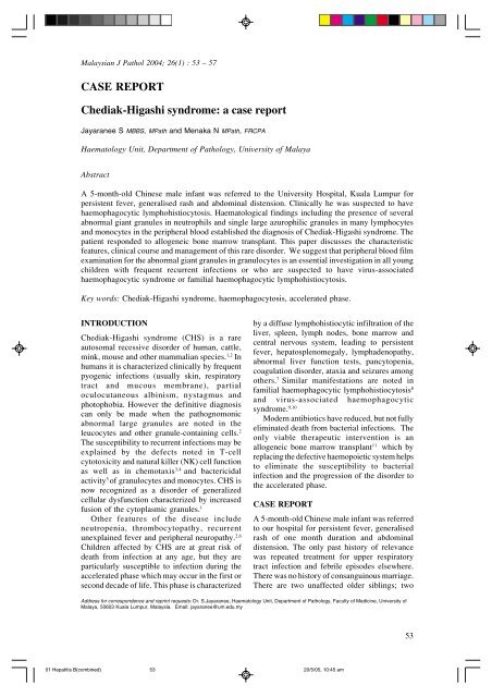
The striking feature in the peripheral blood<br />
film was the presence of several abnormal giant<br />
granules in the neutrophils (Figure 1) and single<br />
large azurophilic granules in many lymphocytes<br />
and in a few monocytes (Figure 2).<br />
Bone marrow aspirate revealed<br />
normocellularity and adequate haemopoietic<br />
cells. Similar giant granules were noted in the<br />
granulocytes and in many of the intermediate<br />
precursors of the granulocytic series. These<br />
granules were strongly myeloperoxidase positive<br />
(Figure 3). Some mononuclear cells had vacuoles<br />
in the cytoplasm (Figure 4). Admixed amongst<br />
the haemopoietic cells were some lymphocytes<br />
and histiocytes, a few of which showed<br />
haemophagocytosis.<br />
Serology for Ebstein Barr virus was positive<br />
for IgG but not for IgM. Parainfluenza virus 3<br />
was isolated from tracheal secretions. Serology<br />
for Toxoplasma, Rubella, Cytomegalovirus,<br />
Dengue and Herpes was negative. Stool<br />
examination too was negative for rota virus,<br />
Salmonella and Shigella. Blood cultures proved<br />
to be negative.<br />
Clinical course<br />
During his hospital stay, the patient had one<br />
episode of croup which was attributed to the<br />
FIG. 1: Neutrophil with abnormal giant granules in<br />
the cytoplasm. MGG x 40<br />
FIG. 2: Monocyte with a single large granule in<br />
cytoplasm. MGG x 40<br />
FIG. 3: Myelocytes with abnormal granules showing<br />
strong myeloperoxidase positivity. Peroxidase<br />
stain x 100<br />
positive culture of Parainfluenza virus 3 from<br />
his tracheal secretions. His persistent fever was<br />
treated with various antibiotics. Since the patient<br />
was diagnosed to be in the accelerated phase at<br />
presentation, he was given dexamethasone and<br />
54<br />
01 Hepatitis B(combined) 54<br />
20/5/05, 10:45 am
CHEDIAK-HIGASHI HEPATITIS SYNDROME B REVIEW<br />
FIG. 4: Monocyte showing large vacuoles in the<br />
cytoplasm. MGG x 40<br />
VP16 which caused only a partial shrinkage in<br />
the size of the liver and spleen. Subsequently an<br />
allogeneic bone marrow transplant was<br />
performed as a matched sibling donor was<br />
available. His post-transplant recovery was<br />
uneventful.<br />
The peripheral blood smear two weeks posttransplant<br />
did not show the characteristic giant<br />
granules in the neutrophils. A sixty days post<br />
transplant bone marrow aspirate too did not<br />
show the typical giant granules in the myeloid<br />
cells. However it took more than six months for<br />
the triglycerides and fibrinogen levels to become<br />
normal. Serum ferritin levels were not repeated<br />
after bone marrow transplantation.<br />
DISCUSSION<br />
<strong>Chediak</strong> <strong>Higashi</strong> <strong>syndrome</strong> (CHS) is a rare<br />
autosomal recessive disorder of lysosomal<br />
granule-containing cells with clinical features<br />
prominently involving the haematologic and<br />
neurologic systems. Majority of the patients<br />
enter an accelerated phase characterized by<br />
lymphohistiocytic infiltration in many organs<br />
leading to haemophagocytosis, pancytopenia and<br />
coagulation disorders. The accelerated phase<br />
may occur shortly after birth or several years<br />
later which, left untreated, proves to be invariably<br />
fatal. The use of etoposide (VP-16) combined<br />
with steroids and intrathecal methotrexate though<br />
resulted in remissions, such remissions were<br />
transient only. Subsequent relapses have been<br />
noted to become increasingly resistant to<br />
conventional therapy, with death occurring due<br />
to haemorrhage and/or infection. 11 Those patients<br />
who survive into young adulthood, develop a<br />
progressive sensorimuscular peripheral<br />
neuropathy which leads them to become wheelchair<br />
bound and they eventually die of infective<br />
complications in their thirties. 12 Uyama et al<br />
after extensive review, have suggested that there<br />
may be two distinct clinical forms: (i) the well<br />
recognized childhood type with the characteristic<br />
marked susceptibility to infection, leading to<br />
early death from overwhelming infection or an<br />
accelerated lymphohistiocytic proliferative<br />
phase, and (ii) the rarer adult form where<br />
neurological defects mimicking parkinsonism,<br />
dementia, or spinocerebellar degeneration and<br />
peripheral neuropathy dominates with a lack of<br />
severe susceptibility to infection. 13 Sung et al’s<br />
autopsy findings lend some credence to this<br />
school of thought. 14 They have shown three<br />
types of pathological changes, (i)<br />
lymphohistiocytic cellular infiltrates in many<br />
organs which appeared to occur late in the<br />
course of the disease since it was observed in the<br />
older patients only, (ii) degenerative changes in<br />
the axons and myelin sheaths, the intensity of<br />
which paralleled the severity of infiltrates, and<br />
(iii) abnormal intracytoplasmic inclusions in a<br />
variety of neurons including nerve cells,<br />
astrocytes, satellite cells of the dorsal spinal<br />
ganglia and schwann cells; interestingly these<br />
were found in all ages (both young and older<br />
individuals) and were thought to be altered<br />
lipofuschin granules, since their presence did<br />
not seem to interfere with the function or survival<br />
of the cells.<br />
Typically patients with CHS present at an<br />
early age with recurrent bacterial infections,<br />
especially with Staphylococcus aureas and beta<br />
haemolyticus streptococcus, partial ocular and<br />
cutaneous albinism, easy bruising due to platelet<br />
dysfunction and severe peridontal disease. 15,16<br />
The diagnosis is made by observing the<br />
characteristic defective giant granules in the<br />
leucocytes in the peripheral blood film. The<br />
susceptibility to infections may be explained by<br />
defects observed in T-cell cytotoxicity and the<br />
NK cell activity as well as in impaired chemotaxis<br />
and bactericidal capacity of the granulocytes<br />
and monocytes. 17<br />
In normal neutrophils, two distinct types of<br />
granules, primary (azurophilic) and secondary<br />
(specific) are present. The azurophilic granule,<br />
formed in promyelocytes is a typical lysosome<br />
containing acid hydrolases whereas the specific<br />
granule formed in myelocytes differs from the<br />
typical lysosome in that it contains enzymes<br />
which act best in alkaline pH. Since the latter is<br />
active in phagocytosis as well, it too can be<br />
considered as a lysosome. Myeloperoxidase is<br />
restricted to the azurophilic granules. Lysosomes<br />
55<br />
01 Hepatitis B(combined) 55<br />
20/5/05, 10:45 am
Malaysian J Pathol June 2004<br />
are membrane-limited organelles that contain a<br />
variety of acid hydrolases. These structures are<br />
present in most cells and their function is related<br />
to intracellular digestion which includes the<br />
hydrolysis of extracellular material and under<br />
certain circumstances, hydrolysis of intracellular<br />
contents. Primary lysosomes may fuse with<br />
cytoplasmic vacuoles or phagosomes, forming<br />
secondary lysosomes that contain partially or<br />
completely digested materials. 18<br />
In <strong>Chediak</strong>-<strong>Higashi</strong> <strong>syndrome</strong>, the large<br />
granules within the neutrophils result from<br />
abnormal fusion of primary (azurophilic)<br />
granules with secondary (specific) granules. 19,20<br />
The fusion of the giant granules with phagosomes<br />
is delayed, contributing to the impaired<br />
immunity. 5,15 Almost all cells of a CHS patient<br />
show some aspect of the abnormal and<br />
dysmorphic lysosomes, storage granules, or<br />
related vesicular structures. For example, the<br />
melanosomes of melanocytes are oversized and<br />
delivery to the keratinocytes in the hair follicles<br />
is partly impaired. When examined<br />
microscopically, the hair shafts have a mixture<br />
of giant melanosomes alternating with small<br />
regions devoid of these pigment granules. This<br />
leads to the macroscopic impression of hair that<br />
is lighter than expected from parental colouration<br />
and has an irregular grayish-silver particulate<br />
sheen. The same defect in melanocytes results<br />
in partial ocular albinism with light sensitivity.<br />
There may be reduced dense bodies in platelets,<br />
which may explain the easy bruising and<br />
prolonged bleeding time found in some CHS<br />
patients. 12<br />
Treatment options for CHS patients are<br />
limited. Previously, the mainstay of treatment<br />
has been mainly symptomatic with antibiotics<br />
and Vitamin C 21 for bacterial infections and<br />
blood product replacement for bleeding<br />
complications. Subsequently when accelerated<br />
phase occurs, etoposide (VP-16), steroids and<br />
intrathecal methotrexate have been tried. Only<br />
recently has allogeneic bone marrow<br />
transplantation become a viable option for these<br />
patients. However, though transplantation has<br />
been shown to correct the haematologic and<br />
immunologic complications of CHS thus halting<br />
the inevitable grim prognosis, it has not been<br />
shown to reverse or prevent further neurological<br />
deficit. 12 This is probably because once the<br />
degenerative changes in the axons and myelin<br />
sheaths occur in the course of the disease, it<br />
cannot be reversed, they being permanent cells<br />
with no regenerative capacity. 14<br />
Since the diagnosis of CHS depends on the<br />
recognition of the characteristic abnormal giant<br />
granules present in the granulocytes in a<br />
peripheral blood film, we suggest that this is an<br />
essential investigation in all young children who<br />
are seen for frequent recurrent infections or who<br />
are suspected to have virus-associated<br />
haemophagocytic <strong>syndrome</strong> or familial<br />
haemophagocytic lymphohistiocytosis. It is<br />
important to offer allogeneic bone marrow<br />
transplant at an early stage of the disease since<br />
the prognosis is uniformly fatal once the disease<br />
progresses to the accelerated phase.<br />
REFERENCES<br />
1. Boxer LA. Neutrophil disorders: Qualitative<br />
abnormalities of neutrophils. In: Williams WJ,<br />
Beutler E, Erslev AJ, Lichtman MA, editors.<br />
Hematology. 4 th ed. McGraw-Hill Publishing<br />
Company; 1991.p.821-24.<br />
2. Blume RS, Wolff SM. The <strong>Chediak</strong>-<strong>Higashi</strong><br />
<strong>syndrome</strong>: Studies in 4 patients and a review of the<br />
literature. Medicine (Baltimore) 1972;51:247-80.<br />
3. Barak Y, Karov Y, Nir E, Wagner Y, Kristal H,<br />
Levin S. <strong>Chediak</strong>-<strong>Higashi</strong> <strong>syndrome</strong>. Expression<br />
of the cytoplasmic defect by in vitro cultures of<br />
bone marrow progenitors. Am J Pediatr Hematol<br />
Oncol 1986;8(2):128-33.<br />
4. Clark RA, Kimball HR. Defective granulocyte<br />
chemotaxis in the <strong>Chediak</strong>-<strong>Higashi</strong> <strong>syndrome</strong>. J<br />
Clin Invest 1971;50:2645-52.<br />
5. Root RK, Rosenthal AS, Balestro DJ. Abnormal<br />
bactericidal, metabolic and lysosomal functions of<br />
<strong>Chediak</strong>-<strong>Higashi</strong> <strong>syndrome</strong>. J Clin Invest<br />
1972;51:649-65.<br />
6. Haliotis T, Roder J, Klein M, et al. <strong>Chediak</strong>-<strong>Higashi</strong><br />
gene in humans. I. Impairment of natural-killer<br />
function. J Exp Med 1980;151(5):1039-48.<br />
7. Amichai B, Zeharia A, Mimouni M, Cohen IJ,<br />
Tunnessen WW. <strong>Chediak</strong>-<strong>Higashi</strong> <strong>syndrome</strong>. Arch<br />
Pediatr Adolesc Med 1997;151: 425-6.<br />
8. Janka GE. Familial haemophagocytic<br />
lymphohistiocytosis. Eur J Pediatr 1983;140(3):221-<br />
30.<br />
9. Risdall RJ, McKenna RW, Nesbit ME, Krivit W,<br />
Balfour HH, Simmons RL, et al. Virus-associated<br />
hemophagocytic <strong>syndrome</strong>: A benign histiocytic<br />
proliferation distinct from malignant histiocytosis.<br />
Cancer 1979;44(3):993-1002.<br />
10. Wilson ER, Malluh A, Stagno S, Crist WM. Fatal<br />
Epstein-Barr virus associated hemophagocytic<br />
<strong>syndrome</strong>. J Pediatr 1981;98(2):260-2.<br />
11. Haddad E, Le Deist F, Blanche S, Benkerrou M,<br />
Rohrlich P, Vilmer E, et al. Treatment of <strong>Chediak</strong>-<br />
<strong>Higashi</strong> <strong>syndrome</strong> by allogeneic bone marrow<br />
transplantation: Report of 10 <strong>case</strong>s. Blood 1995;85<br />
(11):3328-33.<br />
12. Malech HL, Nauseef WM. Primary inherited defects<br />
in neutrophil function: etiology and treatment.<br />
Seminars in hematology 1997;34(4):279-90.<br />
13. Uyama E, Hirano T, Ito K, Nakashima H, Sugimoto<br />
56<br />
01 Hepatitis B(combined) 56<br />
20/5/05, 10:45 am
CHEDIAK-HIGASHI HEPATITIS SYNDROME B REVIEW<br />
M, Naito M et al. Adult <strong>Chediak</strong>-<strong>Higashi</strong> <strong>syndrome</strong><br />
presenting as parkinsonism and dementia. Acta<br />
Neurol Scand 1994;89:173-183.<br />
14. Sung JH, Meyers JP, Stadlan EM, Cowen D, Wolf<br />
A. Neuropathological changes in <strong>Chediak</strong>-<strong>Higashi</strong><br />
disease. J Neuropathol Exp Neurol 1969;28:86-<br />
118.<br />
15. Introne W, Boissy RE, Gahl WA. Clinical, molecular<br />
and cell biological aspects of <strong>Chediak</strong>-<strong>Higashi</strong><br />
<strong>syndrome</strong>. Mol Genet Metab 1999;68:283-303.<br />
16. Wolff SM. The <strong>Chediak</strong>-<strong>Higashi</strong> Syndrome: studies<br />
of host defenses. Ann Intern Med 1972;76:293-<br />
306.<br />
17. Certain S, Barrat F, Pastural E, Le Deist F, Goyo-<br />
Rivas J, Jabado N, et al. Protein truncation test of<br />
LYST reveals heterogenous mutations in patients<br />
with <strong>Chediak</strong>-<strong>Higashi</strong> <strong>syndrome</strong>. Blood<br />
2000;95(3):979-83.<br />
18. Davis WC, Douglas DS. Defective granule<br />
formation and function in the <strong>Chediak</strong>-<strong>Higashi</strong><br />
<strong>syndrome</strong> in man and animals. Semin Hematol<br />
1972;9(4):431-<br />
19. Rausch PG, Pryzwansky KB, Spitznagel JK.<br />
Immunohistochemical identification of azurophilic<br />
and specific granule markers in the giant granules<br />
of <strong>Chediak</strong>-<strong>Higashi</strong> <strong>syndrome</strong> neutrophils. N Engl<br />
J Med 1978;298:693-8.<br />
20. White JG, Clawson CL. The <strong>Chediak</strong>-<strong>Higashi</strong><br />
<strong>syndrome</strong>: The nature of the giant neutrophils and<br />
their interactions with cytoplasm and foreign<br />
particulates. I. Progressive enlargement of the<br />
massive inclusions in mature neutrophils. II.<br />
Manifestations of cytoplasmic injury and<br />
sequestration. III. Interactions between giant<br />
organelles and foreign particulates. Am J Pathol<br />
1980;98:151-96.<br />
21. Weening RS, Schoorel EP, Roos D, van Shaik<br />
MLJ, Voetman AA, Bot AAM, et al. Effect of<br />
ascorbate on abnormal neutrophil, platelet and<br />
lymphocyte function in a patient with the <strong>Chediak</strong>-<br />
<strong>Higashi</strong> <strong>syndrome</strong>. Blood 1981;57(5):856-65.<br />
57<br />
01 Hepatitis B(combined) 57<br />
20/5/05, 10:45 am
Malaysian J Pathol June 2004<br />
58<br />
01 Hepatitis B(combined) 58<br />
20/5/05, 10:45 am


