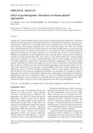
Disseminated histoplasmosis mimicking miliary tuberculosis: a case ...
Disseminated histoplasmosis mimicking miliary tuberculosis: a case ...
Disseminated histoplasmosis mimicking miliary tuberculosis: a case ...

Create successful ePaper yourself
Turn your PDF publications into a flip-book with our unique Google optimized e-Paper software.
<strong>Disseminated</strong> <strong>histoplasmosis</strong> <strong>mimicking</strong> <strong>miliary</strong> <strong>tuberculosis</strong>: a <strong>case</strong> report<br />
K ~ SoonCHAN I MBBS,MPath, Lai Meng LOOI* FRCPath, MDand Siew Pheng CHAN, MBBS, MRCP*"<br />
Puthluh. Ipoh rind D~~partmerzrs of*Patk(~log~ nttd *"M~d/cine, University of ~Wrxluyu<br />
A 35-pcar-old Chincsc man who was known to have insulin-dependent diabetes tnellitus was<br />
admilled for fever and weight loss. During his hospital stay, he fell to his death from his ward at the<br />
t welrth floor. The clinical features, radiological fintlings and gross organ changc.; at autopsy cl(>sely<br />
simulated rniliary <strong>tuberculosis</strong>. Ilistologp, howcver, revealed extensive nccrosis of thc adrcnal<br />
glands, lungs, spleen, kidneys and thyn)id associated with the presence of Histo]~bsnw ccsp.rr~kurunl<br />
organisms. This <strong>case</strong> highlights the similarity hr~th clinically andpathotogically betwwn histoplasmr,sis<br />
and <strong>tuberculosis</strong> and emphasizes the need to hc awarc of this infection in a nonendemic area among<br />
patients with a compromised immune syslcm.<br />
Kcy wurds: Histopla~n~r~sis, granulornatous i~lilaminalion. ti~berculosis. diabetes mellitus.<br />
INTRO~UCTTON<br />
lwo previous admissions four years earlier both<br />
for diabetic ketoacidosis.<br />
Depressiori of cellular immunity can prcdisposc<br />
On admission, the patient was pale, thin and<br />
10 various fungal infections including candidiasis,<br />
emaciated. He had a hlooci pressure uf 1001711<br />
,~spergillosis, nocardiosis and histuplasmosis.<br />
rnn~Hg and a pulse rate uf 108 per minute.<br />
Condilioi~s implicated as causes of imrni~ne<br />
There was a low gr~icie fever. Occasional<br />
depression includecancer chemotherapy, Lhyrnic<br />
crepirations were heard at rhc lung bases. Buth<br />
dyspenesis. corticostero~d lherapy and more<br />
the liver and spleen wcrc cnlargcd. Ucntal<br />
recently, human immunodeficiency virus (HIV)<br />
hygiene was poor bul there wcrc no oral ulccrs<br />
infection. In FUC~ patients, a high index of<br />
or skin Icsions.<br />
suspicion should hc kcpt c~f the rarer infections<br />
Routine haetnatological tests were within<br />
that they arc pronc to. Histoplastnosis is one<br />
normal limils. The random blood sugar level<br />
such infcction. Diagnosis of this infection can<br />
wils elevated (21.6 rnmol/L). There was also<br />
hc difficult because the clinical features arc<br />
glycosuria. ketonuria and a metabolic acidosis.<br />
usually prolean and frequently simulatc othcl-<br />
The blood urea and electrolyte levels were: urea<br />
granulornatous infections. Furthermore, the<br />
3.5 mmol/I, sodium 136 ~nrnol/l, potassium 3.3<br />
organisms ihernselves can be difficult to<br />
mnlolfi and chlonde l03 mmol/l. Ttie serutn<br />
recognise in histological sections.<br />
creatinine level was 84 umnl/l. Hlood cultures<br />
Wc report a <strong>case</strong> of disscrninatcd<br />
failed to grow any organism and serological<br />
<strong>histoplasmosis</strong> in a diabetic patient which<br />
tests for typhoid, typhus and dcnguc fcvcr wcrc<br />
clinically mimicked <strong>tuberculosis</strong>. The diagnonegative.<br />
The chest radiograph showed nusis<br />
was clarified only on histological examinamcrous<br />
tiny <strong>miliary</strong> shadows on which a clii~ical<br />
tion of post mortcm material. This <strong>case</strong><br />
suspicion of <strong>tuberculosis</strong> was aroused. Liver<br />
emphasises the necd for greater clinical awarefunction<br />
tests showed mild elevation of liver<br />
ness of the condition particularly in view of the<br />
enzymes. Sputum examination failed to demhigh<br />
mortality assuciatcd with the disseminated<br />
c~nstratc any acid fast bacillus on staining and<br />
form of this infcction.<br />
Lhe ouimme of cultures for <strong>tuberculosis</strong> was<br />
CASE REPORT<br />
being awai~ed.<br />
The patient was treated with insulin injections<br />
A 35-year-old Chinese rnan was admitted to the and his diabetes improved. Extensive dental<br />
University Hospital, Kuala Lumpur for fever. canes requlred denial rcfcral and extractions<br />
loss of appctitc, loss of weight, polyuria and were done.<br />
polydipsia of one month's duration. He was a Eleven days after admission. he was found<br />
vagahoild froin Jinjang, Kuala Lumpur and had dcad cm the ground floor of the hospital tower<br />
Address for mrresp~~denceandrepnnrrequesrs Or K.S Chan Pathology and Cllnlwl hhratory1M) Sdn. Bhd. 49, JalanOnn Jaafar, 30300 Ipoh, Perak.<br />
Malaysia.
Malassian J Pathol December 1993<br />
block. presumably having jumped from his ward<br />
on the twelfth floor. The Mantoux test was not<br />
yet ready for reading at the time of death. The<br />
HIV antibody status of this patient was not<br />
known. A coroner's autopsy was performed.<br />
Autop.~ finriirigs<br />
. ..<br />
The autopsy examination revealed a markedly<br />
emaciated body with extensive injuries and<br />
fractures involving the head, chest wall, thoracic<br />
and abdominal organs consistent with the<br />
fall. In addition, both lungs were consdidated<br />
and heavy. Their external and cut surfaces<br />
revealed numerous confluent, yellowish white<br />
<strong>case</strong>ous nodules of about 2 mm. diameter each<br />
(Fig. 1). The hilar lymph nodes were not enlarged.<br />
The right adrenal gland contained nodular<br />
yellowish necrotic lesions. The left adrenal<br />
gland was macerated by the trauma, but the<br />
remaining tissue also showed extensive necrosis.<br />
The enlarged liver and spleen were lacerated<br />
but did not show any other gross pathology.<br />
Other visceral organs examined did not show<br />
any gross lesions. No culture of the tissues was<br />
attempted.<br />
Histology<br />
The most striking changes were seen in the<br />
adrenal glands. These showed extensive areas<br />
of necrosis with numerous intracellular and extracellular<br />
yeast cells at the periphery (Fig. 2).<br />
There was very little inflammatory reaction to<br />
the necrosis. The yeast cells were ovoid. measuring<br />
about 4 urn in their long axis and each<br />
exhibited a peripheral halo. The Gomori methenamine<br />
silver (GMS) stain demonstrated budding<br />
in some yeast cells which were typical of<br />
Histoplasma caps~rlatirm (Fig. 3).<br />
Sections of the lungs showed many -. poorlyformed<br />
epithelioid gra~ulomas with large areas<br />
of central <strong>case</strong>ous necrosis. There was minimal<br />
lymphocytic reaction and Langhan's type giant<br />
cells were not present. Moderate numbers of<br />
Histoplasma capsulatirm organisms were found<br />
within the epithelioid cells.<br />
Similar organisms were also found in the<br />
kidneys, thyroid and spleen, with the former two<br />
showing granulomatous lesions as well. In addition<br />
the spleen exhibited marked depletion of<br />
lymphoid cells and marked distension of sinusoids<br />
by histiocytes. The liver revealed numerous<br />
tiny granulomatous lesions although no fungal<br />
organism was found. No acid fast bacilli were<br />
demonstrable in all these organs. The mediastinal<br />
lymph nodes were depleted of lymphoid<br />
follicles.<br />
DISCUSSION<br />
Although <strong>histoplasmosis</strong> was first discovered<br />
by Darling and later described in detail by Parsons<br />
er al.' more than 80 and 40 years ago<br />
respectively, a high mortality rate associated<br />
with missed diagnosis of disseminated<br />
<strong>histoplasmosis</strong> is still common.2 Thus pathologists<br />
and physicians alike must be aware of this<br />
FIG. I: Gross appearancc of upper lobe of Icf~ lung showing numerous tiny necrotic nodules.
DISSEMINATED HISTOPLASMOSlS<br />
FIG. 2: Photomicrograph of right adrenal gland showing groups of yeast cells within the cytoplasm<br />
of necrotic macrophages and parenchyma1 cells. Extracellular forms are seen at lower right<br />
field. Note the poor inflammatory response to the organism. H&E X 300.<br />
disease and the diagnostic methods available in<br />
order to lower its mortality rate. Furthermore,<br />
with the expected increase in incidence of the<br />
acquired immune deficiency syndrome (AIDS)<br />
in Malaysia in the future, the incidence of<br />
<strong>histoplasmosis</strong> is likely to increase. Hence,<br />
there is a further need for greater awareness<br />
among health care workers of this infection.<br />
Immune deficiency associated with diabetes<br />
mellitus probably had played a part in the susceptibility<br />
of this patient to the infection.<br />
This infection, whether in the form.of chronic<br />
pulmonary disease or the disseminated forms,<br />
closely simulate <strong>tuberculosis</strong> ~linically'~"~~' and<br />
sometimes also in histological sections. The<br />
histological diagnosis is hampered by the fact<br />
that the organisms are frequently very scarse.<br />
Because of their small size, they are also very<br />
difficult to identify.' Special techniques such as<br />
the Gomori's methenamine silver method or<br />
periodic acid Schiff reaction are required to<br />
demonstrate the yeast forms in tissue sections.<br />
Diagnosis is usually dependent on isolation<br />
or identification of the organism^,^,^ or, more<br />
recently, on detection of specific antibody or<br />
FIG. 3: Section of ripht adrenal gland showing nuantigen<br />
in the serum or urine.' Rapid diagnosis<br />
merous yeast cells. some with budding charhave<br />
also been achieved in certain centres with<br />
acteristic of Histoplusmu c~aps~tlurun~.<br />
the application of immunofluorescence and Gomori's methenamine silver X 300.
immunoperoxidase techniques on tissue sec- REFEWENCES<br />
tions." Allhough isolation of the fungus was not<br />
I. Pa~son RJ, Zarafonctis CJD. Hjstnplasmusis inMan:<br />
carried out in this <strong>case</strong>, a diagnosis of disscmi- reporl of scven cascs and a review or xventy-one<br />
nated <strong>histoplasmosis</strong> can be made with ceriainty <strong>case</strong>s. Arch Int Med 1945: 75: 1.<br />
because of the characteristic yeasr cells found in 2. Salhapatayavongs B. Batteipr BE. Wheat J, Slama<br />
tissue sections. In histological sections, other 'W, Wass JL. Clinical and labrxatnry fenlures of<br />
infections that need to be distinguished from disseminated <strong>histoplasmosis</strong> during two large urban<br />
<strong>histoplasmosis</strong> are <strong>tuberculosis</strong>, torulopsis, outbreaks. Medicinc (Baltimore) 1983; 62: 263-<br />
70.<br />
leishmaniasis. candidiasis and crypt~coccosis.~~~<br />
Goodwin et crl.klassified histopla.;mosis by<br />
considering the clinical and pathological features.<br />
The ~ntermediate duration of illness,<br />
preserice of' weighk loss, hepatosplenornegaly<br />
and focal destructive lesions, especially in the<br />
adrenal glands, place our patienl into the subacute<br />
disseminated form according to Goodwin's<br />
classification.<br />
Because tuberculos~s is cornmon In Malaysia,<br />
il is invariably thc first differential diagnosis in<br />
a <strong>case</strong> of this naturc. However. whel-le\~er acid<br />
fast bacilli cannot be deinonstratcd or isolated, a<br />
diagnosis of histoplasrnosis should be considered.<br />
Steroids should not be giver] in any<br />
granulomatous disease unless <strong>histoplasmosis</strong> had<br />
been exclrided, as it has been reported Lhal this<br />
frequently results in a fatal o~tcorne.~"<br />
'l'his patient had very severe adrenal gland<br />
involve~nent. Antemortern findings suggestive<br />
of adrcnal insufficiency include a marginally<br />
IOW blood pressurc and serum sodiuln level.<br />
Goodwin et al.' in nn exzensive study reported<br />
adrenal invtllvement at autopsy in 82% of all<br />
forms of clissernina~cd <strong>histoplasmosis</strong>.<br />
3. Goodwin RA, Shapiro JL, Thur~nan GH, Thunnan<br />
SS. Dcs Prez RM. Disscminalcd <strong>histoplasmosis</strong>:<br />
clinica1 and pathologic correlatiuna. Medicine<br />
(Baltimore) 1980; 59: 1-33.<br />
4. Juhnsron AW. Pustlcthwaite R, Ewcn SWB er al.<br />
t)isse~ninated hisroplasmtsis. J Inf 1984: 9. 79-82.<br />
5. ChinTH, McGarry T, Coopers eral. Dis~eminatcd<br />
histoplas~nosis it] the acquired immunodcficiency<br />
aqndrornc. Arch Int Mcd 1987; 147: 1181-4.<br />
6. Wheat J, French MLV, Kohlcr RH, Zimmennan<br />
SE, Smith WR, Nortun JA, Eilzen IIE. Smirh CD,<br />
Slaina TG. The diagthostic laboratory tests for<br />
hihtoplasmosis. A~~alysis of expcrier~ce m a large<br />
urban outbreak. Ann Int Med 1982; 97. h80-5.<br />
7. Wheat LJ, Kohlcr RB, Tewan RP. Diagnos~s of<br />
disseminated histnplasmos~s by detection of<br />
Histoplasma capapsulutum antigen in ser111n and urlnt.<br />
specitnens. N Engl J Mcd 1986; 3 14: 83-8.<br />
8. Klalt EC, Covgrove M.Mcyer PR. Rapid diagnosis<br />
of disseminated h~sluplasmusis in tissues. Arch<br />
Pathol Lab Med 1986; 110: l 173-5.<br />
9. Binford CH, Connor UH. Pathology uf Troplcal<br />
and errraurd~~~ary Discascs, Wasl~itrgton DC, Armed<br />
Forces Institute of Pathology, 1976 vol 2. pp 578-<br />
813.<br />
10. Taylor CD. Fanning E.4, Fcrgusorl JP cr 01. Dis-<br />
aeminnted h~stopla\mnsis in a nonetldernlr: area.<br />
Can Med Assoc 1 1985. 133( 15): 763-5.