CASE REPORT Haemoglobin Lepore in a Malay family: a ... - MJPath
CASE REPORT Haemoglobin Lepore in a Malay family: a ... - MJPath
CASE REPORT Haemoglobin Lepore in a Malay family: a ... - MJPath

Create successful ePaper yourself
Turn your PDF publications into a flip-book with our unique Google optimized e-Paper software.
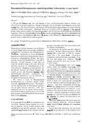
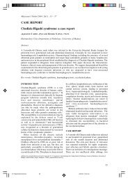
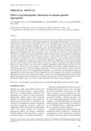
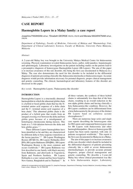
<strong>Malay</strong>sian J Pathol June 2005<strong>in</strong>vestigations revealed the diagnosis to behomozygous <strong>Haemoglob<strong>in</strong></strong> <strong>Lepore</strong>. The cl<strong>in</strong>ical,haematological and laboratory features of thisdisorder are discussed <strong>in</strong> this paper.<strong>CASE</strong> <strong>REPORT</strong>A 2-year-old <strong>Malay</strong> boy was brought toUniversity <strong>Malay</strong>a Medical Centre forthalassemia/ haemoglob<strong>in</strong>opathy screen<strong>in</strong>g.Physical exam<strong>in</strong>ation revealed thalassemiafacies, pallor, mild jaundice, hepatomegaly (3cm below the right subcostal marg<strong>in</strong>) andsplenomegaly (3 cm below the left subcostalmarg<strong>in</strong>). His height was at 3 rd percentile and hisweight was at 3-10 th percentile. Hishaematological profile showed haemoglob<strong>in</strong> 74g/L, red cell count 3.3 x 10 12 L, MCV 77 fl,MCH 22 pg and a reticulocyte count of 5.8%.The white blood cell count and platelet countwere normal. The patient’s blood smear revealedhypochromia, microcytosis, anisopoikilocytosis,polychromasia, basophilic stippl<strong>in</strong>g and a fewtarget cells (Fig.1).<strong>Haemoglob<strong>in</strong></strong> electrophoresis on celluloseacetate at alkal<strong>in</strong>e pH (pH 8.6) showed aprom<strong>in</strong>ent F band and an additional band <strong>in</strong> theS region (Fig. 2). There was no haemoglob<strong>in</strong> Aor haemoglob<strong>in</strong> A2 bands seen. Quantificationof haemoglob<strong>in</strong> F revealed a level of 90% andthe unknown haemoglob<strong>in</strong> constituted 9.3%.Electrophoresis at acid pH (pH 6.0) on citrateagar was carried out to identify the band <strong>in</strong> theS region. There was no Hb S detected. However,a prom<strong>in</strong>ent band was noted <strong>in</strong> the F region anda fa<strong>in</strong>t band <strong>in</strong> the A region. As the patient didnot have any A band on the cellulose acetatestrip, the fa<strong>in</strong>t band <strong>in</strong> the A region on citrateagar was suggestive of Hb D/G or Hb <strong>Lepore</strong>.Further test<strong>in</strong>g to identify and quantify theabnormal haemoglob<strong>in</strong> variant was carried outby high performance liquid chromatography(HPLC) Bio-Rad Variant <strong>Haemoglob<strong>in</strong></strong> test<strong>in</strong>gsystem (Fig. 3). The unknown haemoglob<strong>in</strong> hada retention time (RT=3.59) similar to Hb A2 onthe “Beta Thal Short Program” and wasquantified as 10.7 %. There was no haemoglob<strong>in</strong>A synthesized and the rest of the haemoglob<strong>in</strong>present was hemoglob<strong>in</strong> F. Hb <strong>Lepore</strong> is knownto have the same retention time as Hb A2 on the“Beta Thal Short Program” and therefore apresumptive diagnosis of homozygous Hb<strong>Lepore</strong> was made for the patient.Screen<strong>in</strong>g was carried out for both parents.The father’s blood studies showed haemoglob<strong>in</strong>140 g/L, MCV 72 fl, and MCH 24 pg while themother’s haemoglob<strong>in</strong> was 111g/L, MCV 65.0fl,MCH 19 pg. The blood smear of both parentsrevealed hypochromia, microcytosis, few targetcells and occasional basophilic stippl<strong>in</strong>g (Fig.4). <strong>Haemoglob<strong>in</strong></strong> electrophoresis on celluloseacetate (pH 8.6) of both parents (Fig. 5) showedbands at A, A2, and F regions and an additionalband <strong>in</strong> the S region while electrophoresis oncitrate agar at acid pH showed a prom<strong>in</strong>ent Aband and a fa<strong>in</strong>t band <strong>in</strong> the F region. There wasFIG. 1: Peripheral blood film of patient show<strong>in</strong>ghypochromasia, microcytosis, anisopoikilocytosis,target cells and nucleated red bloodcell. Wright sta<strong>in</strong> X 400FIG. 2: <strong>Haemoglob<strong>in</strong></strong> electrophoresis on cellulose acetateat pH 8.6 Lane 1: normal control show<strong>in</strong>gHb A and Hb A2. Lane 2: patient:absence of Hb A and Hb A2, <strong>in</strong>creased Hb Fand an abnormal Hb (unknown band).34
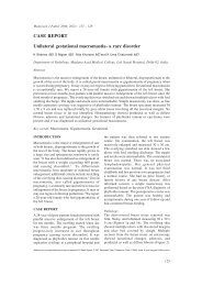
HAEMOGLOBIN LEPOREno Hb S detected. The father’s Hb F level was1.6%, Hb A2 level 2.7% while the mother’s HbF level was 2.1%, Hb A2 level 3.0 %. Highperformance liquid chromatography (Fig. 6)revealed the unknown haemoglob<strong>in</strong> to be Hb<strong>Lepore</strong> (RT= 3.59) and the level <strong>in</strong> the fatherand mother was 13.1% and 13.8% respectively.FIG. 5: <strong>Haemoglob<strong>in</strong></strong> electrophoresis on celluloseacetate at pH 8.6. Lane 1: normal controlshow<strong>in</strong>g Hb A and Hb A2. Lane2: mother,heterozygous show<strong>in</strong>g HbA, Hb A2 and anabnormal Hb (unknown). Lane 3: father,heterozygous show<strong>in</strong>g similar f<strong>in</strong>d<strong>in</strong>gs.FIG. 3: High performance liquid chromatographyanalysis us<strong>in</strong>g the VARIANT Hemoglob<strong>in</strong>Test<strong>in</strong>g System (Bio-Rad Laboratories).Chromatogram of patient (homozygous). NoHb A, <strong>in</strong>creased Hb F, Hb A2 + <strong>Lepore</strong>retention time 3.59FIG. 4: Peripheral blood film (mother) show<strong>in</strong>ghypochromasia, microcytosis and target cells.Wright sta<strong>in</strong> x 400FIG. 6: High performance liquid chromatographyanalysis us<strong>in</strong>g the VARIANT Hemoglob<strong>in</strong>Test<strong>in</strong>g System (Bio-Rad Laboratories) .Chromatogram of mother (heterozygous). HbA2 + <strong>Lepore</strong> retention time 3.5935
<strong>Malay</strong>sian J Pathol June 2005A diagnosis of heterozygous haemoglob<strong>in</strong><strong>Lepore</strong> was made for both parents. The parentsare first degree relatives. The patient’s andparents’ samples were sent for DNA sequenc<strong>in</strong>gwhich confirmed this unknown haemoglob<strong>in</strong> tobe Hb <strong>Lepore</strong> Wash<strong>in</strong>gton Boston. The patientis on regular follow-up and has received 3 bloodtransfusions at 4 -5 monthly <strong>in</strong>tervals s<strong>in</strong>ce thetime of diagnosis.DISCUSSIONHb <strong>Lepore</strong> is a common abnormal haemoglob<strong>in</strong>seen <strong>in</strong> the Mediterranean region. It is the mostcommon abnormal haemoglob<strong>in</strong> <strong>in</strong> Caucasians<strong>in</strong> Central Portugal and <strong>in</strong> the Spanish AltaExtremadura. 1 It is a variant form of humanhaemoglob<strong>in</strong> that conta<strong>in</strong>s delta beta hybridcha<strong>in</strong>s or a fused glob<strong>in</strong> cha<strong>in</strong>, from the productof a hybrid gene: the unequal cross<strong>in</strong>g overbetween the delta and beta glob<strong>in</strong> genes result<strong>in</strong>g<strong>in</strong> a deletion of 7.4kb <strong>in</strong> the beta glob<strong>in</strong> genecluster. 1,2Three different types of Hb <strong>Lepore</strong> have beendescribed so far, each with a different crossoverbreakpo<strong>in</strong>t: Hb <strong>Lepore</strong> Wash<strong>in</strong>gton Boston, Hb<strong>Lepore</strong> Baltimore and Hb Hollandia. 1,2,4 Thebasic defect <strong>in</strong> all the Hb <strong>Lepore</strong> disorders is the<strong>in</strong>efficient synthesis of the delta beta fusioncha<strong>in</strong>s of Hb <strong>Lepore</strong>, which leads to a variabledegree of glob<strong>in</strong> imbalance, excess alpha cha<strong>in</strong>production and the cl<strong>in</strong>ical phenotype of betathalassaemia. The <strong>in</strong>effective erythropoiesis andthe shortened red cell survival results from thedeleterious effects of excess alpha cha<strong>in</strong>s. Theselection of cells which are synthesiz<strong>in</strong>grelatively more gamma cha<strong>in</strong>s follow the samemechanism as occurs <strong>in</strong> beta thalassemia. 5 It hasbeen postulated that the deletion which resulted<strong>in</strong> the delta beta fusion is somehow responsiblefor <strong>in</strong>creas<strong>in</strong>g the absolute output of gammacha<strong>in</strong>s. 6 This is compatible with the observationthat Hb <strong>Lepore</strong> heterozygotes producesignificantly more Hb F than beta thalassaemiaheterozygotes.The Hb <strong>Lepore</strong> heterozygotes are usuallyasymptomatic. In a large Italian series noabnormal physical signs were reported, but afew of the Greek and Yugoslavian heterozygoteshad mild splenomegaly. 7,8 Heterozyogotes aretherefore healthy <strong>in</strong>dividuals with only a slightdecrease <strong>in</strong> haemoglob<strong>in</strong> levels, but with adist<strong>in</strong>ct microcytosis and hypochromia of theirred blood cells. The mean cell haemoglob<strong>in</strong>(MCH) is thought to be the most reliableparameter that is altered <strong>in</strong> the Hb <strong>Lepore</strong>heterozygotes (range 20-25 pg). 9 In this series,the red cell <strong>in</strong>dicies and morphologicalappearances of the blood film of betathalassaemic carriers were compared with thatof heterozygous Hb <strong>Lepore</strong>. It was found to besimilar <strong>in</strong> both conditions. Studies suggest thatthere are no major differences <strong>in</strong> thehaematological f<strong>in</strong>d<strong>in</strong>gs between carriers of thedifferent molecular varieties of Hb <strong>Lepore</strong>. 1 Redcell survival and ferrok<strong>in</strong>etic data available revealred cell survival is slightly shortened and Fe 59utilization is <strong>in</strong>creased. 10The Hb <strong>Lepore</strong> homozygotes vary <strong>in</strong> theseverity of their cl<strong>in</strong>ical manifestations. 6,7,8 Atone end of the spectrum are patients who presentwith<strong>in</strong> the first 5 years of life with severe anaemiawith haemoglob<strong>in</strong> values rang<strong>in</strong>g from 4 to 7 g/dl. Significant splenomegaly, hepatomegaly, andskeletal abnormalities <strong>in</strong>dist<strong>in</strong>guishable fromthose of homozygous beta thalassaemia are seen.These patients require regular blood transfusionand exhibit shortened life span. At the other endof the spectrum are patients who have a milderdisorder <strong>in</strong>dist<strong>in</strong>guishable from beta thalassemia<strong>in</strong>termedia, who are anaemic throughoutchildhood and require only occasional bloodtransfusions. The homozygous state for Hb<strong>Lepore</strong> therefore varies from a transfusiondependent disease like beta thalassaemia majorthrough the milder spectrum of thalasaemia<strong>in</strong>termedia that appears to be compatible withlongevity of life. 5 The haematological f<strong>in</strong>d<strong>in</strong>gs<strong>in</strong> these patients are peripheral blood film features<strong>in</strong>dist<strong>in</strong>guishable from the severe forms of betathalassaemia. Bone marrow smears show markederythroid hyperplasia due to high level oferythropoiet<strong>in</strong> production <strong>in</strong> response to theanaemia. 6The multifaceted approach for thepresumptive identification of haemoglob<strong>in</strong>variants <strong>in</strong>cludes a scrut<strong>in</strong>y of blood counts/redcell <strong>in</strong>dices and haemoglob<strong>in</strong> analysis. Thisapproach easily identifies Hb <strong>Lepore</strong>. Oncellulose acetate electrophoresis at alkal<strong>in</strong>e pH,Hb <strong>Lepore</strong> shows electrophoretic mobilitysimilar to Hb S. 5 The other haemoglob<strong>in</strong>s thatrun <strong>in</strong> this position are Hb D and Hb G, whichare differentiated from Hb S by sickle solubilitytest and electrophoresis at acid pH. Hb <strong>Lepore</strong>has a similar retention time (RT) value as Hb A2on high performance liquid chromatography(HPLC) analysis with the Bio-Rad Variant<strong>Haemoglob<strong>in</strong></strong> test<strong>in</strong>g system. 2 Values greaterthan 10% suggest the presence of variant36
HAEMOGLOBIN LEPOREhaemoglob<strong>in</strong>. In <strong>Malay</strong>sia the most commonhaemoglob<strong>in</strong> variant seen is Hb E that isdifferentiated from Hb <strong>Lepore</strong> by celluloseacetate electrophoresis at alkal<strong>in</strong>e pH. Hb Eruns <strong>in</strong> the same position as HbA2, whereas Hb<strong>Lepore</strong> runs at the position of Hb S. Therefore amultifaceted approach which <strong>in</strong>cludeshaemoglob<strong>in</strong> electrophoresis and highperformance liquid chromatography providessufficient <strong>in</strong>formation for the presumptiveidentification of Hb <strong>Lepore</strong>. The characterisationof the type of Hb <strong>Lepore</strong> requires analysis of theglob<strong>in</strong> cha<strong>in</strong>s by reversed phase HPLC andDNA studies would confirm the diagnosis ofthis variant haemoglob<strong>in</strong>. 4In the homozygous state, Hb A and Hb A2are absent and the haemoglob<strong>in</strong> is made up ofHbs F and <strong>Lepore</strong> only. The level of Hb <strong>Lepore</strong>ranges from 8 to 30% with a mean value ofapproximately 15%, the rema<strong>in</strong>der of thehaemoglob<strong>in</strong> be<strong>in</strong>g Hb F. In the heterozygousstate the haemoglob<strong>in</strong> conta<strong>in</strong>s Hbs A, <strong>Lepore</strong>,A2 and a variable amount of Hb F. The reportedlevel of Hb <strong>Lepore</strong> is between 5 to 15%, with amean level around 10%. The mean level of HbA2 is about 2% and the reported values for HbF range from between 1-14%. 5In conclusion, Hb <strong>Lepore</strong> is a structuralhaemoglob<strong>in</strong> variant coded for by a hybrid geneformed by the fusion of delta and beta genes.The homozygous state is cl<strong>in</strong>ically similar tobeta thalassaemia <strong>in</strong>termedia or major.Presumptive identification can be easily made<strong>in</strong> the laboratory by a multifaceted approach that<strong>in</strong>cludes the methods, scrut<strong>in</strong>y of blood counts/red cell <strong>in</strong>dices, haemoglob<strong>in</strong> electrophoresis atalkal<strong>in</strong>e and acid pH and haemoglob<strong>in</strong> analysisby high performance liquid chromatography.However, confirmation requires DNAcharacterization. A correct diagnosis is essentialfor proper cl<strong>in</strong>ical management and geneticcounsel<strong>in</strong>g.4. Ropero P, Gonzalez FA, Sanchez J et al. Identificationof the Hb <strong>Lepore</strong> phenotype by HPLC.Haematologica 1999; 84: 1081-45. The δ β and related thalassaemias. In: WeatherallDJ and Clegg JB, editors. The Thalassemia Syndromes.4 th ed. Blackwell Science Ltd; 2001. p.361-636. Olivieri NF, Rees DC, G<strong>in</strong>der GD et al. Treatmentof thalassemia major with phenylbutyrate and hydroxyurea.Lancet 1997; 350: 491-27. Duma H, Efremov G, Sadikario A et al. Study ofn<strong>in</strong>e families with haemoglob<strong>in</strong> <strong>Lepore</strong>. Br JHaematol. 1968; 15: 161-728. Quattr<strong>in</strong> N, Luzzatto L, Quattr<strong>in</strong> S. New cl<strong>in</strong>icaland biochemical f<strong>in</strong>d<strong>in</strong>gs from 235 patients withhaemoglob<strong>in</strong> <strong>Lepore</strong>. Ann NY Acad Sci 1980;344: 364 -749. Huisman TH. Compound heterozygosity for HbSand the hybrid Hb S <strong>Lepore</strong>. Comparision ofhaematological and haemoglob<strong>in</strong> composition data.Hemoglob<strong>in</strong> 1997; 21: 249-5710. Pearson HA, Mc Farland W, K<strong>in</strong>g ER.Erythrok<strong>in</strong>etic studies <strong>in</strong> thalassemia trait. J LabCl<strong>in</strong> Med 1960; 56: 866-73REFERENCES1. Ribeiro ML, Cunha E., Goncalves P et al. Hb.<strong>Lepore</strong>- Baltimore and Hb <strong>Lepore</strong>-Wash<strong>in</strong>gton-Boston <strong>in</strong> Central Portugal and Spanish AltaExtremadura. Hum. Genet. 1997; 99: 669-732. Shaji RV, Edison ES, Krishamoorthy R, ChandyM, Srivastava A. Hb <strong>Lepore</strong> <strong>in</strong> the Indian population.Hemoglob<strong>in</strong> 2003; 27:7-143. Viprakasit V, Pung-Amritt P, Suwanthon L, ClarkK, Tanphaichitr VS. Complex <strong>in</strong>teractions of [delta][beta] hybrid haemoglob<strong>in</strong> (Hb <strong>Lepore</strong>- Hollandia)Hb E ([beta] 26G>A ) and [alpha] + thalassaemia <strong>in</strong> aThai <strong>family</strong>. Eu J Haematol 2002; 68:107-12.37