When Healing Becomes Educating, Vol. 6 - Waldorf Research Institute
When Healing Becomes Educating, Vol. 6 - Waldorf Research Institute
When Healing Becomes Educating, Vol. 6 - Waldorf Research Institute

You also want an ePaper? Increase the reach of your titles
YUMPU automatically turns print PDFs into web optimized ePapers that Google loves.
Observations like those of Jacoby Sr. and O. Mueller help us to see the<br />
functions of organs such as the kidney and liver in a new way: as modified<br />
cardiac functions. A system of vessels comparable to the pipes of a ram<br />
thus exists in numerous variations in the vascular system, and the concept<br />
of extracardial circulation appears in a new light: The wave of peripheral<br />
tissue fluid, the lumen of which may be said to be infinitely large, is brought<br />
to a halt and reversed in organ functions and in the heart beat, where the<br />
lumina of the vessels are much smaller.<br />
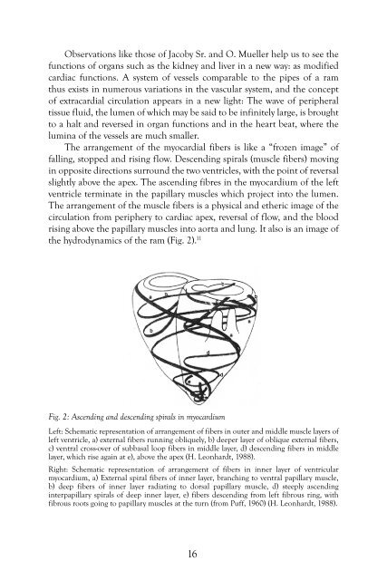
The arrangement of the myocardial fibers is like a “frozen image” of<br />
falling, stopped and rising flow. Descending spirals (muscle fibers) moving<br />
in opposite directions surround the two ventricles, with the point of reversal<br />
slightly above the apex. The ascending fibres in the myocardium of the left<br />
ventricle terminate in the papillary muscles which project into the lumen.<br />
The arrangement of the muscle fibers is a physical and etheric image of the<br />
circulation from periphery to cardiac apex, reversal of flow, and the blood<br />
rising above the papillary muscles into aorta and lung. It also is an image of<br />
the hydrodynamics of the ram (Fig. 2). 11<br />
Fig. 2: Ascending and descending spirals in myocardium<br />
Left: Schematic representation of arrangement of fibers in outer and middle muscle layers of<br />
left ventricle, a) external fibers running obliquely, b) deeper layer of oblique external fibers,<br />
c) ventral cross-over of subbasal loop fibers in middle layer, d) descending fibers in middle<br />
layer, which rise again at e), above the apex (H. Leonhardt, 1988).<br />
Right: Schematic representation of arrangement of fibers in inner layer of ventricular<br />
myocardium, a) External spiral fibers of inner layer, branching to ventral papillary muscle,<br />
b) deep fibers of inner layer radiating to dorsal papillary muscle, d) steeply ascending<br />
interpapillary spirals of deep inner layer, e) fibers descending from left fibrous ring, with<br />
fibrous roots going to papillary muscles at the turn (from Puff, 1960) (H. Leonhardt, 1988).<br />
16