Access article in PDF - Project MUSE - Johns Hopkins University
Access article in PDF - Project MUSE - Johns Hopkins University
Access article in PDF - Project MUSE - Johns Hopkins University

You also want an ePaper? Increase the reach of your titles
YUMPU automatically turns print PDFs into web optimized ePapers that Google loves.
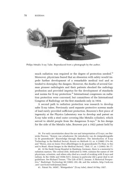
78 BOERSMA<br />
Philips Metalix X-ray Tube. Reproduced from a photograph by the author.<br />
much radiation was required or the degree of protection needed. 39<br />
Moreover, physicians feared that an obsession with safety would impede<br />
further development of a remarkable medical tool and so<br />
tended to downplay the dangers. However, the deaths of several German<br />
pioneer radiologists and their patients shocked the radiology<br />
profession and provided impetus for the development of standards<br />
and norms for X-ray protection. 40 International congresses on radiation<br />
protection were convened, but committees of the International<br />
Congress of Radiology set the first standards only <strong>in</strong> 1934.<br />
A second path to radiation protection was research to develop<br />
safer X-ray tubes. Previously used separate protective screens made<br />
of lead rarely provided sufficient protection. Bouwers’s first piece of<br />
<strong>in</strong>genuity at the Physics Laboratory was to develop and patent an<br />
X-ray tube with a steel outer cover<strong>in</strong>g (the Metalix cyl<strong>in</strong>der), which<br />
served to shield people from the dangerous X-rays. 41 In his design<br />
for the side of the Metalix tube, Bouwers put a 1922 patent held by<br />
39. For early uncerta<strong>in</strong>ties about the use and <strong>in</strong>terpretation of X-rays, see Bernicke<br />
Pasveer, “Kennis van schaduwen: De <strong>in</strong>troductie van de röntgenfotografie<br />
<strong>in</strong> de geneeskunde” [Knowledge through Shadows: The Introduction of X-ray<br />
Technology <strong>in</strong> the Medical Sector], Kennis en Methode 12, nr. 4 (1988): 274–94,<br />
and “Horen, zien en lezen: Over afbeeld<strong>in</strong>gen <strong>in</strong> de geneeskunde [To Hear, to See<br />
and to Read: About Images <strong>in</strong> the Medical Sector],” ibid. 17, nr. 1 (1993): 56–77.<br />
40. At the Sankt Georg Hospital <strong>in</strong> Hamburg, Germany, there is a memorial to<br />
radiation martyrs. The central stele, dedicated <strong>in</strong> 1936, conta<strong>in</strong>s the names of 159<br />
physicians, scientists, and others who died as a result of work<strong>in</strong>g with X-rays and<br />
radium. In the 1920s and 1930s Ed C. Jerman <strong>in</strong> particular did a great deal to set<br />
guidel<strong>in</strong>es. See Richard Terrass, “The Life of Ed C. Jerman: A Historical Perspective,”<br />
Radiologic Technology 66 (1995): 291–98, and the website, http://web.wn<br />
.net/~usr/ricter/web/jerman.html.<br />
41. Patent No. 16507, “Röntgenbuis” [X-ray tube], dated 10 May 1927.