CONNECTIVE TISSUE GRAFT IN THE ... - Journal of IMAB
CONNECTIVE TISSUE GRAFT IN THE ... - Journal of IMAB
CONNECTIVE TISSUE GRAFT IN THE ... - Journal of IMAB

You also want an ePaper? Increase the reach of your titles
YUMPU automatically turns print PDFs into web optimized ePapers that Google loves.
<strong>Journal</strong> <strong>of</strong> <strong>IMAB</strong> - Annual Proceeding (Scientific Papers) 2008, book 2<br />
<strong>CONNECTIVE</strong> <strong>TISSUE</strong> <strong>GRAFT</strong> <strong>IN</strong> <strong>THE</strong><br />
TREATMENT OF MULTIPLE ADJACENT<br />
G<strong>IN</strong>GIVAL RECESSIONS<br />
Christina Popova, K. Kotsilkov<br />
Department <strong>of</strong> Periodontology<br />
Faculty <strong>of</strong> Dental Medicine, Medical University - S<strong>of</strong>ia, Bulgaria<br />
SUMMARY:<br />
Marginal tissue recession is a displacement <strong>of</strong> the s<strong>of</strong>t<br />
tissue margin apical to the cement-enamel junction with<br />
exposure <strong>of</strong> the root surface. The etiology <strong>of</strong> the gingival recessions<br />
is associated with the gingival inflammation, mechanical<br />
factors like improper toothbrushing in the circumstances <strong>of</strong><br />
tooth malposition, bone dehiscence, thin periodontal tissue,<br />
and aberrant frenulum. The treatment <strong>of</strong> the gingival recession<br />
in the last years includes gingival grafting procedures. The<br />
most generally used procedure is the free gingival graft. Recent<br />
studies have demonstrated that connective tissue grafting is<br />
an effective treatment <strong>of</strong> gingival recession. The graft used may<br />
either be an epithelialized graft or a subepithelial connective<br />
tissue graft <strong>of</strong> palatal masticatory mucosa.<br />
The presentation demonstrates an connective tissue<br />
graft procedures for root coverage in a 23 years old patient<br />
with Miller class II recessions on teeth #14, #15, #24, and<br />
Miller class I recessions on teeth #16, #25, #26 and the results.<br />
In the limitations <strong>of</strong> this case the connective tissue<br />
graft procedure led to clinical improvement which is a premise<br />
for better maintenance <strong>of</strong> the achieved root coverage.<br />
Key words: marginal tissue recession, s<strong>of</strong>t tissue graft<br />
procedures, epithelial collar, root coverage.<br />
The most common cause for the marginal tissue<br />
recessions is abrasive and traumatic toothbrushing habits.<br />
Teeth positioned bucally tend to have greater recession.<br />
Recession on the gingival tissue and bone exposes the<br />
cementum surface, which allows abrasion and ditching <strong>of</strong> the<br />
cervical area.<br />
Periodontal inflammation and the consequential loss<br />
<strong>of</strong> attachment results in reduced attached gingiva. Advanced<br />
periodontal involvement in areas <strong>of</strong> minimal attached gingiva<br />
result in the base <strong>of</strong> pocket extending close to, or apical to,<br />
the mucogingival junction.<br />
Frenal and muscle attachments encroach on the<br />
marginal gingiva distend the gingival sulcus, fostering plaque<br />
accumulation, increasing the rate <strong>of</strong> progression <strong>of</strong> periodontal<br />
recession, and causing their recurrence after treatment.<br />
The problem is more common on facial surfaces, but it may<br />
also occur on the lingual surface.<br />
Orthodontic tooth movement through a thin buccal<br />
osseous plate leading to a dehiscence beneath a thin gingival<br />
tissue can cause recession and/or loss <strong>of</strong> the gingiva (1, 3, 9).<br />
One <strong>of</strong> the most generally used procedure for root<br />
coverage is the free s<strong>of</strong>t tissue graft procedure. The graft<br />
used may either be an epithelialized graft or a subepithelial<br />
connective tissue graft <strong>of</strong> palatal masticatory mucosa.<br />
The technique utilizing a subepithelial s<strong>of</strong>t tissue graft,<br />
i.e. the connective tissue, involves the placement <strong>of</strong> the graft<br />
directly over the exposed root and the mobilization <strong>of</strong> a<br />
mucosal flap to be coronally or laterally moved for coverage<br />
<strong>of</strong> the graft (2, 4, 6, 7, 8).<br />
GOAL: The presentation demonstrates an envelope<br />
technique connective tissue graft procedures for root<br />
coverage in a 23 years old patient with Miller class II<br />
recessions on teeth #14, #15, #24, and Miller class I recessions<br />
on teeth #16, #25, #26.<br />
MA<strong>THE</strong>RIALS AND METHODS:<br />
The surgical protocol <strong>of</strong> the both treated sites is<br />
presented on the following photos – Figures 1-6.<br />
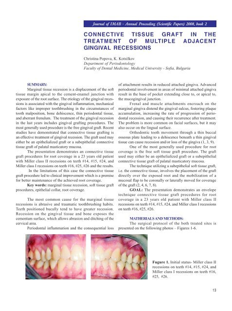
Fugure 1. Initial status- Miller class II<br />
recessions on teeth #14, #15, #24, and<br />
Miller class I recessions on teeth #16,<br />
#25, #26.<br />
13
Fugure 2. Initial horizontal and<br />
intrasulcular incisions<br />
Fugure 3. Partial thickness flaps are<br />
reflected. The prominent root surfaces<br />
and dehiscence are seen.<br />
Fugure 4. The connective tissue graft<br />
taken according the Hurtzeler-Weng<br />
technique (5).<br />
Fugure 5. Connective tissue graft with<br />
epithelial collar on the right side and<br />
connective tissue graft without<br />
epithelial collar on the left side. Both<br />
grafts are positioned and immobilized<br />
with interdental resorbable sutures.<br />
Fugure 6. Fixation <strong>of</strong> the graft and<br />
coronal flap positioning.<br />
14
RESULTS:<br />
On the first month after surgery complete root<br />
coverage was obtained. The gain <strong>of</strong> attached gingiva is<br />
3mm on teeth #16,#15,#14 and 1mm on teeth #26,#25, and<br />
#24.The color and the appearance <strong>of</strong> the connective tissue<br />
grafted area is similar to the adjacent gingiva which leads<br />
to good aesthetic result. The result is stable on the sixth<br />
month after treatment.<br />
Fugure 7. Result at the sixth month -<br />
complete root coverage is achieved.<br />
CONCLUSION:<br />
In the limitations <strong>of</strong> this case the connective tissue<br />
graft procedure led to tissue root coverage, shallow residual<br />
probing depths, gain in clinical attachment and an increase<br />
in gingival height and width, which is a premise for better<br />
maintenance <strong>of</strong> the achieved root coverage. The epithelial<br />
collar approach led to better gain <strong>of</strong> attached gingiva but<br />
the aesthetic result was worse because <strong>of</strong> the rugged<br />
gingival surface.<br />
REFERENCES:<br />
1. Bernimoulin J. P., Loscher B,<br />
Muhlemann H. R. (1975) :Coronally<br />
repositioned flap. J Clin Periodontol 2;1<br />
2. Bruno, J. F. (1994). Connective tissue<br />
graft technique assuring wide root coverage.<br />
International <strong>Journal</strong> <strong>of</strong> Periodontics and<br />
Restorative Dentistry 14, 127- 137.<br />
3. Hall W. B. (1984) Pure mucogingival<br />
problems. Etiology, treatment and<br />
prevention. Chicago, Quintessence.<br />
4. Harris, R. J. (1994). The connective<br />
tissue with partial thickness double pedicle<br />
graft: the results <strong>of</strong> 100 consecutively-<br />
treated defects. <strong>Journal</strong> <strong>of</strong> Periodontology<br />
65, 448-461.<br />
5. Hurtzeler, M. B., Weng, D. (1999):A<br />
single incision technique to harvest<br />
subepithelial connective tissue grafts from<br />
the palate. Int J Periodontics Restorative<br />
Dent 19, 279.<br />
6. Langer, B. & Langer, L. (1985).<br />
Subepithelial connective tissue graft<br />
technique for root coverage. <strong>Journal</strong> <strong>of</strong><br />
Periodontology 56, 715-720.<br />
7. Nelson, S. W. (1987). The subpedicle<br />
connective tissue graft. A bilaminar<br />
reconstructive procedure for the coverage <strong>of</strong><br />
denuded root surfaces. <strong>Journal</strong> <strong>of</strong><br />
Periodontology 58, 95-102.<br />
8. Wennstrom J & Pini Prato G.P.<br />
(2003) “Mucogingival Therapy -Periodontal<br />
Plastic Surgery” in Jan Lindhe’s “Clinical<br />
Periodontology and Implant Dentistry”<br />
Blackwell Munksgaard, a Blackwell<br />
Publishing Company (Fourth Edition)<br />
9. Wo<strong>of</strong>er C. (1969): The prevalence and<br />
etiology <strong>of</strong> gingival recession. Periodont<br />
Abstr 17:45.<br />
Address for correspondence:<br />
Assoc. pr<strong>of</strong>. Christina Popova, PhD<br />
Department <strong>of</strong> Periodontology, Faculty <strong>of</strong> Dental Medicine, Medical University <strong>of</strong> S<strong>of</strong>ia,<br />
1, Georgi S<strong>of</strong>iiski Str., S<strong>of</strong>ia, Bulgaria<br />
Mobile: +359 88 875 90 49; E-mail: hrpopova@yahoo.com<br />
15