PPCO Twist System - Wound Care Advisor
PPCO Twist System - Wound Care Advisor
PPCO Twist System - Wound Care Advisor

Create successful ePaper yourself
Turn your PDF publications into a flip-book with our unique Google optimized e-Paper software.
Chronic venous<br />
insufficiency with<br />
lower extremity<br />
disease: Part 2<br />
CEAP and rescue<br />
By Donald A. Wollheim, MD, WCC, DWC, FAPWCA<br />
To begin appropriate treatment for<br />
chronic venous insufficiency<br />
(CVI), clinicians must be able to<br />
make the correct diagnosis. Part 1<br />
(published in the March-April edition) described<br />
CVI and its presentation. This article<br />
provides details of the CVI diagnosis<br />
(including the differential diagnosis from<br />
other diseases), disease classification to<br />
help assess the extent of CVI, diagnostic<br />
studies used to diagnose CVI, and various<br />
treatment options to “rescue” the patient<br />
from CVI.<br />
CVI diagnosis<br />
CVI is diagnosed mainly from the patient’s<br />
history and physical exam. Typically with<br />
CVI, discomfort in the lower extremity gets<br />
worse toward the end of day, decreases<br />
with leg elevation, and feels the best first<br />
thing in the morning after the patient has<br />
slept with the legs elevated. Physical examination<br />
may reveal edema, acute or chronic<br />
leg changes, or both. Some extremities may<br />
show permanent changes, such as hemosiderin<br />
staining, atrophie blanche, or lipodermatosclerosis.<br />
Varicose veins also may<br />
be present. In general, the more severe<br />
CVI abnormalities usually occur in the older<br />
patients and those with significant comorbidities,<br />
such as peripheral arterial disease<br />
(PAD), diabetes mellitus, and arthritis.<br />
(For a glossary of terms, see Part 1 of this<br />
article in the March-April issue.)<br />
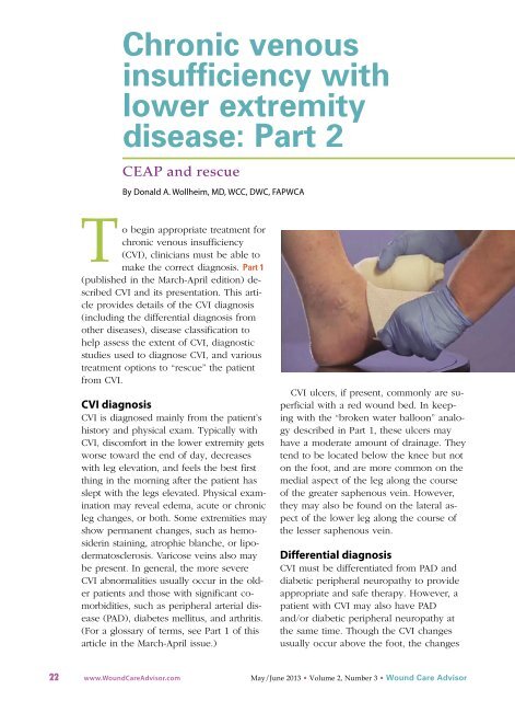
CVI ulcers, if present, commonly are superficial<br />
with a red wound bed. In keeping<br />
with the “broken water balloon” analogy<br />
described in Part 1, these ulcers may<br />
have a moderate amount of drainage. They<br />
tend to be located below the knee but not<br />
on the foot, and are more common on the<br />
medial aspect of the leg along the course<br />
of the greater saphenous vein. However,<br />
they may also be found on the lateral aspect<br />
of the lower leg along the course of<br />
the lesser saphenous vein.<br />
Differential diagnosis<br />
CVI must be differentiated from PAD and<br />
diabetic peripheral neuropathy to provide<br />
appropriate and safe therapy. However, a<br />
patient with CVI may also have PAD<br />
and/or diabetic peripheral neuropathy at<br />
the same time. Though the CVI changes<br />
usually occur above the foot, the changes<br />
22 www.<strong>Wound</strong><strong>Care</strong><strong>Advisor</strong>.com May/June 2013 • Volume 2, Number 3 • <strong>Wound</strong> <strong>Care</strong> <strong>Advisor</strong>
for both peripheral arterial disease and diabetic<br />
neuropathy occur below the ankle.<br />
PAD<br />
In an extremity with PAD, the anklebrachial<br />
index typically is 0.9 or lower. As<br />
noted above, some patients may have an<br />
extremity with both CVI and PAD. In this<br />
case, the therapy selected must accommodate<br />
both diseases, because PAD can hinder<br />
the healing of CVI wounds.<br />
Pain associated with PAD differs from<br />
CVI-related pain. With CVI, pain improves<br />
with elevation, whereas the opposite may<br />
occur with PAD. Typically, PAD patients<br />
require gravity to aid arterial blood flow to<br />
the extremity and thus elevation of the<br />
PAD extremity may aggravate, not improve,<br />
the pain. PAD pain is linked to ischemia,<br />
is mild to severe, and stems from<br />
a more proximal (closer to the heart) vessel<br />
narrowing or occlusion.<br />
PAD-related pain often is called claudication,<br />
an intermittent discomfort occurring<br />
with exercise, which may progress to nocturnal<br />
pain, discomfort caused by elevation<br />
of the nonexercising leg at night. It may<br />
progress to a more severe resting pain,<br />
where the pain is constantly present independent<br />
of the leg’s position or activity.<br />
Resting pain cannot be eliminated or significantly<br />
improved without some form of<br />
surgical intervention (which, unfortunately,<br />
may involve an amputation). Physical examination<br />
may show changes associated<br />
with decreased arterial blood flow, such as<br />
a lack of hair, diminished or absent pulses,<br />
and dystrophic (thick) toenails.<br />
Also, arterial ulcers (from PAD) tend to<br />
be more painful than venous ulcers.<br />
They’re usually deeper than the more superficial<br />
venous ulcers, and are located at<br />
the distal part of the arterial vascular tree,<br />
near the toes or on the lateral aspect of the<br />
foot (the area more subject to footwear<br />
trauma). An arterial ulcer has a pale wound<br />
bed with decreased drainage due to impaired<br />
blood flow to the tissue. Because of<br />
the decreased blood flow, the ulcer may<br />
progress from reversible tissue ischemia to<br />
irreversible necrotic tissue. These ulcers<br />
commonly are also infected due to the<br />
compromised arterial blood flow.<br />
Diabetic peripheral neuropathy<br />
In a patient with diabetes, the leg may show<br />
a combination of the three types of neuropathy—peripheral<br />
motor, peripheral sensory,<br />
and peripheral autonomic neuropathy.<br />
Findings in these legs may differ significantly<br />
from a leg with CVI. However, like PAD,<br />
a CVI extremity may also have concurrent<br />
diabetic peripheral neuropathic changes.<br />
In a patient with diabetic peripheral<br />
neuropathy, the history usually reveals at<br />
least a 10-year history of diabetes. <strong>Wound</strong>s<br />
are within an insensate (numb) foot and<br />
on its plantar aspect.<br />
PAD pain is linked to<br />
ischemia, is mild to<br />
severe, and stems from a<br />
more proximal vessel<br />
narrowing or occlusion.<br />
A diabetic foot ulcer typically starts as a<br />
callous on the bottom of the foot, which<br />
breaks down to create the ulcer. Commonly,<br />
a bony deformity is present due to neuropathy<br />
and acts as a focal point of increased<br />
shearing and pressure with<br />
walking. This leads to the initial callous<br />
formation. These wounds usually occur<br />
within the surrounding callous, in an insensate<br />
area of the plantar aspect of the<br />
foot, and commonly with an overlying<br />
bony foot deformity.<br />
Disease classification<br />
Several classification systems exist for CVI.<br />
One system that helps guide therapy is<br />
called CEAP.<br />
<strong>Wound</strong> <strong>Care</strong> <strong>Advisor</strong> • May/June 2013 • Volume 2, Number 3 www.<strong>Wound</strong><strong>Care</strong><strong>Advisor</strong>.com 23
Other CVI classification systems<br />
The Venous Clinical Severity Scale uses 10<br />
parameters to assess the overall clinical severity of<br />
chronic venous insufficiency (CVI)—pain, varicose<br />
veins, edema, hyperpigmentation, inflammation,<br />
induration, number of ulcers, duration of ulcers,<br />
ulcer size, and patient’s compliance with compression<br />
therapy.<br />
The Venous Disability Score documents whether<br />
the patient has CVI signs or symptoms, extent of<br />
symptoms, and effect of CVI on the patient’s ability<br />
to perform activities. It’s scored on a scale of 0 to 3:<br />
0: asymptomatic<br />
1: symptomatic patient who can perform usual<br />
activities without leg compression<br />
2: symptomatic patient who can carry out activities<br />
but requires leg compression or elevation<br />
3: symptomatic patient who is unable to perform<br />
normal activities even with compression or<br />
elevation.<br />
The Venous Segmental Disease Score combines<br />
anatomic and physiologic classifications changes.<br />
The Villalta Scale measures an associated postthrombotic<br />
syndrome.<br />
CEAP: Clinical Etiology Anatomy<br />
Pathophysiology<br />
CEAP classification should be done for all<br />
lower extremities with CVI. Therapy for<br />
CVI varies with signs and symptoms, disease<br />
extent and duration, and the overall<br />
treatment goal (be it that of symptomatic<br />
relief or cosmetic improvement). CEAP<br />
components include clinical severity, etiology,<br />
anatomy, and pathophysiology.<br />
Clinical severity has eight grades:<br />
• C0: no visible or palpable signs of venous<br />
insufficiency<br />
• C1: telangiectasia, reticular veins, or both<br />
• C2: varicose veins<br />
• C3: edema into the skin and subcutaneous<br />
space, which may be pitting.<br />
(Note: Unlike lymphedema, CVI doesn’t<br />
involve the foot or toes.)<br />
• C4a: pigmentary skin changes, such as<br />
hemosiderin staining or eczema<br />
• C4b: lipodermatosclerosis<br />
• C5: healed venous ulcer with or without<br />
atrophic skin; it may present as a pigmentary<br />
change, usually of lighter discoloration,<br />
within an area of hemosiderin<br />
staining<br />
• C6: active venous ulcer.<br />
Etiology may be classified as:<br />
• Ep: primary<br />
• Es: secondary (due to an etiology other<br />
than CVI, such as trauma)<br />
• En: unknown (nonspecific)<br />
• Ec: congenital.<br />
Anatomy (location) of the pathologic<br />
vein may be classified as:<br />
• As: superficial veins<br />
• Ad: deep veins<br />
• Ap: perforating veins<br />
• An: nonspecific veins.<br />
Pathophysiology may be classified as:<br />
• Pr: Venous reflux due to valve damage<br />
or prior DVT leading to retrograde<br />
blood flow<br />
• Po: Venous obstruction due to ongoing<br />
venous thrombosis<br />
• Po, r: Both reflux and obstruction<br />
• Pn: Neither reflux nor obstruction<br />
Other classification systems<br />
Other systems for classifying CVI include<br />
the Venous Clinical Severity Scale and Venous<br />
Disability Scale. (See Other CVI classification<br />
systems.)<br />
Diagnostic studies<br />
Ultrasonography, a noninvasive study, generally<br />
is less expensive than venography<br />
(the gold standard study for CVI) and carries<br />
no risk. In fact, duplex ultrasonography<br />
has largely replaced invasive venography.<br />
(See Rarely used studies.)<br />
Ultrasonography indications include:<br />
• diagnosis of venous obstruction, venous<br />
reflux, and deep vein thrombosis<br />
• aid in the diagnosis of suspected CVI<br />
when that diagnosis can’t be established<br />
from signs and symptoms alone<br />
24 www.<strong>Wound</strong><strong>Care</strong><strong>Advisor</strong>.com May/June 2013 • Volume 2, Number 3 • <strong>Wound</strong> <strong>Care</strong> <strong>Advisor</strong>
• evaluation of patients with physical findings<br />
of CVI but with atypical symptoms<br />
• evaluation of patients with atypical venous<br />
insufficiency, such as those<br />
younger than age 40<br />
• evaluation of patients who develop CVI<br />
after trauma<br />
• evaluation of patients with clinical CVI<br />
who don’t respond to standard therapy<br />
• to rule out pathologic conditions that<br />
may mimic CVI<br />
• to guide interventions by evaluating the<br />
anatomic and physiologic features of CVI.<br />
Venography<br />
Venography is considered the gold standard<br />
for diagnosing venous insufficiency,<br />
but is rarely needed due to the ultrasonographic<br />
techniques available today. During<br />
venography, contrast dye is injected into<br />
the patient’s venous system to identify the<br />
normal and abnormal anatomy of the<br />
blood flow within and out of the extremity.<br />
Venography is more expensive than ultrasound<br />
studies. Unlike ultrasound, it’s invasive<br />
and can cause complications such<br />
as venous phlebitis, thrombosis, and reactions<br />
to the contrast dye.<br />
Patients with lower-extremity wounds<br />
should undergo an arterial assessment to<br />
identify if the wound has an arterial component<br />
where there is not enough oxygenrich<br />
blood flow to allow healing. This<br />
arterial evaluation is also necessary if compression<br />
therapy is ordered, which often<br />
is for the CVI extremity, to guide the clinician<br />
on how it can safely be applied.<br />
Rarely used studies<br />
Two rarely used studies for chronic venous insufficiency<br />
(CVI) are air plethysmography (APG) and<br />
photoplethysmography (PPG).<br />
APG measures volume changes in the leg, most<br />
likely due to both blood and edema. It compares<br />
volume refill time with the patient supine (when<br />
leg veins are collapsed) to the refill time when the<br />
patient is standing (when leg veins are swollen). In<br />
CVI, this time differs from that of a normal venous<br />
system. However, APG can’t localize reflux; it only<br />
reveals if reflux is occurring.<br />
PPG assesses the overall venous hemodynamics<br />
by evaluating the time it takes for the venous system<br />
to fill after emptying of the veins. Like APG, it can<br />
indicate venous reflux disease. But it can also determine<br />
if the reflux occurs in the superficial venous<br />
system, deep venous system, or both systems.<br />
Ankle-brachial index<br />
Ankle-brachial index (ABI) is a noninvasive<br />
study that can diagnose PAD and determine<br />
how much lower-extremity compression<br />
can be applied safely in patients<br />
with CVI. ABI compares systolic blood<br />
pressure measured at the ankle with the<br />
systolic blood pressure measured at the<br />
arm. Normally, the pressure at the elbow<br />
(brachial artery) is the same as the pressure<br />
in the ankle arteries (dorsalis pedis or<br />
posterior tibial arteries).<br />
The results of the ABI study should be<br />
used as a guide for ordering a safe level of<br />
compression in the CVI patient. If the ABI<br />
is not determined, the patient might develop<br />
significant complications from an incorrect<br />
amount of compression applied, leading<br />
to “downstream” ischemia and possibly<br />
necrosis of tissue.<br />
A normal ABI result is 1. Anything below<br />
1.0 indicates PAD. ABI of 1.3 or higher typically<br />
is a falsely elevated level as a result of<br />
an inability to compress calcified ankle vessels.<br />
This commonly occurs in elderly patients<br />
and those with diabetes or renal failure.<br />
An ABI of 1.3 or higher warrants<br />
another study to evaluate arterial blood<br />
flow to that extremity. (See Interpreting<br />
and applying ABI results in CVI patients.)<br />
Management guidelines<br />
During the first 6 months after the diagnosis<br />
is made, conservative therapy should be<br />
used for CVI patients in an attempt to decrease<br />
the extremity swelling and reduce<br />
the “water balloon” effect—pooling of<br />
blood and intravascular fluid around the<br />
ankle (described in Part 1 of this article).<br />
<strong>Wound</strong> <strong>Care</strong> <strong>Advisor</strong> • May/June 2013 • Volume 2, Number 3 www.<strong>Wound</strong><strong>Care</strong><strong>Advisor</strong>.com 25
Interpreting and applying ABI results in CVI patients<br />
The ankle-brachial index (ABI)<br />
can guide clinicians in ordering<br />
safe levels of compression therapy<br />
for CVI patients with or<br />
without associated peripheral<br />
arterial disease.<br />
• ABI of 0.9 or higher, but less<br />
than 1.3: Full therapeutic<br />
compression up to 40 mm<br />
Hg at the ankle can be applied<br />
without concern for<br />
distal ischemia.<br />
• ABI of 0.6 to 0.8: A modified<br />
amount of compression (23<br />
mm Hg or less at the ankle)<br />
should be used. This ABI indicates<br />
clinically compromised<br />
blood flow to the leg,<br />
to the point where the leg<br />
might not tolerate full therapeutic<br />
compression of 40<br />
mm Hg.<br />
• ABI of 0.5 or lower: With this<br />
result, do not apply any compression;<br />
instead, refer the<br />
patient for a vascular consultation.<br />
This ABI means the<br />
compromised leg is getting,<br />
at best, half as much blood<br />
flow as the arm is receiving;<br />
the leg is at significant risk<br />
for ischemia or necrosis, and<br />
might eventually need to be<br />
amputated unless surgical intervention<br />
is successfully performed.<br />
Applying compression<br />
in this case is likely to<br />
increase the risk of ischemic<br />
or necrotic complications.<br />
• ABI of 1.3 or higher: A different<br />
study is needed to guide<br />
the wound care team. This<br />
result most likely is falsely<br />
elevated and can’t be relied<br />
on to determine a safe<br />
amount of compression to<br />
be applied to the leg.<br />
Important: If the patient has<br />
heart failure, compression of<br />
any kind should not be applied<br />
to the extremity regardless of<br />
the ABI value, because the increase<br />
in blood flow leaving the<br />
leg during compression and returning<br />
to a compromised heart<br />
possibly could worsen the heart<br />
failure.<br />
Other indications for obtaining<br />
ABI to assess the arterial tree<br />
of an extremity include:<br />
• a lower extremity with any<br />
wound (wound healing requires<br />
arterial blood flow)<br />
• a weak or absent lowerextremity<br />
pulse (some patients<br />
with PAD may be<br />
asymptomatic)<br />
• a lower-extremity wound<br />
that isn’t healing despite<br />
therapy (clinicians might be<br />
underestimating the extent<br />
of the PAD or that condition<br />
may be worsening).<br />
Ways to do this include elevating the leg,<br />
initiating an exercise program, and using<br />
safe compression therapy. This conservative<br />
approach increases oxygen to the tissues,<br />
decreases edema (improving subcutaneous<br />
swelling, capillary constriction, and<br />
capillary separation), reduces tissue inflammation,<br />
and compresses the dilated veins.<br />
If the patient has changes consistent<br />
with dermatitis or infection, use an appropriate<br />
topical agent—but be aware that<br />
this might preclude the use of long-wearing<br />
compression wraps to allow daily (or<br />
more frequent) access to the extremity for<br />
various medication applications.<br />
If the patient has a noninfected ulcer,<br />
consider using a prolonged wearing wrap<br />
to provide compression, such as a longstretch<br />
system (for example, multilayered<br />
wraps, single-layered wraps, or the Duke<br />
boot). Consider providing a resistance to<br />
leg swelling for ambulatory patients by<br />
using a short-stretch system, such as the<br />
Unna boot. Other options for these patients<br />
include adjustable compression<br />
wraps and intermittent pneumatic compression<br />
devices.<br />
If the patient has an infected ulcer of<br />
the CVI extremity, consider using leg elevation<br />
plus a topical antimicrobial (with or<br />
without systemic antibiotic therapy), and<br />
postpone compression therapy until the infection<br />
is under control.<br />
If the patient has no ulcers or a healed<br />
ulcer (through the proliferative phase of<br />
full-thickness wound healing), use removable<br />
compression stockings or an intermittent<br />
pneumatic pump system.<br />
Ablative therapy<br />
Unfortunately, the patient will need lifelong<br />
compression therapy if CVI can’t be<br />
corrected effectively with other more permanent<br />
forms of therapy. Clinicians might<br />
26 www.<strong>Wound</strong><strong>Care</strong><strong>Advisor</strong>.com May/June 2013 • Volume 2, Number 3 • <strong>Wound</strong> <strong>Care</strong> <strong>Advisor</strong>
consider ablative therapy if symptoms do<br />
not improve after 6 months of conservative<br />
therapy or if the patient seeks cosmetic<br />
improvement. Ablative therapy refers to<br />
the various types of therapy that destroy<br />
or remove the abnormal veins.<br />
Types of ablative therapy include chemical,<br />
thermal, and mechanical techniques.<br />
• Chemical ablation consists of placing an<br />
irritating fluid directly into the vein,<br />
which damages the endothelial lining<br />
and leads to the destruction of the abnormality.<br />
This method is used mainly<br />
for telangiectasia, reticular veins, and<br />
small varicose veins. To treat larger veins<br />
without having to use large amounts of<br />
fluid, an ablative foam solution can be<br />
used instead; this may be done for larger<br />
varicosities, incompetent saphenous<br />
veins, or incompetent perforating veins.<br />
• Thermal ablation uses a form of heat<br />
energy that destroys the wall of the vein.<br />
It may be used with topical light therapy<br />
applied to the skin for small, superficial<br />
dilated veins (telangiectasia or reticular<br />
veins). For larger veins (greater or lesser<br />
saphenous veins), heat energy may be<br />
applied within the vein itself using laser<br />
or radiofrequency energy.<br />
• Mechanical ablation physically destroys<br />
or removes the abnormal vein. Examples<br />
of this technique include surgical<br />
vein ligation and vein stripping. (See<br />
Ablation success rates.)<br />
Ablation success rates<br />
Here are the overall 3-year success rates for ablative<br />
therapy:<br />
• foam sclerotherapy: 77%<br />
• vein stripping: 78%<br />
• radiofrequency ablation: 84%<br />
• laser ablation: 94%<br />
Correct identification is crucial<br />
In a patient with suspected CVI, wound<br />
care clinicians must be able to correctly<br />
identify the disease process and determine<br />
if significant comorbid diseases are present<br />
before prescribing or administering any<br />
form of therapy. Otherwise, the therapy<br />
ordered may not correct the problem—and<br />
could even cause harm. Various diagnostic<br />
tools are available; in practice, ultrasonography<br />
is most often used for diagnosing<br />
venous disease.<br />
Multiple therapeutic modalities are available<br />
to treat CVI. Unless the disease is corrected,<br />
the patient will need lifelong compression<br />
therapy. How much compression<br />
that can be safely applied depends on the<br />
presence and extent of comorbidities, such<br />
as PAD and heart failure.<br />
n<br />
Selected references<br />
Alguire PC, Mathes BM. Clinical evaluation of lower<br />
extremity chronic venous disease. UpToDate. Last<br />
updated April 18, 2012. www.uptodate.com/contents/<br />
clinical-evaluation-of-lower-extremity-chronic-venousdisease.<br />
Accessed April 11, 2013.<br />
Alguire PC, Mathes BM. Diagnostic evaluation of<br />
chronic venous insufficiency. UpToDate. Last updated<br />
May 7, 2102. www.uptodate.com/contents/<br />
diagnostic-evaluation-of-chronic-venous-insufficiency<br />
?source=search_result&search=Diagnostic+evaluation<br />
+of+chronic+venous+insufficiency&selectedTitle=<br />
1%7E128. Accessed April 11, 2013.<br />
Alguire PC, Scovell S. Overview and management of<br />
lower extremity chronic venous disease. UpToDate.<br />
Last updated June 27, 2012. http://www.uptodate.com/<br />
contents/overview-and-management-of-lowerextremity-chronic-venous-disease?source=search_<br />
result&search=Overview+and+management+of+<br />
lower+extremity+chronic+venous+disease&selected<br />
Title=1%7E150. Accessed April 11, 2013.<br />
Moneta G. Classification of lower extremity chronic<br />
venous disorders. UpToDate. Last updated October<br />
22, 2012. www.uptodate.com/contents/classificationof-lower-extremity-chronic-venous-disorders.<br />
Accessed<br />
April 11, 2013.<br />
Sardina D. Skin and <strong>Wound</strong> Management Course;<br />
Seminar Workbook. <strong>Wound</strong> <strong>Care</strong> Education Institute:<br />
2011;92-112.<br />
Donald A. Wollheim is a practicing wound care<br />
physician in southeastern Wisconsin. He also is<br />
an instructor for <strong>Wound</strong> <strong>Care</strong> Education Institute<br />
and Madison College. He serves on the Editorial<br />
Board for <strong>Wound</strong> <strong>Care</strong> <strong>Advisor</strong>.<br />
<strong>Wound</strong> <strong>Care</strong> <strong>Advisor</strong> • May/June 2013 • Volume 2, Number 3 www.<strong>Wound</strong><strong>Care</strong><strong>Advisor</strong>.com 27