ALC Cover Jan/Feb - Wound Care Advisor
ALC Cover Jan/Feb - Wound Care Advisor
ALC Cover Jan/Feb - Wound Care Advisor

Create successful ePaper yourself
Turn your PDF publications into a flip-book with our unique Google optimized e-Paper software.
Understanding stoma<br />
complications<br />
Learn how to identify and manage stoma hernias,<br />
trauma, mucocutaneous separation, necrosis,<br />
prolapse, retraction, and stenosis.<br />
By Rosalyn S. Jordan, RN, BSN, MSc, CWOCN, WCC, OMS; and Judith LaDonna Burns, LPN, WCC, DFC<br />
About 1 million people in the<br />
United States have either temporary<br />
or permanent stomas.<br />
A stoma is created surgically<br />
to divert fecal material or urine in patients<br />
with GI or urinary tract diseases or<br />
disorders.<br />
A stoma has no sensory nerve endings<br />
and is insensitive to pain. Yet several complications<br />
can affect it, making accurate assessment<br />
crucial. These complications may<br />
occur during the immediate postoperative<br />
period, within 30 days after surgery, or later.<br />
Lifelong assessment by a healthcare<br />
provider with knowledge of ostomy surgeries<br />
and complications is important.<br />
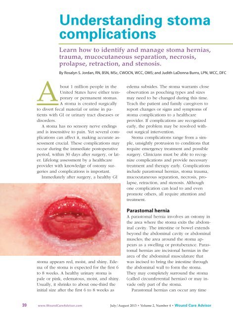
Immediately after surgery, a healthy GI<br />
stoma appears red, moist, and shiny. Edema<br />
of the stoma is expected for the first 6<br />
to 8 weeks. A healthy urinary stoma is<br />
pale or pink, edematous, moist, and shiny.<br />
Usually, it shrinks to about one-third the<br />
initial size after the first 6 to 8 weeks as<br />
edema subsides. The stoma warrants close<br />
observation as pouching types and sizes<br />
may need to be changed during this time.<br />
Teach the patient and family caregivers to<br />
report changes or signs and symptoms of<br />
stoma complications to a healthcare<br />
provider. If complications are recognized<br />
early, the problem may be resolved without<br />
surgical intervention.<br />
Stoma complications range from a simple,<br />
unsightly protrusion to conditions that<br />
require emergency treatment and possible<br />
surgery. Clinicians must be able to recognize<br />
complications and provide necessary<br />
treatment and therapy early. Complications<br />
include parastomal hernias, stoma trauma,<br />
mucocutaneous separation, necrosis, prolapse,<br />
retraction, and stenosis. Although<br />
one complication can lead to and even<br />
promote others, all require attention and<br />
treatment.<br />
Parastomal hernia<br />
A parastomal hernia involves an ostomy in<br />
the area where the stoma exits the abdominal<br />
cavity. The intestine or bowel extends<br />
beyond the abdominal cavity or abdominal<br />
muscles; the area around the stoma appears<br />
as a swelling or protuberance. Parastomal<br />
hernias are incisional hernias in the<br />
area of the abdominal musculature that<br />
was incised to bring the intestine through<br />
the abdominal wall to form the stoma.<br />
They may completely surround the stoma<br />
(called circumferential hernias) or may invade<br />
only part of the stoma.<br />
Parastomal hernias can occur any time<br />
20 www.<strong>Wound</strong><strong>Care</strong><strong>Advisor</strong>.com July/August 2013 • Volume 2, Number 4 • <strong>Wound</strong> <strong>Care</strong> <strong>Advisor</strong>
after the surgical procedure but usually<br />
happen within the first 2 years. Recurrences<br />
are common if the hernia needs to<br />
be repaired surgically. Risk factors may be<br />
patient related or technical. Patient-related<br />
risk factors include obesity, poor nutritional<br />
status at the time of surgery, presurgical<br />
steroid therapy, wound sepsis, and chronic<br />
cough. Risk factors related to technical issues<br />
include size of the surgical opening<br />
and whether surgery was done on an<br />
emergency or elective basis.<br />
Parastomal hernias occur in four types.<br />
(See Types of parastomal hernias.) Initially,<br />
a parastomal hernia begins as an unsightly<br />
distention in the area surrounding the<br />
stoma; the hernia enlarges, causing pain,<br />
discomfort, and pouching problems resulting<br />
in peristomal skin complications that<br />
require frequent assessment. Conservative<br />
therapy is the usual initial treatment. Adjustments<br />
to the pouching system typically<br />
are required so changes in the shape of<br />
the pouching surface can be accommodated.<br />
Also, a hernia support binder or pouch<br />
support belt may be helpful. Avoid convex<br />
pouching systems; if this isn’t possible, use<br />
these systems with extreme caution. If the<br />
patient irrigates the colostomy, an ostomy<br />
management specialist should advise the<br />
patient to discontinue irrigation until the<br />
parastomal hernia resolves.<br />
The image shows stoma injury caused by a poor<br />
fitting appliance. Photo by Connie Johnson. Used with permission.<br />
Stoma trauma<br />
Stoma trauma occurs when the stoma is injured,<br />
typically from a laceration. Lacerations<br />
usually result from the pouch appliance<br />
or clothing. Belt-line stomas are<br />
easily traumatized and injury may occur<br />
from both clothing belts and pouch support<br />
belts. Stoma lacerations commonly result<br />
from a small opening in the flange or<br />
a misaligned pouch opening. Other causes<br />
include parastomal or stomal prolapse with<br />
possible stoma enlargement or edema.<br />
Signs and symptoms of stoma trauma<br />
include bright red bleeding, a visible cut,<br />
and a yellowish-white linear discoloration.<br />
Lacerations may heal spontaneously.<br />
If the culprit is the pouching<br />
system, make sure nothing within the<br />
system comes in contact with the stoma.<br />
Usually direct pressure controls bleeding,<br />
but if bleeding continues, refer the patient<br />
to a physician for treatment.<br />
Mucocutaneous separation<br />
Mucocutaneous separation occurs when<br />
the stoma separates from the skin at the<br />
junction between the skin and the intestine<br />
used to form the stoma. Causes are related<br />
to poor wound-healing capacity, such as<br />
malnutrition, steroid therapy, diabetes, infection,<br />
or radiation of the abdominal area.<br />
Tension or tautness of the suture line also<br />
can cause mucocutaneous separation.<br />
This complication usually arises early<br />
Types of parastomal hernias<br />
The four types of parastomal hernias are based on<br />
hernia location within the abdominal tissue:<br />
• intestinal interstitial, in which the hernia lies<br />
within the layers of the abdominal wall<br />
• subcutaneous, in which the hernia is contained<br />
within subcutaneous tissue<br />
• intrastomal, in which the herniated intestine<br />
penetrates the stoma (usually confined to an<br />
ileostomy)<br />
• peristomal, in which the hernia is located within<br />
a stoma that has prolapsed.<br />
<strong>Wound</strong> <strong>Care</strong> <strong>Advisor</strong> • July/August 2013 • Volume 2, Number 4 www.<strong>Wound</strong><strong>Care</strong><strong>Advisor</strong>.com 21
This temporary ileostomy secondary to colon<br />
cancer has been treated for mucocutaneous<br />
separation.<br />
Closure of the temporary ileostomy.<br />
Photos by Connie Johnson. Used with permission.<br />
and can lead to other serious conditions,<br />
such as infection, peritonitis, and stomal<br />
stenosis. The area of the separation may<br />
completely surround the stoma (known<br />
as a circumferential separation), or the<br />
separation may affect only certain areas<br />
Blood flow and<br />
tissue perfusion<br />
are essential to<br />
stoma health.<br />
of the stoma/skin junction. The separation<br />
may be superficial or deep.<br />
The first sign of mucocutaneous separation<br />
may be induration. Treat the separation<br />
as a wound, and apply woundhealing<br />
principles: Absorb drainage,<br />
reduce dead space, use the proper dressing,<br />
and promote wound healing. The<br />
proper dressing depends on wound<br />
depth and amount of wound drainage.<br />
Be sure to assess the wound, using the<br />
“clock method” to describe location;<br />
measure the wound area in centimeters;<br />
and describe the type of tissue in the<br />
wound bed. Be aware that slough may<br />
be present.<br />
Treatment of the wound dictates how<br />
often the pouch is changed. A two-piece<br />
pouching system commonly is used to reduce<br />
the number of pouch changes. <strong>Cover</strong><br />
the wound dressing with the pouching<br />
system unless the wound is infected. If<br />
infection is present, let the wound drain<br />
into the pouch and heal by secondary intention.<br />
Don’t use a convex pouching<br />
system, because this may cause additional<br />
injury to the mucocutaneous junction.<br />
Stoma necrosis<br />
Blood flow and tissue perfusion are essential<br />
to stoma health. Deficient blood<br />
flow causes stoma necrosis. A stoma may<br />
be affected by both arterial and venous<br />
blood compromise. The cause of necrosis<br />
usually relates to the surgical procedure,<br />
such as tension or too much trimming of<br />
the mesentery, or the vascular system<br />
that provides blood flow to the intestine.<br />
Other causes of vascular compromise include<br />
hypovolemia, embolus, and excessive<br />
edema.<br />
Stoma necrosis usually occurs within<br />
the first 5 postoperative days. The stoma<br />
appears discolored rather than red,<br />
moist, and shiny. Discoloration may be<br />
cyanotic, black, dark red, dusky bluish<br />
purple, or brown. The stoma mucosa<br />
may be hard and dry or flaccid. Also, the<br />
22 www.<strong>Wound</strong><strong>Care</strong><strong>Advisor</strong>.com July/August 2013 • Volume 2, Number 4 • <strong>Wound</strong> <strong>Care</strong> <strong>Advisor</strong>
stoma has a foul odor. Associated complications<br />
may include stoma retraction,<br />
mucocutaneous separation, stoma stenosis,<br />
and peritonitis.<br />
Report signs and symptoms to the primary<br />
care provider immediately. Superficial<br />
necrosis may resolve with necrotic<br />
tissue simply sloughing away. But if tissue<br />
below the fascial level is involved,<br />
surgery is necessary. A transparent twopiece<br />
pouching system is recommended<br />
for frequent stoma assessment. The<br />
pouch may need to be resized often.<br />
Stoma prolapse<br />
A stoma prolapse occurs when the stoma<br />
moves or becomes displaced from its<br />
proper position. The proximal segment of<br />
the bowel intussuscepts and slides through<br />
the orifice of the stoma, appearing to telescope.<br />
This occurs more often in loop<br />
transverse colostomies. A prolapsed stoma<br />
increases in both length and size. Prolapse<br />
may be associated with stoma retraction<br />
and parastomal hernias.<br />
Causes of stoma prolapse include<br />
large abdominal-wall openings, inadequate<br />
bowel fixation to the abdominal<br />
wall during surgery, increased abdominal<br />
pressure, lack of fascial support, obesity,<br />
pregnancy, and poor muscle tone.<br />
Unless the patient complains of pain,<br />
has a circulatory problem, or has signs<br />
or symptoms of bowel obstruction, conservative<br />
treatment is used for uncomplicated<br />
stoma prolapse. The prolapse usually<br />
can be reduced with the patient in a<br />
supine position. After reduction, applying<br />
a hernia support binder often helps.<br />
Also, a stoma shield can be used to protect<br />
the stoma. A prolapsed stoma may<br />
require a larger pouch to accommodate<br />
the larger stoma. Some clinicians use<br />
cold compresses and sprinkle table sugar<br />
on the stoma; the sugar provides osmotic<br />
therapy or causes a fluid shift<br />
across the stoma mucosa and reduces<br />
edema.<br />
Unless the patient<br />
complains of pain, has<br />
a circulatory problem,<br />
or has signs or symptoms<br />
of bowel obstruction,<br />
conservative treatment<br />
is used for uncomplicated<br />
stoma<br />
prolapse.<br />
Stoma retraction<br />
The best-formed stoma protrudes about<br />
2.5 cm, with the lumen located at the<br />
top center or apex of the stoma to guide<br />
the effluent flow directly into the pouch.<br />
In stoma retraction, the stoma has receded<br />
about 0.5 cm below the skin surface.<br />
Retraction may be circumferential or<br />
may occur in only one section of the<br />
stoma.<br />
The usual causes of stoma retraction<br />
are tension of the intestine or obesity.<br />
Stoma retraction during the immediate<br />
postoperative period relates to poor<br />
blood flow, obesity, poor nutritional status,<br />
stenosis, early removal of a supporting<br />
device with loop stomas, stoma<br />
placement in a deep skinfold, or thick<br />
abdominal walls. Late complications usually<br />
result from weight gain or adhesions.<br />
Stoma retraction is most common in patients<br />
with ileostomies.<br />
A retracted stoma has a concave, bowl-<br />
<strong>Wound</strong> <strong>Care</strong> <strong>Advisor</strong> • July/August 2013 • Volume 2, Number 4 www.<strong>Wound</strong><strong>Care</strong><strong>Advisor</strong>.com 23
shaped appearance. Retraction causes a<br />
poor pouching surface, leading to frequent<br />
peristomal skin complications. Typical<br />
therapy is use of a convex pouching<br />
system and a stoma belt. If obtaining a<br />
pouch seal is a problem and the patient<br />
has recurrent peristomal skin problems<br />
from leakage, stoma revision should be<br />
considered.<br />
Most stoma<br />
complications<br />
are preventable.<br />
Stoma stenosis<br />
Stoma stenosis is narrowing or constriction<br />
of the stoma or its lumen. This condition<br />
may occur at the skin or fascial<br />
level of the stoma. Causes include hyperplasia,<br />
adhesions, sepsis, radiation of the<br />
intestine before stoma surgery, local inflammation,<br />
hyperkeratosis, and surgical<br />
technique.<br />
Stoma stenosis frequently is associated<br />
with Crohn’s disease. You may notice a<br />
reduction or other change in effluent<br />
output with both urinary and GI ostomies.<br />
With GI stoma stenosis, bowel<br />
obstruction frequently occurs; signs and<br />
symptoms are abdominal cramps, diarrhea,<br />
increased flatus, explosive stool,<br />
and narrow-caliber stool. The initial sign<br />
is increased flatus. With urinary stoma<br />
stenosis, signs and symptoms include<br />
decreased urinary output, flank pain,<br />
high residual urine in conduit, forceful<br />
urine output, and recurrent urinary tract<br />
infections.<br />
Partial or complete bowel obstruction<br />
and stoma stenosis at the fascial level<br />
require surgical intervention. Conservative<br />
therapy includes a low-residue diet,<br />
increased fluid intake, and correct use<br />
of stool softeners or laxatives for colosto -<br />
mies.<br />
Most stoma complications are preventable<br />
and result from poor stoma placement.<br />
Up to 20% of patients with stoma<br />
complications require surgical revision of<br />
the stoma. All patients with ostomies require<br />
ongoing, accurate assessment and,<br />
if needed, early intervention by trained<br />
clinicians.<br />
n<br />
Selected references<br />
Al-Niaimi F, Lyon CC. Primary adenocarcinoma in<br />
peristomal skin: a case study. Ostomy <strong>Wound</strong> Manage.<br />
2010;56(1):45-7.<br />
Appleby SL. Role of the wound ostomy continence<br />
nurse in the home care setting: a patient case study.<br />
Home Healthc Nurse. 2011:29(3);169-77.<br />
Black P. Managing physical postoperative stoma<br />
complications. Br J Nurs. 2009:18(17):S4-10.<br />
Burch J. Management of stoma complications. Nurs<br />
Times. 2011;107(45):17-8, 20.<br />
Butler DL. Early postoperative complications following<br />
ostomy surgery: a review. J <strong>Wound</strong> Ostomy Continence<br />
Nurs. 2009:36(5):513-9.<br />
Husain SG, Cataldo TE. Late stomal complications.<br />
Clin Colon Rectal Surg. 2008:21(1):31-40.<br />
Jones T, Springfield T, Brudwick M, Ladd A. Fecal<br />
ostomies: practical management for the home health<br />
clinician. Home Healthc Nurse. 2011;29(5):306-17.<br />
Kann BR. Early stomal complications. Clin Colon<br />
Rectal Surg. 2008:21(1):23-30.<br />
Nybaek H, Jemec GB. Skin problems in stoma patients.<br />
J Eur Acad Dermatol Venereol. 2010;24(3):249-57.<br />
Shabbir J, Britton DC. Stomal complications: a literature<br />
overview. Colorectal Dis. 2010;12(10):958-64.<br />
Szymanski KM, St-Cyr D, Alam T, Kassouf W. External<br />
stoma and peristomal complications following<br />
radical cyctectomy and ileal conduit diversion: a systematic<br />
review. Ostomy <strong>Wound</strong> Manage.<br />
2010;56(1):28-35.<br />
<strong>Wound</strong>, Ostomy, Continence Clinical Practice Ostomy<br />
Subcommittee. Stoma Complications: Best Practice<br />
for Clinicians. Mt. Laurel, NJ; 2007.<br />
Rosalyn S. Jordan is the senior director of Clinical<br />
Services at Recover<strong>Care</strong>, LLC, in Louisville,<br />
Kentucky. Judith LaDonna Burns is currently<br />
pursuing a BSN and plans to gain certification<br />
as an Ostomy Management Specialist.<br />
24 www.<strong>Wound</strong><strong>Care</strong><strong>Advisor</strong>.com July/August 2013 • Volume 2, Number 4 • <strong>Wound</strong> <strong>Care</strong> <strong>Advisor</strong>