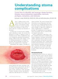
Necrotizing fasciitis - Wound Care Advisor
Necrotizing fasciitis - Wound Care Advisor
Necrotizing fasciitis - Wound Care Advisor

You also want an ePaper? Increase the reach of your titles
YUMPU automatically turns print PDFs into web optimized ePapers that Google loves.
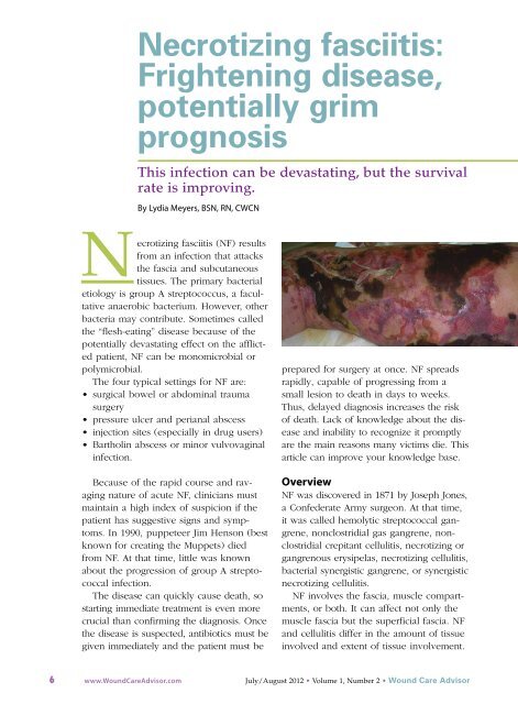
Comparing types of necrotizing <strong>fasciitis</strong>Type Microbe(s) involved FeaturesType 1 Polymicrobial, including group A • Rapid necrosishemolytic streptococci, staphylococci, • Systemic signs and symptomsanaerobic and aerobic Staphylococcuspyogenes, Staphylococcus aureus,Vibrio vulnificus, and AeromonashydrophilaType 2 Group A streptococci, with or without • Often associated with traumastaphylococci• Skin lesion present• Develops over daysType 3 Gram-negative, marine-related • Incubation period of 2 months to 2 yearsorganisms (Vibrio species,• Presents as ulcerated, edematous lesionMycobacterium ulcerans)Type 4 Fungi (commonly Mucor, Rhizopus, • Occurs mainly in immunocompromisedor Absidia species of the Mucoralespersonsorder) • High mortality (70% to 100%)• Progresses rapidly once lesions appear• May result from contaminated dressingor splints applied to skin• Also called mucormycosis, zygomycosis,or phycomysosisType 1 NF typically is associated with multiple bacteria, both aerobic and anaerobic (including facultativebacteria). This rapidly progressing, severe form causes systemic sepsis. Facultative bacteria can changefrom anaerobic to aerobic, depending on the environment.Type 2 NF can occur at any age, and may or may not be linked to other illnesses. The main bacterium involvedis group A β-hemolytic streptococcus, a facultative aerobic bacterium.Type 3 NF is associated with gram-negative, marine-related organisms and is more common near coastalwaters. It commonly appears as a painful edematous lesion, which may indicate bacteria are becoming activeand starting a devastating infection. Signs and symptoms arise 3 to 7 days after exposure related totrauma. Patients need emergency care with immediate antibiotic administration and surgical debridement.Type 4 NF is a rare fungal infection occasionally seen in immunocompromised persons. It may becomenecrotizing and spread quickly. The typical cause is introduction of bacteria from contaminated dressings,splints, and foreign bodies. The causative fungi are found in soil, decaying wood, and other organic matter.I.V. fluids, and oxygen. Limb amputationshould be done only as a last resort.Surgical debridement involves penetratingdeep into the fascia and removing allnecrotic tissue. After the first debridement,release of “dishwater fluid” may occur.Administering hyperbaric oxygen therapy(HBOT) after the first debridement increasestissue oxygenation, thus reducing tissuedestruction by anaerobic bacteria. DuringHBOT (usually given as a 90-minute treatment),the patient breathes 100% oxygenin an environment of increasing atmosphericpressure.HBOT should be given in conjunctionwith surgical debridement (usually aftereach debridement) and should continueuntil necrotic tissue ceases and cell destructionstops. HBOT also promotes collagensynthesis and neoangiogenesis (new<strong>Wound</strong> <strong>Care</strong> <strong>Advisor</strong> • July/August 2012 • Volume 1, Number 2 www.<strong>Wound</strong><strong>Care</strong><strong>Advisor</strong>.com 9
Diagnostic findings in necrotizing <strong>fasciitis</strong>Diagnostic testMagnetic resonance imagingFindings in NF• Underlying pockets of gas and subcutaneous air spacesComplete blood cell count with differential • White blood cell count > 14,000/mm 3• Hemoglobin < 10 g/dL• Thrombocytopenia (platelet count < 150,000/µL)Comprehensive metabolic panelCultures• Blood urea nitrogen > 15 mg/dL• Creatinine > 2 mg/dL• Sodium < 135 mEq/L• Increased blood glucose and lactate levels• Decreased calcium, protein, albumin, and cholesterollevels• Vary with bacteria presentblood vessel growth), which boosts bloodsupply and oxygen to tissues.Adverse effects of HBOT include earpain, oxygen toxicity, and seizures. Earpain can be minimized by swallowing oryawning. If the patient continues to haveear pain, ear tubes may be inserted by anotolaryngologist. During HBOT, air breaks(intervals of breathing room air) are importantin controlling oxygen toxicity (themain cause of seizures).Throughout the HBOT treatment period,wound dressings must be simple. Wellmoistenedgauze dressings and an abdominalpad provide good support. Oncenecrotic destruction occurs, dressings dependon wound size and the need to fillcavities. The patient may require a divertingcolostomy, depending on woundlocation and the amount of uncontrolleddiarrhea. Blood glucose levels must bemonitored before and after HBOT, as thistreatment affects blood glucose.Supportive care and follow-uptreatmentDuring initial treatment, patients need supportivecare and monitoring. Once they’reout of danger, begin teaching them how toprevent NF recurrences. Advise them tocontrol blood glucose levels, keeping theglycated hemoglobin (HbA1c) level to 7%or less. Caution patients to keep needlescapped until use and not to reuse needles.Instruct them to clean the skin thoroughlybefore blood glucose testing or insulin injection,and to use alcohol pads to cleanthe area afterward.Before discharge, help arrange the patient’saftercare, including home health carefor wound management and teaching,social services to promote adjustment tolifestyle changes and financial concerns, andphysical therapy to help rebuild strengthand promote the return to optimal physicalhealth. One helpful patient resource is theNational <strong>Necrotizing</strong> Fasciitis Foundation. TheCenters for Disease Control and Preventionsection on necrotizing <strong>fasciitis</strong> includes“Common sense and great wound care are the bestways to prevent a bacterial skin infection.”The life-threatening nature of NF, scarringcaused by the disease, and in somecases the need for limb amputation canalter the patient’s attitude and viewpoint,so be sure to take a holistic approachwhen dealing with the patient and family.Today, NF has a much better survival ratethan 2 decades ago when Jim Henson(continued on page 19)10 www.<strong>Wound</strong><strong>Care</strong><strong>Advisor</strong>.com July/August 2012 • Volume 1, Number 2 • <strong>Wound</strong> <strong>Care</strong> <strong>Advisor</strong>
tissue when the dressing is removed.Ostomy paste products can serve as effectivefiller. These pliable products can bespread into position to obtain a secure sealunder the transparent drape in hard-to-sealareas, such as the perineum. Pastes remainflexible and can be removed without residue.Temporarily increasing NPWT pressureto a higher setting may help locate asubtle leak or provide enough negativepressure to self-seal the leak. Once theleak resolves, remember to return thepressure to the ordered setting.Knowledge optimizes healingIt’s important to be aware of potentialcomplications of NPWT (See Take carewith NPWT). However, when applied correctly,NPWT is an effective option formanaging complex wounds. Recognizingand managing potential complications atthe wound site, ensuring periwound protection,minimizing epibole formation, andView: NPWT case studyReview a step-by-step process forpreparing a wound for negative pressurewound therapy (NPWT)preventing wound infection can result in abetter-prepared wound bed and promoteoptimal healing.■Selected referencesBaranoski S, Ayello EA. (2012). <strong>Wound</strong> <strong>Care</strong> Essentials:Practice Principles. 3rd ed. Springhouse, PA;Lippincott Williams & Wilkins.Bovill E, Banwell PE, Teot L, et al. Topical negativepressure wound therapy: a review of its role andguidelines for its use in the management of acutewounds. Int <strong>Wound</strong> J. 2008;5:511-529.Sussman C, Bates-Jensen B. <strong>Wound</strong> <strong>Care</strong>: A CollaborativePractice Manual for Health Professionals. 4th ed.Baltimore, MD; Lippincott Williams & Wilkins; 2011.Ronald Rock is an Adult Health Clinical NurseSpecialist in the Digestive Disease Institute atthe Cleveland Clinic in Cleveland, Ohio.<strong>Necrotizing</strong> <strong>fasciitis</strong>(continued from page 10)died. In my practice, I’ve seen four NFcases. Thanks to early identification, goodwound care, and HBOT, these patientssuffered only minimal damage. ■Selected referencesBoyer A, Vargas F, Coste F, et al. Influence of surgicaltreatment timing on mortality from necrotizingsoft tissue infections requiring intensive care management.Intensive <strong>Care</strong> Med. 2009;35(5):847-853.doi:10.1007/s00134-008-1373-4.Cain S. <strong>Necrotizing</strong> <strong>fasciitis</strong>: recognition and care.Practice Nurs. 2010;21(6):297-302.Centers for Disease Control and Prevention. Notes fromthe field: fatal fungal soft-tissue infections after a tornado—Joplin,Missouri, 2011. MMWR. 2011;60(29):992.Chamber AC, Leaper DJ. Role of oxygen in woundhealing: a review of evidence. J <strong>Wound</strong> <strong>Care</strong>. 2011;20(4):160-164.Christophoros K, Achilleas K, Vasilia D, et al. Postraumaticzygomycotic necrotizing abdominal wall <strong>fasciitis</strong> with intraabdominalinvasion in a non immunosuppressed patient.Internet J Surg. 2007;11(1). doi:10.5580/17a8.Ecker K-W, Baars A, Topfer J, Frank J. <strong>Necrotizing</strong><strong>fasciitis</strong> of the perineum and the abdominal wallsurgicalapproach. Europ J Trauma Emerg Surg. 2008;34(3):219-228. doi:10.1007/s00068-008-8072-2.Hunter J, Quarterman C, Waseem M, Wills A. Diagnosisand management of necrotizing <strong>fasciitis</strong>. Br JHosp Med. 2011;72(7):391-395.Magel DC. The nurse’s role in managing necrotizing<strong>fasciitis</strong>. AORN J. 2008;88(6):977-982.Phanzu MD, Bafende AE, Imposo BB, Meyers WM, PortaelsF. Under treated necrotizing <strong>fasciitis</strong> masqueradingas ulcerated edematous Mycobacterium ulcerans infection(Buruli ulcer). Am J Trop Med Hyg. 2012;82(3):478-481.Ruth-Sahd LA, Gonzales M. Multiple dimensions ofcaring for a patient with acute necrotizing <strong>fasciitis</strong>.Dimens Crit <strong>Care</strong> Nurs. 2006;25(1):15-21.Stevens DL, Bisno AL, Chambers HF, et al; InfectiousDiseases Society of America. Practice guidelines forthe diagnosis and management of skin and soft-tissueinfections. Clin Infect Dis. 2005;41(10):1373-1406.Su YC, Chen HW, Hong YC, Chen CT, et al. Laboratoryrisk indicator for necrotizing <strong>fasciitis</strong> score andthe outcomes. ANZ J Surg. 2008;78(11):968-972.Taviloglu K, Yanar H. <strong>Necrotizing</strong> <strong>fasciitis</strong>: strategies for diagnosisand management. World J Emerg Surg. 2007;2:19.Lydia Meyers is a medical reviewer for NationalGovernment Services in Castleton, Indiana, and aclinical liaison at CTI Nutrition in Indianapolis.She has 11 years of wound care experience innursing homes, wound clinics, and home health.<strong>Wound</strong> <strong>Care</strong> <strong>Advisor</strong> • July/August 2012 • Volume 1, Number 2 www.<strong>Wound</strong><strong>Care</strong><strong>Advisor</strong>.com 19