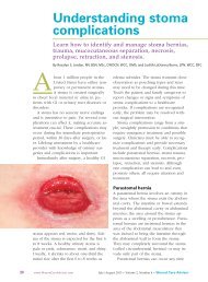
Necrotizing fasciitis - Wound Care Advisor
Necrotizing fasciitis - Wound Care Advisor
Necrotizing fasciitis - Wound Care Advisor

Create successful ePaper yourself
Turn your PDF publications into a flip-book with our unique Google optimized e-Paper software.
Comparing types of necrotizing <strong>fasciitis</strong>Type Microbe(s) involved FeaturesType 1 Polymicrobial, including group A • Rapid necrosishemolytic streptococci, staphylococci, • Systemic signs and symptomsanaerobic and aerobic Staphylococcuspyogenes, Staphylococcus aureus,Vibrio vulnificus, and AeromonashydrophilaType 2 Group A streptococci, with or without • Often associated with traumastaphylococci• Skin lesion present• Develops over daysType 3 Gram-negative, marine-related • Incubation period of 2 months to 2 yearsorganisms (Vibrio species,• Presents as ulcerated, edematous lesionMycobacterium ulcerans)Type 4 Fungi (commonly Mucor, Rhizopus, • Occurs mainly in immunocompromisedor Absidia species of the Mucoralespersonsorder) • High mortality (70% to 100%)• Progresses rapidly once lesions appear• May result from contaminated dressingor splints applied to skin• Also called mucormycosis, zygomycosis,or phycomysosisType 1 NF typically is associated with multiple bacteria, both aerobic and anaerobic (including facultativebacteria). This rapidly progressing, severe form causes systemic sepsis. Facultative bacteria can changefrom anaerobic to aerobic, depending on the environment.Type 2 NF can occur at any age, and may or may not be linked to other illnesses. The main bacterium involvedis group A β-hemolytic streptococcus, a facultative aerobic bacterium.Type 3 NF is associated with gram-negative, marine-related organisms and is more common near coastalwaters. It commonly appears as a painful edematous lesion, which may indicate bacteria are becoming activeand starting a devastating infection. Signs and symptoms arise 3 to 7 days after exposure related totrauma. Patients need emergency care with immediate antibiotic administration and surgical debridement.Type 4 NF is a rare fungal infection occasionally seen in immunocompromised persons. It may becomenecrotizing and spread quickly. The typical cause is introduction of bacteria from contaminated dressings,splints, and foreign bodies. The causative fungi are found in soil, decaying wood, and other organic matter.I.V. fluids, and oxygen. Limb amputationshould be done only as a last resort.Surgical debridement involves penetratingdeep into the fascia and removing allnecrotic tissue. After the first debridement,release of “dishwater fluid” may occur.Administering hyperbaric oxygen therapy(HBOT) after the first debridement increasestissue oxygenation, thus reducing tissuedestruction by anaerobic bacteria. DuringHBOT (usually given as a 90-minute treatment),the patient breathes 100% oxygenin an environment of increasing atmosphericpressure.HBOT should be given in conjunctionwith surgical debridement (usually aftereach debridement) and should continueuntil necrotic tissue ceases and cell destructionstops. HBOT also promotes collagensynthesis and neoangiogenesis (new<strong>Wound</strong> <strong>Care</strong> <strong>Advisor</strong> • July/August 2012 • Volume 1, Number 2 www.<strong>Wound</strong><strong>Care</strong><strong>Advisor</strong>.com 9