View Complete Issue PDF
View Complete Issue PDF
View Complete Issue PDF

Create successful ePaper yourself
Turn your PDF publications into a flip-book with our unique Google optimized e-Paper software.
=J^<br />
(ggf<br />
The Journal of the International Society<br />
for Prosthetics and Orthotics<br />
Prosthetics and<br />
Orthotics<br />
International<br />
April 1987, Vol. 11, No. 1
For your Orthopaedic Workshop: Special Equipment and Machines<br />
CD<br />
1. 758A14 Workbench for Lamination Work<br />
With 758Z5 Benchtop Cabinet and suction channel, workbench<br />
dimensions: 1500 or 2000 mm length, 750 mm<br />
width, available with different workbench tops.<br />
3. 759H4 OTTO BOCK Heating Plate<br />
For heating thermoplastic sheet material for forming. The<br />
cover has a self-regulated counterpressure plate to accommodate<br />
varying thicknesses of material. Heating surface<br />
max. 1100 x 855 mm, temperature adjustment max. 300°C.<br />
2. 701F6 OTTO BOCK Socket Router<br />
With integrated dust collection system and motor. The<br />
Socket Router is also available with two speed router motor<br />
(article no. 701 F8) and with separate dust collection<br />
system (article no. 701F7/9).<br />
4. 701E1 OTTO BOCK Heating Oven -Stainless Steel-<br />
With forced convected airflow, temperature range from 5<br />
to 300 °C, temperature high limit device, thermostat and<br />
timer, volume approx 720 I (other sizes on request).<br />
ORTHOPÄDISCHE INDUSTRIE<br />
GmbH & Co<br />
Industriestraße<br />
• D-3408 Duderstadt<br />
OTTO BOCK A/ASIA, Sydney<br />
OTTO BOCK AUSTRIA, Seekircheπ<br />
OTTO BOCK AUSTRIA, Wien<br />
OTTO BOCK BENELUX, Son en Breugel<br />
OTTO BOCK do BRASIL, Rio de Janeiro<br />
OTTO BOCK CANADA, Winnipeg<br />
OTTO BOCK FRANCE, Les Ulis<br />
OTTO BOCK IBERICA, Très Cantos/Mad.<br />
OTTO BOCK ITALIA, Budrio (BO)<br />
OTTO BOCK SCANDINAVIA, Norrköping<br />
OTTO BOCK U. K, Egham<br />
OTTO BOCK USA, Minneapolis
Prosthetics and<br />
Orthotics<br />
International<br />
Co-editors:<br />
JOHN HUGHES (Scientific)<br />
NORMAN A. JACOBS (Scientific)<br />
RONALD G. DONOVAN (Production)<br />
Editorial Board:<br />
VALMA ANOLISS<br />
RENÉ BAUMGARTNER<br />
RONALD G. DONOVAN<br />
WILLEM H. EISMA<br />
JOHN HUGHES<br />
NORMAN A. JACOBS<br />
ACKE JERNBERGER<br />
MELVIN STILLS<br />
Prosthetics and Orthotics International is published three times yearly by the International Society for<br />
Prosthetics and Orthotics (ISPO), Borgervaenget 5,2100 Copenhagen 0, Denmark, (Tel. (01) 20 72<br />
60). Subscription rate is $52.50 (U.S.) per annum, single numbers $18 (U.S.). The journal is provided<br />
free to Members of ISPO. The subscription rate for Associate Members is $25 (U.S.) per annum.<br />
Remittances should be made payable to ISPO.<br />
Editorial correspondence, advertisement bookings and enquiries should be directed to Prosthetics and<br />
Orthotics International, National Centre for Training and Education in Prosthetics and Orthotics,<br />
University of Strathclyde, Curran Building, 131 St. James' Road, Glasgow G4 0LS, Scotland (Tel.<br />
041-552 4049).<br />
ISSN 0309-3646<br />
Produced by the National Centre for Training and Education in Prosthetics and Orthotics, University of<br />
Strathclyde, Glasgow<br />
Printed by David J. Clark Limited, Glasgow
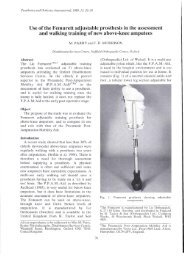
The High Technology Prosthesis<br />
The Eπdoskeletal Stabilised Knee is<br />
very suited to the needs of the active<br />
amputee especially when used in<br />
conjunction with the Pneumatic<br />
Swing Phase Control (PSPC) unit.<br />
These knee mechanisms are part of<br />
the light weight ENDOLITE system.<br />
Using the latest technology,<br />
advanced materials such as carbon<br />
fibre, weight limits of 1 kg for the<br />
below knee and 2 kg for the above<br />
knee prostheses are achieved without<br />
detracting from the strength<br />
requirements for all categories of<br />
patient.<br />
Blatchford<br />
Lister Road Basingstoke Hampshire RG22 4AH England<br />
Telephone: 0256 465771<br />
ENDOLITE is a trademark of Chas A. Blatchford & Son Ltc<br />
ii
The Journal of the International Society<br />
W for Prosthetics and Orthotics<br />
April 1987, Vol 11, No. 1<br />
Contents<br />
Editorial 1<br />
ISPO Accounts 2<br />
Executive Board Meeting Report<br />
Obituary—Fred Forchheimer<br />
The effect of adjuvant oxygen therapy on transcutaneous p0 2<br />
and healing in the<br />
below-knee amputee 10<br />
C. M. BUTLER, R. O. HAM, K. LAFFERTY, L. T. COTTON AND V. C. ROBERTS<br />
The value of revision surgery after initial amputation of an upper or lower limb 17<br />
M. R. WOOD, G. A. HUNTER AND S. G. MILLSTEIN<br />
A versatile hand splint 21<br />
A. S. JAIN AND D. MCDOUGALL<br />
Evaluation of introducing the team approach to the care of the amputee : the Dulwich study 25<br />
R. HAM, J. M. REGAN AND V. C. ROBERTS<br />
Abnormal extension of the big toe as a cause of ulceration in diabetic feet 31<br />
K. LARSEN AND P. HOLSTEIN<br />
Foot loading characteristics of amputees and normal subjects 33<br />
G. D. SUMMERS, J. D. MORRISON AND G. M. COCHRANE<br />
Technical note — a multipurpose orthosis for paralysed children 40<br />
N. G. LAWRENCE<br />
Technical note—new splinting materials 42<br />
R. WYTCH, C. MITCHELL, I. K. RITCHIE, D. WARDLAW AND W. LEDINGHAM<br />
Letters to the editor 46<br />
Book review 48<br />
Calendar of events 49<br />
6<br />
9
Prosthetics and Orthotics International, 1987, 11<br />
ISPO<br />
Elected Members of Executive Board:<br />
J. Hughes (President) UK<br />
W. H. Eisma (President Elect) Netherlands<br />
S. Heim (Vice President) FRG<br />
S. Sawumura (Vice President) Japan<br />
V. Angliss Australia<br />
R. Baumgartner FRG<br />
A. Jernberger Sweden<br />
M. Stills USA<br />
E. Lyquist (Past President) Denmark<br />
G. Murdoch (Past President) UK<br />
J. Steen Jensen (Hon. Treasurer) Denmark<br />
N. A. Jacobs (Hon. Secretary) UK<br />
Standing Committee Chairmen and Task Officers<br />
J. Kjolbye (Finance) Denmark<br />
E. Lyquist (Protocol) Denmark<br />
W. Eisma (Congress. Publications Netherlands<br />
and Membership)<br />
G. Murdoch (Education) UK<br />
H. G. Thyregod (Professional Register) Denmark<br />
B. Klassoπ (Socket Design) Sweden<br />
E. Marquardt (Limb Deficient Child) FRG<br />
Consultants to Executive Board<br />
H. C. Chadderton (Consumer) Canada<br />
J. Van Rolleghem (INTERBOR) Belgium<br />
J. N. Wilson (WOC) UK<br />
International Consultants to Executive Board<br />
P. Kapuma Africa<br />
Wu Zongzhe<br />
China<br />
G. Bousquet France<br />
M. K. Goel India<br />
H. Schmidl Italy<br />
Yongpal Ahn<br />
Korea<br />
E. K. Jensen South America<br />
T. Keokarn South East Asia<br />
R. Lehneis USA<br />
N. Kondrashin USSR<br />
Chairmen of National Member Societies<br />
Australia<br />
W. Doig<br />
Belgium<br />
M. Stehman<br />
Canada<br />
G. Martel<br />
China<br />
Tang Yi-Zhi<br />
Denmark<br />
H. C. Thvregod<br />
FRG<br />
G. Neff<br />
Hong Kong<br />
K. Y. Lee<br />
Israel<br />
T. Steinbach<br />
Japan<br />
K. Tsuchiya<br />
Netherlands<br />
P. Prakke<br />
Norway<br />
G. Veres<br />
Sweden<br />
A. Jernberger<br />
Switzerland<br />
J. Vaucher<br />
UK<br />
D. Condie<br />
USA<br />
Past Presidents<br />
K. Jansen (1974-1977) Denmark<br />
G. Murdoch (1977-1980) UK<br />
A. Staros (1980-1982) USA<br />
E. Lyquist (1982-1983) Denmark<br />
E. G. Marquardt (1983-1986) FRG<br />
Secretary<br />
Aase Larsson<br />
\S<br />
F. Golbranson<br />
Denmark
Prosthetics and Orthotics International, 1987, 11<br />
Editorial<br />
This issue of the Journal displays the financial statement for the year 1986.<br />
Due to alterations in Danish law and regulations, the Society has changed to the State Authorized<br />
Public Accountants, Schøbel & Marholt, who are" internationally represented by Touche Ross<br />
International. Our new accountants also act as investment advisors with respect to Danish tax<br />
legislation and with the purpose of increasing the yield on our long-term investment programme<br />
The accounts show that securities have increased by 1,463,165 DKK since the fiscal year, 1985. This<br />
is mainly generated by reinvestment of interest and the year's surplus, which includes the profit from the<br />
V World Congress. About two thirds of the securities consist of government bonds, which provide a<br />
yearly interest of 10-12% of the face value and are freely negotiable without taxable yield The market<br />
value is influenced by the Danish interest rate. The remainder of our securities are placed in investment<br />
trust units the profit on which is not taxable after three years retention. Consequently none of the<br />
investments should be taxable and they will securely cover the liabilities and the contingency fund of<br />
1,500,000 DKK.<br />
The present financial statement is prepared in accordance with the Danish Articles of Associations.<br />
The accrual concept of accounting has been introduced from January 1,1986 replacing the previous cash<br />
accounting. This provides a more accurate picture of the financial state of the Society, but makes direct<br />
comparison with previously published accounts difficult, as is explained in note 9 to the Accounts.<br />
The Journal, Prosthetics and Orthotics International is identified under a separate account. The<br />
deficit of 12,075 DKK is rather close to the profit of 6,561 DKK for 1985, which appears after<br />
adjustment for prepaid advertising and subscriptions. The Journal is thus being produced at virtually no<br />
cost to the Society.<br />
The total surplus of the Society has increased over the years from 421,219 DKK in 1983, 629,724<br />
DKK in 1984 and 489,306 DKK in 1985 to 1,050,754 DKK in 1986. The major contributions to the latter<br />
increase has been from the income on investments and the profit obtained from the world congress,<br />
which in 1986 was 325,751 DKK.<br />
The Society is very grateful for the contributions totalling 134,424 DKK provided by the Society<br />
and Home for the Disabled and the War Amputations of Canada. These bodies have indicated their<br />
continuing support.<br />
In spite of unchanged individual fees since 1984, our income on membership is constantly<br />
increasing as the Society grows. This contributes sufficiently to run the daily administration of ISPO and<br />
will once more be held at 400 DKK for 1987 and 1988. The costs of our office in Copenhagen amount to<br />
429,265 DKK, including the cost of our one salaried staff member, Aase Larsson. The costs are only<br />
kept down by her enthusiastic spirit and the free office facilities provided by the Society and Home for<br />
the Disabled.<br />
The meeting and travelling expenses for the Executive Board have been kept down to the 1984<br />
level, including our participation in meetings of the International Standard Organizations, of<br />
Rehabilitation International and of task officer visits.<br />
The economy of the Society is sound. We have created the basis for increased future activity, which<br />
the Executive Board is already generating.<br />
Finally we express our gratitude to sponsors for their support and members for their activity. These<br />
contribute to the future successful development of our Society.<br />
J. Steen Jensen<br />
Honorary Treasurer
Prosthetics and Orthotics International, 1987, 11, 2-5<br />
Auditors' Report<br />
I.S.P.O. Statement of Accounts, 1986<br />
We have audited the financial statements for the year ended December 31, 1986.<br />
The audit has been performed in accordance with approved auditing standards and has included<br />
such procedures as we considered necessary. We have satisfied ourselves that the assets shown in the<br />
financial statements exist, have been fairly valued and are beneficially owned by the company and that<br />
all known material liabilities on the balance sheet date have been included.<br />
The financial statements have been prepared in accordance with statutory requirements and the<br />
articles of association and generally accepted accounting principles. In our opinion the financial<br />
statements give a true and fair view of the state of the company's affairs on December 31,1986 and of the<br />
profit for the period then ended.<br />
Copenhagen, February 19, 1987<br />
Schøbel & Marholt<br />
Søren Wonsild Glud<br />
State Authorized Public Accountant<br />
Accounting Policies<br />
The accrual concept of accounting has been used from January 1,1986. In prior periods cash accounting<br />
concept has been used. The effect on the result for the year is explained in note 9.<br />
Securities: Bonds and shares have been valued at the lower cost on market.<br />
Income Statement for the Year 1986<br />
SUMMARY<br />
Society membership and administration (note 1) 130.873<br />
Sponsorship (note 2) 134.424<br />
Conferences, courses etc. (note 3) 254.893<br />
Prosthetics and Orthotics International (note 4) (12.075)<br />
Publications (note 5) 3.206<br />
Investment income (note 6) 539.433<br />
Balance sheet as of December 31, 1986<br />
DKK 1.050.754<br />
ASSETS<br />
Cash DKK 148.742<br />
Accounts due<br />
Advertising due 13.510<br />
Dividend tax due<br />
Accrued interest<br />
779<br />
157.733<br />
Advance funding of World Congress 1980 119.690<br />
DKK 291.712<br />
Securities (at cost) (note 7) 3.317.768<br />
Total Assets DKK 3.758.222<br />
LIABILITIES AND CAPITAL<br />
Liabilities<br />
Provision DKK 119.690<br />
:
I.S.P.O. Statement of Accounts, 1986 3<br />
Capital<br />
Capital January 1, 1986<br />
Provision for Advance Funding of<br />
World Congress 1980<br />
Result for the year<br />
Capital December 31, 1986<br />
Short-term liabilities<br />
Accrued expenses<br />
Prepaid advertising income<br />
Prepaid subscription income<br />
2.495.595<br />
(119.690)<br />
2.375.905<br />
1.050.754<br />
DKK 3.426.659<br />
59.469<br />
72.408<br />
79.996<br />
Total Liabilities and Capital DKK 3.758.222<br />
Contingent liabilities (note 8)<br />
Notes to the Financial Statements<br />
1. SOCIETY MEMBERSHIP AND ADMINISTRATION<br />
Income<br />
Membership — fees<br />
Transfer from Knud Jansen's foundation<br />
Expenditure<br />
Executive Board:<br />
Travel and hotel<br />
Meeting expenses<br />
Meeting in other organizations<br />
Travelling expenses, Honorary Secretary<br />
and Treasurer<br />
Staff salaries<br />
Membership, Rehabilitation International<br />
Data service<br />
Stationery printing<br />
Office supplies<br />
Accountant<br />
Telephone<br />
Postage<br />
Maintenance<br />
Sundries<br />
(110.609)<br />
(40.409)<br />
(5.167)<br />
714.218<br />
2.105<br />
DKK 716.323<br />
(25.256)<br />
(219.174)<br />
(6.188)<br />
(40.111)<br />
(65.900)<br />
(4.154)<br />
(35.008)<br />
(5.789)<br />
(22.465)<br />
(1.845)<br />
(3.375) (585.450)<br />
DKK 130.873<br />
2. SPONSORSHIP<br />
Contribution from the War Amputation of Canada<br />
Contribution from SAHVA<br />
59.424<br />
75.000<br />
DKK 134.424<br />
3. CONFERENCES, COURSES etc.<br />
World congress 1986<br />
Dundee course publication<br />
Toronto Education Symposium<br />
Pakistan<br />
Education Workshop Questionnaire<br />
325.751<br />
(63.167)<br />
(1.829)<br />
(7.499)<br />
(2.021)<br />
DKK 254.893
4 l.S.P.O. Statement of Accounts, 1986<br />
4. PROSTHETICS AND ORTHOTICS INTERNATIONAL<br />
Income<br />
Advertising<br />
Subscriptions<br />
136.018<br />
73.500<br />
DKK 209.518<br />
Expenditure<br />
Printing<br />
Mailing inclusive of labels<br />
Production editor<br />
Committee meeting, Publication<br />
(190.304)<br />
14.1301<br />
15.109)<br />
(2.050)<br />
DKK (221.593)<br />
DKK (12.075)<br />
5. PUBLICATIONS<br />
Book sale<br />
3.206<br />
DKK 3.206<br />
6. INVESTMENT INCOME<br />
Bonds<br />
Maturity yield<br />
Interest<br />
82.024<br />
434.000<br />
DKK 516.024<br />
Shares<br />
Dividend<br />
Bank account<br />
Interest income<br />
Interest expense<br />
DKK 1.095<br />
24.655<br />
(990)<br />
DKK 23.665<br />
Expenditure<br />
Safekeeping fee<br />
(1.351)<br />
Result<br />
DKK 539.433<br />
7. SECURITIES<br />
Bonds<br />
12% Dansk Statslån S.2001<br />
10% Dansk Statslån S.2004<br />
10% Dansk Statslån 1979/89<br />
10% Dansk Statslån ST.L. 1994<br />
10% Dansk Statslàn S.1988<br />
10% Østifternes Krf.<br />
18. série 2003<br />
Rate<br />
31/12-86<br />
101,25<br />
91,50<br />
99,00<br />
94,00<br />
99,75<br />
91,00<br />
Face<br />
value<br />
632.000<br />
400.000<br />
260.000<br />
600.000<br />
269.000<br />
100<br />
DKK 2.161.100<br />
Market<br />
value<br />
639.900<br />
366.000<br />
257.000<br />
564.000<br />
268.328<br />
91<br />
2.095.719<br />
Original<br />
cost<br />
575.292<br />
367.551<br />
239.559<br />
606.909<br />
250.842<br />
71<br />
2.040.224<br />
Investment trust<br />
units<br />
Sparinvest, D<br />
Privatinvest 2<br />
Privatinvest 5<br />
Investor-Maximum<br />
Rate<br />
31/12-86<br />
226,00<br />
611,25<br />
99,50<br />
100,00<br />
Face<br />
value<br />
90.000<br />
38.000<br />
450.000<br />
343.000<br />
Market<br />
value<br />
203.400<br />
232.275<br />
447.750<br />
343.000<br />
Original<br />
cost<br />
224.751<br />
227.371<br />
453.359<br />
349.885<br />
DKK 921.000<br />
1.226.425<br />
1.255.366
I S.P.O. Statement of Accounts, 1986 5<br />
Shares<br />
Københavns<br />
Handelsbank 260,00 8.000 20.800 22.178<br />
DKK 8.000 20.800 22.178<br />
Total result DKK 3.090.100 3.342.944 3.317.768<br />
8. CONTINGENT LIABILITY<br />
The association is involved in a court trial in connection with the World Congress 1980. The association<br />
might be liable to additional cost in this connection. The outcome is at present uncertain.<br />
9. CHANGE IN ACCOUNTING POLICY<br />
Effect from changing accounting policy from cash accounting concept to accrual accounting concept.<br />
Interest income earned up to December 31, 1985,<br />
but received in 1986<br />
Advertising income earned up to December 31, 1985,<br />
215.870<br />
but received in 1986<br />
Advertising income relating to 1986,<br />
27.665<br />
but prepaid as of December 31,1985 (11.124)<br />
1986 subscriptions received in 1985 (60.480)<br />
Net effect DKK 171.931<br />
INTERNATIONAL SOCIETY FOR PROSTHETICS AND ORTHOTICS<br />
FEE REDUCTION<br />
FOR DEVELOPING COUNTRIES<br />
The Executive Board has agreed that individuals from Developing Countries who wish to<br />
join the Society can do so at a reduced annual membership fee of DKK150 (that is one hundred<br />
and fifty Danish Crowns). This reduced fee also applies to existing members. Those<br />
individuals who wish to take advantage of this reduced fee should apply in writing to:<br />
The Secretariat,<br />
ISPO,<br />
Borgervaenget 5,<br />
2100 Copenhagen 0,<br />
DENMARK.
Prosthetics and Orthotics International, 1987, 11, 6-8<br />
Executive Board Meeting<br />
17th and 18th January, 1987<br />
The following paragraphs summarize the major discussions and conclusions of the last Executive Board<br />
Meeting held in Copenhagen. They are based on the draft minutes of that meeting which have not yet<br />
been approved by the Executive Board.<br />
International Committee Meeting<br />
The Executive Board discussed a number of matters which had been raised at the International<br />
Committee Meeting which was held at the time of the Copenhagen Congress:<br />
a) it was agreed that the Executive Board should pursue the possibility of holding an interim meeting<br />
of representatives of the International Committee between Congresses subject to finances being<br />
available.<br />
b) in response to a suggestion from the International Committee, Melvin Stills was working on the<br />
production of a Tape Slide Set which would describe the workings and interests of the Society.<br />
When this work had been completed copies of the set would be made available in order to help<br />
promote the Society.<br />
c) the Executive Board discussed the International Committee's proposal that some form of ISPO<br />
certificate of attendance should be produced for locally organized courses and meetings. The<br />
Protocol Committee were asked to examine this matter and report to the next Executive Board<br />
meeting.<br />
d) the Executive Board discussed the suggestion that the Society should improve its Information<br />
Service. No conclusion was reached, however the President and Honorary Secretary would make<br />
proposals on this matter to the next Executive Board meeting.<br />
e) at the request of the International Committee, George Murdoch had prepared a draft proposal<br />
regarding the protocol for inspection of prosthetic and orthotic education centres. The Executive<br />
Board made further suggestions with regard to this protocol and an amended form of protocol<br />
would be presented to the next Executive Board meeting.<br />
Committee, Task Officer and Consultant Appointments<br />
The Executive Board discussed Committee, Task Officer and Consultant appointments. A<br />
complete list of these appointments can be seen on page iv of this issue of the Journal.<br />
Task Officer Reports<br />
The Honorary Treasurer reported that there would be a surplus available to the Society from the<br />
year 1986. The official accounts for the year are printed elsewhere in this issue of the Journal. A revised<br />
budget for 1987 and a draft budget for 1988 was presented to the Executive Board. It was agreed that the<br />
membership fee for 1988 should remain at 400 DKK.<br />
The Chairman of the Protocol Committee indicated that they had discussed the protocol for the<br />
adjudication and award of the Blatchford Prize and this would be put to the next Board meeting. The<br />
President reported that the Forchheimer family wished to offer a Prize, to be awarded every three years,<br />
in memory of Alfred Forchheimer. It was intended that the prize be awarded through the Society for<br />
outstanding work in clinical assessment, evaluation or measurement. More information regarding these<br />
prizes will be intimated to the membership in due course.<br />
The Executive Board discussed a proposal to hold a Workshop on the Upgrading of Short Course<br />
Trained Technicians and agreed that this workshop should be held in the University of Strathclyde from<br />
the 19th-25th July 1987.<br />
The Board agreed that Ron Donovan should be appointed as Co-editor (Production) of<br />
'Prosthetics and Orthotics International'. It was decided that the Journal be airmailed to all members<br />
and subscribers outside Europe. The Board agreed that the Society should pursue the establishment of<br />
an International Newsletter. At present it should be an integral part of the Journal and if successful,<br />
might eventually become independent. It was agreed that David Condie and Joan Edelstein be invited<br />
to edit the International Newsletter. The Publications Committee had examined the possibility of<br />
publishing a separate volume of selected articles from the Journal, but recommended that this should<br />
not be pursued at present.
Executive Board Meeting 7<br />
The manual on 'The Planning and Installation of Orthopaedic Workshops in Developing<br />
Countries' has now been published and copies are available at a cost of $10 (US) for non-members and<br />
$5 (US) for members.<br />
The Executive Board discussed a proposal from A. Bennet Wilson Jr. and Melvin Stills with regard<br />
to the possibility of holding an ISPO Workshop on 'Above-knee Fitting and Alignment Techniques'.<br />
The Board agreed that there was an urgent need to study the various techniques of above-knee fitting<br />
and that, if possible, a workshop should be arranged as soon as possible. A steering committee,<br />
comprising of A. Bennet Wilson Jr., Melvin Stills, Al Muilenburg and George Murdoch was appointed<br />
to discuss detailed arrangements. It was agreed that the final decision to go ahead with the workshop<br />
should be made by the President in consultation with the Honorary Treasurer and the Honorary<br />
Secretary after submission of a detailed proposal from the Steering Committee.<br />
International Organizations<br />
The President, President Elect and Honorary Secretary attended the last meeting of the<br />
INTERBOR Board of Directors in November 1986. The Society is collaborating with INTERBOR<br />
with regard to their Congress in Barcelona in June 1987, and the President Elect has been appointed to<br />
its Scientific Committee. INTERBOR is interested in establishing standards of education in Europe<br />
and it was agreed that there should be a more formal meeting between representatives of ISPO and<br />
INTERBOR regarding this subject which should take place at the time of the Barcelona Congress.<br />
The President reported on a warm and friendly meeting with officers of the International<br />
Committee of the Red Cross (ICRC) in November 1986. The meeting was attended by the President,<br />
President Elect and Honorary Secretary on behalf of the Society. The Executive Board approved of<br />
these initial contacts and recommended that the Society should continue to develop its collaboration<br />
with ICRC.<br />
The President reported that whilst in Geneva visiting ICRC the group had taken the opportunity to<br />
meet with the World Health Organization (WHO). The meeting had established initial contacts with<br />
WHO and means of further collaboration were being explored.<br />
The Honorary Secretary reported on a recent meeting with the Secretary General of Rehabilitation<br />
International (RI) which explored ways in which closer collaboration could be achieved between the<br />
two organizations. The Board agreed that the Society should seek an RI/ICTA representative to the<br />
Executive Board. The Board further agreed that Willem Eisma should co-ordinate the sectoral meeting<br />
on 'Changes in Prosthetics and Orthotics with regard to new Technology' that ISPO would be<br />
presenting at the RI Congress in Japan in 1988. The President and Honorary Secretary had attended the<br />
RI Assembly meeting held in October 1986 in London on behalf of the Society.<br />
Margaret Ellis had attended the International Commission on Technical Aids (ICTA) meeting in<br />
Newcastle in October 1986 on behalf of the Society during which a number of matters were raised which<br />
were of interest to the Society:<br />
a) the new Wheelchair Manual has been compiled and is now available from I CT A/INFO RM.<br />
b) a Seminar on the Provision of Technical Aids for Third World Countries was held in Bombay in<br />
September 1986.<br />
c) the European Economic Community Group of ICTA are involved in helping develop Handinet, a<br />
computerized system of information related to technical aids.<br />
The Executive Board agreed that Margaret Ellis, Bo Klasson and George Murdoch continue to be<br />
representatives of ISPO to ICTA.<br />
The Honorary Secretary reported on a recent meeting with World Rehabilitation Fund (WRF).<br />
During the meeting, it emerged that WRF would be interested in co-operating with the Society in the<br />
Workshop on the Upgrading of Short Course Trained Technicians by sending representatives to the<br />
meeting.<br />
Proposals from the International Labour Office (ILO) to hold a Pan African Workshop for Experts<br />
and Policy Makers in Prosthetics and Technical Aids had been taken over by a newly established<br />
organization known as the African Rehabilitation Institute (ARI) which is based in Zimbabwe. ARI is<br />
funded by the African community and is actively pursuing arrangements to hold this workshop in the<br />
near future. The Society would keep in contact with ARI and offer to collaborate in the organization of<br />
the workshop.<br />
The Executive Board reviewed its representation to the United Nations (UN). It was agreed that<br />
Jean Vaucher and Sepp Heim should be the Society's representatives in Geneva and Vienna and that<br />
Melvin Stills and Richard H. Lehneis should be its representatives in New York.
Executive Board Meeting<br />
The Society continues to have contact with other organizations such as World Orthopaedic<br />
Concern (WOC), Internationaler Verband der Orthopadie-Schuhtechniker (IVO) and SICOT.<br />
World Congress<br />
The President reported on the Bologna Congress. There have been no developments with the court<br />
case and no new date for a further hearing had been set.<br />
The Secretary General of the Copenhagen Congress reported that the final accounting was almost<br />
complete and would be presented to the next Board Meeting. There had been over 600 active<br />
participants and the Exhibition had been successful and made a substantial surplus. The President<br />
thanked the Secretary General and his colleagues for a very satisfactory outcome.<br />
Seishi Sawamura outlined the draft scientific programme for the Japanese Congress in 1989. This<br />
programme had been discussed by the International Congress Committee prior to the Executive Board<br />
meeting and a number of suggestions had been made. He also indicated that he had been in contact with<br />
the National Member Societies for their comments. One major suggestion is that the Instructional<br />
Courses should not take place in the morning, but the late afternoon and the Japanese programme<br />
committee had been asked to look into the possibility of implementing this suggestion. It was agreed<br />
that the registration fee for INTERBOR members should be the same as that available to ISPO<br />
members.<br />
No invitations had been received as yet to host the 1992 Congress. The Honorary Secretary was<br />
asked to remind National Member Societies that the final date for submission of official invitations is the<br />
end of March 1987 and should be made according to the guidelines outlined in the Society's booklet,<br />
"Information and Guidelines for National Secretaries and other Officers of National Member<br />
Societies".<br />
Future Activities<br />
a) Arrangements for the Symposium on Traumatic Amputation to be held in Herzliya, Israel on<br />
September 6-10 1987 were well underway. It was agreed that there should be a special meeting of<br />
the Executive Board members participating in the Symposium.<br />
b) Willem Eisma reported that the arrangements for the meeting in Netherlands, 28th-30th October<br />
1987 were progressing well and the organization had reached an advanced stage.<br />
c) The Symposium on the Limb Deficient Child will be held in Heidelberg, FRG on August<br />
28th-September 1st 1988.<br />
d. The possibility of the Society collaborating in a conference in the USSR in 1988 is being<br />
investigated.<br />
e) The Swedish National Member Society is discussing the possibility of holding a meeting on the<br />
Deformed Foot, but not until 1990.<br />
Developing Countries<br />
The Executive Board discussed the level of membership fee for developing countries. It was agreed<br />
that a special rate should be instituted, namely 150 DKK. The Honorary Secretary and Honorary<br />
Treasurer would administer this scheme.<br />
George Murdoch reported on a visit to Pakistan at the invitation of the Pakistani Orthopaedic<br />
Association.<br />
The Honorary Secretary informed the Board that he had been in correspondence with the National<br />
Training Centre for Orthopaedic Technologists in Jordan with regard to an inspection visit,<br />
arrangements had yet to be finalized.<br />
Sepp Heim reported that plans for a West African Survey of former students had not yet been<br />
finalized. As soon as information was available he would forward this to the Honorary Secretary.<br />
M. K. Goel presented a video to the Executive Board which illustrated the scale of the disabled<br />
problem in India. This was a very informative presentation.<br />
The Executive Board agreed that Sepp Heim should prepare an overview of activities in Prosthetics<br />
and Orthotics in the Developing World.<br />
Fellowship<br />
Bernard Schievink (FRG) has been elected as a fellow of the Society.<br />
Norman A. Jacobs<br />
Honorary Secretary
Prosthetics and Orthotics International, 1987, II<br />
Obituary - Fred Forchheimer<br />
Fred Forchheimer, Stockholm, passed away on the 17th of November 1986 at the age of 68. He is survived<br />
by his wife Sylvia, physician and PhD, his son Robert, acting professor at Linköping University with wife<br />
Irene M.Sc. Linköping and his daughter Claire MA. Stockholm.<br />
Fred Forchheimer was an unusually productive scientist, philosopher and friend. He came to<br />
Gothenburg, Sweden in the 40's having experienced severe difficulties and the loss of his family in World<br />
War II. In Gothenburg he attained an Engineering degree and his creativity immediately started to<br />
produce results. His work regarding the development of rust protective systems, light consistent ink for<br />
ball-point pens, emulgation techniques, techniques for photography and enlargements as well as<br />
tixotrophic solvents are examples of early achievements which have reached industrial application. In the<br />
early 60's after having moved to Stockholm he started academic studies in measurement techniques and<br />
focused his interest towards medical applications. In a joint effort at The Wallenburg Laboratory, his<br />
expertise in the fields of mechanical and electronic engineering as well as his knowledge in the patent field<br />
were of great importance for the project "Isoelectric Focusing", an electrophoreses technique that has<br />
given rise to a methology and products which are used all over the world today. Towards the end of the 60's<br />
he started, in his spare time, a collaboration with the Department of Tumour Biology at the Karolinska<br />
Institute. He created many technical solutions with regard to the construction of an experimental<br />
radiation system in which oxygen concentration could be measured with an hitherto unachieved accuracy.<br />
His system is still being used and has contributed to discoveries and characterizations of several oxygen<br />
related radiation-biological effects. In the early 70's he participated in a research project with Professor<br />
Britton Chance, Philadelphia to set up a measurement programme for the biological measurement of<br />
oxygen concentration using yeast hemoglobin. The time consuming measurements were made during the<br />
night to avoid disturbing his regular daily work. Many technical problems had to be solved and he also<br />
significantly improved Professor Chance's "Time Sharing Dual Wavelength Spectro-photometer". His<br />
most recent activity was a project related to morphometrical problems, where he worked towards a<br />
computerized solution. In spite of his engineering background he decided to present a PhD thesis in<br />
medicine. At the age of 61 he went back to school and graduated in medicine in 1980 Soon after he<br />
registered as a post graduate student with the Department of Tumour Biology of the Karolinska Institute.<br />
In his "daytime" professional career he was responsible for measurements related to temperature and<br />
heat transfer until in the mid 70's he moved to the Prosthetics Research Laboratory of the Karolinska<br />
Hospital. This included collaboration with the Department of Orthopaedic Surgery at the same hospital.<br />
(In 1980 the management of the laboratory was transferred to the Een-Holmgren Ort. AB.) This was a<br />
very importamt move for prosthetics and orthotics as well as for himself. For the first time he could fully<br />
combine his deep knowledge in engineering, medicine, natural sciences and psychology. His first task was<br />
to organize and conduct the evaluation of upper extremity prostheses. He caused some turbulence by his<br />
strong statement that an evaluation can only be made if the questions "value for whom" and "in which<br />
situation" are carefully considered. At this time a lot of prosthetics and orthotics developments were more<br />
related to engineering ambitions than to the patient's need. He also helped us to understand that the<br />
evaluation itself could influence the acceptance of the aid to be evaluated. Although he worked with very<br />
advanced systems, he always looked for simplicity. He loved the following aphorism coined within his<br />
team: "The most advanced application of technology is not necessarily the same as the application of the<br />
most advanced technology".<br />
He was involved in or responsible for several projects, including gait analysis, ergonometric<br />
measurements, mechanical component testing etc. Very often he made his test-rigs himself. Whatever<br />
project or activity he was involved in, he always took the time to look into the philosophical and ethical<br />
aspects of it. If it did not meet his requirements he preferred not to participate.<br />
He also served as a teacher, covering subjects like mathematics, physics, biology, biomechanics,<br />
computer science, psychology and philosophy.<br />
After retiring he increased his activity level, serving also as a patent consultant—he held several patents<br />
himself.<br />
He was one of the early Swedish ISPO members serving as auditor for ISPO Sweden until his passing.<br />
One of the most important contributions to the Society was, however, his participation in the organizing of<br />
the 1976 workshop on "The Deformed Foot and Orthopaedic Footwear". The book was printed in the<br />
English and German languages which was possible only thanks to Fred's participation in the editorial<br />
group. He was also an invited member of "Professors World Peace Academy" (PWPA).<br />
But first of all he was a friend and a true altruist. His own career was not important. He would rather<br />
spend work on assisting friends in their research than on finishing his own PhD thesis. It was so obvious<br />
that his main sources of energy were friendship and givingness.<br />
His love for life and peace was emotionally as well as scientifically based. He used to say: "How can we<br />
allow ourselves to take lives when we can't even make a substitute for one of the most important<br />
conditions for life — the cell membrane".<br />
And indeed, he did carry insects out of the room instead of killing them!<br />
Bo Klasson.<br />
•••>
Prosthetics and Orthotics International, 1987, II, 10-16<br />
The effect of adjuvant oxygen therapy<br />
on transcutaneus pO 2<br />
and healing in the<br />
below-knee amputee<br />
C. M. BUTLER, R. O. HAM, K. LAFFERTY, L. T. COTTON and V. C. ROBERTS<br />
Department of Medical Engineering and Physics, King's College School of Medicine and Dentistry, London.<br />
Abstract<br />
The effects on tissue oxygenation of postoperative<br />
adjuvant oxygen have been studied in<br />
a group of 20 patients undergoing below-knee<br />
(BK) amputation for vascular disease. Ten<br />
patients received no therapy, the remainder<br />
receiving 28% oxygen for 48 hours following<br />
surgery. The results showed that the<br />
trancutaneous pO 2<br />
in the amputation flaps fell<br />
significantly by some 20 mmHg (p
In fields unrelated to vascular amputation, a<br />
number of animal and human studies have<br />
shown that increasing inspired oxygen<br />
concentration results in increased available<br />
tissue oxygen and that this may be of benefit in<br />
reducing wound infection and improving tissue<br />
healing (Hunt and Pai, 1972; Knighton et al,<br />
1984; Chang et al, 1983). Mustapha and<br />
colleagues have reported that increasing<br />
inspired oxygen concentration results in an<br />
increase in below-knee TcpO 2<br />
in patients with<br />
ischaemic limbs (Mustapha et al, 1984), though<br />
they have not reported on its effect on healing.<br />
This present study was designed firstly to<br />
investigate the effects of amputation surgery on<br />
stump TcpO 2<br />
and to test the hypothesis that<br />
increasing inspired oxygen concentration could<br />
beneficially increase stump transcutaneous<br />
oxygen levels and secondly, to assess<br />
prospectively the value of post-operative<br />
adjuvant oxygen therapy in relation to stump<br />
survival.<br />
Patients and methods<br />
The study was in two parts. The first,<br />
involving 20 patients undergoing BK<br />
amputation, was aimed at gaining an<br />
understanding of the effects of adjuvant oxygen<br />
and amputation surgery on the transcutaneous<br />
oxygen levels in below-knee stumps. The<br />
second, involving 39 patients undergoing BK<br />
amputation, was aimed at evaluating the effects<br />
on healing of post-operative adjuvant oxygen<br />
therapy. All patients admitted consecutively to<br />
the vascular unit of Dulwich Hospital and<br />
requiring major amputation for ischaemia were<br />
considered for entry. The only criteria for<br />
exclusion were visible ischaemic demarcation<br />
above a suitable level for below-knee<br />
amputation or severe disease of the ipsilateral<br />
knee joint precluding satisfactory prosthetic<br />
fitting. All remaining patients were entered<br />
sequentially into the study. These patients<br />
underwent skew-flap myoplastic below-knee<br />
amputation after identical pre-operative ward<br />
preparation and antibiotic prophylaxis using the<br />
regime and operative techniques described by<br />
Robinson et al (1982).<br />
Skin oxygen study<br />
The first 20 patients entering the study were<br />
allocated using a restricted randomization into<br />
either treated or untreated groups. The<br />
operations were performed alternately by one of<br />
two surgeons only (CB or KL). The treated<br />
group (n=10) received 28% adjuvant oxygen by<br />
Ventimask (Vickers Medical) for 48 hours<br />
post-operatively at a rate of 4 litres per minute.<br />
The untreated group (n=10) received nothing.<br />
Only light gauze dressings were used and<br />
conventional stump bandaging or plaster were<br />
not used.<br />
Transcutaneous pO 2<br />
(TcpO 2<br />
) measurements<br />
were made independently (RH) and the results<br />
not examined until the end of the study. A<br />
Roche cutaneous pO 2<br />
monitor (Kontron<br />
Medical) containing two 632 modules, enabling<br />
simultaneous recording at two sites, was used<br />
throughout. Electrode positions were at a<br />
central site 5cm below the clavicle and at<br />
anterior and posterior sites 10cm below the knee<br />
at the centre of the amputation flaps on the<br />
stump and 5cm medial to the tibial shaft and at<br />
the same level on the contralateral limb. The<br />
measurements were made in each case on the<br />
day prior to operation and at 1, 2, 7 and 14 days<br />
post-operatively. The measurement on the<br />
second post-operative day was made at least two<br />
hours after stopping oxygen therapy.<br />
All readings were made with the patient<br />
semi-supine having been resting in bed for at<br />
least 20 minutes. Sensor temperature was 44°C<br />
and the calibration of the modules and<br />
derivation of TcpO 2<br />
was described by Ratliff et<br />
al (1984). All patients had regular physiotherpy,<br />
early ambulation using the P.P.A.M.-Aid<br />
(Vessa Ltd.) and were measured for their<br />
artificial limb in the early post-operative period<br />
on the ward by a team from the Department of<br />
Health and Social Security (DHSS) limb fitting<br />
centre at Roehampton.<br />
Healing study<br />
In this study 39 patients selected as above for<br />
BK amputation were entered consecutively into<br />
a randomized controlled trial of the effects on<br />
healing of post-operative adjuvant oxygen<br />
therapy. Using this randomization 17 patients<br />
fell in the treated group and 22 in the untreated<br />
group. Patient treatment was as for the previous<br />
study, the treated group receiving the adjuvant<br />
oxygen and the untreated group no additional<br />
therapy. Healing was independently assessed as<br />
'primary', 'secondary' when partial or complete<br />
breakdown of the stump occurred with<br />
subsequent satisfactory healing, or 'failed' when
12 C. M. Bmler, R. O. Ham, K. Lufferty. L. T. Cotton and V. C. Roberts<br />
a proximal reamputation was necessary to<br />
achieve healing. Patients were followed up for at<br />
least one year following amputation. The<br />
composition of the two groups is shown in Table<br />
1.<br />
Results<br />
The results from the study of transcutaneous<br />
oxygen have been assessed using the Student-t<br />
test and those for the healing study using the<br />
Chi 2<br />
test with Yates' correction.<br />
There was no morbidity associated with the<br />
use of adjuvant oxygen and the patients and<br />
ward staff were able to manage the Ventimasks<br />
without problems.<br />
The heated TcpO 2<br />
electrode usually produced<br />
a spot of erythema, but no significant skin<br />
damage.<br />
Table 1. Composition of groups<br />
Skin oxygen<br />
The results of the transcutaneous oxygen<br />
study are illustrated in Figure 1. The top figure<br />
shows the TcpO 2<br />
levels for the treated group and<br />
the bottom one the untreated group. The<br />
hatched bar indicates the duration of therapy.<br />
Each point indicates the mean value of each set<br />
of ten readings, and the vertical bars indicate<br />
one SD. For clarity, levels for the posterior flap<br />
have not been shown. They followed the<br />
anterior flap figures, usually at a slightly higher<br />
level.<br />
In the treated group there was significant<br />
(p
Adjuvant oxygen therapy 13<br />
subsequently healed and four required proximal<br />
reamputation, two at one week, one at two<br />
weeks and one at four weeks. Five of the six<br />
deaths in the group occurred within a month of<br />
surgery. The late death occurred at eight months<br />
due to myocardial infarction. Although the<br />
mortality in the untreated group was higher than<br />
in the treated group, the difference was not<br />
significant.<br />
Although overall healing rates appeared<br />
better in the treated (oxygen) group (Table 2),<br />
the numbers are too small to achieve statistical<br />
significance. However, when the pre-operative<br />
TcpO 2<br />
levels are examined, the results are more<br />
revealing. If the two groups are taken as a whole,<br />
regardless of post-operative therapy, the mean<br />
pre-operative value of TcpO 2<br />
in the successful<br />
cases (40 mmHg±ll) was significantly higher<br />
(p
measure skin oxygen, which varies from the<br />
PaO 2<br />
of capillary blood to zero at the epidermis.<br />
With the probe used here at 44°C, the values of<br />
TcpO 2<br />
can reasonably be taken to reflect<br />
capillary oxygen levels. The results of the first<br />
study have highlighted the profound fall in<br />
TcpO 2<br />
which occurs in the amputated limb<br />
following surgery. The results also show how this<br />
fall can be prevented by the use of adjuvant<br />
oxygen therapy. The exact mechanism behind<br />
this fall in TcpO 2<br />
is not well understood and<br />
needs further investigation. Another study<br />
currently being conducted within the authors'<br />
department (Fairs et al, 1986) has shown that<br />
skin blood flow in an amputation stump,<br />
measured with a laser Doppler flowmeter, rises<br />
immediately following surgery, thereafter<br />
falling to a stable baseline as healing progresses.<br />
It is attractive to argue from this evidence that<br />
such a simple treatment as adjuvant oxygen<br />
would be an aid to healing in such patients<br />
(Mustapha et al, 1983) since it would potentiate<br />
an already elevated skin perfusion. However, no<br />
rationale has been established for an optimum<br />
treatment regime. The choice of 48 hours<br />
therapy was based on convenience rather than<br />
on any scientific basis, but nevertheless, it<br />
appears from these present studies that adjuvant<br />
oxygen therapy does affect the operative<br />
outcome.<br />
Although significant increases in healing rates<br />
with adjuvant therapy have not been<br />
demonstrated, the authors have been<br />
encouraged by the significantly lower levels of<br />
pre-operative TcpO 2<br />
at which healing can be<br />
achieved with the help of adjuvant oxygen.<br />
Furthermore, it seems likely that with larger<br />
numbers the authors would also be able to<br />
demonstrate that the TcpO 2<br />
in the oxygentreated<br />
failures was significantly lower than in<br />
the untreated failures. This must await further<br />
studies. It might be argued that "force healing"<br />
by the use of adjuvant oxygen merely delays the<br />
breakdown of wounds for a month or so.<br />
However the long term follow-up has shown no<br />
evidence of this. The fall observed in TcpO 2<br />
after stopping oxygen might suggest a rational<br />
basis for continuing therapy for longer than 48<br />
hours, although difficulties will inevitably occur<br />
in maintaining continuous therapy as patients<br />
become more mobile.<br />
It might reasonably have been expected that if<br />
oxygen therapy improved healing the time to<br />
discharge in the treated group would be less than<br />
that in the untreated group. That it was not is<br />
however more a reflection of the other factors<br />
which determine discharge times (such as the<br />
delay between measuring and fitting the<br />
prosthesis, the completion of home<br />
modifications etc). In any event the discharge<br />
times achieved during this study were less than<br />
the average of 51 days which is typical for the<br />
District (Ham et al, 1987). It is anticipated that<br />
with more extensive use of oxygen therapy and<br />
with a resident prosthetist significantly shorter<br />
rehabilitation times might be achieved.<br />
Many reports have appeared on the use of the<br />
TcpO 2<br />
monitor (Ratcliff et al, 1984; Mustapha et<br />
al, 1983; Dowd et al, 1983; Spence and Walker,<br />
1984; Dowd, 1986). The principle of the<br />
technique has been described elsewhere<br />
(Simpson and Bryan, 1982) and, although<br />
debate continues as to the exact physiological<br />
significance and accuracy of TcpO 2<br />
measurement in ischaemic limbs (Spence and<br />
Walker, 1984), most authors agree that it is of<br />
value as an indicator of skin oxygenation. In the<br />
authors' series no additional useful information<br />
was obtained by measurement at both anterior<br />
and posterior sites. A central measurement is<br />
probably useful to exclude significant central<br />
hypoxia and the results agree with the findings of<br />
Mustapha and his colleagues (1983) that no<br />
additional information can be gained by<br />
examining the chest to below knee ratio.<br />
Most of the published reports on the<br />
prognostic value of pre-operative TcpO 2<br />
monitoring have included patients with<br />
amputations at different levels and more<br />
importantly performed by many different<br />
surgeons using a variety of methods. This makes<br />
the results difficult to interpret. The present<br />
trial, although of small numbers, was carefully<br />
designed to try to keep the treatment of each<br />
patient as identical as possible. The authors were<br />
satisfied to achieve an overall healing rate of<br />
83% for all the below-knee amputations. This is<br />
as good as published series where much more<br />
rigorous criteria for exclusion were used. The<br />
authors now believe that there can be little<br />
expectation of healing of below-knee<br />
amputations below a TcpO 2<br />
level of 20 mmHg at<br />
the anterior 10cm below knee level. Other<br />
authors (Harward et al, 1985) have suggested<br />
levels as low as 10 mmHg to indicate potential<br />
success. Others specify 30 mmHg or higher (Ito
et al, 1984). These differences appear difficult to<br />
reconcile. However, they may be more a<br />
reflection of the performance of the<br />
instrumentation used than of the tissue oxygen<br />
levels (Spence et al, 1985), and each centre must<br />
in the end adopt the level which it finds<br />
appropriate for its own patients and<br />
instrumentation.<br />
It is wrong to assume that a single preoperative<br />
level of TcpO 2<br />
will predict success in<br />
every case and that adequate tissue oxygenation<br />
is the only requirement for a successful<br />
amputation. Some would argue that a better<br />
identification of potential success or failure can<br />
be achieved by measurements following some<br />
circulatory provocation such as exercise or<br />
oxygen inhalation (Harward et al, 1985;<br />
McCollum et al, 1986). This has not yet been the<br />
authors' experience. There is no doubt that<br />
success in amputation surgery and rehabilitation<br />
depends primarily on the interest shown in the<br />
individual patient, the use of careful operative<br />
technique and enthusiastic post-operative care<br />
(Malone et al, 1981). However, the authors<br />
believe that this study, where the above criteria<br />
have been closely followed, has shown that<br />
pre-operative TcpO 2<br />
at an anterior below-knee<br />
site is a useful prognostic indicator.<br />
A strong move to below-knee amputation has<br />
resulted in an overall success ratio of below-knee<br />
to above-knee amputations of nearly 70%<br />
compared with 30% in previous years in this<br />
Unit with similar patients (Ham et al, 1987). It is<br />
expected that use of adjuvant oxygen therapy<br />
will further increase this success rate.<br />
Adjuvant oxygen therapy 15<br />
DOWD, G. S. E , LINGE, K., BENTLEY, G. (1983).<br />
Measurement of transcutaneous oxygen pressure in<br />
normal and ischaemic skin. J. Bone Joint Surg. 65B,<br />
79-83.<br />
FAIRS, S. L. E., HAM, R. O., CONWAY, B. A.,<br />
ROBERTS, V. C, COTTON, L. T., (1986).<br />
Amputation level selection in the lower limb using<br />
a laser Doppler flowmeter. Proc. Intl. Vasc. Symp.<br />
London (Abstract).<br />
FINCH, D. R. A., MCDOUGALL, M., TIBBS, D. J.,<br />
MORRIS, P. J. (1980). Amputation for vascular<br />
disease: the experience of a peripheral vascular unit.<br />
Br. J. Surg. 67, 233-7.<br />
FRANZECK, V. C, TALKE, P., BERNSTEIN, E. F.,<br />
GOLBRANSON, F. L., FRONEK, A. (1982).<br />
Transcutaneous pO 2<br />
measurements in health and<br />
peripheral arterial occlusive disease. Surgery. 91,<br />
156-63.<br />
HAM, R. O., REGAN, J. M., ROBERTS, V. C. (1987).<br />
Evaluation of introducing the team approach to the<br />
care of the amputee—the Dulwich Study. Prosthet.<br />
Orthot. Int. 11, 25-30.<br />
HOLSTEIN, P., SAGER, P., LASSEN, N. A. (1979).<br />
Wound healing in below-knee amputations in<br />
relation to skin perfusion pressure. Acta Orthop.<br />
Scand. 50, 49-58.<br />
HARWARD, T. R., VOLNY, J., GOLBRANSON, F.,<br />
BERNSTEIN, E. F., FRONEK, A , (1985). Oxygen<br />
inhalation—induced transcutaneous pO 2<br />
changes<br />
as a predictor of amputation level. J. Vasc. Surg. 2,<br />
220-227.<br />
HUNT, T. K., PAI, M. P. (1972). The effect of varying<br />
ambient oxygen tensions on wound metabolism and<br />
collagen synthesis. Surg. Gynaecol Obstet. 135,<br />
561-567.<br />
ITO, K., OHGI, S., MORI, T., URBANYI, B.,<br />
SCHLOSSER, V. (1984). Determination of<br />
amputation level in ischaemic legs by means of<br />
transcutaneous oxygen pressure measurement Int.<br />
Surg. 69, 59-61.<br />
REFERENCES<br />
BURGESS, E. M., MATSEN, F. A., WYSS, C. R.,<br />
SIMONS, C. W. (1982). Segmental transcutaneous<br />
measurements of pO 2<br />
in patients requiring belowknee<br />
amputation for peripheral vascular<br />
insufficiency. J. Bone Joint Surg. 64A, 378-382.<br />
CEDERBERG, P. A., PRITCHARD, D. J., JOYCE, J. W.<br />
(1983). Doppler determined segmental pressures<br />
and wound healing in amputations for vascular<br />
disease. J. Bone Joint Surg. 65A, 363—5.<br />
CHANG, N., GOODSON, W. M., GOTTRUN, F., HUNT,<br />
T. K. (1983). Direct measurement of wound and<br />
tissue oxygen tension in postoperative patients.<br />
Ann. Surg. 197, 470-478.<br />
DOWD, G. S. E. (1986). Predicting stump healing<br />
following amputation for peripheral vascular<br />
disease using a transcutaneous oxygen monitor.<br />
Ann. Royal Coll. Surg. 68, 31-35.<br />
JAMIESON, C. W., HILL, D. (1976). Amputation for<br />
vascular disease, Br. J. Surg. 63, 683-690.<br />
KATSAMOURIS, A., BREWSTER, D. C , MEGERMAN, J.<br />
CINA, C, DARLING, R. C, ABBOT, W. M. (1984).<br />
Transcutaneous oxygen tension in selection of<br />
amputation level. Am. J. Surg. 147, 510-517.<br />
KIRK, D., IRVIN, T. T. (1977). The role of oxygen<br />
therapy in the healing of experimental skin wounds<br />
and colonic anastomosis. Br. J. Surg. 64, 100-103.<br />
KNIGHTON, D. R., HALLIDAY, B., HUNT, T. K.<br />
(1984). Oxygen as an antibiotic. The effects of<br />
inspired oxygen on infection. Arch. Surg. 119,<br />
199-204.<br />
LEPANTALO, M. J., HAAJANEN, J., LINDFORS, O.,<br />
PAAVOLAINEN, P., SCHEININ, T. M. (1982).<br />
Predictive value of pre-operative segmental blood<br />
pressure measurements in below-knee amputations.<br />
Acta. Chir. Scand. 148, 581-584.
16 C. M. Butler, R. O. Ham, K. Lafferty, L. T. Cotton and V. C. Roberts<br />
LIM, R. C, BLAISDELL, F. W, HALL, A. D , MOORE,<br />
W. S., THOMAS, A. N. (1967). Below knee<br />
amputation for ischaemic gangrene. Surg. Gynecol<br />
Obstet. 125, 493-501.<br />
MALONE, J. M., MOORE, W., LEAL, J. M., CHILDERS,<br />
S. J. (1981). Rehabilitation for lower extremity<br />
amputation. Arch. Surg. 116, 93-98.<br />
MCCOLL, I. (1986). Review of artificial limb and<br />
appliance centre services. London: HMSO.<br />
MCCOLLUM, P. T., SPENCE, V. A., WALKER, W. F.<br />
(1986). Oxygen inhalation induced changes in the<br />
skin as measured by transcutaneous oximetry. Br. J.<br />
Surg. 73, 882-885.<br />
MOORE, W. S., HENRY, R. E., MALONE, J. M,, DALY,<br />
M. J., PATTON, D., CHILDERS, S. J. (1981).<br />
Prospective use of Xenon 133 clearance for<br />
amputation level selection. Arch. Surg. 116, 86-88.<br />
MUSTAPHA, N. M., REDHEAD, R. G., JAIN, S. K,,<br />
WIELOGORSKI, J. W. J. (1983). Transcutaneous<br />
partial oxygen pressure assessment of the ischaemic<br />
lower limb. Surg. Gynaecol Obstet. 156, 582-584.<br />
MUSTAPHA, N. M., JAIN, S. K., DURDEY, P.,<br />
REDHEAD, R. G. (1984). The effect of oxygen<br />
inhalation and intravenous Naftidrofuryl on the<br />
transcutaneous partial oxygen pressure in<br />
ischaemic lower limbs. Prosthet. Orthot. Int. 8,<br />
135-138.<br />
POLLOCK, S. B., ERNST, C. B. (1980). Use of Doppler<br />
measurements in predicting success in amputation<br />
of the leg. Am. J. Surg. 139, 303-306.<br />
RATCLIFF, D. A., CLYNE, C. A. C., CHANT, A. B.,<br />
WEBSTER, J.H.N. (1984). Prediction of amputation<br />
wound healing: the role of transcutaneous pO2<br />
assessment. Br. J. Surg. 71, 219-222.<br />
ROBINSON, K. P., HOILE, R., CODDINGTON, T. (1982).<br />
Skew flap myoplastic below-knee amputation: a<br />
preliminary report. Br. J. Surg. 69, 554-557<br />
SIMPSON, R. MCD., BRYAN, M. N. (1982).<br />
Transcutaneous oximetry. Br. J. Hosp. Med. 19,<br />
269-272.<br />
SPENCE, V. A., MCCOLLUM, P. T., MCGREGOR, I.<br />
W., SHERWIN, S. J., WALKER, P. F. (1985). The<br />
effect of the transcutaneous electrode on the<br />
variability of dermal oxygen changes. Clin. Phys.<br />
Physiol Meas. 6, 139-145.<br />
SPENCE, V. A., WALKER, W. F. (1984). Tissue oxygen<br />
tension in normal and ischaemic human skin.<br />
Cardiovasc Res. 18, 140-4<br />
SPENCE, V. A., WALKER, W. F. TROUP, I. M.,<br />
MURDOCH, G. (1981). Amputation of the ischaemic<br />
limb: selection of the optimum site by<br />
thermography. Angiology 32, 155-69.
Prosthetics and Orthotics International, 1987, 11, 17-20<br />
The value of revision surgery after initial<br />
amputation of an upper or lower limb<br />
M. R. WOOD*, G. A. HUNTER** and S. G. MILLSTEIN<br />
Amputee Clinic, Ontario Workers' Compensation<br />
Board<br />
*St. Joseph's Health Centre,<br />
Toronto<br />
**Sunnybrook Medical Centre, Toronto<br />
Abstract<br />
The value of revision surgery when carried out<br />
more than six weeks after initial amputation of<br />
the upper or lower limb was assessed. When<br />
performed for stump and/or phantom limb pain<br />
alone, only 33/95 (35%) obtained satisfactory<br />
results after one revision; 25/95 (26%) of the<br />
patients required four or more surgical<br />
procedures without relief of pain. However,<br />
when carried out for local specific pathology, the<br />
results of surgical revision were 100%<br />
successful, even if the procedure had to be<br />
repeated once in 15% (28/189) of this group of<br />
patients. Transcutaneous nerve stimulation<br />
appeared to offer no long lasting relief of pain<br />
following amputation surgery.<br />
Introduction<br />
Revision surgery after initial amputation of an<br />
upper or lower limb is often necessary. The<br />
revision rate at the authors' Amputee Clinic is<br />
25% for all levels of upper and lower limb<br />
amputees. Fifty per cent were revised at the<br />
same level and 50% to higher levels. Indications<br />
for such a procedure include:<br />
1. Stump pain and/or phantom limb pain.<br />
2. Late infection of the stump.<br />
3. Symptomatic bone spurs.<br />
4. Revision of a skin graft used primarily to<br />
conserve stump length.<br />
5. Improvement of the stump for prosthetic<br />
fitting.<br />
It is the purpose of this review to assess the<br />
results of revision surgery performed for the<br />
above indications at least six weeks after the<br />
initial amputation of an upper or lower limb<br />
(excluding partial hand and partial foot<br />
amputations, which have previously been<br />
reported from this clinic. Harris and Silverstein,<br />
1964; Harris and Houston, 1967; Lily, 1974).<br />
Patients and methods<br />
The case histories of patients who had revision<br />
surgery performed at least six weeks after the<br />
initial amputation were reviewed from the files<br />
of the Amputee Clinic of the Workers'<br />
Compensation Board of Ontario. The timing of<br />
six weeks was chosen to exclude minor<br />
debridement and stump closure as surgical<br />
"revision" procedures.<br />
All patients with peripheral vascular disease<br />
(either pre-existing or developing after accident)<br />
were excluded from this study.<br />
Revision surgery for pain in the absence of<br />
local tissue pathology included excision of<br />
neuromata (56%) or proximal amputation<br />
(44%). Either of these two procedures was often<br />
combined with proximal neurectomy and the<br />
nerve was buried into adjacent muscle or soft<br />
tissue away from the suture line.<br />
When carried out for local specific pathology,<br />
surgical treatment included management of late<br />
infection, removal of bone spurs, adjustment of<br />
skin and soft tissues after skin grafts or provision<br />
of a better stump for prosthetic fitting.<br />
After chart review, postal questionnaire,<br />
telephone interview and where necessary,<br />
personal examination, there was sufficient<br />
information to include 284 patients in the study.<br />
The average age of the patients at the time of<br />
accident was 38 years with a range of 17 to 64<br />
years. The period of follow-up after surgical<br />
treatment varied from 1-21 years with a mean of<br />
8 years.
Results<br />
Success after revision surgery was defined as<br />
the relief of the postoperative problem. Failure<br />
was defined as persistence of the preoperative<br />
problem often requiring one or more further<br />
surgical procedures on the stump.<br />
The results (Table 1) indicate that when<br />
revision surgery was carried out for pain alone,<br />
in the absence of local specific pathology, only<br />
33/95 (35%) of patients obtained satisfactory<br />
relief of pain after the first revision operation. In<br />
this group with chronic pain, revision surgery<br />
included excision of the neuroma, proximal<br />
neurectomy and/or proximal amputation. Often<br />
during the prolonged treatment, all of these<br />
procedures had been attempted on one or more<br />
occasions.<br />
A total of 239 procedures had been carried out<br />
on 95 patients for stump and/or phantom limb<br />
pain alone at the time of review and 25 of these<br />
patients had four or more revision procedures<br />
with little ultimate benefit.<br />
However, when revision surgery was carried<br />
out for the treatment of chronic infection,<br />
removal of bone spurs, revision of skin grafts or<br />
to provide a better stump for prosthetic fitting,<br />
the results were successful in 161/189 (85%) of<br />
patients after the first revision and 100%<br />
successful after a second revision procedure.<br />
Regarding the site of amputation and<br />
revisions, 2/3 of patients had lower limb<br />
amputations and 1/3 had upper limb<br />
amputations. There appeared to be no<br />
difference between reasons for revisions or<br />
results of revisions in these two groups.<br />
Approximately three of four amputees were<br />
limb wearers, but there were problems with<br />
recurrent skin breakdowns and problems of<br />
pain. There were more problems in lower limb<br />
amputations than upper limb because of weightbearing<br />
and this prevented full use of a<br />
prosthesis. (Millstein et al, 1985).<br />
Discussion<br />
In this series, local revision surgery was<br />
unsuccessful in relieving stump and/or phantom<br />
limb pain in the absence of local specific<br />
pathology.<br />
Other authors have also found stump revision<br />
for pain to be unsuccessful. Leriche (1939)<br />
emphatically states reamputation must be<br />
avoided, even if the stump is not very<br />
satisfactory. Mitchell (1965) stated that he did<br />
not reamputate any of his patients, but that he<br />
certainly was aware of other people who had<br />
reamputated because of pain and had not been<br />
rewarded in their efforts. Sherman et al (1980)<br />
found non-surgical treatment methods were<br />
more successful than surgical treatment.<br />
Sherman et al (1984) reported that 52% of 27<br />
amputees had only minor temporary<br />
improvement following stump revision.<br />
Following loss of a limb, most amputees will<br />
suffer stump and/or phantom limb pain for a<br />
varying period of time. At this Amputee Clinic,<br />
68% of amputees reported stump and phantom<br />
limb pain with a 14 year follow-up. Millstein et al<br />
(1985) and Sherman et al (1984) found phantom<br />
limb pain was as high as 78% and correlated with<br />
stump pain.<br />
The treatment of pain following loss of a limb<br />
is difficult to assess, because there are many<br />
aetiological factors (Table 2) and there is no<br />
reliable way to measure precisely the intensity of<br />
pain.<br />
In the authors' Amputee Clinic the standard<br />
measures are employed, such as analgesics,<br />
biofeedback, acupuncture, and on occasion<br />
nerve blocks and neurosurgical procedures to<br />
treat the established pain syndrome following<br />
amputation.<br />
Table 1. Number of revisions performed on 284 patients
Table 2. Causes of pain following loss of limb<br />
Table 3. Features of chronic pain syndrome<br />
In retrospect, many of these patients exhibit<br />
features suggestive of a chronic pain syndrome<br />
(Table 3). Psychological assessment prior to<br />
surgery cannot be over-emphasized, but it<br />
should be stressed that this particular group of<br />
patients often deny psychosocial factors and are<br />
resistant to standard psychotherapy.<br />
The development of a neuroma is a natural<br />
response to section of a nerve; it is not surprising<br />
that excision of the neuroma, proximal<br />
neurectomy or proximal amputation would only<br />
be successful in relieving pain in one of three<br />
patients when no local specific pathology was<br />
identified (Table 1). Leriche (1939) stated<br />
nerves wre not meant to be divided and the<br />
effectiveness of neurosurgical techniques for<br />
phantom limb pain have been disappointing<br />
(Sunderland and Kelly, 1948).<br />
The fact that 25/95 (26%) of patients required<br />
four or more revision procedures would indicate<br />
a degree of "Mania Operativa" (Hunter and<br />
Kennard, 1982) as a result of chronic pain<br />
syndrome.<br />
Since all these patients sustained their<br />
amputation as a result of a work related<br />
accident, they were covered by the Ontario<br />
Workers' Compensation Board. Under the<br />
Workers' Compensation Board Act, patients<br />
currently receive benefits for medical expenses<br />
and loss of wages (75% to 90% of their earnings<br />
up to a maximum of $32,100.00). Upon<br />
completion of treatment, and when the patient is<br />
ready to return to work, patients are awarded a<br />
permanent disability pension based on their<br />
level of amputation and their earnings. Under<br />
the Act, patients who accept W.C.B,<br />
compensation benefits relinquish the right to<br />
litigation and the majority of patients do not<br />
pursue litigation except under unusual<br />
circumstances.<br />
Repeated surgery may be due to a simple<br />
desire not to work, coupled with secondary gain,<br />
as patients generally receive benefits almost<br />
equal to their salaries while on medical<br />
treatment (Hunter and Kennard, 1982). It<br />
should be noted, however, that permanent<br />
disability pensions are relatively small by today's<br />
standards, even for proximal limb amputation<br />
and there is no doubt that the patient would be<br />
better off financially returning to the work force.<br />
It is difficult to understand why operations<br />
were performed without any specific reason in<br />
the stump. Often the reason for revision was not<br />
evident and difficult to determine in a<br />
retrospective study. Baumgartner and Riniker<br />
(1981) reported operating on stumps which<br />
externally presented a normal aspect and found<br />
deep scars and thus suggested the amputee has a<br />
chance for relief from his pain by operative<br />
revision, even if an exact diagnosis cannot be<br />
established preoperatively.<br />
The authors' have found that revision surgery<br />
for pain when no specific stump pathology can<br />
be determined, is usually unsuccessful in<br />
relieving the amputee's pain problem.<br />
The results were successful when objective<br />
findings necessitated revision surgery, even if<br />
the operation had to be repeated once in 28/189<br />
(15%) patients (Table 1).<br />
Should non-operative treatment be resorted<br />
to in the hope of relieving pain following<br />
amputation? In a separate review, a group of 35<br />
patients, who had not responded to standard<br />
methods of treatment for stump and/or phantom<br />
limb pain were given a transcutaneous nerve<br />
stimulator (T.N.S. Neuromod) if preliminary
tests indicated relief of symptoms. Most patients<br />
adjusted the use of their machine to suit<br />
individual requirements.<br />
At one year follow-up, 15/35 (43%) reported<br />
improvement in symptoms, but only six months<br />
later, only 4/35 (11%) continued to have relief of<br />
pain. These results are compatible with other<br />
T.N.S. studies that indicate an initial success of<br />
60%, but with a marked tendency to decrease<br />
with time to about 30% or less. (Erikson et al,<br />
1979; Myerson, 1983).<br />
Eleven out of 35 (31 % ) patients were found to<br />
have a significant psychological handicap, but<br />
there did not appear to be any correlation<br />
between the result of the psychological testing<br />
(including the Minnesota Multiphasic<br />
Personality Inventory) and the success or failure<br />
of T.N.S.<br />
Although the effectiveness of T.N.S. is not<br />
impressive, it is a non-invasive treatment, easy<br />
to use and has no side effects and may help some<br />
patients (Miles and Lipton, 1978; Gessler and<br />
Struppler, 1981; Winnem and Amundsen,<br />
1982). The reduction of pain from T.N.S.<br />
appears to be temporary at best. Any treatment<br />
technique that ignores the multifactorial<br />
influences on chronic pain is unlikely to lead to a<br />
satisfactory result. Sherman et al (1984) when<br />
assessing the effectiveness of treatment for<br />
chronic phantom and stump pain found that only<br />
1% reported lasting benefits from any of a<br />
multitude of treatments attempted.<br />
Until the mechanism of pain following<br />
amputation is better understood, the authors<br />
strongly advise against repeated local revision<br />
surgery in the hope of relieving stump and/or<br />
phantom limb pain.<br />
When local specific pathological findings were<br />
present, however, surgical treatment was<br />
successful in relieving the patients' problems and<br />
should allow early prosthetic fitting and<br />
rehabilitation.<br />
Acknowledgements<br />
This study would have been impossible had it<br />
not been for Dr. G. A. Hunter's appointment as<br />
Director of the Amputation Clinic at the<br />
Workers' Compensation Board and<br />
Rehabilitation Centre in Toronto, Canada. The<br />
co-operation and financial support of the Board<br />
are much appreciated.<br />
REFERENCES<br />
BAUMGARTNER, R., RINIKER, C. (1982). Surgical<br />
stump revision as a treatment of stump and phantom<br />
pains: results of 100 cases In: Siegfried, J. and<br />
Zimmermann, M. (eds) Phantom and stump pain.-<br />
Berlin, Heidelberg, New York: Springer-Verlag,<br />
118-122.<br />
ERIKSSON M.B.E., SJOLUND, B. H., NIELZEN, S.<br />
(1979). Long term results of peripheral conditioning<br />
stimulation as an analgesic measure in chronic pain.<br />
Pain. 6, 335-347.<br />
GESSLER, M., SPRUPPLER, A. (1981). Relief of<br />
phantom pain by stimulation of the nerve supplying<br />
the corresponding extensor muscles. Pain (suppl. I),<br />
257.<br />
HARRIS, W. R., SILVERSTEIN, E. A. (1964). Partial<br />
amputations of the foot, Can. J Surg. 7, 6-11.<br />
HARRIS W. R., HOUSTON, I. K. (1967) Partial<br />
amputations of the hand—a follow-up study Can. J.<br />
Surg. 10, 431-438.<br />
HUNTER, G. A., KENNARD, A. B. (1982). Mania<br />
Operativa: an uncommon unrecognized cause of<br />
limb amputation. Can. J. Surg. 25, 92-93.<br />
LERICHE. R. (1939). The pain of amputation stumps.<br />
In: Young, A. (trans, and ed.) The surgery of pain.<br />
London: Balliere, Tindall and Cox. Chap. 8.<br />
LILY, D. (1974). Rehabilitation of patients with partial<br />
amputation of hand. Can. J. Occup. Ther. 41,<br />
72-77.<br />
MILES, J., LIPTON. S. (1978). Phantom limb pain<br />
treated by electrical stimulation. Pain, 5, 373-382.<br />
MILLSTEIN, S. G., BAIN, D., HUNTER, G. A. (1985). A<br />
review of employment patterns of industrial<br />
amputees: factors influencing rehabilitation.<br />
Prosthet. Orthot. Int. 9, 69-78.<br />
MITCHELL, S. W. (1965). Neural maladies of stumps.<br />
In: Injuries of nerves and their consequences-New<br />
York: Dover Publications Inc. (Chap. 14).<br />
MYERSON, B. A. (1983). Electrostimulation<br />
procedures, effects, presumed rationale and<br />
possible mechanisms. In: Bonica, J. J., Linoblom,<br />
U. and Iggo, A. (eds). Advances in pain research<br />
and therapy. Vol. 5. New York; Raven Press.<br />
495-534.<br />
SHERMAN, R. A., SHERMAN, C. J., GALL, N. G.<br />
(1980). A survey of current phantom limb pain<br />
treatment in the United States. Pain 8, 85-99.<br />
SHERMAN, R. A., SHERMAN C. J., PARKER, L. (1984).<br />
Chronic phantom and stump pain among American<br />
Veterans: results of a survey. Pain. 18, 83-95.<br />
SPENGLER, D. M., LOESER, I. D., MURPHY, T. M.<br />
(1980). Orthopaedic aspects of the chronic pain<br />
syndrome. In: American Academy of Orthopaedic<br />
Surgeons, Instructional Course Lectures 29. St.<br />
Louis, Toronto, London: C. V. Mosby. 101-107.<br />
SUNDERLAND, S., KELLY, M. (1948). The painful<br />
sequelae of injuries to peripheral nerves. Aust. NZ.<br />
J. Surg. 18, 75-118.<br />
WINNEM, M. F., AMUNDSEN, T. (1982). Treatment of<br />
phantom limb pain with T.E.N.S.(Letter) Pain 12,<br />
299-300.
Prosthetics and Orthotics International, 1987, 11, 21-24<br />
A versatile hand splint<br />
A. S. JAIN and D. McDOUGALL*<br />
Department of Orthopaedic and Traumatic Surgery, Royal Infirmary, Dundee<br />
Tayside Rehabilitation Engineering Services, Roval Infirmary,<br />
Dundee.<br />
Abstract<br />
Splintage plays a major part in the<br />
management and rehabilitation of the hand<br />
following injury, infection and operation. It is<br />
essential to have a simple, comfortable, and firm<br />
but flexible splint. This should be available "off<br />
the shelf, but be acceptable to the patient and<br />
to the treating clinician.<br />
This paper will describe a splint which not only<br />
has these qualities, but is cheap and is re-usable<br />
after washing.<br />
The splint is made of high density Plastazote<br />
and is available in two sizes for each hand. It is<br />
moulded into the shape of the functional<br />
position of the hand and can easily be trimmed<br />
with bandage scissors to give a precise fitting.<br />
The splint has been evaluated following surgery<br />
on fifty hands. Its fabrication, use and simplicity<br />
are discussed.<br />
4. Be firm but flexible.<br />
5. Be lightweight and durable.<br />
6. Be cost-effective.<br />
7. Be available off the shelf.<br />
8. Be easily modified according to the needs<br />
of the individual.<br />
Position of comfort/position of function<br />
Positioning of the hand in a splint is very<br />
important. There have been very few studies on<br />
this aspect. In most of these (Bateman. 1946;<br />
Bunnell, 1946; Capener, 1946) the emphasis has<br />
been on the status of the collateral ligaments of<br />
MP, PIP and DIP joints of the fingers. It is<br />
generally accepted that these joints should be<br />
placed in a position where the ligaments are in<br />
the taut position; i.e. that the MP joint is in 35° to<br />
40° of flexion whereas the IP joints are almost in<br />
extension.<br />
Introduction<br />
Leading authorities in hand rehabilitation<br />
including Bunnell, Rank, Wackfield, Heuston,<br />
Wynn Parry and others have emphasized the<br />
importance of correct splintage for the recovery<br />
of hand function following operation, injury or<br />
infection.<br />
Choosing a splint is very difficult. Clinicians<br />
have their likes and dislikes with regard to the<br />
varying characteristics of splints, but most will<br />
agree on the following criteria.<br />
The splint should—<br />
1. Maintain the hand in the position of<br />
comfort or the "position of function".<br />
2. Allow the fingers as much mobility as is<br />
permissible in accordance with the<br />
disease or the operation.<br />
3. Be strong enough to support the hand and<br />
forearm without losing its integrity.<br />
Fig. 1 Top, position of comfort/position at rest.<br />
Bottom, position of function.<br />
21
22 A. S. Jain and D. McDougall*<br />
Observation of peoples' hands when they are<br />
walking, sleeping or at leisure shows a certain<br />
posture of wrist, hand and fingers—the attitude<br />
of comfort or rest (Fig. 1, top). This position is<br />
that of maximum relaxation with a<br />
corresponding decrease of energy consumption<br />
and increase in comfort. This position of<br />
physiological rest suggested by Capener closely<br />
resembles the prescribed position of function<br />
with the addition that the wrist is in slight<br />
dorsi-flexion with ulnar deviation and the thumb<br />
in opposition (Pryce, 1980) (Fig. 1, bottom).<br />
The ideal splint, while maintaining this<br />
position, should also allow some movements<br />
within prescribed limits and approximately<br />
satisfy the criteria already listed.<br />
High density Plastazote appeared to fit the<br />
material property criteria most closely being<br />
light weight, firm but flexible, durable, re-usable<br />
and inexpensive.<br />
Fabrication of splint (Fig. 2, top)<br />
A rough-shaped portion is cut from VA" high<br />
density Plastazote sheet, heated in an oven at<br />
200°C for 3 minutes, and then wrapped on the<br />
master cast. (A pair of heat resistant gloves<br />
should be used to handle the material). A<br />
Fig. 2. Top, items required for splint fabrication—<br />
oven; pre-cut Plastazote sheet; spatula; crepe<br />
bandage; scissors; mounted cast. Bottom, use of<br />
spatula to contour the thenar eminence.<br />
Fig. 3. Two sizes for each hand-large and small.
Fig. 5. Free movement of fingers and thumb with splint<br />
in position.<br />
Fig. 4. Individually trimmed splint being applied.<br />
spatula is used for contouring the thenar<br />
eminence (Fig. 2, bottom). A crepe bandage is<br />
used to wrap the splint round the master cast.<br />
After proper pressure the bandage is<br />
removed. The final trimming of the splint is<br />
carried out simply by using bandage scissors.<br />
Two standard sizes of splints for right and left<br />
hands are stocked and are then readily available,<br />
off the shelf (Fig. 3). The use of the splint in<br />
many patients with varying clinical conditions<br />
has suggested that two sizes meet the needs of<br />
most adult patients. This is due to the flexibility<br />
and maleability of the splint.<br />
The larger size is sufficient to cater for all<br />
hands having bulky dressings i.e. post-operative<br />
patients and traumatic hands with bandages etc.<br />
The smaller size is used for splintage of hands<br />
where there is no bandage or minimal dressing.<br />
The contour of the splint can be adjusted by<br />
squeezing or stretching to fit the patient's<br />
forearm. Further individual trimming is easily<br />
performed at the time of application (Fig. 4).<br />
Evaluation of splint<br />
This splint has been evaluated on fifty hands<br />
(47 patients) following operation for<br />
Dupuytren's contracture. Forty-four were males<br />
and only three were females. All the patients<br />
were operated on by open McCash technique<br />
and were splinted for two weeks.<br />
At two weeks a subjective evaluation of the<br />
patients' impressions was carried out.<br />
They were asked to comment on—<br />
1. Pain—increase in pain after first day.<br />
2. Movements of fingers and thumb (Fig. 5).<br />
3. Weight of splint.<br />
4. Acceptability of splint.<br />
5. Any other comment.<br />
None of the patients complained of any<br />
increase in pain after the day of operation. This<br />
was confirmed by the prescription of analgesics.<br />
All the patients required simple oral analgesics<br />
after twenty-four hours of surgery. All the<br />
patients but one were discharged home within<br />
forty-eight hours of surgery. Unfortunately one<br />
developed pneumothorax following brachial<br />
block regional anaesthesia. This was treated by<br />
chest drain, but he did not need any injectable<br />
analgesics. Most patients took a mild analgesic<br />
occasionally at home after discharge. All the<br />
patients were able to move the fingers and<br />
thumb freely and all agreed it was very light.<br />
None rejected the splint.<br />
Six patients complained of the smell of the<br />
splint. It seemed that sweating and bleeding<br />
within the bandage caused an odour which was
unpleasant, but not serious enough to warrant<br />
rejection. This problem can easily be solved by<br />
using carbon impregnated dressings; carbon<br />
particles absorb odours.<br />
At follow-up all the splints were examined for<br />
any breakage. Except for one or two creases in a<br />
few splints nothing abnormal was noted. All of<br />
the splints could be re-used twice with some<br />
being used three times.<br />
Since the survey of patients operated on for<br />
Dupuytren's contracture the splint is also being<br />
used in the following conditions—<br />
1. Post-operative patients following—<br />
a—correction of flexion contracture of<br />
fingers.<br />
b—repair of extensor tendons.<br />
c—surgery for rheumatoid arthritis.<br />
2. Infected hands.<br />
3. Metacarpal fractures.<br />
4. Neuro-muscular problems.<br />
Discussion<br />
Splintage is generally accepted as being<br />
necessary in the recovery of the hand following<br />
trauma, infection and operation, but positioning<br />
of the hand within the splint has always been a<br />
matter for discussion. The views of Capener,<br />
Bunnel and Bateman have been followed for<br />
years. Recently, biomechanical studies by Smidt<br />
et al (1975) and Pryce (1979) have confirmed<br />
that to gain maximum grip power the wrist<br />
should be placed in 30° of dorsiflexion with ulnar<br />
deviation. It is known that mobilization of the<br />
fingers with forceful grip will help in reducing<br />
swelling, thus relieving pain. This in turn helps<br />
the patient to increase his active mobilization.<br />
Reduction of swelling, relief of pain and active<br />
mobilization are the keys for the successful<br />
outcome for the hand under treatment.<br />
Heavy density Plastazote is an excellent<br />
material. It has all the required characteristics,<br />
provides easy fabrication in the desired hand,<br />
position and can be contoured according to the<br />
shape of the forearm. It has the added advantage<br />
of off the shelf fitting by trimming using simple<br />
bandage scissors.<br />
REFERENCES<br />
BATEMAN, J. E. (1946). A universal splint for<br />
deformities of the hand. J. Bone Joint Surg. 28A,<br />
169-173.<br />
BUNNELL, S. (1946). Active splintage of the hand. J.<br />
Bone Joint Surg. 28A, 732-735.<br />
CAPENER, N. (1946). Physiological rest with special<br />
reference to arthritis and nerve lesions and to the<br />
manufacture of appliances. Brit. Med. J. 2,761-766.<br />
PRYCE, J. C. (1979). The wrist position between<br />
neutral and ulnar deviation that facilitates the<br />
maximum power grip strength. J. Biomech. 13,<br />
505-511.<br />
RANK, B. K., WACKFIELD, A. R., HUESTON, J. T.<br />
(1973). Surgery of repair as applied to hand injuries.<br />
4th ed. Edinburgh, London: Churchill Livingstone.<br />
SMIDT, G. L., FLATT, A. E., STEPHENS, R. I. (1975).<br />
The influence of wrist position on the force<br />
produced by finger flexors. J. Biomech. 8, 301-306.<br />
WYNN PARRY, C. B. (1981). Rehabilitation of the<br />
hand. 4th ed. London: Butterworth.
Prosthetics and Orthotics International, 1987, 11, 25-30<br />
Evaluation of introducing the team approach<br />
to the care of the amputee: the Dulwich study<br />
R. HAM, J. M. REGAN* and V. C. ROBERTS<br />
Department of Medical Engineering and Physics, King's College<br />
School of Medicine and Dentistry<br />
* Vessa Ltd., Limb Fitting Centre, Roehampton Lane, London<br />
Abstract<br />
The effects of introducing the Team<br />
Approach to the management of the lower limb<br />
amputee has been assessed in a consecutive<br />
series of 233 patients over a five year period.<br />
During the first year, baseline data was<br />
collected and during the subsequent yearly<br />
phases the effects of introducing a<br />
physiotherapist co-ordinator, visiting prosthetist<br />
and medical officer from the local Artificial<br />
Limb and Appliance Centre (ALAC), and<br />
finally trained surgeons were studied. During<br />
the final phases of the study, the effects of<br />
changing team staff were monitored.<br />
The results have shown that only when the full<br />
Team Approach is adopted are the best results<br />
achieved, but that, once this approach is<br />
established, staff changes can be made without<br />
serious reductions in effectiveness. The study<br />
has shown that the team can reduce in-patient<br />
stay by 20 days; reduce the need for postdischarge<br />
physiotherapy by 94%; increase the<br />
proportion of patients discharged with a<br />
prosthesis more than fivefold and increase the<br />
effectiveness of long term rehabilitation<br />
threefold.<br />
Introduction<br />
In spite of advances in vascular surgery over<br />
the last decade, patients with advanced<br />
arteriosclerosis are still coming to major<br />
amputation when vascular reconstruction and<br />
radiological procedures are of no help. At the<br />
end of 1984 there were more than 51,000 lower<br />
limb amputees in England and Wales (McColl,<br />
1986). Of these, 88% were civilian (rather than<br />
war pensioners) and 68% were over 60 years of<br />
age.<br />
Many authors (Murdoch, 1984; Little, 1975)<br />
have stressed the need for good surgical<br />
techniques, ward management, and intense,<br />
co-ordinated rehabilitation so that these patients<br />
can quickly and successfully return to the<br />
community. Rehabilitation of the amputee is not<br />
complete however, unless every patient has had<br />
the opportunity to be assessed and try an<br />
artificial leg. In the majority of the United<br />
Kingdom, amputations are carried out at<br />
District General Hospitals (DGH) in which<br />
typically only 5-10 are carried out annually. The<br />
surgery is performed by junior surgeons who<br />
have little experience in amputations and<br />
generally less knowledge of prosthetic<br />
prescription (McColl, 1986). The patients can<br />
wait up to 60 days to visit their local ALAC and<br />
this is often a distance of 60 miles, involving a<br />
2-3 hour ambulance journey (Hutton and<br />
Rothnie, 1977). It is only at the few specialist<br />
centres such as Roehampton, London and<br />
Dundee, Scotland that staff are knowledgeable<br />
about correct surgical management, the team<br />
approach to rehabilitation and modern<br />
prosthetics. These centres are in close contact<br />
and proximity to an ALAC which makes<br />
prosthetic supply, alteration and repair a<br />
quicker process than it is for the rest of the<br />
country (McColl, 1986).<br />
The authors' centre is a busy vascular unit<br />
without an ALAC on site (and therefore typical<br />
of many DGH's). In 1981, a general<br />
dissatisfaction with the services provided to the<br />
hospital by the local ALAC prompted moves to<br />
improve the management of patients. These<br />
moves have included improving both the patient<br />
25
26 R Ham, J. M. Regan and V. C. Roberts<br />
assessment and preparation, the surgery and the<br />
rehabilitation, as well as introducing the team<br />
approach and the visiting services of the ALAC<br />
to the hospital. The changes in management<br />
which were introduced in distinct phases and the<br />
subsequent results of their introduction are<br />
reported here.<br />
Patients and methods<br />
Patients with peripheral vascular disease who<br />
are referred to the Camberwell Health District<br />
in London are dealt with either at King's College<br />
or at Dulwich Hospital. Of the patients requiring<br />
amputation, the majority (81%) come from this<br />
health district and 95% are from the lower social<br />
classes. Over the five year period 1981-86, 233<br />
patients have undergone lower limb amputation<br />
for vascular insufficiency in this district, the<br />
greater majority on the Dulwich site. All the<br />
patients were managed throughout their entire<br />
stay on acute general surgical wards at either<br />
site.<br />
The five year period was divided into annual<br />
phases so that the specific changes in<br />
management could be evaluated (Table 1).<br />
During Phase 1 (1981-82) base line data were<br />
collected from the two sites. Patients were<br />
managed by either the general or vascular<br />
consultant surgeon under whom they were<br />
referred and the amputation was performed<br />
under a general anaesthetic by a surgical<br />
registrar in training. Following surgery, a crepe<br />
bandage was used to cover the wound and the<br />
stump was often immobilized by sand bags or<br />
sheets to prevent flexion contractures<br />
developing. There was no routine antibiotic or<br />
analgesia cover and the post-operative<br />
management varied with each consultant.<br />
Physiotherapy sessions generally lasted half an<br />
hour and were given either by a junior or student<br />
physiotherapist. Early mobility walking aids<br />
were not used routinely and consultants were<br />
often actively opposed to their use. Patients<br />
were referred to the ALAC using a standard<br />
Department of Health and Social Security<br />
(DHSS) form (AOF3) and they generally<br />
attended for their first appointment while they<br />
were still in-patients. Once the patients were<br />
medically stable, they were discharged either on<br />
crutches or using a wheelchair. In a few cases<br />
only had a home visit been carried out by the<br />
occupational therapist. Patients received their<br />
prostheses as out-patients and then reattended<br />
the physiotherapy department until they were<br />
either walking independently or had discarded<br />
their prostheses totally.<br />
During Phase II, changes were implemented<br />
on both sites. At Dulwich a full-time senior<br />
physiotherapist was appointed to co-ordinate<br />
the amputee rehabilitation. The amount of<br />
physiotherapy the patients received increased<br />
and they were treated together as a group. The<br />
physiotherapist co-ordinated the post-operative<br />
management, communicated with the other staff<br />
members and started a weekly multidisciplinary<br />
team meeting. The physiotherapy on the King's<br />
site remained unchanged. Also in Phase II, the<br />
visiting services of an ALAC Medical Officer<br />
(MO) and a prosthetist from Vessa Ltd. was<br />
started on a weekly basis to both the hospitals.<br />
Table 1.
The Dulwich study 27<br />
The MO and prosthetist assessed the patients,<br />
measured and delivered the prostheses to the<br />
patients while they were still in hospital. Patients<br />
on the Dulwich site were not discharged until<br />
they were fully rehabilitated, either on a<br />
prosthesis or from a wheelchair.<br />
During Phase III, and on the Dulwich site<br />
only, the involvement of the surgeons completed<br />
the team approach. Senior registrars (surgeons<br />
in the final stages or training) who had an<br />
interest in amputee rehabilitation visited<br />
Roehampton and gained up to date knowledge<br />
regarding surgical techniques and modern<br />
prosthetic needs (McColl, 1986). In addition,<br />
improvements were made in the diagnostic tests<br />
provided by the Vascular Laboratory for the<br />
assessment of amputation level. Particular<br />
emphasis was placed on the use of<br />
transcutaneous oxygen (TcpO 2<br />
) measurements<br />
(Butler, 1986). The amount of day to day<br />
teaching and education by the physiotherapist<br />
and surgeon on the modern management of<br />
these patients also increased during this phase.<br />
The service on the King's site remained<br />
unchanged from Phase II.<br />
During Phase IV, there were changes in staff<br />
on both sites. At King's there was a change in the<br />
surgical registrar and a physiotherapist began to<br />
act as co-ordinator for the amputees on that site.<br />
At Dulwich there were three staff changes. The<br />
surgical registrar in training rotated to another<br />
surgical firm; the physiotherapist co-ordinator<br />
left for another department and as a<br />
consequence there was a four month interval<br />
preceding the appointment of a new<br />
physiotherapist co-ordinator (which occurred at<br />
the start of Phase V) during which time the team<br />
was co-ordinated by a junior physiotherapist as<br />
part of her normal workload; lastly there was a<br />
change in the ALAC MO.<br />
During Phase V, patients were transferred<br />
wherever possible from King's to Dulwich<br />
pre-operatively so that they could benefit from<br />
the full-time physiotherapist and the team<br />
approach to rehabilitation. Due to these<br />
transfers, Phase V results are reported for the<br />
Dulwich site only. At Dulwich during this phase<br />
a new full time physiotherapist co-ordinator was<br />
appointed and there was the annual change of<br />
surgical registrar.<br />
Table 2.
28 R Ham, J M. Regan and V. C. Roberts<br />
Results<br />
To evaluate the introduction of the team<br />
approach, the base line data (Phase I) from<br />
King's and Dulwich is compared with Phase II<br />
and then Phase III. To evaluate the effects of<br />
staff changes comparison should be made<br />
between the results in Phases IV and V and the<br />
earlier phases (Table 2).<br />
In Phase I, all the patients were of a similar<br />
age, 73% suffering from arteriosclerosis and<br />
20% diabetes. The majority of amputations<br />
(74% at Kings and 88% at Dulwich) were at the<br />
above-knee level and the hospital stay was 60<br />
and 72 days respectively. Only three patients<br />
(one at Kings and two at Dulwich) received a<br />
prosthesis while they were in-patients. The<br />
patients re-attended for out-patient<br />
physiotherapy following prosthetic delivery an<br />
average of 47 times/patient at Kings and 29.8<br />
times/patient at Dulwich. The percentage of<br />
patients using their prostheses functionally at<br />
one year was 29% and 36% respectively (these<br />
figures are affected by the deaths which occurred<br />
following discharge, three for the King's group<br />
and one for the Dulwich group).<br />
In Phase II on the King's site, with the<br />
addition of the visiting services of the ALAC<br />
MO and prosthetist to the hospital, the results<br />
show that there was an increase in the number of<br />
patients discharged with a prosthesis (100%).<br />
There was also an increase in the hospital stay<br />
(from 60 to 96 days) and in the percentage using<br />
their prosthesis functionally at one year (29% to<br />
50%)—(these figures take into account one<br />
death in the 12 months following discharge).<br />
There was also a decrease in outpatient<br />
physiotherapy reattendance (from 47 to 24.5<br />
visits/patients).<br />
In Phase II, at the Dulwich site (with the<br />
addition of the visiting ALAC and the<br />
physiotherapist co-ordinator) there was a similar<br />
increase in patients discharged with prostheses<br />
(17% to 93%) in its subsequent functional use<br />
(36% to 91%) (again these figures take into<br />
account the 4 deaths which occurred following<br />
discharge). There was also a decrease in the<br />
hospital stay (63.3 from 71.5 days) and<br />
outpatient re-attendance (1.8 from 29.8 visits/<br />
patient).<br />
The percentage of above-knee amputations<br />
on both sites remained high but the days to<br />
prosthetic assessment and delivery began to<br />
decrease.<br />
In Phase III on the King's site, the services<br />
remained unchanged from Phase II and the<br />
results were similar. On the Dulwich site, as the<br />
surgical involvement completed the team, the<br />
percentage of amputations at the above-knee<br />
level decreased to 39% and the hospital stay and<br />
out-patient re-attendance were reduced further.<br />
Prosthetic measurement and delivery times from<br />
operation were also further reduced, especially<br />
on the Dulwich site with both the improved<br />
surgery and with the more careful co-ordination<br />
of the treatment of the patients. Functional use<br />
of the prosthesis in the team approach situation<br />
at Dulwich continued to increase as the patients<br />
were fully rehabilitated at discharge. This figure<br />
remained low at King's.<br />
In Phase IV at King's, with the greater level of<br />
education in the District and a change in surgical<br />
staff, there was a dramatic reduction in the final<br />
percentage of above-knee amputations (23%).<br />
A physiotherapist acted as a part-time coordinator<br />
for these patients during this phase<br />
and there was a further reduction in hospital<br />
stay, days from operation to prosthetic delivery<br />
and functional use of the prostheses. Outpatient<br />
re-attendance increased. The ALAC<br />
MO increased the prescription of P.T.B, rather<br />
than AK/BK prostheses with a subsequent<br />
increase in days from operation to measurement<br />
at the Dulwich site. At Dulwich there was also a<br />
change in surgical staff and this may account for<br />
the increase in hospital mortality and increase in<br />
amputations at the above-knee level. The<br />
physiotherapist co-ordinator also changed<br />
during this phase and the team was co-ordinated<br />
by a junior physio. This could account for the<br />
increase in outpatient re-attendance and<br />
decrease in functional use of the prostheses.<br />
During Phase V at Dulwich, a new senior<br />
physiotherapist co-ordinator was appointed and<br />
the surgeon in training also rotated. The hospital<br />
mortality remained high and the number of days<br />
between operation and measurement increased<br />
by almost five days. These results could be a<br />
reflection on both the surgeon and the prosthetic<br />
prescription by the MO. As the in-patient stay<br />
remained constant the number of days available<br />
for prosthetic training fell to 28.6 days from 37.5<br />
days (a reduction of 8.9 days), a level at which it<br />
has been in Phase III with experienced team<br />
members.<br />
Delivery times of the prostheses have<br />
remained almost constant (around one week)
The Dulwich study 29<br />
during the four phases that the prosthetist has<br />
been visiting the hospital.<br />
Discussion<br />
The results show the value of the<br />
multidisciplinary groups that are so often<br />
mentioned in the make-up of the team approach<br />
(Murdoch, 1977).<br />
Amputees who are not rehabilitated while in<br />
hospital have to await long periods of time to<br />
receive their prostheses (McColl, 1986). Once<br />
they take delivery of the artificial limb, many<br />
outpatient physiotherapy sessions are required<br />
and few gain good functional use of their<br />
prostheses (Phase I). When the cost of the<br />
hospital stay and the outpatient reattendance<br />
are added together, "rehabilitation" for these<br />
patients is very expensive.<br />
Providing the patients with an artificial limb<br />
while they are inpatients, but without adequate<br />
physiotherapy is again of little benefit either<br />
functionally or as a cost saving exercise (King's<br />
Phase II). Providing patients with prostheses,<br />
the appropriate physiotherapy and coordinating<br />
their rehabilitation, reduces the<br />
hospital stay and out-patient re-attendance,<br />
increases the functional use of the prosthesis and<br />
is a cost saving exercise for the Health District.<br />
When surgical involvement is added (Dulwich<br />
Phase III), patients benefit as both their knees<br />
and subsequent expenditure during walking<br />
energy are saved (Finch et al, 1980; Romano et<br />
al, 1971; Robinson, 1972). As the stumps are<br />
also technically better, the ALAC staff can<br />
supply the prostheses sooner, which also helps to<br />
reduce the hospital stay. The percentage of<br />
amputations at the below-knee level increased<br />
during this study though the patient type<br />
remained unchanged. With the reduction in the<br />
energy required for walking which this produces<br />
(Waters et al, 1976), more patients were able to<br />
continue to use their prosthesis functionally.<br />
The present study clearly shows that even with<br />
the inevitable staff changes in a National Health<br />
Service, which uses staff-in-training to help run<br />
the service, these positive results can be<br />
maintained and continued (Phase IV & V<br />
Dulwich) as long as there is at least one<br />
experienced person who has the energy and<br />
enthusiasm to continue organizing the team, and<br />
the ability to continue the staff training and<br />
education programmes.<br />
Conclusion<br />
For a long time, the amputee had been sadly<br />
neglected in this Health District as in many<br />
others throughout the United Kingdom<br />
(D.H.S.S., 1972). The morale, interest and<br />
knowledge of the staff dealing with these<br />
patients was low as was the morale of the<br />
patients themselves.<br />
The team approach to the management of<br />
these patients and to other disabilities alike, has<br />
been well documented since the second World<br />
War. Up to now it has only been practised in a<br />
few specialist centres in this country.<br />
Implementing the team approach in phases has<br />
shown both the value of each team member and<br />
how the results alter as each professional<br />
interacts in amputee rehabilitation. For its<br />
successful adoption and implementation<br />
however, it must have the support of the<br />
consultant in charge of vascular surgery. There<br />
are some benefits with improved physiotherapy<br />
and intensive, co-ordinated rehabilitation, some<br />
with the improved surgery and improved<br />
medical management. The introduction of the<br />
visiting services of the ALAC MO and<br />
prosthetist to the hospital site to assess, measure<br />
and deliver the prostheses also had some<br />
benefits, but it is only when all these disciplines<br />
work together that the maximum is achieved for<br />
the patient and staff alike (Hutton and Rothnie,<br />
1977).<br />
Continuous education and training<br />
programmes are essential and must be the<br />
responsibility of every teaching hospital where<br />
medical, physiotherapy and nursing students are<br />
trained for the benefit of the rest of the country.<br />
Perhaps the most important point to emerge<br />
from this study is the fact that the team approach<br />
can and does work in a hospital that is not a<br />
specialist amputee centre and should be possible<br />
and feasible in every Health District carrying out<br />
more than 20 amputations annually.<br />
Acknowledgements<br />
The authors gratefully acknowledge the help<br />
of many colleagues in this study. In particular<br />
thanks are due to Mr. L. T. Cotton and<br />
Professor V. V. Kakkar under whose care the<br />
patients were admitted, Messrs K. Lafferty and<br />
C. M. Butler the principal surgeons in training<br />
and Drs. D. Thornberry and R. Luff the visiting<br />
ALAC medical officers. The financial support of<br />
the King's College Joint Research Committee,
30 R. Ham, J. M. Regan and V. C. Roberts<br />
the Rank Trust and BLESMA is also gratefully<br />
acknowledged.<br />
REFERENCES<br />
BUTLER, C. M. (1986). The vascular amputee. MS<br />
thesis, University of London.<br />
DEPARTMENT OF HEALTH AND SOCIAL SECURITY<br />
(1972). Central Health Services Council,<br />
Rehabilitation; report of a sub-committee of the<br />
Standing Medical Advisory Committee. Chairman<br />
Sir Ronald Tunbridge.—London—H.M.S.O.<br />
FINCH, D. R. A., MACDOUGAL, M., TIBBS, D. J.,<br />
MORRIS, P. J. (1980). Amputation for vascular<br />
disease: the experience of a peripheral vascular unit<br />
Br. J. Surg. 67, 233-237.<br />
HUTTON, I. M., ROTHNIE, N. G. (1977). The early<br />
mobilisation of the elderly amputee Br. J. Surg. 64,<br />
267-270.<br />
MCCOLL, I. (1986). Review of Artificial Limb and<br />
Appliance Centre Services.—London: D.H.S.S.<br />
MURDOCH G. (1977). Amputation surgery in the lower<br />
extremity—part II Prosthet. Orthot. Int. 1, 183-192.<br />
MURDOCH, G. (1984). Amputation revisited:<br />
Prosthet. Orthot. Int. 8, 8-15.<br />
ROBINSON, K. (1972). Long posterior flap myoplastic<br />
below-knee amputation in ischaemic disease.<br />
Review of experience. Lancet I, 193-195.<br />
ROMANO, R. L., BURGESS, E. M. (1971). Level<br />
Selection in lower extremity amputations. Clin.<br />
Orthop. 74, 177-184.<br />
WATERS, R. L. PERRY, J., ANTONELL, D., HISLOP, H.<br />
(1976). Energy cost of walking of amputees: the<br />
influence of level of amputation J. Bone Joint Surg.<br />
58A, 1, 42-46.<br />
LITTLE, J. M. (1975). Major amputations for vascular<br />
disease—Edinburgh: Churchill Livingstone.
Prosthetics and Orthotics International, 1987, 11, 31-32<br />
Abnormal extension of the big toe as a cause of<br />
ulceration in diabetic feet<br />
K. LARSEN and P. HOLSTEIN<br />
Steno Memorial Hospital, Gentofte,<br />
Copenhagen<br />
Abstract<br />
Relief from external pressure and repetitive<br />
stress is a main concept in treating diabetic<br />
neuropathic ulcers. Ulcers on the tip of the big<br />
toe may be caused by abnormal extension of the<br />
big toe, which can be diagnosed only by<br />
observing the patient during barefoot walking.<br />
A flexor pad underneath the big toe eliminates<br />
the phenomenon, but in case of rigidity of the<br />
toe, extremely roomy toe boxes must be<br />
prescribed. Out of a series of 272 diabetic<br />
patients with skin lesions on the feet 18 suffered<br />
this phenomenon, in one case with bilateral<br />
ulcerations. Fifteen ulcers healed and below the<br />
knee amputation had to be carried out in 4 legs<br />
due to ischaemia.<br />
Introduction<br />
The degree and duration of the extension of<br />
the toes during walking is, like the walking<br />
pattern itself, highly individual (Saunders et al,<br />
1953). The authors have observed that problems<br />
from the big toe may be ascribed to excessive<br />
and constant extension during walking found in<br />
some patients. The phenomenon stresses the tip<br />
of the toe as it repeatedly bumps against the<br />
upper of the shoe. In patients with peripheral<br />
neuropathy, e.g. diabetics, there is a high risk of<br />
ulceration. In case of occlusive arterial disease,<br />
gangrene of the toe may be initiated (Fig. 1,<br />
top). A callosity on the tip of the toe or under the<br />
rim of the nail indicates the occurence of stress<br />
and will precede ulceration or necrosis of the<br />
skin.<br />
The phenomenon of abnormal extension can<br />
be diagnosed only by observing the patient<br />
during barefoot walking. A normal pattern of<br />
walking implies that the big toe is slightly<br />
extended during swingphase and at heelstrike.<br />
Fig. 1 Top, skin necrosis of the tip of the big toe.<br />
Centre, abnormal extension of the big toe at heel<br />
strike. Bottom, flexor pad placed underneath the big<br />
toe causing flexion of the interphalangeal joint.<br />
Abnormal extension is easily recognized as the<br />
big toe is markedly extended during the whole<br />
walking cycle. (Fig. 1, centre). The<br />
phenomenon is usually bilateral and connected<br />
with cavus foot deformity.<br />
Compensation for the abnormality can be<br />
provided by hand made shoes with extremely<br />
roomy toe-boxes. Another solution is a "flexorpad"<br />
which consists of a soft bar, about 5 to<br />
6 mm in height placed under the first phalanx of<br />
31
32 K. Larsen and P Holstein<br />
the big toe and transversely to its longitudinal<br />
axis (Fig. 1, bottom). This bar will cause the big<br />
toe to be slightly flexed during heel rise and<br />
push-off. It is a condition for employing a flexor<br />
pad that the flexor hallucis longus muscle and<br />
the joints of the hallux are mobile.<br />
Patients and methods<br />
During a 3 years period (01.12.79 to 30.11.82)<br />
272 diabetic patients with skin lesions on the feet<br />
were treated. The clinical examination included<br />
an evaluation of the foot as regards function of<br />
the joints and muscles; the pattern of walking;<br />
peripheral neuropathy and arterial supply as<br />
well as an examination of the shoes with special<br />
attention to the roominess of the toe caps.<br />
Eighteen patients (7%) had ulcers on the tip of<br />
the big toe, in one case on both feet, caused by<br />
abnormal functional extension. The patients<br />
were supplied with insoles made from soft<br />
materials using functional imprints (Holstein et<br />
al, 1976; Larsen et al, 1982). In 13 patients flexor<br />
pads were placed underneath the big toes. In<br />
three cases hand-made shoes with extremely<br />
roomy toe-boxes were prescribed. This<br />
treatment was chosen because of hallux rigidus<br />
in two of the patients and because of paralysis of<br />
the flexor hallucis muscle in the third patient. In<br />
the last two patients no local treatment was given<br />
because the lesions developed into frank<br />
gangrene.<br />
Results<br />
The duration of the treatment period<br />
averaged 3 months (range 1 to 10 months) and 15<br />
out of the 19 ulcers healed. In the four other legs<br />
a below-knee amputation became necessary. In<br />
these cases the systolic ankle and toe pressures<br />
demonstrated severe ischaemia. The legs in<br />
which the big toe lesions healed were followed<br />
up for on average 30 months (range 6 to 47<br />
months). None of the skin lesions on the tip of<br />
the big toes recurred. Four patients died and<br />
three legs were amputated. The cause of<br />
amputation was progressive occlusive arterial<br />
disease resulting in heel necrosis in one leg and<br />
heel necrosis in combination with gangrene of<br />
more than one toe in the other two legs.<br />
Discussion<br />
Abnormal extension of the big toe during<br />
walking as a cause of ulceration of the tip of the<br />
big toe is not described in current text-books.<br />
Reumert (1976) has described abnormal<br />
extension as a cause of unguis incarnatus in<br />
non-diabetic patients, and has recommended<br />
treatment with a flexor pad as a supplement to<br />
nail braces.<br />
In diabetic patients the extension<br />
phenomenon is probably related to the presence<br />
of peripheral neuropathy. It is well known that<br />
weakness of the intrinsic musculature of the foot<br />
predisposes to claw-toe deformity (Wagner,<br />
1981). In patients with big toe extension the<br />
clinical examination suggests weakness of the<br />
peroneal muscles compensated during walking<br />
by dorsiflexion of the foot by means of the long<br />
extensor muscles, especially the extensor<br />
hallucis longus muscle. The subsequent<br />
extension of the big toe is then a risk due to the<br />
neuropathic sensory loss. The phenomenon is<br />
easily overlooked unless observation of barefoot<br />
walking is included in the clinical examination.<br />
Treatment with the flexor pad, or in case of<br />
rigidity with extremely roomy toe caps has<br />
proved successful in cases where the blood<br />
supply is adequate for healing. In theory there is<br />
a risk of pressure ulcer underneath the big toe<br />
due to the flexor pad but the authors have not so<br />
far encountered such cases.<br />
In the four legs which had to be amputated<br />
primarily the pressure lesions developed into<br />
gangrene. This demonstrates the risk of the<br />
abnormal extension especially in case of<br />
ischaemia. The presence of a skin lesion on a<br />
diabetic foot always requires a careful analysis of<br />
the pathomechanism. Correction for abnormal<br />
extension of the big toe must be part of the<br />
armamentarium in the relief of intolerable local<br />
external pressure and stress to the neuropathic<br />
foot.<br />
REFERENCES<br />
HOLSTEIN, P., LARSEN, K., SAGER, P. (1976).<br />
Decompression with the aid of insoles in the<br />
treatment of diabetic neuropathic ulcers Acta<br />
Orthop. Scand. 47, 463-468.<br />
LARSEN, K., CHRISTIANSEN, J. S., EBSKOV, B. (1982).<br />
Prevention and treatment of ulcerations of the foot<br />
in unilaterally amputated diabetic patients. Acta<br />
Orthop. Scand 53, 481-485.<br />
REUMERT, L. N. (1976). Unguis incarnatus hallucis.<br />
Fodterapeuten 124-127.<br />
SAUNDERS, J. B. de CM., INMAN, V. T., EBERHARDT,<br />
H. D. (1953). The major determinants in normal<br />
and pathological gait. J. Bone Joint Surg. 35A,<br />
543-558.<br />
WAGNER, F. W. JR. (1981). The dysvascular foot: A<br />
system for diagnosis and treatment. Foot and Ankle<br />
2, 64-122.
Prosthetics and Orthotics International, 1987, 11, 33-39<br />
Foot loading characteristics of amputees<br />
and normal subjects<br />
G. D. SUMMERS, J. D. MORRISON* and G. M. COCHRANE<br />
Mary Marlborough Lodge and Artificial Limb and Appliance Centre*<br />
Nuffield Orthopaedic Centre, Oxford<br />
Abstract<br />
The foot loading characteristics of 100<br />
consecutively attending amputees wearing their<br />
definitive prostheses were studied during their<br />
routine visits to the Artificial Limb and<br />
Appliance Centre. Results were compared with<br />
observations on 100 age and sex matched<br />
controls who were free from any locomotor<br />
disability. The parameters measured were the<br />
percentage of body weight borne on each foot,<br />
the positions of the centres of pressure under<br />
each foot and the position of the overall centre of<br />
foot pressure. The results demonstrate the range<br />
of variability of these parameters in normal<br />
subjects of different ages and provide<br />
preliminary indications of the patterns<br />
associated with different types of prostheses and<br />
different levels of amputation. The information<br />
was collected using the Double Video<br />
Forceplate (DVF) a tool developed for the rapid<br />
assessment of stance, at University College<br />
London Bioengineering Centre. It is proposed<br />
that the DVF may be useful in assisting<br />
prosthetic alignment, in clinical teaching of<br />
prosthetists, physiotherapists and doctors and in<br />
monitoring of patients with lower limb<br />
amputation.<br />
Introduction<br />
To attain good function, the lower limb<br />
amputee must strive to achieve a pattern of<br />
standing and walking that approaches normality<br />
(Murdoch, 1969). His success in this will depend<br />
upon effective and pain free transference of<br />
body weight through the stump to the socket, as<br />
well as upon optimum alignment of the<br />
prosthetic limb. Achieving an even distribution<br />
of body weight between the limbs during<br />
standing is especially important for the elderly<br />
vascular amputee to prevent the deleterious<br />
effects of excessive load upon the remaining<br />
limb. When interpreting the foot loading pattern<br />
of an amputee it is necessary to bear in mind the<br />
range of variation in weight distribution between<br />
the feet in subjects free from locomotor<br />
disorders. This has been determined for a group<br />
of predominantly young males (Murray et al,<br />
1975) but not for more elderly subjects.<br />
The positions of the centres of pressure under<br />
each individual foot are important determinants<br />
of stance. These are henceforth referred to as<br />
the individual centres of foot pressure (ICFP).<br />
In the "ideal" symmetrical subject the left and<br />
right ICFP should be in identical positions within<br />
the respective foot outline. The patterns of these<br />
individual central foot pressures has not been<br />
systematically studied in normal subjects<br />
although preliminary data indicates that the<br />
ideal situation is true on average (Lord and<br />
Smith, 1984). When asymmetry is caused by<br />
amputation or deformity the position of the<br />
centres of pressure of the feet will be altered with<br />
respect to one another according to the type of<br />
deformity or the alignment of the prosthesis<br />
(Lord and Smith, 1984). The forward shift of one<br />
ICFP will then cause a corresponding backward<br />
shift of the contralateral ICFP. A straight line<br />
joining the two ICFPs will pass through the<br />
overall centre of foot pressure (CFP) which<br />
bears a close relationship to the vertical<br />
projection of the body centre of gravity. The<br />
exact location of the CFP along the line is<br />
determined by the relative weight distribution<br />
between the feet. The present study was<br />
undertaken to delineate the characteristic foot<br />
pressure patterns associated with different levels<br />
of amputation and compare them with foot<br />
33
34 G. D. Summers, J. D. Morrison and G. M. Cochrane<br />
pressure patterns of a control population of<br />
similar age and sex free from locomotor<br />
disorders.<br />
Methods<br />
Measurements were carried out using the<br />
double video forceplate (DVF), an instrument<br />
that was developed at the University College<br />
London Bioengineering Centre (Lord and<br />
Smith, 1984). The DVF consists of a low<br />
platform with separate force plates for each foot,<br />
a BBC microcomputer, a video display and a<br />
printer. The patients were asked to stand<br />
'normally' on the foreceplates with hands by the<br />
side and facing forwards for 15 seconds without<br />
using any support after which a time-averaged<br />
display of foot loading was produced. The video<br />
monitor showed the foot outlines and the<br />
percentage of body weight borne on either foot.<br />
A (+) within each foot outine denoted the<br />
ICFPs and a (+) between the foot outlines the<br />
CFP. The screen display was printed out to form<br />
a permanent record.<br />
Patients<br />
One hundred consecutively attending patients<br />
were studied during their routine visits to the<br />
Artificial Limb and Appliance Centre.<br />
All patients were wearing their definitive<br />
prostheses. Only patients with temporary<br />
prostheses and those unable to stand<br />
unsupported for 15 seconds were excluded from<br />
the study. There were 80 male and 20 female<br />
amputees. The mean age of the population was<br />
57 years, with an age range of 10-83 years. More<br />
than half the subjects were over the age of 60<br />
years. Fifty-one patients had amputation of the<br />
right leg, 44 of the left leg and five both legs.<br />
There were 29 above-knee amputees, 56 belowknee<br />
amputees (including four bilateral), four<br />
through-knee amputees, four Gritti-Stokes<br />
amputees, three through-hip amputees and four<br />
Symes amputees (including one bilateral). In the<br />
above knee groups 10 patients wore<br />
endoskeletal limbs and 19 metal limbs. Types of<br />
socket, knee mechanism and prosthetic ankle<br />
are shown in Tables 1 and 2. Thirty-three<br />
unilateral below-knee amputees and three<br />
bilateral below-knee amputees wore patellar<br />
tendon bearing prostheses, whilst 19 unilateral<br />
below-knee amputees and one bilateral belowknee<br />
amputee wore metal limbs with a thigh<br />
corset. Three Gritti-Stokes patients and two<br />
through-knee amputees wore endoskeletal<br />
limbs and the others in these groups wore metal<br />
limbs. All three through-hip patients wore<br />
endoskeletal type tilting table prostheses. The<br />
Symes amputees wore end-bearing prostheses.<br />
Table 2. Types of prosthetic foot and ankle joints in<br />
above and below-knee prostheses<br />
Controls<br />
One hundred age and sex matched individuals<br />
free from any locomotor disability were selected<br />
randomly from members of staff, local clubs and<br />
community centres. None of the subjects used<br />
any walking aids. The mean age of the control<br />
subjects was 56 years. Twelve control subjects<br />
stated they were left handed and 88 that they<br />
were right handed.<br />
Analysis<br />
The position of the ICFP within each foot<br />
outline was expressed as a percentage of foot<br />
length and width (Fig. 1). To make valid<br />
comparisons between individuals, data on the<br />
anteroposterior locations of the ICFPs were<br />
expressed as a percentage of foot length anterior<br />
or posterior to the CFP. When the ICFP was<br />
anterior to the CFP it was arbitrarily assigned a<br />
positive value. A negative value denoted an<br />
ICFP posterior to the CFP.<br />
Table 1. Types of socket and knee joint mechanism in above-knee amputees
Foot loading in amputees and normal subjects 35<br />
Fig. 1. Calculation of the position of the ICFP within<br />
the foot outline and with respect to the CFP.<br />
Results<br />
1. Weight distribution between the feet<br />
For control subjects the average foot loading<br />
on the left side was 49% body weight and on the<br />
right side 51% body weight. There was no<br />
difference in males and females in this respect<br />
and no significant difference between right and<br />
left handed persons. The average difference in<br />
the percentage of body weight borne by the two<br />
feet was 7.5%±5.8% for the whole group.<br />
Females showed a greater range of variation in<br />
weight distribution between the feet with a mean<br />
difference of 10% of body weight compared with<br />
7% of body weight for male subjects.<br />
For amputee subjects the mean percentage of<br />
body weight transmitted through the prosthetic<br />
limb was 39.5% for above-knee amputees, 46%<br />
for below-knee amputees wearing limbs with<br />
thigh corsets, and 45% for PTB prosthesis<br />
wearers. Preliminary data on smaller groups of<br />
patients with other types of amputations show<br />
corresponding values of 40% of body weight for<br />
through-hip patients, 46% for through-knee or<br />
Gritti-Stokes and 51% for Symes amputees.<br />
Four double below-knee amputees bore an<br />
average of 43% of body weight on the left leg<br />
and 57% on the right leg, and one bilateral<br />
Symes amputee had 45% of body weight on the<br />
left foot and 55% on the right foot. The ranges of<br />
weight distribution under the prosthetic foot are<br />
shown in Figure 2 and are compared with that<br />
under the left and right feet in normal subjects.<br />
For none of the amputee groups was any<br />
relationship found between prosthetic weightbearing<br />
and age, sex or time since amputation.<br />
Using the students' t-test there was a significant<br />
difference (p
Fig. 5. Mean weight distributions and foot pressure<br />
patterns in amputees. For the purpose of illustration<br />
the amputee side is shown as the right side.<br />
Fig. 4. The anteroposterior distribution of ICFPs in<br />
normal subjects and amputees expressed as a<br />
percentage of foot length with respect to the CFP<br />
position. Anteriorly placed ICFPs are assigned a<br />
positive value.<br />
and this tendency was more marked in female<br />
subjects. In 80% of subjects a difference in the<br />
antero-posterior locations of the ICFPs<br />
compared to the CFP was less than or equal to<br />
5% of foot length. The number of left handed<br />
persons was too small to detect a significant<br />
effect of dominance although left handed<br />
subjects appeared more likely to have anterior<br />
left ICFPs than right handed persons (56% of<br />
subjects versus 33% of subjects respectively). In<br />
normal subjects the centres of foot pressure<br />
were centrally placed within the foot outline.<br />
The antero-posterior distributions of foot<br />
pressure placements in amputee subjects is<br />
shown in Figure 4 and the mean positions in<br />
Figure 5. In above-knee amputees two types of<br />
foot pressure patterns were found. Twenty-one<br />
patients had anterior placement of the prosthetic<br />
ICFP compared to CFP whilst in seven patients<br />
the prosthetic ICFP was posterior. In one<br />
patient all the pressures were at the same<br />
antero-posterior location. All four patients who<br />
possessed a stabilized knee had anterior<br />
placements of their prosthetic ICFPs. There was<br />
no obvious difference in foot pressure patterns<br />
between those who walked with free or rigid<br />
knee. On average patients with quadrilateral or<br />
H sockets produced prosthetic ICFPs that were<br />
markedly posterior to the CFP (—12% and<br />
-12.4% of foot length respectively). Patients<br />
with conventional sockets and total surface<br />
bearing suction sockets had an average<br />
prosthetic ICFP only slightly posterior to the<br />
CFP (-3% and -2% of foot length<br />
respectively). Three of the five patients with<br />
TSB suction sockets also had stabilized knees.<br />
Patients were not formally examined for hip<br />
flexion deformities since they were attending a<br />
busy outpatient department. However two<br />
patients recently recorded as having flexion<br />
deformities of 15° had posteriorly placed<br />
prosthetic ICFPs whilst one patient with a 5° hip<br />
flexion deformity had an anteriorly placed<br />
prosthetic ICFP. Of four further patients who<br />
were said not to have hip flexion deformities,<br />
three had posteriorly placed and one an<br />
anteriorly placed ICFP.<br />
The lateral placement of the ICFP within the
foot outline differs between those above-knee<br />
amputees who carried their weight<br />
predominantly on the anterior part of the foot<br />
and those who carried their weight<br />
predominantly on the posterior part of the foot.<br />
The anteriorly placed prosthetic ICFPs tended<br />
to be central within the foot outline and the<br />
ICFP on the normal side was then shifted<br />
laterally (Fig. 5). When the prosthetic ICFP was<br />
posterior it tended to be laterally placed within<br />
the foot outline and the corresponding ICFP on<br />
the normal side was centrally placed (Fig. 5).<br />
In below-knee amputees wearing PTB limbs<br />
the prosthetic ICFP was anterior to the CFP (24<br />
patients) or only slightly posterior (nine<br />
patients). The prosthetic ICFP is also more<br />
medial within the foot outline than the average<br />
position for normal controls (Fig. 5). The foot<br />
pressure pattern in below-knee amputees<br />
wearing limbs with thigh corsets was more<br />
variable. Prosthetic ICFPs were anterior to the<br />
CFP in 12 patients, posterior in six and neutral in<br />
one. Compared to normal controls the ICFP of<br />
the unaffected foot was rather laterally placed<br />
within the foot outline (Fig. 5). The type of<br />
prosthetic ankle joint did not appear to have an<br />
influence on the foot loading pattern. The only<br />
consistent abnormality for double below-knee<br />
amputees was that all four patients tended to<br />
lean forward with a mean CFP of 49% of foot<br />
length.<br />
The foot pressure patterns of through-knee<br />
and Gritti-Stokes amputees resembled those of<br />
the majority of above-knee amputees with six<br />
prosthetic ICFPs that were posterior to the CFP,<br />
one neutral and one anterior. The prosthetic<br />
ICFP tended to be shifted laterally and<br />
posteriorly within the foot outline (Fig. 5).<br />
Two of the three through-hip amputees had<br />
prosthetic ICFPs that were markedly anterior to<br />
the CFP while in the other patient the prosthetic<br />
ICFP was markedly posterior.<br />
Each of the three unilateral Symes amputees<br />
had prosthetic ICFPs that were shifted slightly<br />
posteriorly and laterally compared with normal<br />
controls.<br />
Discussion<br />
This study has indicated the limb loading<br />
characteristics of normal subjects and provided<br />
the preliminary data on the abnormalities shown<br />
by amputees.<br />
For normal controls the variability in weight<br />
distribution between the feet is very similar to<br />
that described by Murray and Peterson (1973).<br />
The average difference in the percentage of<br />
body weight borne by the feet was 7% for men<br />
which compares well with Murray's figure of<br />
7.3% for her groups of 21 younger men. It is of<br />
interest that women show a greater variability in<br />
weight bearing between the feet with an average<br />
difference of 10% of body weight. Foot loading<br />
was assessed in this study in a clinical setting with<br />
a rapid 15 second analysis on the DVF. In the<br />
study of Murray and Peterson (1973) subjects<br />
underwent a more complicated testing<br />
procedure of four separate one minute<br />
assessments on a forceplate, with analysis being<br />
limited to the last 15 seconds of each test period.<br />
The fact that the results of this study compare<br />
well with those obtained under more rigorous<br />
laboratory conditions tends to confirm the<br />
usefulness of the DVF as a clinical tool. Murray<br />
and Peterson (1973) also noted no significant<br />
differences in the distribution of weights for tests<br />
repeated on the same day or on consecutive<br />
days. Lord and Smith (1984) have shown that<br />
repeated testing of 14 healthy subjects on the<br />
DVF produced measurements that varied less<br />
than 7%. The authors' repeat tests on a few<br />
normal subjects and amputees showed similar<br />
reproducibility.<br />
Some reduction in weight bearing on the<br />
prosthetic side is to be expected since the weight<br />
of the prosthesis is lower than the weight of the<br />
amputated limb. Using anthropomorphic data<br />
Arsenault and Valiquette (1981) were able to<br />
calculate the likely weights of the amputated<br />
limbs in ten above-knee and ten below-knee<br />
amputees. Using their data, the theoretical foot<br />
pressure under the prosthesis during<br />
symmetrical standing should amount to 48% of<br />
body weight for above-knee amputees and 49%<br />
for below-knee amputees. These values would<br />
be approximately 1% body weight lower for<br />
patients wearing the lighter type of prostheses.<br />
A similar calculation reveals a value of between<br />
44% and 46% of body weight for the "ideal"<br />
prosthetic foot pressure of a through-hip<br />
amputee wearing a light endoskeletal prosthesis.<br />
It is clear from this study that the majority of<br />
amputees, particularly those with above-knee<br />
prostheses do not achieve ideal weight bearing.<br />
Lack of confidence in the limb, poor balance and<br />
discomfort in the stump might well lead to<br />
reduced weight bearing on the prosthetic side. It
has been noted that weight bearing by belowknee<br />
amputees on the prosthetic limb increases<br />
from 20% of body weight to 50% of body weight<br />
during walking training (Stolov et al, 1971). It<br />
appears from analysis of the data that age, sex<br />
and time since amputation did not influence the<br />
percentage of weight carried on the prosthetic<br />
limb. None of the patients had had their<br />
amputation less than six months before this<br />
study and only 9% had their amputations less<br />
than one year before the study. It seems likely<br />
that weight bearing on the prosthetic limb does<br />
not change significantly after the first six months<br />
although a prospective study would be needed to<br />
confirm this. The average weight bearing by<br />
patients with Gritti-Stokes or through-knee<br />
amputations was significantly higher than that of<br />
above-knee amputees and this is probably<br />
accounted for by the partial end-bearing<br />
capabilities of these longer stumps.<br />
One of the most immediately apparent<br />
features of the foot pressure patterns is the<br />
antero-posterior distribution of the ICFPs with<br />
respect to each other and with respect to the<br />
resultant CFP. Figures 4 and 5 show the<br />
considerable overlap of the anterior-posterior<br />
distribution of foot pressures between amputees<br />
and normal subjects. Only 30% of above-knee<br />
amputee patients, 5% of below-knee, 25% of<br />
through-knee or Gritti-Stokes and all the<br />
through-hip amputations could be recognised as<br />
definitely abnormal on the basis of their foot<br />
pressure patterns alone. The above-knee<br />
amputees appeared to fall into two groups—<br />
those with prosthetic ICFPs which were anterior<br />
to the CFP (25% of patients) and those whose<br />
prosthetic ICFPs were posterior to the CFP<br />
(75% of patients). Anteriorly placed ICFPs<br />
representing the ground reaction force<br />
predominantly under the front of the foot<br />
tended to be associated with stabilized knee<br />
joints and total surface bearing or conventional<br />
sockets. Quadrilateral and H sockets were more<br />
often associated with markedly posterior<br />
prosthetic ICFPs. All above-knee prostheses are<br />
ischially seated although this may be relatively<br />
less important in patients with total surface<br />
bearing or conventional sockets. The presence<br />
or absence of flexion deformities at the hip did<br />
not appear to influence the foot pressure<br />
pattern, although this was not formally studied.<br />
It seems likely that many above-knee amputees<br />
would have at least minor degrees of hip flexion<br />
deformity, although this may well be<br />
compensated for by the socket alignment and<br />
thus not be obvious in the foot pressure pattern.<br />
The type of knee joint may have a major<br />
influence on the foot pressure pattern. All<br />
patients in this study with stabilized knees had<br />
anteriorly placed foot pressures on their<br />
prosthetic side reflecting the fact that these types<br />
of knee joints are aligned in several degrees of<br />
flexion. It was a little surprising that patients<br />
who walked with a free knee did not have more<br />
anteriorly placed foot pressures than those<br />
patients who walked with rigid knees in view of<br />
the necessity for maintaining a load-line anterior<br />
to the prosthetic knee to achieve stability during<br />
standing. It is likely however that the degree of<br />
plantar flexion of the prosthetic foot is important<br />
and it is possible that a relatively large degree of<br />
plantar flexion was responsible for some of the<br />
anterior foot pressures in both free and rigid<br />
knee walkers.<br />
Below-knee amputees wearing PTB sockets<br />
has prosthetic ICFPs that were anterior or only<br />
slightly posterior to the CFP reflecting the<br />
anterior placement of load at the knee and the<br />
setting of the PTB socket in flexion. The<br />
distribution of foot pressures in patients wearing<br />
limbs with thigh corsets was more variable with a<br />
significant proportion of posteriorly placed<br />
ICFPs. This is probably because this type of<br />
prosthesis allows full extension of the knee in the<br />
stance phase.<br />
The pattern of foot pressures for throughknee<br />
and Gritti-Stokes amputees resembled that<br />
of above-knee amputees with predominantly<br />
posterior ICFPs on the prosthetic side reflecting<br />
the ischial seating of the limbs. All patients with<br />
the Symes prosthesis had the expected<br />
posteriorly placed ICFPs on the prosthetic side.<br />
The divergent foot pressure patterns<br />
produced by three patients with hip<br />
disarticulation prostheses cannot be accurately<br />
interpreted, although it is likely that the<br />
alignment of the prosthetic limb has a major<br />
influence on the prosthetic ICFP position.<br />
The authors feel that rapid analysis of weight<br />
distribution between the feet and foot pressure<br />
patterns using the DVF may be useful in the<br />
clinical situation. Preliminary observations<br />
concerning the influence of socket type, knee<br />
mechanism and type of prosthetic ankle joint on<br />
foot pressure patterns need to be confirmed by a<br />
larger study. Having identified the average
patterns for different levels of amputation and<br />
different types of prosthesis such measurements<br />
may help in the analysis of alignment problems<br />
and allow rapid documentation of the effects of<br />
alteration or adjustments to the limbs. It appears<br />
that the lateral placement of the ICFP within the<br />
foot outline is a particularly sensitive indicator of<br />
malalignment.<br />
During walking training in the early postoperative<br />
phase, serial assessment of the<br />
amputees' foot pressure patterns can provide an<br />
objective measure of improvement and may well<br />
act as a source of encouragement for the patient<br />
(unpublished data).<br />
The DVF is also a live interactive device and<br />
the patient can monitor his weight bearing and<br />
foot pressure patterns by watching the screen.<br />
This form of external proprioception or<br />
biofeedback may be used by patient and<br />
therapist during rehabilitation to enable the<br />
patient to achieve symmetrical standing and<br />
increase confidence in putting weight through<br />
the prosthetic limb in a controlled fashion.<br />
The DVF may also have a role in the clinical<br />
teaching of doctors, therapists and prosthetists<br />
enabling them to gain an understanding of<br />
prosthetic design, alignment and fitting by<br />
visualisation of the ground reaction force.<br />
Acknowledgements<br />
The authors would like to thank Dr. M. Lord<br />
and Mr. D. M. Smith, UCL Bioengineering<br />
Centre for their help and advice regarding the<br />
double video forceplate. They are grateful to<br />
Mr. D. Deacon, Manager of Vessa Limited and<br />
Mr. A. Butcher, Manager of Hanger Ltd at the<br />
Oxford Artificial Limb and Appliance Centre<br />
for their kind co-operation throughout this<br />
study.<br />
REFERENCES<br />
ARSENAULT, A. B., VALIQUETTE, C. (1981). Etude de<br />
la statique posturale des amputés du membre<br />
inferieur: correlations theoriques et pratiques de la<br />
mise en charge. Physiotherapy Canada, 33, 17-23.<br />
LORD, M., SMITH, D. M. (1984). Foot loading in<br />
amputee stance. Prosthet. Orthot. Int. 8, 159-164.<br />
MURDOCH, G. (1969). Balance in the amputee.<br />
Physiotherapy, 55, 405-408.<br />
MURRAY, M. P., PETERSON, R. M. (1973). Weight<br />
distribution and weight shifting activity during<br />
normal standing posture. Phys. Ther. 53, 741-748.<br />
MURRAY, M. P., SEIREG, A. A., SEPIC, S. B. (1975).<br />
Normal postural stability and steadiness:<br />
quantitative assessment. J. Bone Jt. Surg. 57A,<br />
510-516.<br />
STOLOV, W. C, BURGESS, E. M., ROMANO, R. L.<br />
(1971). Progression of weight bearing after<br />
immediate prosthesis fitting following below-knee<br />
amputation. Arch. Phys. Med. Rehabil. 52,<br />
491-502.
Prosthetics and Orthotics International, 1987, 11, 40-41<br />
Technical note<br />
A multipurpose orthosis for paralysed children<br />
N. G. LAWRENCE<br />
National Institute of Rehabilitation Training and Research, Orissa, India<br />
Abstract<br />
A severely paralysed child whose trunk as well<br />
as both lower limbs is affected needs an aid for<br />
the basic needs of sitting, standing and other<br />
activities of daily living. Often a number of aids<br />
such as standing table, adapted chairs, commode<br />
etc., is required to meet basic needs. Special<br />
equipment has been designed for this<br />
multipurpose use, which does not occupy much<br />
space in a house and is portable. This equipment<br />
enables the parents to manage the handicapped<br />
child at home.<br />
Introduction<br />
The child who has both lower limbs paralysed<br />
undergoes intensive rehabilitation training for<br />
his potential sitting, standing, ambulation and<br />
other activities. Children with spinal cord<br />
injuries, progressive conditions such as<br />
myopathy etc. are being given rehabilitation<br />
training and treatment in specialized centres,<br />
where equipment such as standing tables, special<br />
chairs, walkers, inclined tables etc. are<br />
available. As soon as the child leaves the<br />
rehabilitation centre, the parents often find it<br />
very difficult to have this equipment at home to<br />
continue the management of their child.<br />
Often either the parents are not able to<br />
continue the rehabilitation training of their child<br />
in the specialized centre due to socio-economic<br />
factors, or the rehabilitation centres may not be<br />
able to keep the patient for long due to the<br />
demand for places.<br />
Whatever the situation, the rehabilitation<br />
process should be completed. In such<br />
circumstances the multipurpose orthosis<br />
described here provides an answer.<br />
Salient features<br />
The device can be easily operated by a family<br />
member. One can make the child comfortable in<br />
Fig. 1. The multipurpose orthosis is easily adjusted for<br />
any desired position.<br />
the desired position such as sitting (Fig. 1, top<br />
left), standing (Fig. 1, top right), or lying supine<br />
(Fig. 1, bottom), without shifting or lifting the<br />
child. Provision has also been made for toilet<br />
activities.<br />
The multipurpose orthosis<br />
The device has two wheels, so that the<br />
attendant can easily move the equipment with<br />
the patient. The over-head sling is provided to<br />
support the paralysed deltoid, to facilitate upper<br />
arm exercises and activities. The lap board<br />
(detachable) can be fixed to undertake writing,<br />
reading or any other work which has to be done<br />
at the level of the chest (Fig. 2, left).
Multipurpose orthosis 4!<br />
Fig. 2. Left, the detachable lap board. Right, fitted<br />
commode.<br />
Appropriate straps are provided to support<br />
the trunk and lower extremities, and an<br />
adjustable head support is also provided at the<br />
back. If one needs to have graded, inclined<br />
sitting/standing positions in the early stages of<br />
bringing the patient to the standing position, this<br />
can be done without extra effort.<br />
Design details<br />
The base which is "U" shaped is made of<br />
45 mm diameter G.I. tube and has two 150 mm<br />
diameter castor wheels at the back. The centre<br />
width of the base can accommodate a pedestal<br />
type commode (Fig. 2, right).<br />
A moveable 20 mm diameter G. I. tube frame<br />
(length 1500 mm, width 380 mm) is fitted on the<br />
middle arm of the "U" base. Two semicircular<br />
40 mm width 6 mm thick mild steel plates are<br />
welded either side of the base to guide the frame.<br />
Locking clamps are also welded on the<br />
semicircular flat.<br />
Adjustable monobar orthoses made of high<br />
strength aluminium alloy with joint units at<br />
ankle, knee and hip are inter connected by<br />
12 mm width 3 mm thick mild steel flat bars;<br />
3 mm thick aluminium sheet is placed to serve as<br />
a backrest and a seat. The seat is perforated with<br />
an oval shaped hole at the centre for toilet<br />
activities.<br />
The frame is fitted with two 25 mm diameter,<br />
125 mm G.I. tubes having handles at both sides<br />
which are moveable in both upward and<br />
downward directions to the stopper provided at<br />
the sitting position. These are inserted on both<br />
sides of the frame. Locking stoppers for stability<br />
are provided to lock the frame in the standing<br />
position so that the child with involuntary body<br />
movements can also safely use it. A single piece<br />
curved calf band and resting stirrups are<br />
provided at the foot region of the frame, along<br />
with a leg shifting lever.<br />
Appropriately positioned straps are provided<br />
to support the trunk and the lower extremities.<br />
For the weight and height of the patients for<br />
which this multipurpose orthosis is designed the<br />
centre of gravity will fall within the base for all<br />
possible positions of the patient. For patients of<br />
greater weight and height the base can be<br />
modified by providing a telescopic arrangement<br />
in the base tubes.<br />
Durability and maintenance<br />
The frame has been designed to withstand<br />
static and dynamic loads of 80 kg. The frame has<br />
to be painted once a year to resist corrosion.<br />
Periodic lubrication must be applied to the<br />
moveable parts. This frame is in use in the<br />
National Institute of Rehabilitation Training<br />
and Research. The doctors, engineers,<br />
prosthetists, physiotherapists and occupational<br />
therapists of the Institution consider this<br />
equipment to be an excellent indigenous<br />
adapted device for the handicapped. The frame<br />
is an improved design based on an earlier version<br />
designed and described by Balakrishnan (1978).<br />
Acknowledgements<br />
The author is thankful fo Shri. A.<br />
Balakrishnan, Director NIRTAR for his<br />
encouragement and suggestions. Shri. M. M.<br />
Sangoi Asst. Professor for his assistance in<br />
preparing this paper and to Shri. Gangasagar<br />
Chaudhary, Prosthetic and Orthotic Technician<br />
ALIMCO-Kanpur and Shri. Sahadev Moharana<br />
who fabricated this device.<br />
REFERENCE<br />
BALAKRISHNAN, A. (1978). A Multipurpose Orthosis<br />
in: International Society for Prosthetics and<br />
Orthotics, Abstracts: of papers presented during<br />
I.S.P.O. World Congress 1977.—I.S.P.O.
Prosthetics and Orthotics International, 1987, 11, 42-45<br />
Technical note<br />
New splinting materials<br />
R. WYTCH, C. MITCHELL, I. K. RITCHIE*, D. WARDLAW* and W. LEDINGHAM*<br />
Department of Bio-Medical Physics and Bioengineering, University of Aberdeen.<br />
*Grampian Health Board, Foresterhill, Aberdeen.<br />
Abstract<br />
Plaster of Paris (P.O.P.) bandage has been the<br />
pre-eminent external splinting material for over<br />
150 years and from time to time synthetic<br />
alternatives have been tried. So far none has<br />
seriously challenged the dominance of P.o.P. as<br />
a primary or secondary material in the<br />
management of fractures. The recent<br />
introduction of Polyurethane coated fibreglass<br />
bandage appears to offer a more serious<br />
challenge than previous contenders. This<br />
technical note reviews bandage type splinting<br />
materials and explains some of the properties of<br />
the PU materials.<br />
Background<br />
Over the past few years, there has been an<br />
increase in the number of splinting materials<br />
available. Plaster of Paris (P.o.P.) which is still<br />
the most popular splinting material, has a long<br />
history stretching back to early times in India<br />
and Arabia. It was first used in the form of an<br />
impregnated cotton bandage by Mathijsen in<br />
1854. P.o.P. combines ease of application and<br />
conformability with rapid setting time. It is also<br />
very cheap.<br />
In spite of its popularity P.o.P. bandage is not<br />
the ideal splinting material. It has a poor<br />
strength-to-weight ratio, and a rapid loss of<br />
strength when brought into contact with water.<br />
Owing to its capacity to absorb and scatter<br />
X-rays, P.o.P. can adversely affect the definition<br />
of radiographs. It is messy in application and a<br />
mobile patient will have to use crutches for two<br />
days until the cast is cured before it can be fully<br />
weight bearing. Despite this, the cast may break<br />
down when subjected to full weight bearing.<br />
To overcome the obvious disadvantages of<br />
P.o.P. bandages, numerous attempts have been<br />
made to impregnate various fabric bandages<br />
with polymeric solutions. Combination casts<br />
have been tried using an outer layer of a rapidly<br />
curing synthetic bandage, for example,<br />
*Crystona or Deltacast around P.o.P. cast. The<br />
philosophy of this approach is to combine the<br />
unrivalled moulding properties of P.o.P. with<br />
the stronger, faster curing, more water resistant,<br />
resin coated bandages. This should mean a<br />
saving on the more expensive resin coated<br />
bandages. The authors' experience with the<br />
P.o.P./Crystona combination is that it offers no<br />
advantage in terms of durability for weight<br />
bearing casts. Furthermore it does not allow<br />
earlier weight bearing than for P.o.P. alone,<br />
because body weight forces are transmitted<br />
through the single layer of Crystona to the<br />
uncured P.o.P., resulting in breakdown of the<br />
P.o.P.<br />
Polyurethane coated bandages<br />
(a) Decription<br />
There is currently available a range of knitted<br />
fabrics (cotton, polyester, or more commonly<br />
fibreglass) coated with a moisture curing<br />
polyurethane pre-polymer which hardens after<br />
immersion in water. When correctly wrapped<br />
they produce a strong, durable cast that is weight<br />
bearing within thirty minutes.<br />
The basic chemistry is shown in the diagram.<br />
*Trade names
New splinting materials<br />
The isocyanate end groups may react with any<br />
molecule containing an active hydrogen.<br />
These polyurethane (PU) coated bandages<br />
derive a large proportion of their strength from<br />
the fibreglass substrates. Typical proportions of<br />
fibreglass to resin would be 55% fibreglass and<br />
45% PU resin. The fibreglass substrate typically<br />
has a mesh size of forty five openings per square<br />
centimetre and a thickness of 0.75mm (Fig. 1).<br />
(b) Cast application and removal<br />
The technique of applying PU coated fabric<br />
bandages is similar to P.O.P. bandages.<br />
However the manufacturers instructions should<br />
be closely adhered to in all circumstances.<br />
Casts can be removed using plaster shears or a<br />
plaster saw. Bivalving is usually necessary.<br />
(c) Mechanical properties<br />
In general PU coated fabrics are much<br />
stronger and tougher than plaster based<br />
materials. They are more hard wearing and have<br />
a greater fatigue strength. Their strength-toweight<br />
ratio is higher than plaster based<br />
materials and the incidence of cast breakdown is<br />
greatly reduced. PU coated bandages regain<br />
most of their strength after immersion in water;<br />
This will mean that casts should need<br />
replacement for clinical reasons only, for<br />
example, remanipulation or reduction of<br />
swelling, and therefore minimize the potential<br />
risk associated with frequent cast changes.<br />
(d) Radiolucency<br />
Plaster of Paris absorbs and scatters X-rays<br />
due to its crystalline structure and the thickness<br />
of the cast. This means that the fine detail of<br />
bone structure is obscured. The current range of<br />
PU coated bandages are radiolucent due to their<br />
lower density and amorphous structure;<br />
Furthermore, fewer layers are required in cast<br />
construction. This results in sharper definition of<br />
the bony structures. Some of the fibreglass<br />
weaves may provide a distracting pattern when<br />
viewing the films closely.<br />
Fig.<br />
1. Examples of polyurethane impregnated fibreglass bandages.
(e) Potential hazards<br />
Some of the potential hazards associated with<br />
the current range of PU coated bandages are<br />
listed below:<br />
(i) Chemical<br />
The Polyurethane resin coated bandages<br />
described in this article are made from<br />
methylene bisphenyl di-isocyanate (MDI). This<br />
substance might present an inhalation toxicity<br />
hazard before it is exposed to moisture. Upon<br />
exposure to moisture the MDI is converted to a<br />
non-toxic, non-volatile polymeric urea<br />
substance. Free isocyanates, however, represent<br />
a small proportion of the pre-polymer element<br />
of the uncured bandage and are unlikely to be a<br />
skin or inhalation hazard to the patient or the<br />
applicator. The applicator is advised to use<br />
gloves or barrier creams to prevent the resin<br />
sticking to the hands.<br />
(ii) Flammability<br />
All polyurethane resins burn easily. When<br />
coated on a fibreglass bandage and wrapped to<br />
form a cast the ability of the bandage to burn<br />
may be modified by one or more of the following<br />
factors:<br />
(a) Uneven distribution of resin on the fabric.<br />
(b) Number of layers of bandage in the cast.<br />
(c) Rucking of the bandage in the cast.<br />
(d) "Heat sink" effect of the fibreglass<br />
substrate resulting in a reduced local<br />
temperature.<br />
(e) The angle of the incident flame to the cast.<br />
In practice the rate of heat transfer through<br />
the cast is such that the patient would be aware<br />
of the raised temperature before the cast had<br />
ignited.<br />
(iii) Toxic gas emission<br />
During the partial combustion of<br />
Table 1 Comparison of currently available bandages for splinting
New splinting materials 45<br />
polyurethane resins, hydrogen cyanide, carbon<br />
monoxide and oxides of isocyanates may be<br />
produced. The quantity of the gases released<br />
from a PU coated bandage when ignited is likely<br />
to be insignificant and would only prove to be a<br />
hazard if released in an unventilated, confined<br />
space. Under these circumstances carbon<br />
monoxide is likely to present the most serious<br />
problem.<br />
(iv) Dust generation<br />
Entrapment of airborne dusts in the lungs could<br />
represent a respiratory hazard leading to<br />
fibrosis. Particles of up to 5μ may be deposited in<br />
the alveolar passages (respirable dust) while<br />
larger particles retained in the mucous<br />
membranes of the respiratory tract are<br />
eliminated by means of the lung clearance<br />
mechanism.<br />
Airborne dust generation during cast removal<br />
with a power saw could represent a respiratory<br />
hazard if the concentration of total dust exceeds<br />
a threshold limit value (TLV) of 10mg/m 3<br />
or if<br />
the respirable fraction exceeds a TLV of<br />
5mg/m 3 .<br />
Plaster of Paris is classified as a nuisance dust<br />
by the Health and Safety Executive, however, it<br />
is anticipated that dust generation from knitted<br />
fibreglass fabric coated with PU resin would be<br />
less severe.<br />
The majority of particles are larger than<br />
P.o.P. since they contain bandage and resin<br />
combined and are, therefore, less mobile. A<br />
particle size of less than five microns (5μ) could<br />
cause respiratory problems.<br />
BIBLIOGRAPHY<br />
FLEETCROFT, J. (1982). Plastering: a combination of<br />
old and new, Injury, 13, 131.<br />
GOLDBERG, L. A. (1980). Determination of the<br />
flammability of commercial splinting bandages,<br />
Pharm. J. 225, 528.<br />
GREEN, A. R., PHILIPS, G. E. (1982). New fracture<br />
immobilization materials—a fire hazard? Injury 14,<br />
263.<br />
HEALTH AND SAFETY EXECUTIVE (1985).<br />
Occupational Exposure Limits, Guidance Note EH<br />
40/85.<br />
HUNT, D. M. (1980). New materials for the<br />
immobilization of fractures. Brit. J. Hosp. Med. 24,<br />
273.<br />
MUNRO, J. K. (1935). The history of plaster of Paris in<br />
the treatment of fractures. Brit. J. Hosp. Med. 24,<br />
273.<br />
ROWLEY, D. I., PRATT, D., POWELL, E. S. (1985). The<br />
comparative properties of plaster of Paris and<br />
plaster of Paris substitutes, Arch. Orthop. Trauma<br />
Surg., 103, 402.
Prosthetics and Orthotics International. 1987, 11, 46-47<br />
Dear Sir,<br />
Letters to the Editor<br />
Re: Angliss V.E, "Holte revisited — a review of the quality of prosthetic treatment"<br />
Prosthetics and Orthotics International, Vol 10, No 1, April 1986<br />
The Australian Orthotic Prosthetic Association whilst agreeing in principle to the thrust and<br />
recommendations of the article, consider that some innaccuracies need to be addressed.<br />
Primarily our association expresses concern that the article was published with no reference to the<br />
date of writing nor the date of the survey upon which the article was based. To our knowledge the survey<br />
was conducted in early 1983. The results as published are therefore over 3 years out of date.<br />
The AOPA believes that given the progressive development in many aspects of Prosthetics and<br />
Orthotics in Australia the article is quite out of context.<br />
More specifically our association holds in contention comment made in Method and Results,<br />
Section 3, Paragraph 2: —<br />
"Largely due to employment of inexperienced staff and lack of adequate<br />
monitoring this evaluation has not been completed ..."<br />
This appears to be an unsubstantiated personal comment. In actual fact both of the pilot Prosthetic<br />
and Orthotic units to which the author is referring, were reviewed, evaluated and favourably reported<br />
upon to the Health Commission within 18 to 24 months of each units establishment. The staff of these<br />
pilot units constantly worked towards 'Holte type' goals and their results reported on several occasions<br />
(1) (3) (4).<br />
Further corrections in section 3 of Methods and Results should be noted. The author reports 20<br />
Prosthetic and Orthotic diplomates being employed. To our knowledge 27 were employed in 1985 (2)<br />
and currently in 1986 there are some 35 graduates employed in Victoria which is in line with Holte<br />
recommendations, although not all are employed in clinical or production settings (e.g. some are<br />
involved with education, administration and research.)<br />
Here in 1986 several hospitals and most private facilities employ technicians. A Prosthetic and<br />
Orthotic technicians course is currently under consideration in Victoria. The establishment of further<br />
Prosthetic and Orthotic units (at least three to date), employment of more graduates and use of<br />
technicians are not reported in the article.<br />
This quiet revolution in Prosthetics and Orthotics over the last few years has, however, evolved on<br />
an ad-hoc within individual hospital basis, often without health commission financial support. The<br />
AOPA agree with the author that government commitment with policy and guidelines and also a<br />
community education programme in regard to Prosthetic and Orthotic needs to be implemented.<br />
(1) Hurley Dr. J., Bauer D. Queen Elizabeth Geriatric Centre Ballarat — What is the value of a<br />
Prosthetist/Orthotist to a hospital based rehabilitation team?<br />
Community Needs in Prosthetics and Orthotics: Notes abstracts and short papers from a National<br />
Seminar held on August 24 and 25 1984. (Lincoln Institute.)<br />
(2) Radford D. Where have all the Graduates gone? AOPA Magazine Vol. 1 No. 1 February 1986.<br />
(3) Twist Dr. E. Caulfield Hospital — What is the value of a Prosthetist/Orthotist to a hospital based<br />
rehabilitation team?<br />
Community Needs in Prosthetics and Orthotics: Lincoln Institute, Notes, abstracts and short<br />
papers from a National Seminar held on August 24 and 25 1984.<br />
(4) Woolley D. Role of the Prosthetist and Orthotist in Australia.<br />
AOPA Magazine Vol. 1 No. 1 February 1986.<br />
Yours sincerely,<br />
Damian P. Wooley.<br />
Australian Orthotic Prosthetic Association,<br />
P.O. Box 2305V, Melbourne,<br />
Victoria 3001, Australia.<br />
Editor's Note: The above paper was submitted for publication in March, 1985.<br />
46
Sirs,<br />
Letters to the Editor 47<br />
Following the issue of Prosthetics and Orthotics International Vol. 10 No. 1, April, 1986, in which my<br />
article appeared, I was approached by members of the Australian Orthotic Prosthetic Association<br />
(AOPA) who criticized points made regarding the quality of Prosthetic-Orthotic services in the State of<br />
Victoria.<br />
In reply I suggested that an appropriate setting for discussion would be in Prosthetics and Orthotics<br />
International. I explained that the article emanated from a paper delivered at the 4th ISPO World<br />
Congress in 1983, at which time the information was valid.<br />
Facts and figures were supplied by the Health Commission in each State and from personnel in<br />
Prosthetics Pilot studies in a rural and urban hospital in the State of Victoria.<br />
Although P/O practice in Australia has developed since the time of presenting the paper (1983), it<br />
would be of interest if the Honorary Secretary of AOPA would provide specific comment on the<br />
progress made in overcoming any differences (facts) recorded in this article.<br />
It is considered that such discussion through the Journal would provide a stimulus to the<br />
Prosthetic/orthotic service generally.<br />
Yours sincerely,<br />
Valma Angliss<br />
Therapist, Central Development Unit<br />
Prosthetics and Orthotics<br />
Yarra Boulevard,<br />
Kew, 3101, Victoria,<br />
Australia<br />
Dear Sir,<br />
World Orthopaedic Concern (UK) is hoping to produce a small handbook on varieties of simple<br />
appropriate techniques which can be used in the Third World, based on the experience and financial<br />
confines of those working in these restricted health services. It is felt that there must be a number of<br />
simple orthopaedic procedures and instrument improvisations which have proved useful in these<br />
conditions, but which may be unknown to others working elsewhere in the Thirld World. WOC hope to<br />
be able co collate these into a small paper-back and is looking for suitable material. Anyone who has<br />
experience in the Third World and has an orthopaedic, prosthetic or orthotic "wrinkle" which they feel<br />
could be used in such a book, please send it to me at the address below.<br />
Yours faithfully<br />
J. N. Wilson, ChM FRCS<br />
World Orthopaedic Concern<br />
The Chequers<br />
Waterdell<br />
Nr Watford<br />
Herts WD2 7LP
Prosthetics and Orthotics International, 1987, 11, 48<br />
Book Review<br />
Physical Therapy Management of Lower<br />
Extremity Amputations.<br />
Gertrude Mensch and Patricia Ellis.<br />
Published by Aspen Publications Inc.<br />
37Spp, $35.<br />
Amputee care and rehabilitation form a<br />
relatively small part of modern medical<br />
treatment. However it is a major problem for<br />
those unfortunate enough to require it. It is for<br />
this reason that a publication regarding the<br />
subject is more than welcome. Gertrude Mensch<br />
and Patricia Ellis have recognised a need and<br />
produced a publication of considerable value.<br />
The fact that they have set themselves this task is<br />
refreshing.<br />
The presentation is unique, the editing<br />
excellent and the illustrations apt. Much<br />
research has gone into the publication and<br />
although bibliographic quotes are extensive,<br />
personal views are expressed on all subject<br />
matter.<br />
The authors do not hesitate to comment on<br />
related professional activities and this must<br />
indicate a closely integrated caring team, the<br />
essence of good amputee care. Further, this<br />
makes the volume very acceptable to all<br />
professionals involved and the views of physical<br />
therapists must be considered and related to<br />
one's personal views regardless of discipline.<br />
The publication is an excellent addition to<br />
those already in print and forms a very firm basis<br />
for continuing education in amputee care,<br />
particularly in the field of physical therapy.<br />
It is hoped that not only therapists but<br />
doctors, nurses, prosthetists and engineers<br />
involved in amputee care, read and refer to this<br />
publication. In my view they will expand their<br />
personal knowledge and improve the integration<br />
of their own professional activities within the<br />
caring team to the benefit of our common<br />
interest, the patient.<br />
I. M. Troup,<br />
Dundee.<br />
For Immediate Release<br />
"Manual Wheelchairs — a guide" is the title of a guide recently issued by the ICTA<br />
Information Centre. It is the result of a project carried out by the ICTA Information Centre<br />
with different specialists involved.<br />
The guide is mainly intended for wheelchair users and relatives and friends of people<br />
using manually propelled wheelchairs.<br />
It describes various types of manual wheelchairs — with rearwheel versus frontwheel<br />
drive, fixed versus folding frame, wheelchairs for children as well as accessories. A chapter<br />
on wheelchair practice gives examples of how to transfer to and from the wheelchair, how to<br />
drive a wheelchair including examples of how to manage obstacles like thresholds, kerbs and<br />
stairs.<br />
Useful advice is given on the maintenance of different parts of the wheelchair. Another<br />
chapter explains how high-quality functional wheelchairs can be produced in any country —<br />
not only in industrialized countries but also in countries in the Third World where resources<br />
are scarce and wheelchairs must be of a sturdy construction in order to withstand the stress of<br />
being manoeuvred over rough terrain.<br />
Advice is given on what to think of when starting up a Third World wheelchair business.<br />
It also gives hints on how to adapt a home to make it more accessible for wheelchair users.<br />
The guide contains 44 pages and the text is richly illustrated with drawings, photos and<br />
tables.<br />
The guide is available at the price of SEK 45 from:<br />
ICTA Information Centre, Box 303<br />
S-161 26 BROMMA, Sweden<br />
P.S. ICTA members will receive one copy of the publication free of charge upon written<br />
request to the ICTA Information Centre.<br />
48
Prosthetics and Orthotics International, 1987, 11, 49-52<br />
Calendar of events<br />
3-5 June, 1987<br />
3rd Canadian Congress on Rehabilitation, Quebec, Canada.<br />
Information: Secretariat, Canadian Rehabilitation Council for the Disabled, One Yonge Street, Suite<br />
2110, Toronto, Ontario M5E 1E5, Canada.<br />
4-6 June, 1987<br />
Annual Meeting of the Association of Children's Prosthetic Orthotic Clinics, Vancouver, British<br />
Columbia.<br />
Information: Sidney Fishmen M.D., c/o NYU PGMS, 317 E.34th St., New York, New York 10016,<br />
U.S.A.<br />
5-7 June, 1987<br />
4th International Independent Living and Polio Conference, St Louis, Missouri.<br />
Information: Rehabilitation International, 432 Park Avenue South, New York, New York 10016,<br />
U.S.A.<br />
9-12 June, 1987<br />
13th Canadian Medical and Biological Engineering Conference, Nova Scotia, Canada.<br />
Information: CMBEC-13-CCGB Secretariat, c/o The Nova Scotia Association of Health<br />
Organisations, 5614 Fenwick Street, Halifax, Nova Scotia B3H 9Z9, Canada.<br />
9-13 June, 1987<br />
Canadian Physiotherapy Association Congress, New Brunswick, Canada.<br />
Information: Nora Scanlon, Canadian Physiotherapy Association, 44 Eglinton Avenue West, Suite<br />
201, Toronto, Ontario M4R 1A1, Canada.<br />
12-14 June, 1987<br />
Rehabilitation International Assembly Meeting, Helsinki, Finland.<br />
Information: Rehabilitation International, 432 Park Avenue South, New York, New York 10016,<br />
U.S.A.<br />
12-14 June, 1987<br />
4th International Conference on Biomedical Engineering, Singapore.<br />
Information: 4th Biomed., Faculty of Engineering, National University of Singapore, Kent Ridge,<br />
Singapore 0511, Republic of Singapore.<br />
14-17 June, 1987<br />
Biomechanics symposium, Cincinatti, Ohio.<br />
Information: Dr. David Butler, Dept. of Aeronautic Engineering and Engineering Mechanics,<br />
University of Cincinatti, Cincinatti, Ohio 45221-070, U.S.A.<br />
14-17 June, 1987<br />
2nd International Congress on Back Pain, Hong Kong.<br />
Information: Congress Team International, 30 Deane Way, Ruislip, Middlesex HA4 8SX, England.<br />
14-20 June, 1987<br />
11th Congress of European League against Rheumatism, Athens, Greece.<br />
Information: Prof. G. Fostiropolous, 57 Ipsilanti St., 11521 Athinai, Greece.<br />
49
50 Calendar of Events<br />
19-23 June, 1987<br />
10th Annual Conference on Rehabilitation Technology, San Jose, California.<br />
Information: RESNA, Suite 700, 1101 Connecticut Avenue NW, Washington, DC 20036, U.S.A.<br />
20-24 June, 1987<br />
6th World Symposia of International Federation of Adapted Physical Activity, Brisbane, Australia.<br />
Information: Jean Claude de Potter Ph.D., Secretary General, Université Libre de Bruxelles, Institut<br />
Superier d'Education Physique, Avenue Paul Heger 28, 1050 Brussels, Belgium.<br />
24-27 June, 1987<br />
10th Interbor World Congress, Barcelona, Spain.<br />
Information: BRP—Barcelona Relaciones Püblicas, Cl. Pau Claris, n.° 138,7.° 4. a , 08009 Barcelona,<br />
Spain.<br />
25-28 June, 1987<br />
North American Spine Society Annual Meeting, Banff, Canada.<br />
Information: A. H. White, NASS Program Chairman, 2225 Prospect, Suite 127, Park Ridge, Illinois<br />
60068, U.S.A.<br />
28 June-2 July, 1987<br />
American Physical Therapy Association Annual Meeting, San Antonio, Texas.<br />
Information: American Physical Therapy Association, 1111 N. Fairfax Street, Alexandria, VA 22314,<br />
U.S.A.<br />
29 June-3 July, 1987<br />
11th International Congress of Biomechanics, Amsterdam, Netherlands.<br />
Information: Congress Secretariat, 11th International Congress of Biomechanics, Interfaculty of<br />
Physical Education, Free University, PO Box 7161, 1007 MC Amsterdam, The Netherlands.<br />
5-8 July, 1987<br />
3rd International Consultation on Counselling Disabled People and their Families, Vienna, Austria.<br />
Information: Allgemeine Unfallversicherungsanstalt, Kongressburo, Adalbert—Stifter—Strasse 65,<br />
A-1200 Vienna, Austria.<br />
26-31 July, 1987<br />
International Symposium on Disability Education, Jerusalem, Israel.<br />
Information: Secretariat—Disability Education, Peltours Ltd.—Conventions, P.O. Box 394, Tel. Aviv<br />
61003, Israel.<br />
2-7 August, 1987<br />
5th World Congress on Pain, Hamburg, FRG.<br />
Information: 5th World Congress on Pain, c/o Hamburg Messe und Congress GmbH Congress<br />
Organization, Postfach 30 24 80, 2000 Hamburg 36, FRG.<br />
7-13 August, 1987<br />
15th International Conference on Medical and Biological Engineering and 8th International<br />
Conference on Medical Physics, San Antonio, Texas.<br />
Information: Mr J. A. Hopps, IFMBE Secretariat, c/o National Research Council of Canada, Room<br />
164 Building M50, Ottawa, Ontario K1A 0R8, Canada.
Calendar of Events 51<br />
16-21 August, 1987<br />
SICOT '87, Munich, West Germany.<br />
Information: SICOT-Secretariat, Frau Renate Berger, Postfach 60, D-8501 Schwarzenbruck,<br />
Nürnberg, West Germany.<br />
31 August-1 September, 1987<br />
Inaugural Meeting of the International Society for Fracture Repair, Helsinki, Finland.<br />
Information: International Society of Fracture Repair Meeting 1987, c/o Kreab Congrex AB., Box<br />
5619, S-11486 Stockholm, Sweden.<br />
6-10 September, 1987<br />
International Seminar on Prosthetics and Orthotics, Herzliya, Israel.<br />
Information: Secretariat ISPO 1987, PO Box 50006, Tel Aviv 61500, Israel.<br />
9-11 September, 1987<br />
6th Annual Advance Course in Lower Extremity Amputation and Prosthetics, New York, U.S.A.<br />
Information: Daniel Shapiro M.D., Nassau County Medical Center, East Meadow, New York 11554,<br />
U.S.A.<br />
16-17 September, 1987<br />
Biomechanics in Sport, Leeds, England.<br />
Information: Paul Wright, Conference Dept. C3409,The Institute of Mechanical Engineers, 1 Birdcage<br />
Walk, Westminster, London SW1H 9JJ, England,<br />
16-18 September, 1987<br />
11th American Society of Biomechanics Annual Meeting, Davis, California.<br />
Information: R. Bruce Martin, Secretary/Treasurer, University of California, Davis, CA 95616,<br />
U.S.A.<br />
21-27 September, 1987<br />
American Academy of Orthotists and Prosthetists Annual National Assembly, San Francisco,<br />
California.<br />
Information: American Academy of Orthotists and Prosthetists, 717 Pendleton St., Alexandria,<br />
VA22314, U.S.A.<br />
1-3 October, 1987<br />
Multidisciplinary International Meeting on Care of Babies Hips, Belgrade, Yugoslavia.<br />
Information: Meeting on Care of Babies Hips, SA VA Centre, PO Box 1,11070 Belgrade, Yugoslavia.<br />
3-8 October, 1987<br />
Western Orthopaedic Association, Colorado Springs, CO.<br />
Information: 2975 Treat Blva,-E5, Concord 94518, U.S.A.<br />
7-9 October, 1987<br />
International Meeting of Rehabilitation Centres for the Physically Handicapped, Madrid, Spain.<br />
Information: Secretariat, Fundacion Mapbre, International Meeting of Rehabilitation Centres for the<br />
Physically Handicapped, Apartado de Correos 36.237, Madrid, Spain.<br />
7-9 October, 1987<br />
3rd International Symposium on Biomedical Engineering, Madrid, Spain.<br />
Information: 3rd International Symposium on Biomedical Engineering, Lab. de Bioengenieπ'a, Clfnica<br />
Puerta de Hierro, c/ San Martin de Porres 4, 28035 Madrid, Spain.
52 Calendar of Events<br />
7-10 October, 1987<br />
American Academy of Cerebral Palsy and Development Medicine Annual Meeting, Boston, U.S.A.<br />
Information: AACPDM, PO Box 11083, Richmond, VA 23230, U.S.A.<br />
28-30 October, 1987<br />
2nd Protech International Congress, Groningen, Netherlands.<br />
Information: Dept. of Rehabilitation, University Hospital, Oostersingel 59, 9713 E Z Groningen,<br />
Netherlands.<br />
1988<br />
4-9 February, 1988<br />
American Academy of Orthopaedic Surgeons Annual Meeting, Atlanta, Georgia.<br />
Information: AAOS, 222 South Prospect, Park Ridge, II 60068, U.S.A.<br />
18-20 April, 1988<br />
ISPO UK Scientific Meeting, Bath, England.<br />
Information: Dr R. G. S. Platts, Institute of Orthopaedics, Brockley Hill, Stanmore, Middlesex HA7<br />
4LP, England.<br />
May, 1988<br />
Quadrennial Congress of the International Federation of Physical Medicine and Rehabilitation,<br />
Toronto, Canada.<br />
Information: Dr John M. Melvin, 1000 N 92 St., Milwaukee, Wisconsin 53226, U.S.A.<br />
5-8 May, 1988<br />
Pediatric Orthopaedic Society of North America, Colorado Springs, Colorado.<br />
Information: AAOS, 222 South Prospect, Park Ridge, IL 60068, U.S.A.<br />
June, 1988<br />
Congress of the World Federation of Occupational Therapists, Australia.<br />
Information: Ms. S. Degilio, Plaistow Hospital, Samson St., London E13, U.K.<br />
June, 1988<br />
3rd European Conference on Research in Rehabilitation, Rotterdam, The Netherlands.<br />
Information: Office for Post Graduate Medical Education, Erasmus University Rotterdam, PO Box<br />
1738, 3000 DR Rotterdam, The Netherlands.<br />
5-9 September, 1988<br />
16th World Congress of Rehabilitation International, Tokyo, Japan.<br />
Information: Secretary General, 16th World Congress of Rehabilitation International, Japanese<br />
Society for Rehabilitation of the Disabled, 3-13-15, Higashi, Ikebukuro, Toshima-ku, Tokyo 170,<br />
Japan.<br />
12-16 September, 1988<br />
Dundee '88 — International Conference on Wheelchairs and Special Seating, Dundee, Scotland.<br />
Information: Dundee '88 Secretariat, c/o Dundee Limb Fitting Centre, 133 Queen Street, Broughty<br />
Ferry, Dundee DD5 1AG, Scotland.<br />
25-30 October, 1988<br />
American Academy of Orthotists and Prosthetists Annual National Assembly, Washington, DC.<br />
Information: American Academy of Orthotists and Prosthetists, 717 Pendleton St., Alexandria, VA<br />
22314, U.S.A.
SMITHERS BIO-MEDICAL SYSTEMS OFFERS THE<br />
"TOTAL IMPRESSION FOAM SYSTEM"<br />
O-FOAM® KITS<br />
or those who desire everything in one convenient<br />
ackage. Each BIO-FOAM KIT contains all the essenals<br />
for accurate casting — two trays impression<br />
bam, detailed casting instructions, orthotic form for<br />
pboratory use — all in sturdy mailing carton.<br />
product<br />
No. Description Color<br />
Kits/<br />
Case<br />
fSC-01 BIO-FOAM STANDARD Blue 6<br />
İPL-20 BIO-FOAM SPECIAL Cray 6<br />
f<br />
O-FOAM® BULK-PAK<br />
:<br />
or those who do not send out their<br />
impressions.<br />
•XTRA SAVINGS — BIO-FOAM Standard Compression<br />
iULK-PAK provided without the special handling,<br />
istructions or mailing carton.<br />
Product<br />
No. Description Color<br />
&SC-12<br />
BIO-FOAM STANDARD<br />
COMPRESSION<br />
Slabs/<br />
Carton<br />
Blue 12<br />
İO-FOAM® CASTING TRAY<br />
=or use with BIO-FOAM BULK-PAK product.<br />
Reusable tray holds one slab impression foam. Made<br />
\i washable, non-porous USFDA material. Designed<br />
last.<br />
Product<br />
No. Description Color<br />
Pieces/<br />
Carton<br />
kCC-10 BIO-FOAM CASTING Gray<br />
TRAY<br />
O-FOAM® IMPRESSION MARKING PEN<br />
πe line BIO-FOAM PEN to highlight bunions,<br />
lesions or other required orthotic accommodations<br />
ight in the foam. Fast drying, water soluable blue ink.<br />
Excellent for marking positive casts.<br />
Product<br />
No Description Color<br />
ACC-12 BIO-FOAM IMPRESSION Blue<br />
MARKING PEN<br />
Pens/<br />
Pkg<br />
12<br />
CONTACT YOUR LOCAL DISTRIBUTOR, OR;<br />
NEW DISTRIBUTORS WELCOME!<br />
SMITHERS BIO-MEDICAL SYSTEMS<br />
P.O. BOX 118 • Kent, OH 44240<br />
Copyright 1986, Smithers Bio-Medical Systems<br />
TELEX: 810-437-2336
Medically two-way-stretch elastic stockings,<br />
of world renown.<br />
JUZO - medically correct elastic stockings have been<br />
leading in top qualities for more than 70 years.<br />
They combine therapeutic effectiveness and<br />
wearing comfort as well as cosmetic and fashionable<br />
elegance<br />
Their optimum components such as compression,<br />
elasticity and sheerness are convincing<br />
Julius Zorn GmbH<br />
8890 Aichach/West Germany<br />
ROBox1280 Tel 08251/901111 Telefax08251/901121<br />
Telex 539405<br />
Elastically<br />
Fixing<br />
Orthosis<br />
for the hip joint<br />
according to<br />
Prof. H.THOM MD<br />
alleviates pain<br />
caused by<br />
activated<br />
coxarthroses<br />
m<br />
Erlanger<br />
Orthosis<br />
according to<br />
Prof. D. HOHMANN<br />
M. D., and<br />
R. UHLIG<br />
Indications:<br />
coxarthroses,<br />
painful loosening<br />
of implantation,<br />
unilateral or bilateral<br />
THR removal.<br />
A product of<br />
Wilh. Jul.Teufel GmbH<br />
US-distributors:<br />
REHABILITATION TECHNICAL COMPONENTS, INC.<br />
1256, Liberty Avenue, Hillside, N.J. 07205<br />
American ORTHOTIC & PROSTHETIC Laboratory, Inc.<br />
1356 Norton Avenue, Columbus, Ohio 43212
Precisely measured<br />
forces and<br />
torques - the key<br />
to biomechanics.<br />
Over 500 KISTLER<br />
force plates are used<br />
by leading institutions<br />
in 33 countries<br />
around the world.<br />
Please ask for detailed information.<br />
Piezo-lnstrumentation
Dynamic<br />
Knee-Brace<br />
New Product<br />
^cellar B a<br />
— Japanese Patern, Design Registration, Trademark Registration and U.-S-A Patent—Being Applied for.<br />
(Product Nami-1 Patellar Band<br />
(Model No.)<br />
703N (P.B.-R.L.)<br />
Size S M L<br />
Peripheral Dia.<br />
30-34 35-39 40-44<br />
of Knee Joint (cm)<br />
(Applicable to both left and right knees)<br />
Anatomy Physiology Support<br />
I NÂKAMURA BRACE Co.,LTD.<br />
OHMORI OHDA TEL NATIONAL 08548.9.0231<br />
SHIMANE 694-03 JAPAN INTERNATIONAL+81. 8548. 9. 0231
We Have A Lot To Offer..<br />
SUPER-SOCK<br />
PROSTHETIC<br />
SOCKS<br />
100%<br />
Washable<br />
Wool<br />
Compressogrip<br />
STUMP SHRINKERS<br />
Tubular elastic compression material<br />
for control or prevention of<br />
edema following amputation<br />
surgery.<br />
NEW STYLE<br />
SHEATHS<br />
Sheath sizes<br />
correspond<br />
to prosthetic<br />
sock sizes<br />
CAST-RITE<br />
TORSO<br />
INTERFACE<br />
CAST-RITE<br />
FRACTURE<br />
CAST SOCKS<br />
For wear<br />
under body<br />
jackets,<br />
girdles,<br />
plaster<br />
casts, etc.<br />
Compressogrip<br />
TUBULAR ELASTIC<br />
BANDAGE<br />
CAST-RITE<br />
FRACTURE<br />
BRACE SOCKS<br />
With Over 10,000 Items In Our Line!<br />
Prosthetic Socks, Fracture Cast and Bracing Supplies, Knees,<br />
Ankle Joints, Stockinette, Cosmetic Underhose, Arm Sleeves,<br />
Feet, Orthotic Supplies, and Much More.<br />
WRITE FOR THE 550 PAGE KNIT-RITE CATALOG TODAY.<br />
MII-RhT> 7 * I N C -<br />
2020 Grand Avenue • P.O. Box 208 • Kansas City, MO 64141<br />
Phone: (816) 221-5200<br />
TWX #9107710513<br />
Cable Code: KNIT-RITE
Controlled Ankle<br />
Motion<br />
A new and unique way to<br />
provide functional range of<br />
motion at the ankle in the<br />
rehabilitation of injuries to the<br />
ankle or foot.<br />
Adjustable Double Action Ankle Joints<br />
• Fixed Position or Range of Motion<br />
• Lock-in Dorsiflexion or Plantarflexion<br />
• Al low up lo 45-degrees T( >tal Range of Motion<br />
Lightweight—Strong<br />
• High Technology Plastic<br />
• Stirrup Molded into Footplate<br />
Smooth Rocker Action<br />
• Eliminates Flat Spots for Smooth Gait<br />
United States Manufacturing Company<br />
180 North San Gabnel Boulevard, Post Office Box 5030<br />
Pasadena, California 91107 U.S.A. (818) 796-0477<br />
Cable: LIMBRACE, TWX No.: 910-588-1973. Telex: 466-302<br />
USMC Product Number: F20-000-0001<br />
CŞ1986. United S<br />
Patent Pending