View Complete Issue PDF
View Complete Issue PDF
View Complete Issue PDF

You also want an ePaper? Increase the reach of your titles
YUMPU automatically turns print PDFs into web optimized ePapers that Google loves.
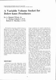
34 G. D. Summers, J. D. Morrison and G. M. Cochrane<br />
pressure patterns of a control population of<br />
similar age and sex free from locomotor<br />
disorders.<br />
Methods<br />
Measurements were carried out using the<br />
double video forceplate (DVF), an instrument<br />
that was developed at the University College<br />
London Bioengineering Centre (Lord and<br />
Smith, 1984). The DVF consists of a low<br />
platform with separate force plates for each foot,<br />
a BBC microcomputer, a video display and a<br />
printer. The patients were asked to stand<br />
'normally' on the foreceplates with hands by the<br />
side and facing forwards for 15 seconds without<br />
using any support after which a time-averaged<br />
display of foot loading was produced. The video<br />
monitor showed the foot outlines and the<br />
percentage of body weight borne on either foot.<br />
A (+) within each foot outine denoted the<br />
ICFPs and a (+) between the foot outlines the<br />
CFP. The screen display was printed out to form<br />
a permanent record.<br />
Patients<br />
One hundred consecutively attending patients<br />
were studied during their routine visits to the<br />
Artificial Limb and Appliance Centre.<br />
All patients were wearing their definitive<br />
prostheses. Only patients with temporary<br />
prostheses and those unable to stand<br />
unsupported for 15 seconds were excluded from<br />
the study. There were 80 male and 20 female<br />
amputees. The mean age of the population was<br />
57 years, with an age range of 10-83 years. More<br />
than half the subjects were over the age of 60<br />
years. Fifty-one patients had amputation of the<br />
right leg, 44 of the left leg and five both legs.<br />
There were 29 above-knee amputees, 56 belowknee<br />
amputees (including four bilateral), four<br />
through-knee amputees, four Gritti-Stokes<br />
amputees, three through-hip amputees and four<br />
Symes amputees (including one bilateral). In the<br />
above knee groups 10 patients wore<br />
endoskeletal limbs and 19 metal limbs. Types of<br />
socket, knee mechanism and prosthetic ankle<br />
are shown in Tables 1 and 2. Thirty-three<br />
unilateral below-knee amputees and three<br />
bilateral below-knee amputees wore patellar<br />
tendon bearing prostheses, whilst 19 unilateral<br />
below-knee amputees and one bilateral belowknee<br />
amputee wore metal limbs with a thigh<br />
corset. Three Gritti-Stokes patients and two<br />
through-knee amputees wore endoskeletal<br />
limbs and the others in these groups wore metal<br />
limbs. All three through-hip patients wore<br />
endoskeletal type tilting table prostheses. The<br />
Symes amputees wore end-bearing prostheses.<br />
Table 2. Types of prosthetic foot and ankle joints in<br />
above and below-knee prostheses<br />
Controls<br />
One hundred age and sex matched individuals<br />
free from any locomotor disability were selected<br />
randomly from members of staff, local clubs and<br />
community centres. None of the subjects used<br />
any walking aids. The mean age of the control<br />
subjects was 56 years. Twelve control subjects<br />
stated they were left handed and 88 that they<br />
were right handed.<br />
Analysis<br />
The position of the ICFP within each foot<br />
outline was expressed as a percentage of foot<br />
length and width (Fig. 1). To make valid<br />
comparisons between individuals, data on the<br />
anteroposterior locations of the ICFPs were<br />
expressed as a percentage of foot length anterior<br />
or posterior to the CFP. When the ICFP was<br />
anterior to the CFP it was arbitrarily assigned a<br />
positive value. A negative value denoted an<br />
ICFP posterior to the CFP.<br />
Table 1. Types of socket and knee joint mechanism in above-knee amputees