

download full issue - Our Dermatology Online Journal
download full issue - Our Dermatology Online Journal
download full issue - Our Dermatology Online Journal

Create successful ePaper yourself
Turn your PDF publications into a flip-book with our unique Google optimized e-Paper software.
was no history suggestive of any constitutional<br />
symptoms, tingling and numbness elsewhere over the<br />
body. There was no history of seizures, deafness, tinnitus<br />
or any other neurological deficit. The past medical and<br />
surgical histories were unremarkable.There was no<br />
history of similar disease in any of the family members<br />
or first degree relatives. There was no known evidence of<br />
any hereditary disease in the family or first degree<br />
relatives.<br />
General physical examination revealed an averagely built<br />
adult female with a steady gait, and satisfactory vital<br />
signs with no signs of pallor, icterus, cyanosis or<br />
lymphadenopathy. Systemic examination was also noncontributory.<br />
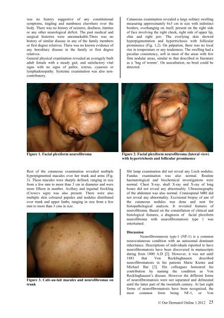
Cutaneous examination revealed a large solitary swelling<br />
measuring approximately 6x5 cm in size with indistinct<br />
borders, overhanging on itself, present on the right side<br />
of face involving the right cheek, right side of upper lip,<br />
chin and right jaw. The overlying skin showed<br />
hyperpigmentation and hypertrichosis with follicular<br />
prominence (Fig. 1,2). On palpation, there was no local<br />
rise in temperature or any tenderness. The swelling had a<br />
peculiar consistency, soft in most of the areas with few<br />
firm nodular areas, similar to that described in literature<br />
as a ‘bag of worms’. On auscultation, no bruit could be<br />
detected.<br />
Figure 1. Facial plexiform neurofibroma<br />
Figure 2. Facial plexiform neurofibroma (lateral view)<br />
with hypertrichosis and follicular prominence<br />
Rest of the cutaneous examination revealed multiple<br />
hyperpigmented macules over her trunk and arms (Fig.<br />
3). These macules were sharply defined, ranging in size<br />
from a few mm to more than 3 cm in diameter and were<br />
more fifteen in number. Axillary and inguinal freckling<br />
(Crowe , s sign) was also present. There were also<br />
multiple skin coloured papules and nodules distributed<br />
over trunk and upper limbs, ranging in size from a few<br />
mm to more than 3 cms in size.<br />
Figure 3. Cafe-au-lait macules and neurofibromas on<br />
trunk<br />
Slit lamp examination did not reveal any Lisch nodules.<br />
Fundus examination was also normal. Routine<br />
haematological and biochemical investigations were<br />
normal. Chest X-ray, skull X-ray and X-ray of long<br />
bones did not reveal any abnormality. Ultrasonography<br />
of the abdomen was also normal. Craniospinal MRI did<br />
not reveal any abnormality. Excisional biopsy of ane of<br />
the cutaneous nodules was done and sent for<br />
histopathological analysis. It revealed features of<br />
neurofibroma. Based on the constellation of clinical and<br />
histological features, a diagnosis of facial plexiform<br />
neurofibroma with neurofibromatosis type 1 was<br />
entertained.<br />
Discussion<br />
Neurofibromatosis type-1 (NF-1) is a common<br />
neurocutaneous condition with an autosomal dominant<br />
inheritance. Descriptions of individuals reported to have<br />
neurofibromatosis have been discovered in manuscripts<br />
dating from 1000 A.D [2]. However, it was not until<br />
1881 that Von Recklinghausen described<br />
neurofibromatosis in his patients Marie Kientz and<br />
Michael Bar [3]. His colleagues honoured his<br />
contribution by naming the condition as Von<br />
Recklinghausen’s disease. However the different forms<br />
of neurofibromatosis were not separated and delineated<br />
until the latter part of the twentieth century. At last eight<br />
forms of neurofibromatosis have been recognized, the<br />
most common form being NF-1, or Von<br />
© <strong>Our</strong> Dermatol <strong>Online</strong> 1.2012<br />
25


