

download full issue - Our Dermatology Online Journal
download full issue - Our Dermatology Online Journal
download full issue - Our Dermatology Online Journal

You also want an ePaper? Increase the reach of your titles
YUMPU automatically turns print PDFs into web optimized ePapers that Google loves.
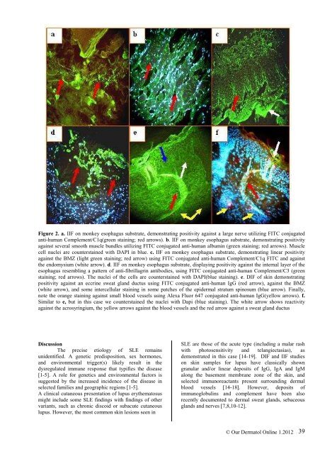
Figure 2. a. IIF on monkey esophagus substrate, demonstrating positivity against a large nerve utilizing FITC conjugated<br />
anti-human Complement/C1q(green staining; red arrows). b. IIF on monkey esophagus substrate, demonstrating positivity<br />
against several smooth muscle bundles utilizing FITC conjugated anti-human albumin (green staining; red arrows). Muscle<br />
cell nuclei are counterstained with DAPI in blue. c. IIF on monkey esophagus substrate, demonstrating linear positivity<br />
against the BMZ (light green staining; red arrow) using FITC conjugated anti-human Complement/C1q FITC and against<br />
the endomysium (white arrow). d. IIF on monkey esophagus substrate, displaying positivity against the internal layer of the<br />
esophagus resembling a pattern of anti-fibrillagrin antibodies, using FITC conjugated anti-human Complement/C3 (green<br />
staining; red arrows). The nuclei of the cells are counterstained with DAPI(blue staining). e. DIF of skin demonstrating<br />
positivity against an eccrine sweat gland ductus using FITC conjugated anti-human IgG (red arrow), against the BMZ<br />
(white arrow), and some intercellular staining in some patches of the epidermal stratum spinosum (blue arrow). Finally,<br />
note the orange staining against small blood vessels using Alexa Fluor 647 conjugated anti-human IgG(yellow arrows). f.<br />
Similar to e, but in this case we counterstained the nuclei with Dapi (blue staining). The white arrow shows reactivity<br />
against the acrosyringium, the yellow arrows against the blood vessels and the red arrow against a sweat gland ductus<br />
Discussion<br />
The precise etiology of SLE remains<br />
unidentified. A genetic predisposition, sex hormones,<br />
and environmental trigger(s) likely result in the<br />
dysregulated immune response that typifies the disease<br />
[1-5]. A role for genetics and environmental factors is<br />
suggested by the increased incidence of the disease in<br />
selected families and geographic regions [1-5].<br />
A clinical cutaneous presentation of lupus erythematosus<br />
might include some SLE findings with findings of other<br />
variants, such as chronic discoid or subacute cutaneous<br />
lupus. However, the most common skin lesions seen in<br />
SLE are those of the acute type (including a malar rash<br />
with photosensitivity and telangiectasias), as<br />
demonstrated in this case [14-19]. DIF and IIF studies<br />
on skin samples for lupus have classically shown<br />
granular and/or linear deposits of IgG, IgA and IgM<br />
along the basement membrane zone of the skin, and<br />
selected immunoreactants present surrounding dermal<br />
blood vessels [14-18]. However, deposits of<br />
immunoglobulins and complement have been also<br />
recently documented to dermal sweat glands, sebaceous<br />
glands and nerves [7,8,10-12].<br />
© <strong>Our</strong> Dermatol <strong>Online</strong> 1.2012<br />
39


