MHC class II transactivator CIITA is a recurrent ... - Ohio University
MHC class II transactivator CIITA is a recurrent ... - Ohio University
MHC class II transactivator CIITA is a recurrent ... - Ohio University

Create successful ePaper yourself
Turn your PDF publications into a flip-book with our unique Google optimized e-Paper software.
RESEARCH<br />
LETTER<br />
Next, we validated three fusion predictions by direct sequencing and<br />
fluorescence in situ hybridization (FISH): the first involving the genes<br />
BAT2L1 (chromosome 9q34.13) and MGMT (chromosome 10q26.3) in<br />
KM-H2 (Supplementary Fig. 2), the second involving the genes<br />
SLCO3A1 (chromosome 15q26.1) and ELMO1 (chromosome 7p14.1–<br />
14.2) in L428 (Supplementary Fig. 2), and lastly the previously mentioned<br />
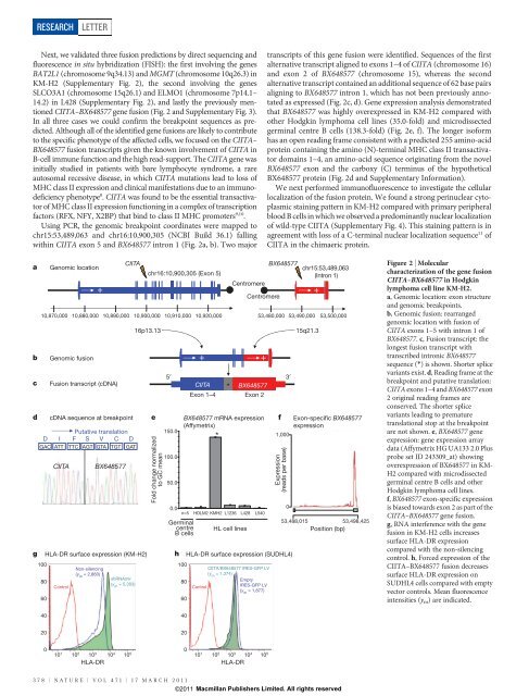
C<strong>II</strong>TA–BX648577 gene fusion (Fig. 2 and Supplementary Fig. 3).<br />
In all three cases we could confirm the breakpoint sequences as predicted.<br />
Although all of the identified gene fusions are likely to contribute<br />
to the specific phenotype of the affected cells, we focused on the C<strong>II</strong>TA–<br />
BX648577 fusion transcripts given the known involvement of C<strong>II</strong>TA in<br />
B-cell immune function and the high read-support. The C<strong>II</strong>TA gene was<br />
initially studied in patients with bare lymphocyte syndrome, a rare<br />
autosomal recessive d<strong>is</strong>ease, in which C<strong>II</strong>TA mutations lead to loss of<br />
<strong>MHC</strong> <strong>class</strong> <strong>II</strong> expression and clinical manifestations due to an immunodeficiency<br />
phenotype 8 . C<strong>II</strong>TA was found to be the essential <strong>transactivator</strong><br />
of <strong>MHC</strong> <strong>class</strong> <strong>II</strong> expression functioning in a complex of transcription<br />
factors (RFX, NFY, X2BP) that bind to <strong>class</strong> <strong>II</strong> <strong>MHC</strong> promoters 9,10 .<br />
Using PCR, the genomic breakpoint coordinates were mapped to<br />
chr15:53,489,063 and chr16:10,900,305 (NCBI Build 36.1) falling<br />
within C<strong>II</strong>TA exon 5 and BX648577 intron 1 (Fig. 2a, b). Two major<br />
transcripts of th<strong>is</strong> gene fusion were identified. Sequences of the first<br />
alternative transcript aligned to exons 1–4 of C<strong>II</strong>TA (chromosome 16)<br />
and exon 2 of BX648577 (chromosome 15), whereas the second<br />
alternative transcript contained an additional sequence of 62 base pairs<br />
aligning to BX648577 intron 1, which has not been previously annotated<br />
as expressed (Fig. 2c, d). Gene expression analys<strong>is</strong> demonstrated<br />
that BX648577 was highly overexpressed in KM-H2 compared with<br />
other Hodgkin lymphoma cell lines (35.0-fold) and microd<strong>is</strong>sected<br />
germinal centre B cells (138.3-fold) (Fig. 2e, f). The longer <strong>is</strong>oform<br />
has an open reading frame cons<strong>is</strong>tent with a predicted 255 amino-acid<br />
protein containing the amino (N)-terminal <strong>MHC</strong> <strong>class</strong> <strong>II</strong> <strong>transactivator</strong><br />
domains 1–4, an amino-acid sequence originating from the novel<br />
BX648577 exon and the carboxy (C) terminus of the hypothetical<br />
BX648577 protein (Fig. 2d and Supplementary Information).<br />
We next performed immunofluorescence to investigate the cellular<br />
localization of the fusion protein. We found a strong perinuclear cytoplasmic<br />
staining pattern in KM-H2 compared with primary peripheral<br />
blood B cells in which we observed a predominantly nuclear localization<br />
of wild-type C<strong>II</strong>TA (Supplementary Fig. 4). Th<strong>is</strong> staining pattern <strong>is</strong> in<br />
agreement with loss of a C-terminal nuclear localization sequence 11 of<br />
C<strong>II</strong>TA in the chimaeric protein.<br />
a<br />
b<br />
c<br />
d<br />
Genomic location<br />
C<strong>II</strong>TA<br />
+ +<br />
10,870,000 10,880,000 10,890,000 10,900,000 10,910,000 10,920,000 53,480,000 53,490,000 53,500,000<br />
Genomic fusion<br />
Fusion transcript (cDNA)<br />
cDNA sequence at breakpoint<br />
Putative translation<br />
D I F S V C D<br />
GAC ATT TTC AGT GTA TGT GAT<br />
16p13.13<br />
g HLA-DR surface expression (KM-H2)<br />
100<br />
Non-silencing<br />
(y m = 2,860)<br />
shRNAmir<br />
80<br />
(y<br />
Control<br />
m = 5,303)<br />
60<br />
C<strong>II</strong>TA<br />
BX648577<br />
chr16:10,900,305 (Exon 5)<br />
e<br />
Fold change normalized<br />
to GC mean<br />
5′<br />
150.0<br />
100.0<br />
50.0<br />
+ +<br />
*<br />
C<strong>II</strong>TA BX648577<br />
Exon 1–4 Exon 2<br />
BX648577 mRNA expression<br />
(Affymetrix)<br />
*<br />
Centromere<br />
Centromere<br />
0.0<br />
GC<br />
n=5 HDLM2 KMH2 L1236 L428 L540<br />
Germinal<br />
centre<br />
B cells<br />
HL cell lines<br />
BX648577<br />
h HLA-DR surface expression (SUDHL4)<br />
100<br />
C<strong>II</strong>TA/BX648577 IRES-GFP LV<br />
(y m = 1,374)<br />
80<br />
Empty<br />
Control<br />
IRES-GFP LV<br />
(y m = 1,877)<br />
60<br />
3′<br />
chr15:53,489,063<br />
(Intron 1)<br />
15q21.3<br />
f Exon-specific BX648577<br />
expression<br />
1,000<br />
Expression<br />
(reads per base)<br />
0<br />
53,488,015<br />
53,498,425<br />
Position (bp)<br />
Figure 2 | Molecular<br />
characterization of the gene fusion<br />
C<strong>II</strong>TA–BX648577 in Hodgkin<br />
lymphoma cell line KM-H2.<br />
a, Genomic location: exon structure<br />
and genomic breakpoints.<br />
b, Genomic fusion: rearranged<br />
genomic location with fusion of<br />
C<strong>II</strong>TA exons 1–5 with intron 1 of<br />
BX648577. c, Fusion transcript: the<br />
longest fusion transcript with<br />
transcribed intronic BX648577<br />
sequence (*) <strong>is</strong> shown. Shorter splice<br />
variants ex<strong>is</strong>t. d, Reading frame at the<br />
breakpoint and putative translation:<br />
C<strong>II</strong>TA exons 1–4 and BX648577 exon<br />
2 original reading frames are<br />
conserved. The shorter splice<br />
variants leading to premature<br />
translational stop at the breakpoint<br />
are not shown. e, BX648577 gene<br />
expression: gene expression array<br />
data (Affymetrix HG UA133 2.0 Plus<br />
probe set ID 243309_at) showing<br />
overexpression of BX648577 in KM-<br />
H2 compared with microd<strong>is</strong>sected<br />
germinal centre B cells and other<br />
Hodgkin lymphoma cell lines.<br />
f, BX648577 exon-specific expression<br />
<strong>is</strong> biased towards exon 2 as part of the<br />
C<strong>II</strong>TA–BX648577 gene fusion.<br />
g, RNA interference with the gene<br />
fusion in KM-H2 cells increases<br />
surface HLA-DR expression<br />
compared with the non-silencing<br />
control. h, Forced expression of the<br />
C<strong>II</strong>TA–BX648577 fusion decreases<br />
surface HLA-DR expression on<br />
SUDHL4 cells compared with empty<br />
vector controls. Mean fluorescence<br />
intensities (y m ) are indicated.<br />
40<br />
40<br />
20<br />
20<br />
0<br />
0<br />
10 1 10 2 10 3 10 4 10 5 10 1 10 2 10 3 10 4 10 5<br />
HLA-DR<br />
HLA-DR<br />
378 | NATURE | VOL 471 | 17 MARCH 2011<br />
©2011 Macmillan Publ<strong>is</strong>hers Limited. All rights reserved