MHC class II transactivator CIITA is a recurrent ... - Ohio University
MHC class II transactivator CIITA is a recurrent ... - Ohio University
MHC class II transactivator CIITA is a recurrent ... - Ohio University

You also want an ePaper? Increase the reach of your titles
YUMPU automatically turns print PDFs into web optimized ePapers that Google loves.
LETTER<br />
doi:10.1038/nature09754<br />
<strong>MHC</strong> <strong>class</strong> <strong>II</strong> <strong>transactivator</strong> C<strong>II</strong>TA <strong>is</strong> a <strong>recurrent</strong> gene<br />
fusion partner in lymphoid cancers<br />
Chr<strong>is</strong>tian Steidl 1 *, Sohrab P. Shah 1 *, Bruce W. Woolcock 1 , Lixin Rui 2 , Masahiro Kawahara 3 , Pedro Farinha 1 , Nathalie A. Johnson 1 ,<br />
Yongjun Zhao 4 , Adele Telenius 1 , Susana Ben Neriah 1 , Andrew McPherson 1 , Barbara Me<strong>is</strong>sner 1 , Ujunwa C. Okoye 3 , Arjan Diepstra 5 ,<br />
Anke van den Berg 5 , Mark Sun 1 , Gillian Leung 1 , Steven J. Jones 4 , Joseph M. Connors 6 , David G. Huntsman 1 , Kerry J. Savage 6 ,<br />
L<strong>is</strong>a M. Rimsza 7 , Douglas E. Horsman 1 , Lou<strong>is</strong> M. Staudt 2 , Ulrich Steidl 3 , Marco A. Marra 4,8 & Randy D. Gascoyne 1<br />
Chromosomal translocations are critically involved in the molecular<br />
pathogenes<strong>is</strong> of B-cell lymphomas, and highly <strong>recurrent</strong> and specific<br />
rearrangements have defined d<strong>is</strong>tinct molecular subtypes linked to<br />
unique clinicopathological features 1,2 . In contrast, several wellcharacterized<br />
lymphoma entities still lack d<strong>is</strong>ease-defining translocation<br />
events. To identify novel fusion transcripts resulting from<br />
translocations, we investigated two Hodgkin lymphoma cell lines by<br />
whole-transcriptome paired-end sequencing (RNA-seq). Here we<br />
show a highly expressed gene fusion involving the major h<strong>is</strong>tocompatibility<br />
complex (<strong>MHC</strong>) <strong>class</strong> <strong>II</strong> <strong>transactivator</strong> C<strong>II</strong>TA (<strong>MHC</strong>2TA)<br />
in KM-H2 cells. In a subsequent evaluation of 263 B-cell lymphomas,<br />
we also demonstrate that genomic C<strong>II</strong>TA breaks are highly<br />
<strong>recurrent</strong> in primary mediastinal B-cell lymphoma (38%) and<br />
<strong>class</strong>ical Hodgkin lymphoma (cHL) (15%). Furthermore, we find<br />
that C<strong>II</strong>TA <strong>is</strong> a prom<strong>is</strong>cuous partner of various in-frame gene<br />
fusions, and we report that C<strong>II</strong>TA gene alterations impact survival<br />
in primary mediastinal B-cell lymphoma (PMBCL). As functional<br />
consequences of C<strong>II</strong>TA gene fusions, we identify downregulation of<br />
surface HLA <strong>class</strong> <strong>II</strong> expression and overexpression of ligands of the<br />
receptor molecule programmed cell death 1 (CD274/PDL1 and<br />
CD273/PDL2). These receptor–ligand interactions have been shown<br />
to impact anti-tumour immune responses in several cancers 3 ,<br />
whereas decreased <strong>MHC</strong> <strong>class</strong> <strong>II</strong> expression has been linked to<br />
reduced tumour cell immunogenicity 4 . Thus, our findings suggest<br />
that <strong>recurrent</strong> rearrangements of C<strong>II</strong>TA mayrepresentanovel<br />
genetic mechan<strong>is</strong>m underlying tumour–microenvironment interactions<br />
across a spectrum of lymphoid cancers.<br />
In Hodgkin lymphoma, translocations or chromosomal breakpoints<br />
have only rarely been described, whereas in PMBCL no <strong>recurrent</strong><br />
translocation events have been reported 5 . Massively parallel,<br />
paired-end sequencing of expressed transcripts (RNA-seq) provides<br />
an analytical platform suitable for genome-wide mapping of translocation<br />
breakpoints, sequence variants and quantitative expression 6,7 .<br />
Thus, we used th<strong>is</strong> technology to detect novel gene fusions in the<br />
two Hodgkin lymphoma cell lines KM-H2 and L428, including<br />
82.9 million paired-end reads for KM-H2, of which 71.1 million<br />
mapped to the reference human genome (86%, 3.6 gigabases), and<br />
61.5 million paired-end reads for L428, of which 55.5 million (90%,<br />
2.8 gigabases) mapped to the reference genome (Supplementary Fig.<br />
1). Using a novel gene-fusion d<strong>is</strong>covery method, we obtained 14 d<strong>is</strong>tinct<br />
fusion transcript predictions for KM-H2 and five for L428<br />
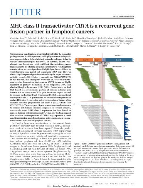
(Supplementary Table 1). Figure 1 shows the fusion prediction with<br />
the highest read support involving C<strong>II</strong>TA and an uncharacterized gene<br />
BX648577 (FLJ27352/hypothetical LOC145788) found in KM-H2.<br />
10896786<br />
10897027<br />
C<strong>II</strong>TA<br />
Exon 3 Exon 4 Exon 2<br />
Chromosome 16<br />
10897122 10900090 53497713<br />
10900028<br />
53497249<br />
53497188<br />
BX648577<br />
Chromosome 15<br />
5′ 3′<br />
GGAACTGGACCAGTATGTCTTCCAGGACTCCCAGCTGGAGGGCCTGAGCAAGGACATTTTCA|GTGTATGTGATTTTAAGGGTAGTCTTCAACGTCAAGAGAAGAGACCACCAAACAGGCTTTGTGATAA<br />
TATGTCTTCCAGGACTCCCAGCTGGAGGGCCTGAGCAAGGACATTTTCA|G<br />
ATGTCTTCCAGGACTCCCAGCTGGAGGGCCTGAGCAAGGACATTTTCA|GT<br />
TGTCTTCCAGGACTCCCAGCTGGAGGGCCTGAGCAAGGACATTTTCA|GTG<br />
GTCTTCCAGGAATCCCAGCTGGAGGGCCTGAGCAAGGACATTTTCA|GTGT<br />
TCTTCCAGGACTCCCAGCTGGAGGGCCTGAGCAAGGACATTTTCA|GTGTA<br />
CTTCCAGGACTCCCAGCTGGAGGGCCTGAGCAAGGACACTTTCA|GTGTAT<br />
TTCCAGGACTCCCAGCTGGAGGGCCTGAGCAAGGACATTTTCA|GTGTATG<br />
TCCAGGACTCCCAGCTGGAGGGCCTGAGCAAGGTCATTTTCA|GATAAGAA<br />
CCAGGACTCCCAGCTGGAGGGCCTGAGCAAGGACATTTTCA|GTGTATGTG<br />
CAGGACTCCCAGCTGGAGGGCCTGAGCAAGGACATTTTCA|GTGTATGTGA<br />
AGGACTCCCAGCTGGAGGGCCTGAGCAAGGACATTTTCA|GTGTATGTGAT<br />
GGACTCCCAGCTGGAGGGCCTGAGCAAGGACATTTTCA|GTGTATGTGATT<br />
GACTCCCAGCTGGAGGGCCTGAGCAAGGACATTTTCA|GTGTATGTGATTT<br />
ACTCCCAGATGGAGGGCCTGAGCAAGGACATTTTCA|GTGTATGTGAATTT<br />
CTCCCAGCTGGAGGGCCTGAGCAAGGACATTTTCA|GTGTATGTGATTTTA<br />
TCCCAGCTGGAGGGCCTGAGCAAGGACATTTTCA|GTGTATGTGATTTTAA<br />
CCCAGCTGGAGGGCCTGAGCAAGGACATTTTCA|GTGTATGTGATTTTAAG<br />
CCAGCTGGACGGCCTGAGCAAGGACATTTTCA|GTGTATGTGATTTTAAGG<br />
CAGCTGGAGGGCCTGAGCAAGGACATTTTCA|GTGTATGTGATTTTAAGGG<br />
AGCTGGAGGGCCTGAGCAAGGACATTTTCA|GTGTATGTGATTTTAAGGGT<br />
GCTGGAGGGCCTGAGCAAGGACATTTTCA|GTGTATGTGATTTTAAGGGTA<br />
CTGGAGGGCCTGAGCAAGGACATTTTCA|GTGTATGTGATTTTAAGGGTAG<br />
TGGAGGGCCTGAGCAAGGTCATTGTCA|GTGGAGGTGAGTTTAAGGGTAGT<br />
GGAGGGCCTGAGCAAGGACATTTTCA|GTGTATGTGATTTTAAGGGTAGTC<br />
GAGGGCCTGAGCAAGGACATTTTCA|GTGTATGTGATTTTAAGGGTAGTCT<br />
AGGGCCTGAGCAAGGACATTTTCA|GTGTATGTGATTTTAAGGGTAGTCTT<br />
GGGCCTGAGCAAGGACATTTTCA|GTGTATGTGATTTTAAGGGTAGTCTTC<br />
GGCCTGAGCAAGGACATTTTCA|GTGTATGTGATTTTAAGGGTAGTCTTCA<br />
GCCTGAGCAAGGACATTTTCA|GTGTATGTGATTTTAAGGGTAGTCTTCAA<br />
CTGAGCAAGGACATTTTCA|GTGTATGTGATTTTAAGGGTAGTCTTCAACG<br />
TGAGCAAGGACATTTTCA|GTGTATGTGATTTTAAGGGTAGTCTTCAACGT<br />
GAGCAAGGACATTTTCA|GTGTATGTGATTTTAAGGGTAGTCTTCAACGTC<br />
AGCAAGGACATTTTCA|GTGTATGTGATTTTAAGGGTAGTCTTCAACGTCA<br />
GCAAGGACATTTTCA|GTGTATGTGATTTTAAGGGTAGTCTTCAACGTCAA<br />
CAAGGACATTTTCA|GTGTATGTGATTTTAAGGGTAGTCTTCAACGTCAAG<br />
AAGGACATTTTCA|GTGTATGTGATTTTAAGGGCAGTCTTCAACGTCAAGA<br />
AGGACATTTTCA|GTGTATGTGATTTTAATGGTAGTCTTCAACGTCAAGAG<br />
GGACATTTTCA|GTGTATGTGATTTTAAGGGTAGTCTTCAACGTCAAGAGA<br />
GAGATTTTCA|GTGTATGTGATTTTAAGGGTAGTCTTCAGCGTCAAGAGAA<br />
ACATTTTCA|GTGTATGTGATTTTAAGGGTAGTCTTCAACGTCAAGAGAAG<br />
CATTTTCA|GTGTATGTGATTTTAAGGGTAGTCTTCAACGTCAAGAGAAGA<br />
ATTTTCA|GTGTATGTGATTTTAAGGGTAGTCTTCAACGTCAAGAGAAGAG<br />
TTTTCA|GTGTATGTGATTTTAAGGGTAGTCTTCAACGTCAAGAGAAGAGA<br />
TTTCA|GTGTATGTGATTTTAAGGGTAGTCTTCAACGTCAAGAGAAGAGAC<br />
TCA|GCGTATGTGATTTTAAGGGTAGTCTTCAACGTCAAGAGAAGAGACCA<br />
CA|GTGTATGTGATTTTAAGGGTAGTCTTCAACGTCAAGAGAAGAGACCAC<br />
A|GTGTATGTGATTTTAAGGGTAGTCTTCAACGTCAAGAGAAGAGACCACC<br />
0 2 4 6 8 10<br />
Figure 1 | C<strong>II</strong>TA–BX648577 gene fusion observed using paired-end<br />
massively parallel whole transcriptome sequencing. In the upper panel, 468<br />
mate-pair sequences are shown aligning on either side of the breakpoint<br />
(pairing C<strong>II</strong>TA and BX648577). The genomic coordinates of the exon<br />
boundaries are given. In light blue, the transcribed intronic BX648577 sequence<br />
<strong>is</strong> shown as part of a transcript variant resulting from the fusion. In the lower<br />
panel, 191 split-reads are depicted lying on the breakpoint (in blue: merged<br />
reference sequence of C<strong>II</strong>TA and BX648577). The h<strong>is</strong>togram on the right<br />
describes the absolute frequency of each sequence read spanning the<br />
breakpoint.<br />
1 Department of Pathology and Laboratory Medicine, Centre for Lymphoid Cancers and the Centre for Translational and Applied Genomics (CTAG), Vancouver, Brit<strong>is</strong>h Columbia, V5Z4E6, Canada.<br />
2 Metabol<strong>is</strong>m Branch, Center for Cancer Research, National Cancer Institute, Bethesda, Maryland, 20892, USA. 3 Department of Cell Biology and Albert Einstein Cancer Center, Albert Einstein College of<br />
Medicine, Bronx, New York, 10461, USA. 4 Genome Sciences Centre, BC Cancer Agency, Vancouver, Brit<strong>is</strong>h Columbia, V5Z4S6, Canada. 5 Department of Pathology and Medical Biology, <strong>University</strong> Medical<br />
Center Groningen, <strong>University</strong> of Groningen, 9700, The Netherlands. 6 Div<strong>is</strong>ion of Medical Oncology, BC Cancer Agency Centre for Lymphoid Cancer, Vancouver, Brit<strong>is</strong>h Columbia, V5Z4E6, Canada.<br />
7 Department of Pathology, <strong>University</strong> of Arizona, Tucson, Arizona, 85724, USA. 8 Department of Medical Genetics, <strong>University</strong> of Brit<strong>is</strong>h Columbia, Vancouver, Brit<strong>is</strong>h Columbia, V6T1Z3, Canada.<br />
*These authors contributed equally to th<strong>is</strong> work.<br />
17 MARCH 2011 | VOL 471 | NATURE | 377<br />
©2011 Macmillan Publ<strong>is</strong>hers Limited. All rights reserved
RESEARCH<br />
LETTER<br />
Next, we validated three fusion predictions by direct sequencing and<br />
fluorescence in situ hybridization (FISH): the first involving the genes<br />
BAT2L1 (chromosome 9q34.13) and MGMT (chromosome 10q26.3) in<br />
KM-H2 (Supplementary Fig. 2), the second involving the genes<br />
SLCO3A1 (chromosome 15q26.1) and ELMO1 (chromosome 7p14.1–<br />
14.2) in L428 (Supplementary Fig. 2), and lastly the previously mentioned<br />
C<strong>II</strong>TA–BX648577 gene fusion (Fig. 2 and Supplementary Fig. 3).<br />
In all three cases we could confirm the breakpoint sequences as predicted.<br />
Although all of the identified gene fusions are likely to contribute<br />
to the specific phenotype of the affected cells, we focused on the C<strong>II</strong>TA–<br />
BX648577 fusion transcripts given the known involvement of C<strong>II</strong>TA in<br />
B-cell immune function and the high read-support. The C<strong>II</strong>TA gene was<br />
initially studied in patients with bare lymphocyte syndrome, a rare<br />
autosomal recessive d<strong>is</strong>ease, in which C<strong>II</strong>TA mutations lead to loss of<br />
<strong>MHC</strong> <strong>class</strong> <strong>II</strong> expression and clinical manifestations due to an immunodeficiency<br />
phenotype 8 . C<strong>II</strong>TA was found to be the essential <strong>transactivator</strong><br />
of <strong>MHC</strong> <strong>class</strong> <strong>II</strong> expression functioning in a complex of transcription<br />
factors (RFX, NFY, X2BP) that bind to <strong>class</strong> <strong>II</strong> <strong>MHC</strong> promoters 9,10 .<br />
Using PCR, the genomic breakpoint coordinates were mapped to<br />
chr15:53,489,063 and chr16:10,900,305 (NCBI Build 36.1) falling<br />
within C<strong>II</strong>TA exon 5 and BX648577 intron 1 (Fig. 2a, b). Two major<br />
transcripts of th<strong>is</strong> gene fusion were identified. Sequences of the first<br />
alternative transcript aligned to exons 1–4 of C<strong>II</strong>TA (chromosome 16)<br />
and exon 2 of BX648577 (chromosome 15), whereas the second<br />
alternative transcript contained an additional sequence of 62 base pairs<br />
aligning to BX648577 intron 1, which has not been previously annotated<br />
as expressed (Fig. 2c, d). Gene expression analys<strong>is</strong> demonstrated<br />
that BX648577 was highly overexpressed in KM-H2 compared with<br />
other Hodgkin lymphoma cell lines (35.0-fold) and microd<strong>is</strong>sected<br />
germinal centre B cells (138.3-fold) (Fig. 2e, f). The longer <strong>is</strong>oform<br />
has an open reading frame cons<strong>is</strong>tent with a predicted 255 amino-acid<br />
protein containing the amino (N)-terminal <strong>MHC</strong> <strong>class</strong> <strong>II</strong> <strong>transactivator</strong><br />
domains 1–4, an amino-acid sequence originating from the novel<br />
BX648577 exon and the carboxy (C) terminus of the hypothetical<br />
BX648577 protein (Fig. 2d and Supplementary Information).<br />
We next performed immunofluorescence to investigate the cellular<br />
localization of the fusion protein. We found a strong perinuclear cytoplasmic<br />
staining pattern in KM-H2 compared with primary peripheral<br />
blood B cells in which we observed a predominantly nuclear localization<br />
of wild-type C<strong>II</strong>TA (Supplementary Fig. 4). Th<strong>is</strong> staining pattern <strong>is</strong> in<br />
agreement with loss of a C-terminal nuclear localization sequence 11 of<br />
C<strong>II</strong>TA in the chimaeric protein.<br />
a<br />
b<br />
c<br />
d<br />
Genomic location<br />
C<strong>II</strong>TA<br />
+ +<br />
10,870,000 10,880,000 10,890,000 10,900,000 10,910,000 10,920,000 53,480,000 53,490,000 53,500,000<br />
Genomic fusion<br />
Fusion transcript (cDNA)<br />
cDNA sequence at breakpoint<br />
Putative translation<br />
D I F S V C D<br />
GAC ATT TTC AGT GTA TGT GAT<br />
16p13.13<br />
g HLA-DR surface expression (KM-H2)<br />
100<br />
Non-silencing<br />
(y m = 2,860)<br />
shRNAmir<br />
80<br />
(y<br />
Control<br />
m = 5,303)<br />
60<br />
C<strong>II</strong>TA<br />
BX648577<br />
chr16:10,900,305 (Exon 5)<br />
e<br />
Fold change normalized<br />
to GC mean<br />
5′<br />
150.0<br />
100.0<br />
50.0<br />
+ +<br />
*<br />
C<strong>II</strong>TA BX648577<br />
Exon 1–4 Exon 2<br />
BX648577 mRNA expression<br />
(Affymetrix)<br />
*<br />
Centromere<br />
Centromere<br />
0.0<br />
GC<br />
n=5 HDLM2 KMH2 L1236 L428 L540<br />
Germinal<br />
centre<br />
B cells<br />
HL cell lines<br />
BX648577<br />
h HLA-DR surface expression (SUDHL4)<br />
100<br />
C<strong>II</strong>TA/BX648577 IRES-GFP LV<br />
(y m = 1,374)<br />
80<br />
Empty<br />
Control<br />
IRES-GFP LV<br />
(y m = 1,877)<br />
60<br />
3′<br />
chr15:53,489,063<br />
(Intron 1)<br />
15q21.3<br />
f Exon-specific BX648577<br />
expression<br />
1,000<br />
Expression<br />
(reads per base)<br />
0<br />
53,488,015<br />
53,498,425<br />
Position (bp)<br />
Figure 2 | Molecular<br />
characterization of the gene fusion<br />
C<strong>II</strong>TA–BX648577 in Hodgkin<br />
lymphoma cell line KM-H2.<br />
a, Genomic location: exon structure<br />
and genomic breakpoints.<br />
b, Genomic fusion: rearranged<br />
genomic location with fusion of<br />
C<strong>II</strong>TA exons 1–5 with intron 1 of<br />
BX648577. c, Fusion transcript: the<br />
longest fusion transcript with<br />
transcribed intronic BX648577<br />
sequence (*) <strong>is</strong> shown. Shorter splice<br />
variants ex<strong>is</strong>t. d, Reading frame at the<br />
breakpoint and putative translation:<br />
C<strong>II</strong>TA exons 1–4 and BX648577 exon<br />
2 original reading frames are<br />
conserved. The shorter splice<br />
variants leading to premature<br />
translational stop at the breakpoint<br />
are not shown. e, BX648577 gene<br />
expression: gene expression array<br />
data (Affymetrix HG UA133 2.0 Plus<br />
probe set ID 243309_at) showing<br />
overexpression of BX648577 in KM-<br />
H2 compared with microd<strong>is</strong>sected<br />
germinal centre B cells and other<br />
Hodgkin lymphoma cell lines.<br />
f, BX648577 exon-specific expression<br />
<strong>is</strong> biased towards exon 2 as part of the<br />
C<strong>II</strong>TA–BX648577 gene fusion.<br />
g, RNA interference with the gene<br />
fusion in KM-H2 cells increases<br />
surface HLA-DR expression<br />
compared with the non-silencing<br />
control. h, Forced expression of the<br />
C<strong>II</strong>TA–BX648577 fusion decreases<br />
surface HLA-DR expression on<br />
SUDHL4 cells compared with empty<br />
vector controls. Mean fluorescence<br />
intensities (y m ) are indicated.<br />
40<br />
40<br />
20<br />
20<br />
0<br />
0<br />
10 1 10 2 10 3 10 4 10 5 10 1 10 2 10 3 10 4 10 5<br />
HLA-DR<br />
HLA-DR<br />
378 | NATURE | VOL 471 | 17 MARCH 2011<br />
©2011 Macmillan Publ<strong>is</strong>hers Limited. All rights reserved
LETTER<br />
RESEARCH<br />
Using high-resolution single nucleotide polymorph<strong>is</strong>m analys<strong>is</strong>, we<br />
found that the rearrangement involving chromosomes 15 and 16 was<br />
associated with relative genomic imbalances at the breakpoints (Supplementary<br />
Fig. 5). Multicolour-FISH in representative metaphases of<br />
KM-H2 and interphase-FISH showed complex chromosomal rearrangements<br />
with various marker chromosomes and subclonal variation as<br />
shown by others for th<strong>is</strong> cell line 12 (Supplementary Fig. 6). Locusspecific<br />
FISH using Fosmid probes for C<strong>II</strong>TA and BX648577 confirmed<br />
co-localization of the C<strong>II</strong>TA and BX648577 gene loci on two derivative<br />
chromosomes 16, both harbouring insertions of chromosome 15<br />
material. These complex rearrangements are likely to place the fusion<br />
gene in a different positional context that might add to the high promoter<br />
activity of C<strong>II</strong>TA driving expression of the fusion transcripts in<br />
th<strong>is</strong> cell line.<br />
To investigate the functional consequence of the C<strong>II</strong>TA–BX648577<br />
gene fusion in KM-H2 cells, we generated stable KM-H2 fusion transcript<br />
knockdowns by RNA interference. Quantitative reverse transcriptase<br />
PCR (RT–PCR) showed reduction of fusion transcript levels<br />
by 85%, and western blotting demonstrated a marked reduction in<br />
C<strong>II</strong>TA–BX648577 fusion protein compared with non-silencing controls<br />
(Supplementary Fig. 7). As C<strong>II</strong>TA <strong>is</strong> an essential <strong>transactivator</strong> of<br />
<strong>MHC</strong> <strong>class</strong> <strong>II</strong> expression 9 and deletion mutants have been described<br />
that inhibit wild-type C<strong>II</strong>TA function in vitro 13,14 , we studied transcriptional<br />
changes of short hairpin RNA (shRNA)-transduced cells<br />
using gene expression microarrays (Supplementary Table 2). Most<br />
strikingly, overrepresentation analys<strong>is</strong> identified genes of the ‘antigen<br />
presentation pathway’ significantly enriched among the upregulated<br />
genes (P 5 0.03), and flow cytometry confirmed increased surface<br />
expression of HLA-DR in KM-H2 cells with fusion transcript knockdown<br />
(Fig. 2g). We also transduced diffuse large B-cell lymphoma<br />
(DLBCL) cell line SUDHL4 with a lentiviral vector containing the<br />
full-length coding complementary DNA (cDNA) of the C<strong>II</strong>TA–<br />
BX648577 fusion (second alternative transcript), which led to decreased<br />
surface HLA-DR expression (Fig. 2h). These data demonstrate that the<br />
C<strong>II</strong>TA–BX648577 fusion suppresses expression of HLA <strong>class</strong> <strong>II</strong> genes in<br />
a<br />
BAC alignment RP11-109M19<br />
Telomere<br />
b<br />
10,500,000<br />
10,600,000<br />
10,700,000<br />
Breakpoint in KM-H2<br />
10,800,000<br />
C<strong>II</strong>TA<br />
10,900,000<br />
16p13.13<br />
RP11-66H6<br />
11,000,000<br />
11,100,000<br />
Centromere<br />
bp<br />
c<br />
d<br />
Cumulative survival<br />
1.0<br />
0.8<br />
0.6<br />
0.4<br />
0.2<br />
0.0<br />
0<br />
KM-H2 and SUDHL4. Dimin<strong>is</strong>hed expression of HLA <strong>class</strong> <strong>II</strong> in B-cell<br />
lymphomas has been linked to reduced immunogenicity, escape from<br />
immunosurveillance and inferior survival 15,16 .<br />
To determine if breakpoints within C<strong>II</strong>TA are <strong>recurrent</strong> in cases of<br />
primary Hodgkin lymphoma, we used FISH on t<strong>is</strong>sue microarrays and<br />
found eight out of 55 cases (15%) rearranged (Supplementary Table 3).<br />
Representative FISH images of the break-apart assay are shown in Fig. 3,<br />
demonstrating involvement of all cells of the malignant cell compartment<br />
(Hodgkin Reed–Sternberg cells that carry the CD30 antigen). In<br />
four of the eight cases we detected an unbalanced rearrangement.<br />
Because PMBCL shares clinical and biological features with cHL, we<br />
also studied PMBCL and found 29 (38%) of 77 cases positive for<br />
break-apart of the C<strong>II</strong>TA locus. Only four out of 131 studied cases<br />
of DLBCL (3%) showed break-apart of the C<strong>II</strong>TA locus. Hence, the<br />
occurrence of C<strong>II</strong>TA rearrangements in PMBCL and cHL was significantly<br />
higher than in DLBCL (P , 0.0001).<br />
We also studied the correlation of C<strong>II</strong>TA translocation with clinical<br />
outcome in 57 patients with PMBCL who were treated with curative<br />
intent using multi-agent chemotherapy with or without radiotherapy 17 .<br />
The clinical character<strong>is</strong>tics of th<strong>is</strong> cohort are summarized in Supplementary<br />
Table 4. Presence of a C<strong>II</strong>TA rearrangement significantly<br />
correlated with a shorter d<strong>is</strong>ease-specific survival (10-year d<strong>is</strong>easespecific<br />
survival 63.6% compared with 85.0%, P 5 0.044) (Fig. 3d).<br />
Multivariate testing using a Cox regression model including the<br />
International Prognostic Index 18 score and the individual clinical<br />
prognostic factors showed that the presence of C<strong>II</strong>TA rearrangement<br />
had independent prognostic significance (P 5 0.013) for decreased<br />
d<strong>is</strong>ease-specific survival together with elevated LDH serum levels<br />
(P 5 0.001) (Supplementary Table 5).<br />
To determine fusion partners involved in C<strong>II</strong>TA rearrangements, we<br />
used 39 rapid amplification of cDNA ends (RACE) in eight cases of<br />
PMBCL that were positive for C<strong>II</strong>TA translocation by FISH. In total, we<br />
identified five new translocation partners (CD274, CD273, RALGDS,<br />
RUNDC2A and C16ORF75) (Supplementary Table 6). Prec<strong>is</strong>e mapping<br />
revealed that the corresponding genomic breakpoints in C<strong>II</strong>TA all<br />
No C<strong>II</strong>TA breakapart<br />
C<strong>II</strong>TA breakapart<br />
10<br />
20<br />
D<strong>is</strong>ease-specific survival (years)<br />
30<br />
Figure 3 | FISH on t<strong>is</strong>sue<br />
microarrays showing <strong>recurrent</strong><br />
C<strong>II</strong>TA break-apart in cHL and<br />
PMBCL. Representative images are<br />
shown. a, Design of the break-apart<br />
assay using bacterial artificial<br />
chromosome probes RP11-109M19-<br />
SpG (green signals) plus RP11-<br />
66H6-SpO (red signals).<br />
b, Combined immunofluorescence<br />
for CD30 (red staining) and FISH<br />
shows C<strong>II</strong>TA rearrangement in<br />
Hodgkin Reed–Sternberg cells that<br />
carry the CD30 antigen. Upper<br />
image: Hodgkin Reed–Sternberg<br />
cell-rich area with C<strong>II</strong>TA breakapart,<br />
lower images: individual<br />
Hodgkin Reed–Sternberg cells with<br />
break-apart. c, PMBCL case with<br />
C<strong>II</strong>TA break-apart in almost all cells<br />
represented in the section. Signal<br />
constellation indicates C<strong>II</strong>TA<br />
polyploidy and rearrangement of<br />
multiple alleles. d, D<strong>is</strong>ease-specific<br />
survival of 57 patients with PMBCL<br />
treated with multi-agent<br />
chemotherapy (with or without<br />
radiation) according to C<strong>II</strong>TA<br />
rearrangement status. The presence<br />
of a C<strong>II</strong>TA rearrangement<br />
significantly correlated with shorter<br />
d<strong>is</strong>ease-specific survival (P 5 0.044)<br />
17 MARCH 2011 | VOL 471 | NATURE | 379<br />
©2011 Macmillan Publ<strong>is</strong>hers Limited. All rights reserved
RESEARCH<br />
LETTER<br />
fell within a 1.6-kilobase (kb)-spanning breakpoint cluster region in<br />
intron 1, indicating a ‘hot spot’ area for chromosomal breaks (Supplementary<br />
Fig. 8). In the case of CD274 (PDL1), CD273 (PDL2) and<br />
RUNDC2A, the fusion transcripts merged exon 1 of C<strong>II</strong>TA with exon 2<br />
of the respective fusion partners, resulting in open reading frames<br />
(Supplementary Information). Of note, in both cases with C<strong>II</strong>TA–<br />
CD273 fusions, mutations to the C<strong>II</strong>TA start codon were detected<br />
resulting in translational reading frames beginning at the original<br />
CD273 start site (Supplementary Information). Overexpression of<br />
CD274 and CD273 closely linked to copy number gain has been<br />
previously described in PMBCL and Hodgkin lymphoma 19–21 , and<br />
a<br />
b<br />
c<br />
d<br />
CD2-PE<br />
CD69 MFI<br />
f<br />
1 2 3 10 4<br />
U2940 (PMBCL) U2932 (ABC) SUDHL4 (GCB) U2940 (PMBCL) U2932 (ABC) SUDHL4 (GCB)<br />
100<br />
100<br />
80<br />
80<br />
60<br />
60<br />
40<br />
40<br />
20<br />
20<br />
0 10<br />
0<br />
10 0 10 1 10 2 10 3 10 4 10 0 10 1 10 2 10 3 10 4 0<br />
10 0 10 1 10 2 10 3 10 4 10 0 10 1 10 2 10 3 10 4 10 0 10 1 10 2 10 3 10 4<br />
10 0 10 10 10<br />
CD274<br />
CD273<br />
Anti-CD3/CD28<br />
No Ab<br />
no PMBCL 1:1 1:2 1:4 (T/PMBCL) no PMBCL 1:1 (T/PMBCL)<br />
10 4<br />
30.1<br />
13.4 36.6 11<br />
11<br />
98.8<br />
42.5<br />
10 3<br />
10 2<br />
10 1<br />
10 0 10 1 10 2 10 3 10 4 10 0 10 1 10 2 10 3 10 4 10 0 10 1 10 2 10 3 10 4 10 0 10 1 10 2 10 3 10 4 10 0 10 1 10 2 10 3 10 4 10 0 10 1 10 2 10 3 10 4<br />
CD69-FITC<br />
Counts<br />
Counts<br />
Counts<br />
100<br />
80<br />
60<br />
40<br />
20<br />
0<br />
no PMBCL 1:1 1:2 1:4<br />
T cells : PMBCL<br />
100<br />
80<br />
60<br />
40<br />
20<br />
0<br />
IL-2 (pg ml –1 )<br />
100<br />
80<br />
60<br />
40<br />
20<br />
0<br />
Relative CD274 mRNA<br />
expression (log 2 )<br />
Relative CD274<br />
mRNA expression<br />
10 0 10 1 10 2 10 3 10 4<br />
CD274<br />
0<br />
Anti-CD3/CD28<br />
PMBCL cells<br />
Inhibitor<br />
10 0 10 1 10 2 10 3 10 4<br />
CD273<br />
10 0 10 1 10 2 10 3 10 4 10 0 10 1 10 2 10 3 10 4<br />
500<br />
400<br />
300<br />
200<br />
100<br />
0<br />
PMBCL<br />
5<br />
4<br />
3<br />
2<br />
1<br />
0<br />
–1<br />
–2<br />
600<br />
500<br />
400<br />
300<br />
200<br />
100<br />
0<br />
CB1<br />
CB2<br />
CD69<br />
* *<br />
ABC<br />
DLBCL<br />
PM1<br />
PM2<br />
PM3<br />
** **<br />
GCB<br />
DLBCL<br />
4 h 24 h 24 h<br />
0<br />
* ** **<br />
PM4<br />
PM5<br />
PM6<br />
CD77 + PMBCL samples<br />
centroblasts<br />
PM7<br />
U2940<br />
e<br />
CD69 MFI<br />
100<br />
80<br />
60<br />
40<br />
20<br />
Counts<br />
Relative CD273 mRNA<br />
expression (log 2 )<br />
Relative CD273<br />
mRNA expression<br />
6<br />
5<br />
4<br />
3<br />
2<br />
1<br />
0<br />
–1<br />
–2<br />
5,000<br />
4,000<br />
3,000<br />
2,000<br />
1,000<br />
PMBCL<br />
**<br />
* * *<br />
– + + + – + +<br />
– – + + – + +<br />
– – Ctrl CTLA4 PD-1 CD274<br />
Ig -Fc -Fc -Fc<br />
+<br />
+<br />
CD273<br />
-Fc<br />
U2932 cells expressing C<strong>II</strong>TA-CD274 (left)<br />
or CD273 fusion (right)<br />
U2932 cells expressing empty vector<br />
T cells stimulated with CD3/CD28 antibodies<br />
Plus 6x U2932 cells expressing C<strong>II</strong>TA–CD274<br />
(left, MFI: 20.6) or CD273 fusion (right, MFI: 20.4)<br />
Plus 6x U2932 cells expressing empty vector<br />
(MFI: 35.4)<br />
Unstimulated T cells<br />
T cells stimulated with CD3/CD28 antibodies<br />
Plus 6x U2932 cells expressing empty vector<br />
Plus 6x U2932 cells expressing C<strong>II</strong>TA–CD274 fusion<br />
Plus 6x U2932 cells expressing C<strong>II</strong>TA–CD273 fusion<br />
Unstimulated T cells<br />
ABC<br />
DLBCL<br />
CB1<br />
CB2<br />
PM1<br />
PM2<br />
PM3<br />
GCB<br />
DLBCL<br />
PM4<br />
PM5<br />
PM6PM7<br />
CD77 + PMBCL samples<br />
centroblasts<br />
Un<strong>class</strong>.<br />
Un<strong>class</strong>.<br />
U2940<br />
expression of CD274 has been shown to correlate with poor prognos<strong>is</strong><br />
in other cancers 22 . Furthermore, re-analys<strong>is</strong> of gene expression profiling<br />
data confirmed overexpression of both genes in over 50% of cases of<br />
PMBCL (Fig. 4a) 23 . Here, we could show by quantitative RT–PCR that<br />
in all three cases CD274 and CD273 were highly overexpressed (fold<br />
changes 451, 4,115 and 1,729, respectively) as part of the respective<br />
C<strong>II</strong>TA fusions compared with CD77 1 centroblasts (Fig. 4b).<br />
Importantly, expression levels markedly exceeded the levels of those<br />
in cases of PMBCL without translocations. Overall, these results indicate<br />
that substitution of active C<strong>II</strong>TA promoters 24 can lead to overexpression<br />
of partner genes in a B-cell context.<br />
Next, we sought to study the functional consequences of CD274 and<br />
CD273 wild-type overexpression. We selected cell line U2940 (ref. 25)<br />
(cons<strong>is</strong>tent with PMBCL features) expressing high levels of both<br />
CD274 and CD273 messenger RNA (mRNA) and membrane protein<br />
(Fig. 4b, c). To study the effects of CD274 and CD273 surface expression<br />
on T-cell activation in particular, we analysed a co-culture system<br />
in which Jurkat T cells were co-incubated with U2940 cells. First, we<br />
confirmed that PD-1 expression was induced in Jurkat T cells upon<br />
T-cell receptor stimulation (Supplementary Fig. 9a). After 4 h of coculture,<br />
Jurkat T-cell activation, measured by CD69 expression, was<br />
markedly decreased with increasing admixture of U2940 cells (Fig. 4d).<br />
In contrast, inhibition of T-cell activation was not observed when<br />
U2932 and SUDHL4 cells were co-incubated that did not express<br />
CD274 or CD273 on their surface (Supplementary Fig. 9b). We<br />
could also show that inhibition of T-cell activation by U2940 cells<br />
was reversible by incubation with inhibitors against PD-1, CD274<br />
and CD273, but not against CTLA4 (Fig. 4e). Furthermore, overexpression<br />
of PD-1 in Jurkat cells further augmented T-cell inhibition,<br />
indicating dependence on the PD-1 pathway (Supplementary Fig. 9c).<br />
We also investigated the effects of forced expression of the C<strong>II</strong>TA–<br />
CD274 and C<strong>II</strong>TA–CD273 gene fusions in U2932 cells, which do not<br />
express wild-type PD-1 ligands on their surface. Ectopic expression of<br />
the full-length coding cDNA of the fusions in U2932 markedly<br />
increased surface expression of the respective proteins in transduced<br />
cells (Fig. 4f) and, similar to U2940, decreased CD69 and interleukin-2<br />
(IL-2) expression of PD-1-overexpressing Jurkat T cells compared<br />
with empty vector controls. In summary, these data show that both<br />
wild-type PD-1 ligand expression and the identified C<strong>II</strong>TA–CD274/<br />
CD273 gene fusions negatively regulate Jurkat T-cell activation.<br />
Taken together, we have d<strong>is</strong>covered breakpoints in the master<br />
regulatorof<strong>MHC</strong><strong>class</strong><strong>II</strong>expressionC<strong>II</strong>TA that are novel and highly<br />
Figure 4 | CD274/CD273 expression on PMBCL cells inhibits T-cell<br />
activation. a, mRNA overexpression of CD274 and CD273 in molecular<br />
subtypes of DLBCL including PMBCL. Normalized relative log 2 ratios are<br />
shown (Affymetrix gene expression profiling) 23 . Probeset intensities of CD274<br />
and CD273 correlate with each other (Pearson coefficient 0.541). b,Foldchange<br />
of CD274 and CD273 mRNA expression in PMBCL compared with germinal<br />
centre B cells including cases with C<strong>II</strong>TA–CD274 (*), C<strong>II</strong>TA–CD273 (**)<br />
fusions and cell line U2940. c, Flow cytometric analys<strong>is</strong> of CD274/CD273<br />
expression. Expression levels of CD274 or CD273 (blue h<strong>is</strong>togram) and <strong>is</strong>otype<br />
controls (red h<strong>is</strong>togram) are shown. d, Inhibition of T-cell activation by PMBCL<br />
cells. Dot plots and bar graph show that CD69 expression on Jurkat T cells (CD2<br />
positive) <strong>is</strong> increasingly reduced by co-incubation with increasing numbers of<br />
U2940 cells. e, T-cell inhibition <strong>is</strong> PD1 dependent. CD274-Fc, CD273-Fc, PD1-<br />
Fc, CTLA4-Fc or control Ig were added to the co-cultures, respectively, in which<br />
Jurkat T cells were mixed with U2940 cells. Mean fluorescence intensities (MFI)<br />
are shown. Each bar <strong>is</strong> the mean of triplicate cultures, the error bars indicating<br />
the standard deviation (Student’s t-test, *P , 0.05, **P , 0.01, compared with<br />
control Ig). f, Forced expression of C<strong>II</strong>TA–CD274 and C<strong>II</strong>TA–CD273 fusion<br />
transcripts in U2932. The top panel shows the surface protein expression in<br />
transduced cells compared with empty vector control cells. The middle and<br />
bottom panels show significantly reduced CD69 expression on Jurkat T cells and<br />
IL-2 levels in the supernatant after admixture of C<strong>II</strong>TA–CD274 and –CD273<br />
fusion-expressing U2932 cells. Each bar <strong>is</strong> the mean of triplicate cultures, the<br />
error bars indicating the standard deviation (Student’s t-test, *P , 0.05,<br />
**P , 0.01, compared with the empty vector control).<br />
380 | NATURE | VOL 471 | 17 MARCH 2011<br />
©2011 Macmillan Publ<strong>is</strong>hers Limited. All rights reserved
LETTER<br />
RESEARCH<br />
<strong>recurrent</strong> in PMBCL. Moreover, the occurrence of C<strong>II</strong>TA breaks in cHL<br />
further substantiates the relatedness of cHL and PMBCL 19,26 . We<br />
observed C<strong>II</strong>TA breaks in only 3% of cases of DLBCL, contrasting with<br />
38% in PMBCL, which highlights the relative specificity of th<strong>is</strong> genetic<br />
event. In PMBCL, C<strong>II</strong>TA breaks were significantly correlated with outcome,<br />
demonstrating the potential clinical importance of these rearrangements,<br />
a finding that will require validation in additional cohorts of<br />
patients. Consequences of C<strong>II</strong>TA rearrangements appear to be diverse<br />
as evidenced by multiple fusion partners, and concomitant chromosomal<br />
imbalances as described previously in PMBCL. However, functional<br />
study of the most frequent specific gene fusions suggests that escape from<br />
immunosurveillance through various mechan<strong>is</strong>ms might play an important<br />
role in the pathogenes<strong>is</strong> of these lymphomas. Furthermore, our data<br />
ra<strong>is</strong>e the possibility that deletion of tumour suppressor genes 27 ,overexpression<br />
of oncogenes resulting from gene fusion, and C<strong>II</strong>TA loss of<br />
function might be concurrent consequences of a single genetic event.<br />
METHODS SUMMARY<br />
RNA-seq data were generated from the Hodgkin lymphoma cell lines KM-H2 and<br />
L428 as previously described 28 , and fusion transcripts were detected using deFuse<br />
(http://compbio.bccrc.ca). The validation of fusion transcripts was performed by<br />
FISH and PCR. Protein expression of the C<strong>II</strong>TA–BX648577 fusion protein was<br />
detected by western blotting and immunofluorescence. Fusion transcript knockdown<br />
in KM-H2 cells was performed by lentiviral transduction of a vector expressing<br />
a shRNAmir interfering with BX648577 sequences. Forced expression of the<br />
C<strong>II</strong>TA gene fusions was achieved using a lentiviral 29 and an inducible retroviral<br />
vector 30 . Gene expression in KM-H2 cells was measured using gene expression<br />
profiling and flow cytometry. Primary paraffin t<strong>is</strong>sue samples of 263 patients with<br />
B-cell lymphoma were studied using FISH. For eight selected cases of PMBCL with<br />
C<strong>II</strong>TA rearrangements, we performed 39 RACE. Quantitative RT–PCR was performed<br />
to measure expression of C<strong>II</strong>TA–BX648577 fusion transcripts, CD274 and<br />
CD273. For in vitro studies we used U2940, U2932 and SUDHL4 cells. We used<br />
Jurkat T cells for co-culture experiments measuring T-cell activation by CD69 flow<br />
cytometric analys<strong>is</strong> and IL-2 enzyme-linked immunosorbent assays (ELISA).<br />
Full Methods and any associated references are available in the online version of<br />
the paper at www.nature.com/nature.<br />
Received 21 April; accepted 14 December 2010.<br />
Publ<strong>is</strong>hed online 2 March 2011.<br />
1. Dyer, M. J. The pathogenetic role of oncogenes deregulated by chromosomal<br />
translocation in B-cell malignancies. Int. J. Hematol. 77, 315–320 (2003).<br />
2. Kerckaert, J. P. et al. LAZ3, a novel zinc-finger encoding gene, <strong>is</strong> d<strong>is</strong>rupted by<br />
recurring chromosome 3q27 translocations in human lymphomas. Nature Genet.<br />
5, 66–70 (1993).<br />
3. Blank, C., Gajewski, T. F. & Mackensen, A. Interaction of PD-L1 on tumor cells with<br />
PD-1 on tumor-specific T cells as a mechan<strong>is</strong>m of immune evasion: implications<br />
for tumor immunotherapy. Cancer Immunol. Immunother. 54, 307–314 (2005).<br />
4. Rimsza,L.M.etal.Lossof<strong>MHC</strong><strong>class</strong><strong>II</strong>geneandproteinexpressionindiffuselargeB-cell<br />
lymphoma <strong>is</strong> related to decreased tumor immunosurveillance and poor patient<br />
survival regardless of other prognostic factors: a follow-up study from the Leukemia<br />
and Lymphoma Molecular Profiling Project. Blood 103, 4251–4258 (2004).<br />
5. Martin-Subero, J. I. et al. Chromosomal breakpoints affecting immunoglobulin loci<br />
are <strong>recurrent</strong> in Hodgkin and Reed-Sternberg cells of <strong>class</strong>ical Hodgkin<br />
lymphoma. Cancer Res. 66, 10332–10338 (2006).<br />
6. Mortazavi,A.,Williams,B.A.,McCue,K.,Schaeffer,L.&Wold,B.Mappingandquantifying<br />
mammalian transcriptomes by RNA-Seq. Nature Methods 5, 621–628 (2008).<br />
7. Palan<strong>is</strong>amy, N. et al. Rearrangements of the RAF kinase pathway in prostate<br />
cancer, gastric cancer and melanoma. Nature Med. 16, 793–798 (2010).<br />
8. Reith, W. & Mach, B. The bare lymphocyte syndrome and the regulation of <strong>MHC</strong><br />
expression. Annu. Rev. Immunol. 19, 331–373 (2001).<br />
9. Silacci, P., Mottet, A., Steimle, V., Reith, W. & Mach, B. Developmental extinction of<br />
major h<strong>is</strong>tocompatibility complex <strong>class</strong> <strong>II</strong> gene expression in plasmocytes <strong>is</strong><br />
mediated by silencing of the <strong>transactivator</strong> gene C<strong>II</strong>TA. J. Exp. Med. 180,<br />
1329–1336 (1994).<br />
10. Masternak, K. et al. C<strong>II</strong>TA <strong>is</strong> a transcriptional coactivator that <strong>is</strong> recruited to <strong>MHC</strong><br />
<strong>class</strong> <strong>II</strong> promoters by multiple synerg<strong>is</strong>tic interactions with an enhanceosome<br />
complex. Genes Dev. 14, 1156–1166 (2000).<br />
11. Cressman, D. E., Chin, K. C., Taxman, D. J. & Ting, J. P. A defect in the nuclear<br />
translocation of C<strong>II</strong>TA causes a form of type <strong>II</strong> bare lymphocyte syndrome.<br />
Immunity 10, 163–171 (1999).<br />
12. Joos, S. et al. Hodgkin’s lymphoma cell lines are characterized by frequent<br />
aberrations on chromosomes 2p and 9p including REL and JAK2. Int. J. Cancer<br />
103, 489–495 (2003).<br />
13. Chin, K. C., Li, G. & Ting, J. P. Activation and transdominant suppression of <strong>MHC</strong><br />
<strong>class</strong> <strong>II</strong> and HLA-DMB promoters by a series of C-terminal <strong>class</strong> <strong>II</strong> <strong>transactivator</strong><br />
deletion mutants. J. Immunol. 159, 2789–2794 (1997).<br />
14. Zhou, H., Su, H. S., Zhang, X., Douhan, J., <strong>II</strong>I & Glimcher, L. H. C<strong>II</strong>TA-dependent and<br />
-independent <strong>class</strong> <strong>II</strong> <strong>MHC</strong> expression revealed by a dominant negative mutant.<br />
J. Immunol. 158, 4741–4749 (1997).<br />
15. Roberts, R. A. et al. Loss of major h<strong>is</strong>tocompatibility <strong>class</strong> <strong>II</strong> gene and protein<br />
expression in primary mediastinal large B-cell lymphoma <strong>is</strong> highly coordinated<br />
and related to poor patient survival. Blood 108, 311–318 (2006).<br />
16. Diepstra, A. et al. HLA <strong>class</strong> <strong>II</strong> expression by Hodgkin Reed-Sternberg cells <strong>is</strong> an<br />
independent prognostic factor in <strong>class</strong>ical Hodgkin’s lymphoma. J. Clin. Oncol. 25,<br />
3101–3108 (2007).<br />
17. Savage, K. J. et al. Favorable outcome of primary mediastinal large B-cell<br />
lymphoma in a single institution: the Brit<strong>is</strong>h Columbia experience. Ann. Oncol. 17,<br />
123–130 (2006).<br />
18. The International Non-Hodgkin’s Lymphoma Prognostic Factors Project. A<br />
predictive model for aggressive non-Hodgkin’s lymphoma. N. Engl. J. Med. 329,<br />
987–994 (1993).<br />
19. Rosenwald, A. et al. Molecular diagnos<strong>is</strong> of primary mediastinal B cell lymphoma<br />
identifies a clinically favorable subgroup of diffuse large B cell lymphoma related<br />
to Hodgkin lymphoma. J. Exp. Med. 198, 851–862 (2003).<br />
20. Yamamoto,R.et al.PD-1-PD-1ligandinteractioncontributestoimmunosuppressive<br />
microenvironment of Hodgkin lymphoma. Blood 111, 3220–3224 (2008).<br />
21. Green, M. R. et al. Integrative analys<strong>is</strong> reveals selective 9p24.1 amplification,<br />
increased PD-1 ligand expression, and further induction via JAK2 in nodular<br />
sclerosing Hodgkin lymphoma and primary mediastinal large B-cell lymphoma.<br />
Blood 116, 3268–3277 (2010).<br />
22. Thompson, R. H. et al. Costimulatory B7–H1 in renal cell carcinoma patients:<br />
Indicator of tumor aggressiveness and potential therapeutic target. Proc. Natl<br />
Acad. Sci. USA 101, 17174–17179 (2004).<br />
23. Lenz, G. et al. Molecular subtypes of diffuse large B-cell lymphoma ar<strong>is</strong>e by d<strong>is</strong>tinct<br />
genetic pathways. Proc. Natl Acad. Sci. USA 105, 13520–13525 (2008).<br />
24. Lennon, A. M. et al. Isolation of a B-cell-specific promoter for the human <strong>class</strong> <strong>II</strong><br />
<strong>transactivator</strong>. Immunogenetics 45, 266–273 (1997).<br />
25. Sambade, C. et al. U-2940, a human B-cell line derived from a diffuse large cell<br />
lymphoma sequential to Hodgkin lymphoma. Int. J. Cancer 118, 555–563 (2006).<br />
26. Savage, K. J. et al. The molecular signature of mediastinal large B-cell lymphoma<br />
differs from that of other diffuse large B-cell lymphomas and shares features with<br />
<strong>class</strong>ical Hodgkin lymphoma. Blood 102, 3871–3879 (2003).<br />
27. Melzner, I. et al. Biallelic deletion within 16p13.13 including SOCS-1 in<br />
Karpas1106P mediastinal B-cell lymphoma line <strong>is</strong> associated with delayed<br />
degradation of JAK2 protein. Int. J. Cancer 118, 1941–1944 (2006).<br />
28. Morin, R. et al. Profiling the HeLa S3 transcriptome using randomly primed cDNA<br />
and massively parallel short-read sequencing. Biotechniques 45, 81–94 (2008).<br />
29. Steidl, U. et al. A d<strong>is</strong>tal single nucleotide polymorph<strong>is</strong>m alters long-range<br />
regulation of the PU.1 gene in acute myeloid leukemia. J. Clin. Invest. 117,<br />
2611–2620 (2007).<br />
30. Ngo, V. N. et al. A loss-of-function RNA interference screen for molecular targets in<br />
cancer. Nature 441, 106–110 (2006).<br />
Supplementary Information <strong>is</strong> linked to the online version of the paper at<br />
www.nature.com/nature.<br />
Acknowledgements Th<strong>is</strong> work <strong>is</strong> supported by a postdoctoral fellowship of the Cancer<br />
Research Society (Steven E. Drabin Fellowship) to C.S., the Michael Smith Foundation<br />
for Health Research to C.S. and S.P.S., the Lymphoma Research Foundation to C.S. and<br />
the Canadian Breast Cancer Foundation to S.P.S. Operational funds were available<br />
through the Canadian Institutes of Health Research, grant number 178536 to R.D.G.<br />
R.D.G., J.M.C., M.A.M. and D.E.H. are also supported by the Terry Fox Foundation<br />
(number 019001). Th<strong>is</strong> work was in part supported by an infrastructure grant of<br />
Genome Canada/Genome BC. U.S. <strong>is</strong> the recipient of a Howard Temin Award of the<br />
National Institutes of Health/National Cancer Institute (R00CA131503), a new<br />
investigator award of the Leukemia Research Foundation, and <strong>is</strong> the Diane and Arthur<br />
B. Belfer Faculty Scholar in Cancer Research of the Albert Einstein College of Medicine.<br />
We thank G. Simkin, C. Polumbo and T. Vogler for technical support. L.R. <strong>is</strong> the recipient<br />
of a CJ Martin Fellowship from the National Health and Medical Research Council of<br />
Australia.<br />
Author Contributions C.S. designed the research, performed FISH, PCR and direct<br />
sequencing, interpreted results and wrote the paper. S.P.S. designed the research,<br />
analysed the transcriptome data and wrote the paper. B.W.W. performed PCR and<br />
interpreted results. M.K., U.C.O. and L.R. performed in vitro functional analyses. P.F.<br />
reviewed pathology and constructed the t<strong>is</strong>sue microarrays. N.A.J. performed single<br />
nucleotide polymorph<strong>is</strong>m analyses. Y.Z. performed library construction and RNA-seq.<br />
A.T. performed nucleic acid extraction and quantitative RT-PCR. B.M. and S.B.N.<br />
performed FISH. A.M., M.S., G.L. and S.J.J. analysed the transcriptome data. A.D., A.B.,<br />
L.R. and D.E.H. interpreted results. J.M.C. and K.J.S. provided clinical data. D.G.H.<br />
designed the research. L.M.S. and U.S. designed the research and interpreted results.<br />
M.A.A. designed the research. R.D.G. designed the research, constructed the t<strong>is</strong>sue<br />
microarrays, interpreted results and wrote the paper.<br />
Author Information Array data are deposited in NCBI Gene Expression Omnibus under<br />
accession number GSE25990. Reprints and perm<strong>is</strong>sions information <strong>is</strong> available at<br />
www.nature.com/reprints. The authors declare no competing financial interests.<br />
Readers are welcome to comment on the online version of th<strong>is</strong> article at<br />
www.nature.com/nature. Correspondence and requests for materials should be<br />
addressed to R.D.G. (rgascoyn@bccancer.bc.ca).<br />
17 MARCH 2011 | VOL 471 | NATURE | 381<br />
©2011 Macmillan Publ<strong>is</strong>hers Limited. All rights reserved
RESEARCH<br />
LETTER<br />
METHODS<br />
Materials and patient samples. The Hodgkin lymphoma cell lines KM-H2 and<br />
L428 were selected for whole transcriptome paired-end sequencing (RNA-seq)<br />
analys<strong>is</strong>. Both cell lines originated from patients with cHL (Epstein Barr virus<br />
negative mixed cellularity and nodular scleros<strong>is</strong>, respectively). Additionally, cHL<br />
cell lines L540, L1236, HDLM2 and microd<strong>is</strong>sected germinal centre cells were used<br />
for compar<strong>is</strong>on of gene expression levels. All cell lines were obtained from the<br />
German Collection of Microorgan<strong>is</strong>ms and Cell Cultures and cultures were grown<br />
following standard recommendations (http://www.dsmz.de). DNA and RNA were<br />
extracted using Allprep extraction kits (Qiagen).<br />
For validation in primary t<strong>is</strong>sue samples, we studied paraffin material from 263<br />
patients using FISH. All cases were identified through the Lymphoid Cancer<br />
Database of the Brit<strong>is</strong>h Columbia Cancer Agency including 131 patients with<br />
DLBCL, 77 with PMBCL and 55 with cHL. Patients with PMBCL were treated<br />
with CHOP and CHOP-like chemotherapy as previously described 17 . Additionally,<br />
we studied eight cases of PMBCL using RACE. All cases included in th<strong>is</strong> study were<br />
independently reviewed by two haematopatholog<strong>is</strong>ts (R.D.G. and P.F.) before analys<strong>is</strong>.<br />
Ethical approval for th<strong>is</strong> study was obtained from the <strong>University</strong> of Brit<strong>is</strong>h<br />
Columbia – Brit<strong>is</strong>h Columbia Cancer Agency Research Ethics Board. Previously<br />
publ<strong>is</strong>hed gene expression profiling data of 203 biopsy samples with defined<br />
molecular subtypes of DLBCL, including PMBCL, were re-analysed 23 .<br />
For functional in vitro studies, we used cell line U2940, derived from a DLBCL<br />
with features cons<strong>is</strong>tent with PMBCL 25 , U2932 (DLBCL, activated B-cell phenotype)<br />
31 and SUDHL4 (DLBCL, germinal centre B-cell phenotype) 32 . Jurkat T cells<br />
were used for co-culture experiments measuring T-cell activation.<br />
Whole transcriptome paired-end sequencing (RNA-seq). RNA-seq was performed<br />
as previously described 28,33 . In brief, double-stranded cDNA was synthesized<br />
from polyadenylated RNA and sheared. The 190–210 base-pair fraction was <strong>is</strong>olated<br />
and amplified with ten cycles of PCR using the Illumina Genome Analyser<br />
paired-end library protocol (Illumina). The resulting libraries were then sequenced<br />
on an Illumina Genome Analyser <strong>II</strong>.<br />
Short read sequences obtained from the Genome Analyser <strong>II</strong> were mapped to<br />
the reference human genome (NBCI build 36.1, hg18, http://genome.ucsc.edu/<br />
cgi-bin/hgGateway), and spliced (cDNA) and unspliced gene sequences (Ensembl<br />
version 54) were aligned using the Bowtie algorithm 34 . Fusion transcript d<strong>is</strong>covery<br />
was performed using deFuse (http://compbio.bccrc.ca). Briefly, deFuse identifies<br />
fusion transcripts by clustering d<strong>is</strong>cordantly aligning paired-end reads as potential<br />
evidence of reads that span a fusion breakpoint. Clusters of d<strong>is</strong>cordantly aligning<br />
reads are then used to inform a targeted search for reads split by fusion breakpoints.<br />
The results produced by deFuse were further filtered to reduce the number<br />
of false positives: (1) predictions had to be supported by at least eight reads<br />
spanning a fusion breakpoint and five reads split by a fusion breakpoint; (2)<br />
fusions between adjacent genes were removed unless implied in genomic inversion<br />
or eversion; (3) predictions involving ribosomal proteins or small nuclear ribosomal<br />
proteins were removed; (4) the results were filtered against fusion predictions<br />
from 41 ovarian cancer libraries in an attempt to remove systematic technical<br />
artefacts. After these series of filters were applied, a candidate l<strong>is</strong>t of fusions was<br />
carried forward for further analys<strong>is</strong>.<br />
Gene expression levels were determined according to the total coverage of a<br />
gene, which was defined as the sum of the coverage of each non-redundant exonic<br />
nucleotide normalized by total mapped nucleotides.<br />
Gene expression profiling of Hodgkin lymphoma cell lines. RNA-seq data were<br />
compared with gene expression data of Hodgkin lymphoma cell lines (KM-H2,<br />
L428, L540, L1236 and HDLM2) and microd<strong>is</strong>sected germinal centre cells (n 5 5)<br />
derived from Affymetrix HG 2.0 Plus gene expression arrays using two-cycle<br />
labelling reactions according to the standard protocol. For compar<strong>is</strong>on of KM-<br />
H2 knockdown cultures (see below) we used freely available software (DCHIP,<br />
http://biosun1.harvard.edu/complab/dchip). Overrepresentation and pathway<br />
analys<strong>is</strong> was performed using Ingenuity Pathway Analys<strong>is</strong> (Ingenuity Systems).<br />
High-resolution single nucleotide polymorph<strong>is</strong>m analys<strong>is</strong>. Genome-wide copy<br />
number analys<strong>is</strong> was performed using Human SNP 6.0 arrays (Affymetrix) following<br />
the standard protocol and the manufacturer’s instructions. Hybridized<br />
arrays were washed, stained and scanned using Affymetrix Fluidics Station 450<br />
and an Affymetrix GeneChip Scanner. The Affymetrix SNP 6.0 data were processed<br />
using the R-based aroma.affymetrix package. 35 Total copy number was<br />
estimated according to the CRMA procedure, calibrating for allelic cross talk,<br />
nucleotide-position probe sequence effects, robust probe summarization and correction<br />
for fragment length effects. CRMA-normalized data were then compared<br />
with a reference derived from the HapMap 270 set of arrays downloaded from the<br />
Affymetrix website. Breakpoints were determined by a modified version of a<br />
hidden Markov model described before 36 . Software (including v<strong>is</strong>ualization routines)<br />
<strong>is</strong> available as part of the CNA-HMMer package available at http://compbio.bccrc.ca.<br />
Genomic and RT–PCR and FISH. The validation of fusion transcripts was performed<br />
using both genomic and RT–PCR with forward and reverse primer combinations<br />
designed within the margins of the paired-end read sequences detected<br />
by RNA-seq (Supplementary Table 7). Expressed fusion transcript variants were<br />
cloned into pCR 2.1-TOPO vectors for amplification in chemically competent<br />
Escherichia coli (TOPO TA cloning kit, Invitrogen). Consensus sequences were<br />
determined from at least five separate colonies. For further validation by FISH,<br />
fusion probes were used to confirm gene fusions in interphase and metaphase<br />
nuclei of KM-H2 and L428. For probe design see Supplementary Fig. 10a–c. For<br />
detailed cytogenetic characterization of KM-H2, we used multicolour-FISH using<br />
24Xcyte multi-colour probes (MetaSystems) as previously described 37 and locusspecific<br />
FISH using Fosmid probes available from the Children’s Hospital &<br />
Research Center of Oakland for the gene loci of C<strong>II</strong>TA (WI2-1388I10,<br />
G248P83694E5) and BX648577 (WI2-2329H16, G248P88223D8).<br />
Lentiviral transductions. RNA interference with the C<strong>II</strong>TA–BX648577 fusion<br />
transcript was performed using lentiviral transduction of a vector (pGIPZsh.FLJ27352,<br />
clone V2LHS_212659, Thermo Scientific Open Biosystems) expressing<br />
an shRNA (TGCTGTTGACAGTGAGCGCGACAGCCACCTCACTATC<br />
AAATAGTGAAGCCACAGATGTATTTGATAGTGAGGTGGCTGTCTTGCC<br />
TACTGCCTCGGA) that, after processing to mature siRNA, interferes with<br />
BX648577 exon 2 sequence as part of the C<strong>II</strong>TA–BX648577 fusion transcript. A<br />
vector with a non-interfering shRNA insert (non-silencing control) was used for<br />
compar<strong>is</strong>on. In brief, the shRNAmir constructs were co-transfected along with the<br />
helper and packaging plasmids (pHDM-G, pHDM-Hgpm2, pHDM-tat1b and<br />
pRC/CMV-rev1b) into 293T cells by calcium-phosphate-mediated transfection<br />
(CalPhos Mammalian Transfection kit (Clontech)). Supernatant was collected<br />
48 h and 72 h after transfection and concentrated by ultracentrifugation for 2 h<br />
at 60,000g, at4uC. The lentiviral particles were resuspended in IMDM and then<br />
used for transduction of KM-H2 cells. KM-H2 parental cells were transduced by<br />
adding 8 mgml 21 polybrene, with both pGIPZ-sh.FLJ27352 and non-silencing<br />
control virus, and green fluorescent protein (GFP)-positive cells were sorted on a<br />
BD FACSAria cell sorter (BD Biosciences) 3 days after transduction and cultured<br />
using standard recommendations (http://www.dsmz.de). Purity (GFP positivity)<br />
was greater 93% for all experiments. Efficiency of RNA interference was evaluated<br />
by measuring residual expression of the fusion transcript by quantitative RT–PCR<br />
(see Supplementary Table 7 for primer sequences). Samples were run in triplicate<br />
on an iQ5 real-time PCR instrument (BioRad) under standard conditions using<br />
SYBR Green (Applied Biosystems) for detection. Measurements were quantified<br />
using the DDCT method (Pfaffl) and expressed relative to the expression in parental<br />
(wild type) KM-H2 cells.<br />
For forced expression of the C<strong>II</strong>TA–BX648577 gene fusion, we PCR-amplified<br />
the full-length cDNA from KM-H2 cells, and cloned it into the pGEM-T easy TA<br />
cloning vector (Promega). The fusion gene was then transferred into the pCAD-<br />
IRES-GFP lentiviral vector 29 . An empty vector was used as a negative control. For<br />
lentivirus production, we co-transfected lentiviral vectors and helper plasmids into<br />
293T cells, harvested the supernatant and concentrated the lentiviral particles by<br />
ultracentrifugation (L7-65, Beckman) at 60,000g, 2 h, 4 uC. The resultant concentrated<br />
lentiviral suspension was then used to transduce SUDHL4 target cells. Three<br />
to five days after transduction, we sorted GFP-positive cells, which were used for<br />
subsequent experiments.<br />
Western blotting and immunofluorescence. Semi-quantitative measurement of<br />
fusion protein expression was performed by semi-dry western blotting using a mouse<br />
monoclonalanti-C<strong>II</strong>TA primaryantibody (7-1H, SantaCruz Biotechnology)directed<br />
againstthe NterminusofC<strong>II</strong>TA, andagoatanti-mouseIgG-HRPsecondaryantibody<br />
(Santa Cruz). An anti-b-actin goat polyclonal antibody (C-11) followed by a donkey<br />
anti-goat IgG-HRP (both Santa Cruz) served as a control. RNA extraction of cell<br />
cultures and gene expression profiling using Affymetrix GeneChip HG 133 2.0 plus<br />
arrays was performed as previously described 38 .<br />
KM-H2 cells and primary human B cells (CD20 1 ) were attached to poly-lysinecoated<br />
slides. To fix the cells, we incubated them with 4% PFA for 10 min at room<br />
temperature and treated with 0.15% Triton X in PBS for 2 min at room temperature.<br />
After blocking, cells were incubated with an anti-C<strong>II</strong>TA antibody (clone<br />
7-1H, Santa Cruz Biotechnology) overnight at 4 uC. After washing, cells were<br />
incubated with Alexa Fluor 594 rabbit anti-mouse IgG (Invitrogen) for 45 min at<br />
37 uC. After stringent washing, cells were counterstained with DAPI and covered.<br />
Slides were analysed on a laser confocal microscope (Leica SP5 AOBS).<br />
FISH on t<strong>is</strong>sue microarrays. T<strong>is</strong>sue microarrays were constructed using archival,<br />
formalin-fixed, paraffin-embedded diagnostic biopsy specimens and 0.6-mm<br />
duplicate cores for DLBCL and PMBCL cases, and 1.5-mm cores for cHL, respectively.<br />
Optimal areas of the biopsies were chosen containing frequent large B or<br />
Hodgkin Reed–Sternberg cells, respectively. FISH was performed as previously<br />
described 39 using in-house bacterial artificial chromosome break-apart and<br />
fusion probes for the loci of C<strong>II</strong>TA and BX648577 (Supplementary Fig. 10a).<br />
©2011 Macmillan Publ<strong>is</strong>hers Limited. All rights reserved
LETTER<br />
RESEARCH<br />
For fluorescence immunophenotyping and interphase cytogenetics as a tool for<br />
the investigation of neoplasms (FICTION) experiments, we used a primary<br />
monoclonal mouse CD30 antibodies (Ber-H2, Dako) and Alexa Fluor 594, goat<br />
anti-mouse (IgG, Invitrogen, Molecular Probes), following a standard protocol as<br />
previously described 40 . Commercially available Vys<strong>is</strong> LSI BCL6 Dual Colour,<br />
Break Apart Rearrangement probes (Abbott Molecular) were used in selected<br />
samples. All cases were independently scored by C.S. and S.B.N. For DLBCL<br />
and PMBCL, cases were recorded as rearranged for a certain locus if break-apart<br />
occurred in more than 5% of nuclei in three different high-power fields. All other<br />
signal constellations were regarded as negative. cHL cases were scored positive if at<br />
least ten cells with a signal split were detected in typically large cells d<strong>is</strong>tributed<br />
across the biopsy core. Unbalanced rearrangements were recorded if unequal<br />
numbers of red or green signals were seen in most nuclei.<br />
RACE and quantitative RT–PCR. We used 39 RACE to identify aberrant transcript<br />
variants of C<strong>II</strong>TA in selected PMBCL samples. Total RNA was extracted<br />
from eight cases with PMBCL with fresh-frozen lymph node material available<br />
after mechanical homogenization using Allprep extraction kits (Qiagen). cDNA<br />
was prepared from 150 ng to 1 mg of total RNA using the SMARTer RACE cDNA<br />
Amplification Kit (Clontech Laboratories) following the manufacturer’s protocol.<br />
39 RACE PCR amplification of the cDNA was performed using gene-specific<br />
primers for C<strong>II</strong>TA exon 1 and Universal Primer Mix. Aberrant lengths products<br />
were gel-purified and sequenced directly or were cloned into pCR 2.1-TOPO<br />
vectors (TOPO TA cloning kit, Invitrogen) and fully sequenced. Chimaeric<br />
mRNAs were confirmed by RT–PCR of randomly primed cDNA of the respective<br />
cases using gene-specific primers.<br />
Quantitative RT-PCT was performed to measure expression levels of CD274<br />
and CD273 mRNA using an Applied Biosystems 7900HT real-time PCR system.<br />
Primers were designed for SYBR Green (Applied Biosystems) detection of both<br />
wild-type and fusion transcripts for each gene (Supplementary Table 7). RNA was<br />
extracted from cell line U2940 and seven PMBCL lymph node samples (cell d<strong>is</strong>aggregates)<br />
using Allprep extraction kits (Qiagen). PMBCL cases included one<br />
with previously identified C<strong>II</strong>TA–CD274 fusion and two with C<strong>II</strong>TA–CD273<br />
fusions. As expression controls, magnetically enriched CD77 1 cells were used<br />
(benign tonsillar cell d<strong>is</strong>aggregates) (Miltenyi Biotec) as previously described 41 .<br />
Flow cytometric analys<strong>is</strong> and ELISA. U2940, U2932 and SUDHL4 cell pellets<br />
were washed in 13 PBS with 1% FCS buffer and incubated with antibodies against<br />
CD69, CD2, CD274, CD273 and PD-1 (all from BD Pharmingen). Jurkat T cells<br />
were stimulated with anti-CD3 and anti-CD28 antibodies (BD Pharmingen).<br />
Recombinant human PD-1-Fc, CD274-Fc, CD273-Fc, CTLA4-Fc and control<br />
IgG (R&D Systems) were used for specific inhibition of surface molecules. KM-<br />
H2 and SUDHL4 cells were incubated with PE-Cy5-conjugated anti-human HLA-<br />
DR antibody (Beckman Coulter), and analysed on a BD FACSAria <strong>II</strong> instrument<br />
(BD Biosciences). IL-2 levels were measured using the Human IL-2 ELISA Kit<br />
(R&D Systems, number D2050) according to the manufacturer’s instructions.<br />
Plates were read at 405 nm using an ELISA reader (Molecular Devices).<br />
Retroviral transduction. PD-1 cDNA (Invitrogen) was subcloned into an<br />
pBMN-IRES-EGFP vector, which was modified from the original Moloney LTR<br />
vector pBMN-IRES-Lyt2 (provided by the G. Nolan laboratory, Stanford<br />
<strong>University</strong>) by replacing Lyt2 with EGFP.<br />
Lipofectamine 2000 (Invitrogen) was used to transfect 293T producer cells with<br />
a plasmid mixture for gag and pol, a mutant ecotropic env and each particular<br />
retrovirus. After 2 days, supernatant was passed through a 0.45-mm filter, mixed<br />
with polybrene (8 mgml 21 ) and used for centrifugal transduction of target cells<br />
expressing the ecotropic retroviral receptor. In some instances a second infection<br />
was performed, using fresh supernatant collected 3 days after transfection of<br />
producer cells.<br />
U2932 cells were first transduced with a feline endogenous virus expressing<br />
the ecotropic retroviral receptor. Ecotropic receptor-expressing cells were then<br />
transduced with a retrovirus expressing the bacterial tetracycline repressor using<br />
blasiticidin as the selectable marker. Single-cell clones were screened for tetracycline<br />
repressor expression. An inducible retroviral vector was used to drive expression of<br />
C<strong>II</strong>TA–CD274 or C<strong>II</strong>TA–CD273 fusions (for full-length sequences see Supplementary<br />
Information) under the control of a cytomegalovirus promoter containing<br />
tetracycline repressor binding sites as described previously 30 .Amixtureof<br />
C<strong>II</strong>TA–CD274- or C<strong>II</strong>TA–CD273-containing plasmid DNA, the mutant ecotropic<br />
envelope-expressing plasmid pHIT/EA6x3* and gag-pol expressing plasmid<br />
pHIT60 was used to transfect 293T cells using the Lipofectamine 2000 reagent<br />
(Invitrogen). Two days after transfection, retrovirus supernatants were collected<br />
to infect the engineered U2932 cells in the presence of 8 mg ml 21 polybrene (Sigma)<br />
in a single spin infection, and puromycin was used to select for stable integrants over<br />
6 days. Fusion gene expression was induced by doxycycline (20 ng ml 21 ) for 2 days<br />
before flow cytometry analys<strong>is</strong> and Jurkat T-cell co-culture experiments. For flow<br />
cytometric analys<strong>is</strong> of intracellular cleaved poly(ADP-ribose) polymerase (PARP)<br />
and active caspase 3 in U2932, cells were fixed in PBS containing 2% paraformaldehyde,<br />
then permeabilized and co-stained with anti-PARP (BD Pharmingen, clone<br />
F21-852) and anti-caspase-3 (BD Pharmingen number 51-68655X) antibodies<br />
diluted in FACS buffer containing 0.25% saponin (Sigma).<br />
Stat<strong>is</strong>tical analys<strong>is</strong>. Group compar<strong>is</strong>ons were performed using Student’s t-tests<br />
and x 2 analys<strong>is</strong>. For time-to-event analyses, we used d<strong>is</strong>ease-specific survival as the<br />
primary endpoint. D<strong>is</strong>ease-specific survival was defined as the time from initial<br />
diagnos<strong>is</strong> to lymphoma- or treatment-related death. Time to event analyses using<br />
the Kaplan–Meier method was performed using SPSS version 11.0.0. Multivariate<br />
analys<strong>is</strong> was performed using a Cox regression model (forward stepw<strong>is</strong>e) with the<br />
input variables of age, tumour stage, serum lactate dehydrogenase concentration,<br />
World Health Organization performance status, number of extranodal sites and<br />
presence of C<strong>II</strong>TA rearrangement.<br />
31. Amini, R. M. et al. A novel B-cell line (U-2932) establ<strong>is</strong>hed from a patient with<br />
diffuse large B-cell lymphoma following Hodgkin lymphoma. Leuk. Lymphoma 43,<br />
2179–2189 (2002).<br />
32. Epstein, A. L. et al. Biology of the human malignant lymphomas. IV. Functional<br />
characterization of ten diffuse h<strong>is</strong>tiocytic lymphoma cell lines. Cancer 42,<br />
2379–2391 (1978).<br />
33. Shah, S. P. et al. Mutational evolution in a lobular breast tumour profiled at single<br />
nucleotide resolution. Nature 461, 809–813 (2009).<br />
34. Langmead, B., Trapnell, C., Pop, M. & Salzberg, S. L. Ultrafast and memory-efficient<br />
alignment of short DNA sequences to the human genome. Genome Biol. 10, R25<br />
(2009).<br />
35. Bengtsson, H., Wirapati, P. & Speed, T. P. A single-array preprocessing method for<br />
estimating full-resolution raw copy numbers from all Affymetrix genotyping arrays<br />
including GenomeWideSNP 5 & 6. Bioinformatics 25, 2149–2156 (2009).<br />
36. Shah, S. P. et al. Integrating copy number polymorph<strong>is</strong>ms into array CGH analys<strong>is</strong><br />
using a robust HMM. Bioinformatics 22, e431–e439 (2006).<br />
37. Lestou, V. S. et al. Multicolour fluorescence in situ hybridization analys<strong>is</strong> of<br />
t(14;18)-positive follicular lymphoma and correlation with gene expression data<br />
and clinical outcome. Br. J. Haematol. 122, 745–759 (2003).<br />
38. Steidl, C. et al. Tumor-associated macrophages and survival in <strong>class</strong>ic Hodgkin’s<br />
lymphoma. N. Engl. J. Med. 362, 875–885 (2010).<br />
39. Shustik, J. et al. Correlations between BCL6 rearrangement and outcome in<br />
patients with diffuse large B-cell lymphoma treated with CHOP or R-CHOP.<br />
Haematologica 95, 96–101 (2010).<br />
40. Martinez-Ramirez, A. et al. Simultaneous detection of the immunophenotypic<br />
markers and genetic aberrations on routinely processed paraffin sections of<br />
lymphoma samples by means of the FICTION technique. Leukemia 18, 348–353<br />
(2004).<br />
41. Klein, U. et al. Transcriptional analys<strong>is</strong> of the B cell germinal center reaction. Proc.<br />
Natl Acad. Sci. USA 100, 2639–2644 (2003).<br />
©2011 Macmillan Publ<strong>is</strong>hers Limited. All rights reserved