application of x-ray diffraction and scanning electron ... - OpenDrive
application of x-ray diffraction and scanning electron ... - OpenDrive
application of x-ray diffraction and scanning electron ... - OpenDrive

Create successful ePaper yourself
Turn your PDF publications into a flip-book with our unique Google optimized e-Paper software.
Nuclear Science Journal <strong>of</strong> Malaysia, Vol. 18, No. 2, Dec 2000 40<br />
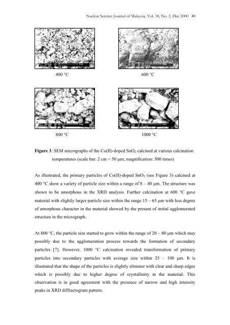
400 C 600 C<br />
800 C 1000 C<br />
Figure 3: SEM micrographs <strong>of</strong> the Co(II)-doped SnO 2 calcined at various calcination<br />
temperatures (scale bar: 2 cm = 50 m; magnification: 500 times)<br />
As illustrated, the primary particles <strong>of</strong> Co(II)-doped SnO 2 (see Figure 3) calcined at<br />
400 C show a variety <strong>of</strong> particle size within a range <strong>of</strong> 8 – 40 m. The structure was<br />
shown to be amorphous in the XRD analysis. Further calcination at 600 C gave<br />
material with slightly larger particle size within the range 15 – 65 m with less degree<br />
<strong>of</strong> amorphous character in the material showed by the present <strong>of</strong> initial agglomerated<br />
structure in the micrograph.<br />
At 800 C, the particle size started to grow within the range <strong>of</strong> 20 – 80 m which may<br />
possibly due to the agglomeration process towards the formation <strong>of</strong> secondary<br />
particles [7]. However, 1000 C calcination revealed transformation <strong>of</strong> primary<br />
particles into secondary particles with average size within 25 – 100 m. It is<br />
illustrated that the shape <strong>of</strong> the particles is slightly slimmer with clear <strong>and</strong> sharp edges<br />
which is possibly due to higher degree <strong>of</strong> crystallinity in the material. This<br />
observation is in good agreement with the presence <strong>of</strong> narrow <strong>and</strong> high intensity<br />
peaks in XRD diffractogram pattern.