application of x-ray diffraction and scanning electron ... - OpenDrive
application of x-ray diffraction and scanning electron ... - OpenDrive
application of x-ray diffraction and scanning electron ... - OpenDrive

You also want an ePaper? Increase the reach of your titles
YUMPU automatically turns print PDFs into web optimized ePapers that Google loves.
Nuclear Science Journal <strong>of</strong> Malaysia, Vol. 18, No. 2, Dec 2000 41<br />
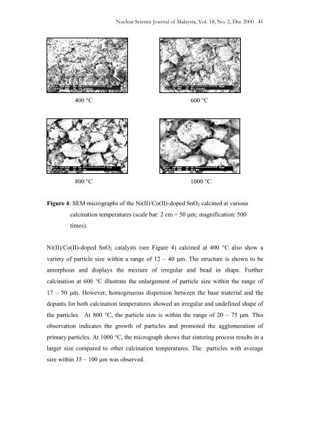
400 C 600 C<br />
800 C 1000 C<br />
Figure 4: SEM micrographs <strong>of</strong> the Ni(II)/Co(II)-doped SnO 2 calcined at various<br />
calcination temperatures (scale bar: 2 cm = 50 m; magnification: 500<br />
times).<br />
Ni(II)/Co(II)-doped SnO 2 catalysts (see Figure 4) calcined at 400 C also show a<br />
variety <strong>of</strong> particle size within a range <strong>of</strong> 12 – 40 m. The structure is shown to be<br />
amorphous <strong>and</strong> displays the mixture <strong>of</strong> irregular <strong>and</strong> bead in shape. Further<br />
calcination at 600 C illustrate the enlargement <strong>of</strong> particle size within the range <strong>of</strong><br />
17 – 50 m. However, homogeneous dispersion between the base material <strong>and</strong> the<br />
dopants for both calcination temperatures showed an irregular <strong>and</strong> undefined shape <strong>of</strong><br />
the particles. At 800 C, the particle size is within the range <strong>of</strong> 20 – 75 m. This<br />
observation indicates the growth <strong>of</strong> particles <strong>and</strong> promoted the agglomeration <strong>of</strong><br />
primary particles. At 1000 C, the micrograph shows that sintering process results in a<br />
larger size compared to other calcination temperatures. The particles with average<br />
size within 35 – 100 m was observed.