Singing cells, art, science and the noise in between - Users - UCLA
Singing cells, art, science and the noise in between - Users - UCLA
Singing cells, art, science and the noise in between - Users - UCLA
Create successful ePaper yourself
Turn your PDF publications into a flip-book with our unique Google optimized e-Paper software.
2.0 The dark side of <strong>the</strong> cell<br />
2.1 Cell Sonics <strong>and</strong> <strong>the</strong> Invisible<br />
In 2002, Prof. James Gimzewski <strong>and</strong> Andrew Pell<strong>in</strong>g of <strong>the</strong> <strong>UCLA</strong> Dep<strong>art</strong>ment of<br />
Chemistry made <strong>the</strong> discovery that yeast <strong>cells</strong> oscillate on a nanoscale. S<strong>in</strong>ce sound<br />
derives from vibration, Prof. Gimzewski amplified <strong>the</strong> oscillation <strong>and</strong> found that <strong>the</strong> cell<br />
sound lies with<strong>in</strong> <strong>the</strong> audible range of humans. Inspired by this discovery, Prof.<br />
Gimzewski <strong>and</strong> Pell<strong>in</strong>g set forth to explore this phenomena, <strong>and</strong> decided to name this<br />
cutt<strong>in</strong>g edge field of research “Sonocytology”.<br />
When Christa Sommerer <strong>and</strong> Laurent Mignonneau developed <strong>the</strong>ir piece, NanoScape, a<br />
table on which an “<strong>in</strong>visible” sculpture can be experienced through a magnetic forcefeedback<br />
<strong>in</strong>terface attached to <strong>the</strong> h<strong>and</strong>, <strong>the</strong>y emphasized one of <strong>the</strong> most important<br />
characteristics of nano<strong>science</strong>: it cannot be experienced through sight. 1 At first, this<br />
statement might seem obvious <strong>and</strong> unnecessary, as we know that matter on a nanoscale is<br />
too small to be observed by <strong>the</strong> human eye alone. But <strong>the</strong> importance <strong>and</strong> depth of this<br />
realization becomes clear when one considers <strong>the</strong> tools of <strong>the</strong> nanoscientist.<br />
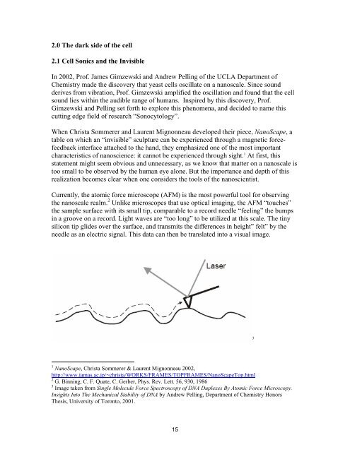
Currently, <strong>the</strong> atomic force microscope (AFM) is <strong>the</strong> most powerful tool for observ<strong>in</strong>g<br />
<strong>the</strong> nanoscale realm. 2 Unlike microscopes that use optical imag<strong>in</strong>g, <strong>the</strong> AFM “touches”<br />
<strong>the</strong> sample surface with its small tip, comparable to a record needle “feel<strong>in</strong>g” <strong>the</strong> bumps<br />
<strong>in</strong> a groove on a record. Light waves are “too long” to be utilized at this scale. The t<strong>in</strong>y<br />
silicon tip glides over <strong>the</strong> surface, <strong>and</strong> transmits <strong>the</strong> differences <strong>in</strong> height” felt” by <strong>the</strong><br />
needle as an electric signal. This data can <strong>the</strong>n be translated <strong>in</strong>to a visual image.<br />
3<br />
1 NanoScape, Christa Sommerer & Laurent Mignonneau 2002,<br />
http://www.iamas.ac.jp/~christa/WORKS/FRAMES/TOPFRAMES/NanoScapeTop.html<br />
2 G. B<strong>in</strong>n<strong>in</strong>g, C. F. Quate, C. Gerber, Phys. Rev. Lett. 56, 930, 1986<br />
3 Image taken from S<strong>in</strong>gle Molecule Force Spectroscopy of DNA Duplexes By Atomic Force Microscopy.<br />
Insights Into The Mechanical Stability of DNA by Andrew Pell<strong>in</strong>g, Dep<strong>art</strong>ment of Chemistry Honors<br />
Thesis, University of Toronto, 2001.<br />
15


