infectious diseases
infectious diseases
infectious diseases

Create successful ePaper yourself
Turn your PDF publications into a flip-book with our unique Google optimized e-Paper software.
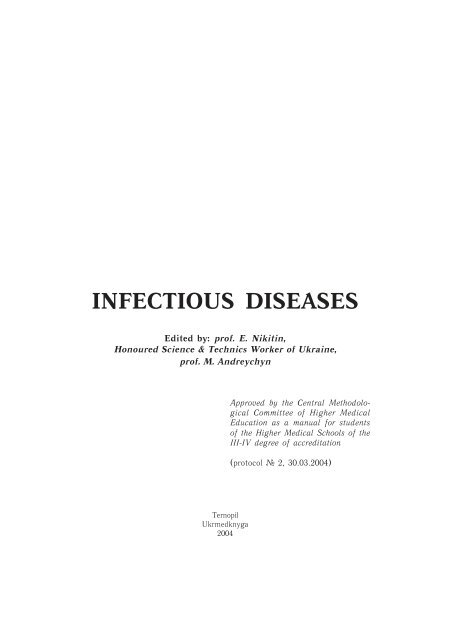
INFECTIOUS DISEASES<br />
Edited by: prof. E. Nikitin,<br />
Honoured Science & Technics Worker of Ukraine,<br />
prof. M. Andreychyn<br />
Approved by the Central Methodological<br />
Committee of Higher Medical<br />
Education as a manual for students<br />
of the Higher Medical Schools of the<br />
III-IV degree of accreditation<br />
(protocol ¹ 2, 30.03.2004)<br />
Ternopil<br />
Ukrmedknyga<br />
2004
ББК 51.9я73<br />
І 60<br />
УДК 616.9(075.8)<br />
Authors: E. Nikitin, M.D., Ph. D.; M. Andreychyn, M.D., Ph. D.; K. Servetskyy,<br />
M.D., Ph. D.; V. Kachor, M.D.; A. Holovchenko, M.D.; E. Usychenko, M.D.<br />
Колектив авторів: д-р мед. наук, проф. Нікітін Є.В., засл. діяч науки і техніки<br />
України, д-р мед. наук, проф. Андрейчин М.А., д-р мед. наук,<br />
проф. Сервецький К.Л., канд. мед. наук, доц. Качор В.О., канд. мед.<br />
наук, доц. Головченко А.М., канд. мед. наук, доц. Усиченко Є.М.<br />
Reviewers: A. Rudenko, Honoured Doctor of Ukraine; I. Sytnyk, M.D., Ph. D.;<br />
L. Shevchenko, M.D., Ph. D.<br />
Рецензенти: А.О. Руденко, д-р мед. наук, заслужений лікар України; І.О. Ситник<br />
д-р мед. наук, проф.; Л.Ю. Шевченко, д-р мед. наук, проф.<br />
І 60<br />
Інфекційні хвороби: Підручник / Є.В. Нікітін, М.А. Андрейчин, К.Л.Сервецький,<br />
В.О. Качор, А.М. Головченко, Є.М. Усиченко: За ред. Є.В. Нікітіна та<br />
М.А. Андрейчина. – Тернопіль: Укрмедкнига, 2004. – 364 с.<br />
ISBN 966-673-012-Х<br />
The manual reveals etiology, pathogenesis, clinic, diagnosis, medical treatment, and<br />
prophylaxis of widespread <strong>infectious</strong> <strong>diseases</strong> considering modern data. The authors of the<br />
book generalized experience of this subject being tought at home and foreign medical<br />
universities and academies, as well as <strong>infectious</strong> <strong>diseases</strong> departments, where they work.<br />
Attention is paid to presentation of material, which has important clinical value.<br />
This manual corresponds to the program of Ministry of Health of Ukraine and is designed<br />
for the students of medical higher education institutions of III-IY accreditation levels.<br />
У підручнику, з урахуванням сучасних відомостей, висвітлено етіологію, патогенез,<br />
клініку, діагностику, лікування та профілактику розповсюджених інфекційних<br />
хвороб. Узагальнено досвід викладання цього предмету у вітчизняних і зарубіжних<br />
медичних університетах та академіях, а також кафедр інфекційних хвороб, на<br />
яких працюють автори книги. Акцент зроблено на подання матеріалу, який має важливе<br />
клінічне значення.<br />
Підручник відповідає програмі, затвердженій Міністерством охорони здоров’я<br />
України, й адресується студентам медичних навчальних закладів III-IV рівнів<br />
акредитації.<br />
ББК 51.9я73<br />
УДК 616.9(075.8)<br />
ISBN 966-673-012-Х © Нікітін Є.В. та ін., 2004
Contents<br />
INTRODUCTION OF INFECTIOUS DISEASES ............................................................... 5<br />
THE PRINCIPLES OF THE DIAGNOSTICS, TREATMENT AND PREVENTION<br />
OF INFECTIOUS DISEASES............................................................................................... 22<br />
TYPHOID FEVER AND PARATYPHOID ......................................................................... 37<br />
BRUCELLOSIS ....................................................................................................................... 47<br />
SHIGELLOSIS......................................................................................................................... 57<br />
SALMONELLOSIS ................................................................................................................. 67<br />
CHOLERA ............................................................................................................................... 77<br />
BOTULISM. TOXIC FOOD-BORNE DISEASES. CAMPYLOBACTERIOSIS ............ 88<br />
PSEUDOTUBERCULOSIS.................................................................................................... 99<br />
LEPTOSPIROSIS .................................................................................................................. 111<br />
HERPETIC DISEASES ........................................................................................................ 122<br />
ERYSIPELAS. ERYSIPELOTRIX ...................................................................................... 135<br />
ANTHRAX............................................................................................................................. 142<br />
VIRAL HEPATITIS .............................................................................................................. 149<br />
INFLUENZA......................................................................................................................... 163<br />
ACUTE RESPIRATORY VIRAL DISEASES ................................................................... 179<br />
MYCOPLASMA PNEUMONIAE INFECTION .............................................................. 198<br />
MENINGOCOCCAL INFECTION .................................................................................... 202<br />
DIPHTHERIA........................................................................................................................ 216<br />
RICKETTSIOSIS ................................................................................................................... 233<br />
VIRAL ENCEPHALITIS ..................................................................................................... 253<br />
RABIES ................................................................................................................................... 263<br />
TETANUS .............................................................................................................................. 272<br />
MALARIA .............................................................................................................................. 279<br />
PLAGUE ................................................................................................................................ 301<br />
TULAREMIA ........................................................................................................................ 319<br />
AQUIRED IMMUNODEFICIENCY SYNDROME ........................................................ 332<br />
SEPSIS .................................................................................................................................... 352<br />
Literature................................................................................................................................. 364
4 Infectious <strong>diseases</strong>
Introduction of <strong>infectious</strong> <strong>diseases</strong><br />
5<br />
INTRODUCTION<br />
OF INFECTIOUS DISEASES<br />
Nowadays there are about 2,500 well-known microorganisms that cause<br />
<strong>infectious</strong> <strong>diseases</strong>. About 300 nosological forms have been distinctly described<br />
(“nosos” means “disease” in Greek). According to the international classification<br />
the <strong>infectious</strong> <strong>diseases</strong> relate to 13 classes and 975 rubrics. They constitute up<br />
to 60-70 % of the total morbidity. In polyclinics 4-6 patients out of 10 suffer<br />
from <strong>infectious</strong> <strong>diseases</strong>. The responsibility for the exposure of the <strong>infectious</strong><br />
morbidity is taken by the physicians of the “first line” – therapeutists, surgeons,<br />
gynecologists and other specialists. Now it is distinctly ascertained that the<br />
<strong>infectious</strong> agents are the basic or leading ethiological factor of the different<br />
branches of the medical science.<br />
Such fields of the internal <strong>diseases</strong> as rheumatology, pulmonology or<br />
hepatology cannot be conceived without taking into consideration the <strong>infectious</strong><br />
factor. The <strong>infectious</strong> factor often determines the outcome of the surgery. The<br />
gynecological, urological and eye <strong>diseases</strong> cannot be treated without considering<br />
it. The association of the viral hepatitis B and C with the primary liver cancer is<br />
undoubted, there is a certain connection between the cancer of the cervix of the<br />
uterus and the virus of the herpes simplex, leucosis and Bercet’s lymphosarcoma.<br />
The significance of the infections agents in bronchial asthma, arthritis,<br />
meningoencephalitis is undoubted.<br />
Besides these many new earlier unknown discuses have appeared. Only<br />
during the last years such infections <strong>diseases</strong> as HIV, Legionnaires disease,<br />
cryptosporidiosis, SARS, hemorrhagic fevers, caused by Marburg or Ebola virus,<br />
hantavirus and others have appeared, they are responsible for the development<br />
of the ulcer disease of the stomach, pneumonia, meningoencephalitis, cutaneous<br />
<strong>diseases</strong>, lymphoadenopathy, heart and vessels <strong>diseases</strong>. Many researchers think<br />
that the world is standing on the verge of the T-cell leucosis epidemic, which is<br />
already widely spread in Japan and in some regions of Latin America. That is<br />
why a physician of any speciality will more or less often encounter with the<br />
<strong>infectious</strong> pathology.<br />
The origin of the <strong>infectious</strong> <strong>diseases</strong> dates back from the ancient times. The<br />
old archives that contain the man’s first descriptions of his thoughts with the<br />
help of signs tell us that he already suffered from such <strong>diseases</strong> as leprosy,<br />
hydrophobia, malaria, trachoma, fungous, helmintic and some other <strong>diseases</strong>.<br />
Although the <strong>infectious</strong> <strong>diseases</strong> exist as long as life itself, their studying<br />
started comparatively not long ago. It is one of the youngest branches of science.
6 Infectious <strong>diseases</strong><br />
The scientific history of the <strong>infectious</strong> <strong>diseases</strong> started at the end of the 19th<br />
century when the term “<strong>infectious</strong> <strong>diseases</strong>” was introduced, and it was determined<br />
that they were caused by the microorganisms i.e. the organisms that could be<br />
found only with the help of a microscope.<br />
The common feature of the majority of the <strong>infectious</strong> <strong>diseases</strong> is the possibility<br />
of transmission from the affected organism to a healthy one, and the ability of<br />
massive (epidemic) spreading. During the study of the <strong>infectious</strong> <strong>diseases</strong> the<br />
terms “infection” and “<strong>infectious</strong> process” are usually used. They both originate<br />
from the Latin words – “infectio” – “pollution”, “contamination” and “infecio”<br />
– “to pollute”. At the modern level of the science development it is impossible<br />
to give an exhaustive definition of the terms “infection” and “<strong>infectious</strong> process”<br />
which would open all the sides of this conception. The term “infection” means<br />
the penetration of a microorganism into another organism and their following<br />
interaction under various conditions connected both with the microbe itself and<br />
the qualities of the organism which receives it at the various stages of the<br />
development of the organic world.<br />
The term “<strong>infectious</strong> process” means the totality of the physiological, defensive<br />
and pathologic reactions, which appear under the certain conditions of the<br />
environment as an answer to the affection of the pathogenic microorganisms.<br />
An <strong>infectious</strong> disease is the extreme stage of the <strong>infectious</strong> process<br />
development, which manifests in different signs and changes of the biological,<br />
chemical and epidemiological order.<br />
According to all of these definitions it is obvious that the term “infection”<br />
cannot be identified with the “intectious process”. The contents which we put<br />
into the term “<strong>infectious</strong> process” does not let us make a complete image of the<br />
infection as a general biological phenomenon. The concept “infection” is much<br />
wider as infection is common to all the beings. The <strong>infectious</strong> process includes<br />
the patterns common to the complicated organisms. The term “<strong>infectious</strong> process”<br />
is used to identify all the dynamics of the pathologic changes connected with the<br />
infection irrespective of the fact that they develop into a special qualitative<br />
condition called an <strong>infectious</strong> disease or not.<br />
The origin of the <strong>infectious</strong> <strong>diseases</strong> and their nature were discovered<br />
owing to the brilliant success of bacteriology. That is why for a long period of<br />
time the <strong>infectious</strong> process was identified only with the activity of the<br />
microorganism without any consideration of the physiological aspects of the<br />
macroorganism.<br />
Later when the scientists started to pay more attention to the study of<br />
pathogenesis i.e. the mechanism of the sickness processes development, they<br />
advanced a thesis of the domination of the macroorganism in the <strong>infectious</strong><br />
process. It was a second mistake as it is impossible to separate something that<br />
is by the nature closely connected and can be understood only in correlation.<br />
Besides, the pathological process also has to be examined under the influence of
Introduction of <strong>infectious</strong> <strong>diseases</strong><br />
the environment. So if the disease develops as a result of the violation of this<br />
certain form of the organism’s adaptation to the environment at the change of its<br />
conditions, then the <strong>infectious</strong> disease develops as the result of the influence of<br />
the part of environment which belongs to the living organisms. In other words,<br />
all the participants of the pathologic process are noted for the biological activity,<br />
functional mobility and the ability to develop.<br />
The penetration of a certain number of microorganisms into the macroorganism<br />
is necessary for the <strong>infectious</strong> disease to develop. Besides, it has to be of a certain<br />
quality i.e. pathogenicity and virulence. However, the development of the<br />
pathological process depends on the general condition of the macroorganism<br />
and its immune status. In case of the weak immune status the pathologic process<br />
develops rapidly and the disease takes a severe course, in case of the comparatively<br />
strong immune status the disease takes a mild course or may not develop at all.<br />
Spreading of the disease and its severity depends on the environment – both on<br />
the geographical position (in the tropics – overheating, in cold countries –<br />
supercooling), and on the social sphere (a luxury villa and overcrowded facilities).<br />
All these processes can be expressed in the formula: the <strong>infectious</strong> disease is pro<br />
rate to the number and quality of the microbe (pathogenecity, virulence) and<br />
invasively to the immune status and the environment. Each of the mentioned<br />
factors is variable and they should be considered as dynamically developing with<br />
the changing of the cause and effect.<br />
Comparing an <strong>infectious</strong> disease with a non-<strong>infectious</strong> one we can point out<br />
their mutual signs – intoxication and functional disorders of the organs and<br />
systems, morphological changes and others. However, with all the similarities we<br />
should mention the peculiarities of the infection <strong>diseases</strong>. This is, first of all, the<br />
cyclical course with expressed periods (incubative, prodromal, high point, fading<br />
healing). The second peculiarity is that the pathogen that caused the disease as a<br />
living agent has its own “interests” – it lives, multiplies as it tries to preserve its<br />
species – at the same time it adjusts, changes or remains in its constancy – the<br />
death of the macroorganism is not “profitable” for it as the pathogen can die<br />
with it as well. The third peculiarity is that the affected organism can become a<br />
source of infection for healthy people. The common feature of the majority of<br />
the <strong>infectious</strong> <strong>diseases</strong> is the possibility of transmission from the affected organism<br />
to healthy one and the ability of massive (epidemic) spreading. The fourth peculiarity<br />
consists of the immune processes which make the organism insensible to the<br />
later affections in case of the same etiologic factor.<br />
Different interrelations occur when organisms contact with one another, it<br />
happens in nature all the time. To understand the <strong>infectious</strong> <strong>diseases</strong> we should<br />
mention the basic types of such interrelations.<br />
1. The meeting and contact of the organisms do not have any consequences,<br />
any reaction. No symbiotic relations appear after it. In such cases we talk about<br />
species inherited immunity. For example, a human immunity to the horned<br />
livestock’s plague, to the hemorrhage septicemia of cats and others.<br />
7
8 Infectious <strong>diseases</strong><br />
2. The meeting of the organisms results in the symbiotic commonwealth (in<br />
Greek „symbiosis” means state of living together). There may be no reaction at<br />
all on the part of both partners, the condition called saprophytosis („saprobe”<br />
means microorganism that lives in the dead organic remains) appears. Some<br />
researchers consider symbiosis to be any form of living together between the<br />
representatives of different species. To this symbiosis they refer:<br />
a) synoikia (Greek) – neutral living together during which one species<br />
uses the other one as a place to live without harming it;<br />
b) mutualism – the symbiosis that is profitable for both organisms;<br />
e) commensalism (Lat. “com” – with, “mensa” – table) also (French –<br />
“commensa” – dependent) interrelation when one organism gets a benefit<br />
from the other without harming it;<br />
d) parasitism – a microorganism feeds with the saps or tissues of the<br />
master harming it. Most of the <strong>infectious</strong> <strong>diseases</strong> belong to this kind of<br />
symbiosis.<br />
Analyzing the pathogenic processes scientists divide the <strong>infectious</strong> <strong>diseases</strong><br />
into endogenous and exogenous ones. The endogenous <strong>diseases</strong> or autoinfections<br />
develop from their own microflora which is situated on the skin, respiratory and<br />
alimentary tracts, conjunctive, genital tracts. Because of the disorder of the<br />
regulatory processes which provide the physiologic symbiotic balance, there<br />
develop local, widely spread or even general <strong>infectious</strong> processes. Such<br />
microorganisms are called conditionally pathogenic or half-parasites.<br />
The <strong>diseases</strong> caused by the penetration of the microorganisms from the<br />
environment and to which a macroorganism is not resistant are called exogenous<br />
<strong>infectious</strong> <strong>diseases</strong>.<br />
Such division is pretty relevant and only relatively right. Sometimes the<br />
endogenous <strong>infectious</strong> disease becomes dangerous to the others because of<br />
itself and because the symbionit acquires new biological qualities.<br />
If the <strong>infectious</strong> disease is caused by one species of microorganism it is<br />
called simple. If two or more microbe agents participate in the disease, then we<br />
talk about mixed-infection. Joining one infection to the other may affect the<br />
<strong>infectious</strong> process in different directions sometimes intensifying it, sometimes<br />
decreasing its activity and manifestations. So while studying the <strong>infectious</strong><br />
pathology we should consider not only the pathogen itself but their associations.<br />
Salmonella infection especially bent to join other <strong>infectious</strong> <strong>diseases</strong> and start<br />
the secondary pathological process, this phenomena is called nosoparasitism (in<br />
Greek “nosos” – disease).<br />
The growing number of the <strong>diseases</strong> caused by the conditionally-pathogenic<br />
pathogens is mostly connected with the changed reactivity of the organism and<br />
especially immune response which as a rule in such cases forms very slowly and<br />
is not valuable. The autoimmune processes activate and take a leading role in<br />
pathogenesis and clinical manifestations.
Introduction of <strong>infectious</strong> <strong>diseases</strong><br />
Many conditionally pathogenic as new discovered microorganisms are<br />
characterized with intracellular localization of the pathogens. Such infections<br />
can cause widely spread pathological disorders and they are more difficult with<br />
their diagnosis and treatment.<br />
An immune system is one of the major targets of the affection of the<br />
environment negative factors according to the modern conception. There are six<br />
basic factors:<br />
Human demographics and behavior. The important factors in changing<br />
human demographics include increases in the number of susceptible persons,<br />
the use of day care and immigration. A number of factors cause a rise in<br />
number of susceptible persons and the greater the population percentage that is<br />
susceptible to the <strong>infectious</strong> <strong>diseases</strong>, the greater is the potential for the disease<br />
transmission. In many countries in the developed world the number of seniors<br />
is growing. Since aging is associated with an increased susceptibility to the<br />
<strong>infectious</strong> <strong>diseases</strong>, the potential for the disease transmission is also increasing<br />
in these countries. In the USA, the percentage of the population over 65 years<br />
was about 4 % in 1900 and will reach almost 25 % in 2040. Certain underlying<br />
<strong>diseases</strong> also place more patients at risk for various <strong>infectious</strong> <strong>diseases</strong>, and<br />
these have also increased. For example, the reported incidence of diabetes mellitus<br />
in the USA increased from 0.5 % of the nation’s population in 1935 to over 3 %<br />
in 1995. It is estimated that there are actually 16 million persons with diabetes in<br />
the USA, so the true incidence of this disease may be greater than 5 % of the<br />
population. The rates for many malignancies are also increasing, and these patients<br />
have increased susceptibility to <strong>infectious</strong> <strong>diseases</strong> from the disease process,<br />
during chemotherapy and, in some cases, lifelong even after the cure. Some of<br />
the most highly susceptible patients are those receiving immunosuppressive<br />
therapy following organ transplantation. Almost 20,000 organ transplants were<br />
performed in the USA in 1995. Worldwide the greatest factor increasing<br />
susceptibility may be the spread of HIV, which has led to millions of persons at<br />
increased risk for a variety of <strong>infectious</strong> <strong>diseases</strong>.<br />
Such social changes as the increased use of day care also affect the emergence<br />
of <strong>infectious</strong> <strong>diseases</strong>. The increasing frequency of both parents working outside<br />
home or of single parent families led to a greater use of day care. The combination<br />
of susceptible children, inadequate hygiene, frequent infections, and frequent<br />
antimicrobial use is the perfect setting for the emergence of antimicrobial<br />
resistance. Thus, it is no surprise that day care attendance has been an important<br />
factor associated with the emergence of penicillin-resistant Streptococcus<br />
pneumoniae. A recent study demonstrated a 4-fold greater relative risk for<br />
colonization with a high-level penicillin-resistant Streptococcus pnenmoniae<br />
among children attending day care.<br />
The increase of immigration and changing the patterns of immigration also<br />
contribute to the emergence of the <strong>infectious</strong> <strong>diseases</strong>. Between 1984 and 1992,<br />
9
10 Infectious <strong>diseases</strong><br />
0.5-1.5 million immigrants and refugees were admitted to the USA each year. In<br />
contrast to the previous waves of immigration many of these individuals came<br />
from the parts of the world where certain infections such as tuberculosis are<br />
common. This is an important factor in the resurgence of tuberculosis in the<br />
USA as the percentage of patients who were foreign-born increased from 22 %<br />
in 1986 to 37 % in 1996.<br />
A variety of human behaviors also influence the emergence of the <strong>infectious</strong><br />
<strong>diseases</strong>. The impact of the sexual revolution on the frequency of gonorrhoea,<br />
syphilis, and HIV is evident. Perhaps less evident is the impact of other changes<br />
such as changes in eating habits. There are changes in the types of food that<br />
people eat, how this food is prepared, and where the food is prepared. This can<br />
result in the new exposures to unfamiliar food or the dependency upon others<br />
to handle and prepare food safely. All these factors have contributed to the<br />
emergence of some of the newer food borne <strong>diseases</strong>. Another important factor<br />
influencing the emergence of resistance has been the unnecessary use of<br />
antimicrobial agents. In 1992 over 110 million courses of antimicrobial drugs<br />
were prescribed to outpatients in the USA. Since three-quarters of these drugs<br />
are prescribed for upper respiratory infections that are often caused by viruses,<br />
over half of these 110 million courses may be unnecessary.<br />
Technology and industry. The impact of technology and industry falls<br />
into three general areas. These include new technologies and products, changes<br />
in food production processing and preservation, and changes in industrial<br />
demographics. New technologies and products may have unexpected disease<br />
implications such as the association of air conditioning and whirlpool spase<br />
with Legionnaires disease, or new tampons with toxic shock syndrome, or the<br />
fast-food hamburger with E. coli O157:H7.<br />
The second area, the changes in how food is produced, processed, and preserved<br />
has also been important. For example, in the last 50 years many of the new<br />
agricultural production strategies involve intensive rearing of young animals<br />
under environmental conditions that are conducive to the transmission of the<br />
<strong>infectious</strong> <strong>diseases</strong>. These production strategies often depend upon increased<br />
antimicrobial use. Thus, only substituting young animals for children, the situation<br />
is similar to the day care setting and has resulted in an increase in antimicrobial<br />
resistance for organisms that are transmitted through the food chain from animals<br />
to humans. Between 1979 and 1989, the frequency of drug resistance in human<br />
Salmonella isolates almost doubled from 17 % to 31 %. Today the resistance<br />
in humans is the result of antimicrobial use in animals. The many changes in<br />
food processing and preservation are also influencing disease emergence. The<br />
recent emphasis on “natural” foods has led to use of fewer preservatives or<br />
secondary barriers to prevent spoilage. Thus, some foods are protected only by<br />
refrigeration. This has resulted in increasing problems with organisms that grow<br />
in the cold, such as Listeria or Yersinia. The lack of secondary barriers also
Introduction of <strong>infectious</strong> <strong>diseases</strong><br />
increases the risk of food handling errors leading to <strong>diseases</strong> such as botulism.<br />
In the last 10 years, several outbreaks of botulism have occurred when “keep<br />
refrigerated” foods were not kept refrigerated.<br />
The third change, that of industrial demographics, is characterized by<br />
consolidations of industry, larger market size, and wider geographic distribution<br />
for a variety of food products. Although these changes have the potential for<br />
greater quality control and better safety, when something goes wrong, it can really<br />
go wrong. Thus, in 1994, an ice cream product produced by a single company in<br />
Minnesota led to thousands of cases of salmonellosis in over 41 states.<br />
Economic development and land use. Changes in economic development<br />
and land use are often cited in discussions of emerging viral <strong>diseases</strong>.<br />
Encroachment on rain forests, for instance, may lead to exposure to new agents<br />
such as Ebola or Marburg viruses. However, such changes are also influencing<br />
the emergence of other <strong>infectious</strong> <strong>diseases</strong>. For example, population growth and<br />
spread lead to environmental change and pollution. The inadequacies of hygiene<br />
and sanitation that exist in many of the “mega cities” in the developing world are<br />
potential ticking time bombs for the emergence of <strong>infectious</strong> <strong>diseases</strong>. Other<br />
types of development and land use practices are contributing to specific problems.<br />
Conservation activities, such as those directed toward deer populations, have<br />
contributed to the emergence of Lyme disease. Coastal agriculture expansion is<br />
leading to blooms of toxic microorganisms, while coastal population growth is<br />
leading to human faecal contamination of shellfish beds and transmission of a<br />
variety of viral and bacterial pathogens.<br />
International travel and commerce. Advances in technology have had<br />
a rapid impact on international travel and commerce. A person or a food can be<br />
almost anywhere in the world in 24-48 hours. This facility in travel and commerce<br />
has increased the potential for the introduction of emerging pathogens to new<br />
geographic areas by infected travellers, by contaminated food, or even by<br />
transporting vehicles.<br />
Microbial adaptation and change. As humankind is instituting a number<br />
of changes, the microbes themselves are changing. This is leading to the evolution<br />
of new pathogens, the development of new virulence factors, the development of<br />
antimicrobial resistance, and tolerance to adverse environmental conditions. A<br />
good example of this microbial change has been the emergence of E. coli 0157:H7,<br />
which probably evolved from an entero-pathogenic E. coli that acquired Shigella<br />
genes. As a food borne pathogen, it combines the worst of Shigella and Salmonella.<br />
Like Shigella, this organism has a low <strong>infectious</strong> dose, requiring only a few<br />
organisms to cause disease. This leads to subsequent person-to-person<br />
transmission once the organism is introduced into a community and also poses a<br />
high risk for cross-contamination in the kitchen. This organism is more similar<br />
to Salmonella in its tolerance to adverse environmental conditions. Thus, it has<br />
been associated with outbreaks that were caused by foods with pH 4.0, conditions<br />
that are usually inhibitory to most bacterial pathogens.<br />
11
12 Infectious <strong>diseases</strong><br />
The breakdown of public health measures. The breakdown of public<br />
health measures has been the result of a series of often unrelated factors. Earlier<br />
successes in the war against <strong>infectious</strong> <strong>diseases</strong> led to complacency. Thus, coupled<br />
with limited resources and competing priorities in public health, often led to the<br />
transfer of resources from <strong>infectious</strong> <strong>diseases</strong> to other areas or to newly emerging<br />
infections.<br />
The impact of the breakdown of public health measures can easily be seen<br />
during wars, population movements, and natural disasters. One such example has<br />
been the emergence of epidemic shigellosis in Africa. Since 1979, massive epidemics<br />
of shigellosis caused by Shigella dysenteriae type 1 have occurred in cities,<br />
rural areas, and refugee camps in Central and Southern Africa. The epidemics<br />
have affected all age groups, often with case-fatality ratios greater than 10 %. In<br />
1991 alone, the disease caused 60,000 deaths in Burundi and at least 200,000<br />
deaths in the rest of Africa. In contrast to Shigella species, which are more<br />
common in parts of the developed world, this organism is essentially resistant to<br />
all available oral antimicrobial drugs. Some of the newer fluoroquinolones are<br />
the last remaining effective oral agents.<br />
Now even in developed countries approximately for 40 % of adult population<br />
tap different imunopathologic states that explains atypical and lingering How<br />
even of classic <strong>infectious</strong> <strong>diseases</strong>, more often development of mixed-infection,<br />
superinfection, continuous, microbe carrying.<br />
The diagnostic of the <strong>infectious</strong> disease should be as early as possible. Such<br />
haste is connected not only with the necessity of assignina the conforming<br />
treatment but also with the demand of carring out urgent preventive actions,<br />
especially, if the disease has arisen in the collective. The diagnosis is grounded<br />
on the combination of symptoms characteristic for this or that <strong>infectious</strong> process.<br />
As in case with other <strong>diseases</strong>, the symptoms should be collected beginning with<br />
the complains of a sick person, anamnestic information of the development of<br />
illness symptoms, the nature of the epidemiological situation. Objective data<br />
should be taking during the physical examination of the patient, and then at<br />
auscultation, percussion and laboratory investigations.<br />
At the identification of the <strong>infectious</strong> <strong>diseases</strong> as well as other <strong>diseases</strong>,<br />
anamnesis is of great importance. It is necessary to point out one of the most<br />
important peculiarities of the anamnestic data in <strong>infectious</strong> <strong>diseases</strong>, it is<br />
epidemiological anamnesis. The epidemiological anamnesis should be extremely<br />
careful and full. When the patient himself cannot give the necessary data (grave<br />
condition, age), the information should be obtained from relatives or the people<br />
around him.<br />
Collecting the epidemiological anamnesis is as difficult as obtaining the<br />
disease anamnesis, and the skill of its collecting needs to be developed just as the<br />
skill of objective examining, the more so as collecting the correct anamnesis is<br />
considered to be more difficult to learn than the procedure of the objective
Introduction of <strong>infectious</strong> <strong>diseases</strong><br />
examination. At the inept approach to the patient and frivolous attitude to the<br />
epidemiological anamnesis obtaining, the doctor cannot get the necessary<br />
information. Sometimes it is difficult to take the correct anamnesis because in<br />
case of the disease with a long incubative period the patient and his relatives can<br />
forget some data, which are of the diagnostical and epidemiological value.<br />
The following points are the most important for the epidemiological<br />
anamnesis:<br />
1. Way of living and living conditions of the patient. It is necessary to<br />
explicitly find out whether the ambient situation could have promoted the intrusion<br />
of an <strong>infectious</strong> agent. If during the last three weeks before the onset of the<br />
disease the patient lived at the place of spreading of the <strong>infectious</strong> <strong>diseases</strong>, the<br />
patient can have developed a similar disease. The information about the cases of<br />
this or that infection in the patient’s house confirms this idea even more. The<br />
use of unboiled water, milk, dirty fruit, pot herb, meat and fish products of poor<br />
quality can be a source of intestinal infection. The wounds, bruises, splinters are<br />
characteristic features of erypsipelas, tetanus, septic <strong>diseases</strong>.<br />
2. The patient’s occupation. Thus the workers of cattle-breeding farms can<br />
more often get sick with brucellosis, the agricultural workers – with leptospirosis,<br />
hemorrhagic fever, tick epidemic typhus, the workers of rice fields are subject to<br />
the infection of ankylostomidosis and strongyloidosis.<br />
3. The previous <strong>diseases</strong> and preventive vaccinations. This information is<br />
necessary as the previous <strong>diseases</strong> in a number of cases is evident against the<br />
disease which is suspected at this moment. However, it is always necessary to take<br />
into consideration that there is not a single <strong>infectious</strong> disease, which would not<br />
repeat, though in rare cases. Such <strong>diseases</strong> as flu, malaria, shigellosis, diphtheria,<br />
erypsipelas are the most recurring. And vice versa measles, epidemic typhus<br />
results in a strong and continuous immunity which guarantees from the recurrence<br />
of the <strong>diseases</strong>. Vaccination in anamnesis does not eliminate a possibility of the<br />
disease caused by the same infection, but there are often distorted, atypical forms of<br />
the disease, the so-called deleted forms in case of vaccination. Having taken the<br />
epidemiological anamnesis one starts to inquire about the main complaints and<br />
symptoms, paying attention to every detail in the sequence of their development.<br />
The temperature rise is one of the earliest symptoms which gives ground to<br />
think of an infection when there are still no other clinical manifestations of the<br />
illness. The temperature which rises in the morning or in the evening up to 37 °C<br />
usually is not considered normal. However, it is necessary to take into consideration<br />
the individual peculiarities of the patient, as for some patients the normal<br />
temperature limits are 37.0-37.3 °C, and on the contrary for a number of patients<br />
the normal temperature does not rise above 36.2-36.3 °C and a slightest rise<br />
even on some tenths of a degree already is evidence of abnormal temperature.<br />
The temperature rise can be fast (acute), when the patient clearly marks<br />
even the hour of the onset of the disease (ornithosis, leptospirosis, etc.). In case<br />
13
14 Infectious <strong>diseases</strong><br />
of the fast temperature rise, as a rule, the patient marks the chills of different<br />
grade – from slight chills up to severe chills (malaria). The temperature may<br />
rise gradually in other <strong>diseases</strong> (typhoid, paratyphoids).<br />
According to the grade of the fever there are distinguished the following<br />
conditions: a subfebrile condition (37.0-37.9 °C), a moderate fever (38.0-39.9 °C),<br />
a high fever (40.0-40.9 °C) and hyperpyrexia (41 °C and higher). Taking into<br />
consideration the pathogenesis of the fever, the subfebrile condition should also<br />
be considered as a fever.<br />
The nature of the temperature curve. In some <strong>infectious</strong> <strong>diseases</strong> the<br />
temperature curve is so characteristic that it determines the diagnosis (malaria,<br />
typhoid fever). It is accepted to determine several types of a temperature curve,<br />
which are of a diagnostic value.<br />
The constant fever (febris continua) is characterized by the permanently<br />
high body temperature often up to 39 °C and higher, the daily temperature<br />
fluctuations are less than 1°C and observed in typhoid-paratyphoid <strong>diseases</strong>, Q- fever,<br />
epidemic typhus, etc.<br />
The remittent fever (f. remittens) is distinguished by daily fluctuations of the<br />
body temperature from more than 1°C but not more than 2 °C (ornithosis, etc.).<br />
The intermittent fever (f. intermittens) is manifested by the correct change<br />
of the high or very high and normal temperature with daily fluctuations of 3-4 °C<br />
(malaria, etc.).<br />
The relapsing fever (f. recurrens) is characterized by the correct change of<br />
the high-fever and fever-free periods that last several days (typhoid fever, etc.).<br />
The undulating or undulant fever (f. undulans) is distinguished by a gradual<br />
increase of the temperature up to the high points and then its gradual decrease<br />
to the subfebrile and sometimes normal temperature; in 2-3 weeks the cycle is<br />
repeated (visceral leishmaniasis, brucellosis, lymphogranulomatosis).<br />
The hectic (exhausting) fever (f. hectica) – a prolonged fever with<br />
considerable daily fluctuations (3-5°C) with the decrease to the normal or<br />
subnormal temperature (sepsis, generalized virus infection, etc.).<br />
The irregular (atypical) fever (f. irregularis) is characterized by large<br />
daily amplitude, a various degree of a temperature increase, an indeterminate<br />
duration. It stands closer to the hectic fever, but does not have a regular rythm<br />
(sepsis, etc.).<br />
The distorted (inverted) fever (f. inversa) is distinguished by a higher<br />
morning temperature than the evening one.<br />
Besides these generally accepted types we consider expedient to mention<br />
two more: an acute undulating fever and a relapsing one.<br />
The acute undulating fever (f. undulans acuta) in contrast to the undulate<br />
one is characterized by relatively short waves (3-5 days) and by the absence of<br />
remissions between the waves; the usual temperature curve represents a series<br />
of damped waves, i.e. each subsequent wave is less expressed (in the altitude
Acute respiratory viral <strong>diseases</strong><br />
and duration) than the previous one (typhoid, ornithosis, mononucleosis, etc.):<br />
when the subsequent wave is caused by adding of a complication, the revertive<br />
interrelations are observed, i.e. the second wave is more expressed, than the first<br />
one (epidemic paratitis, flu, etc.).<br />
The relapsing fever (f. recidiva) in contrast to the recurrent fever (the<br />
regular alternating of the fever waves and apyrexy) is characterized by a relapse<br />
(usually one) of the fever which develops in different terms (from 2 days to one<br />
month and more) after the termination of the first temperature wave (typhoid,<br />
ornithosis, etc.). The relapses develop in some of the patients (10-20 %). In this<br />
connection if the relapse has an important diagnostic value, the absence of it does<br />
not eliminate a possibility of the mentioned above <strong>diseases</strong> at all.<br />
Each <strong>infectious</strong> disease can have different variants of a temperature curve,<br />
among which some are more frequent, they are typical for this or that nosological<br />
form. Sometimes they even allow to diagnose the disease quite accurately (tetrian<br />
fever, etc.).<br />
The duration of a feveris is an important value for the differential diagnosis.<br />
A number of <strong>diseases</strong> are characterized by a short-term fever (herpangina, acute<br />
shigellosis, etc.). And if, for example, the fever lasts more than 5 days, it already<br />
allows to eliminate such frequently encountered <strong>diseases</strong>, as flu and other acute<br />
respiratory virus <strong>diseases</strong>, angina (certainly, if there are no complications).<br />
On the contrary, the long-term fever (more than a month) is observed<br />
rather seldom and only in some <strong>infectious</strong> <strong>diseases</strong> which lend to the lingering<br />
or chronic flow (brucellosis, toxoplasmosis, visceral leishmaniasis, tuberculosis).<br />
Thus the grade and characteristic of a temperature curve and the duration of a<br />
fever allow to differentiate certain groups of <strong>infectious</strong> <strong>diseases</strong>, among which<br />
the differential diagnosis is based on other parameters.<br />
For the differential diagnosis the interval between the onset of a fever and<br />
the appearance of organic lesions is particularly important. In some <strong>infectious</strong><br />
<strong>diseases</strong> this period is less than 24 hours (herpetic infection, scarlatina, rubella,<br />
meningococcemia), in others it lasts from 1 up to 3 days (measles, shigellosis),<br />
and, at last, in a number of <strong>diseases</strong> it lasts more than 3 days (typhoid fever, virus<br />
hepatitis).<br />
The nature and level of the <strong>infectious</strong> <strong>diseases</strong> rate also matters. For example,<br />
any feverishness during the epidemic of flu suggests a possibility of flu. The<br />
indication to the contact with the people sick with measles, scarlatina, water-pox,<br />
rubella and other droplet infections is important. These data are compared to<br />
the terms of an incubation period. Other epidemiological data (the stay on the<br />
territory which is endemic on malaria, and other <strong>diseases</strong>) also matters.<br />
For the differential diagnosis the change of a temperature curve under the<br />
influence of etiotropic medications is important. Delagilum stops malarial attacks,<br />
in endemic typhus the temperature quickly becomes normal after the reception<br />
of tetracyclines and others. There are a number of peculiarities of the fever<br />
15
16 Infectious <strong>diseases</strong><br />
syndrome, which one can use for the differential diagnosis. The differential diagnosis<br />
of a fever needs to be done to distinguish it from the body temperature rising of<br />
another nature (thermal shock, hyperthyroidism).<br />
The second component which is not less important for the diagnosis and<br />
differential diagnosis of the <strong>infectious</strong> <strong>diseases</strong> is a rash on the skin –exanthema. It<br />
is because the rashes are a symptom of many <strong>infectious</strong> <strong>diseases</strong>, besides, they are<br />
well visible, quite often catch one’s eye even at the first examination of the patient.<br />
There are exanthemas, characteristic of this or that <strong>infectious</strong> disease and<br />
they are an obligatory component of a clinical symptomatology of this or that<br />
<strong>infectious</strong> disease.<br />
The expressiveness and nature of exanthemas can be miscellaneous and are<br />
not always observed in other <strong>infectious</strong> <strong>diseases</strong>. Due to this factor their presence<br />
or absence in different <strong>infectious</strong> <strong>diseases</strong> essentially differs.<br />
The exanthemas in <strong>infectious</strong> <strong>diseases</strong> are rather diverse. They differ in<br />
nature of different elements of an eruption, localization, terms of appearance,<br />
stages of a rash, the dynamics of development of separate elements, etc. All these<br />
features are taken into consideration while making a differential diagnosis. In the<br />
diagnostics process the legible definition of separate elements of an eruption and<br />
unified comprehension of the terms are very important. Dermatologists and<br />
infectionists do not always define some elements of an eruption in the same way.<br />
The following nomenclature is generally accepted in <strong>infectious</strong> <strong>diseases</strong>.<br />
Roseola – is a small spot (diameter 2-5 mm) of pink, red or purple-red<br />
color, more often with a spherical form. It is formed as a result of a local<br />
vasodilatation of a papillary layer of skin. The roseola disappears when pressing<br />
the eruption area with a transparent glass-spreading rod or when stretching a<br />
skin and comes up again after the stopping of pressure (stretching) that is the<br />
main distinctive feature of it.<br />
The so-called punctate rash is close to roseola. It consists of a set of shallow<br />
(in a diameter of about 1 mm) elements of the red color. At stretching the skin<br />
these elements, as well as roseolas, disappear. Each element rises a little above the<br />
level of the skin that stipulates a special “velvety” of the skin at the eruption area.<br />
The macule (macula) represents an element of the eruption similar to the<br />
roseola, but larger (5-20 mm), it does not stick above the level of the skin, its color is<br />
the same as roseola’s. The development of the spot, as well as in case of roseola, is<br />
based on vasodilatation. The form of maculae can be oval, spherical or more often<br />
small or with festoon edges. Unlike the dermatologists the infectionists distinguish -<br />
spot eruption, in which the elements of an eruption vary in diameter from 5 up to 10<br />
mm, and “large-spot eruption” with the elements’ diameter of 11-20 mm. This distinction<br />
has a differential – diagnostic value. For example, in a patient with rubella there is a<br />
small-spot eruption, and in a patients with measles – a large-spot one.<br />
Papule (papula) – a superficial formation without a cavity, rising above the<br />
level of the skin. It has a mild or dense consistence, the reverse development<br />
ends without the formation of a scar. There are inflammatory and noninflammatory
Introduction of <strong>infectious</strong> <strong>diseases</strong><br />
papules. In the <strong>infectious</strong> <strong>diseases</strong> only inflammatory ones occur. They are caused<br />
by the proliferation of epidermis and infiltrate development in the papillary layer<br />
of derma with vasodilatation and limited edema. Papulas have the same color as<br />
roseola and macula. There are papulas of a different size (1-20 mm). Small<br />
papulas (1-1.5 mm) are called milliar ones, the larger (2-3 mm) papulars –<br />
lenticularis ones. The confluence of separate papules results in the formation of<br />
the eruptions elements called plaque.<br />
Erythema – is vast fields of the bloodshot skin which are red, purple-red or<br />
magenta. The erythema is formed as a result of large maculae joining. Therefore the<br />
erythema has festoon blurry edges. Inside arythmetic fields there can be separate<br />
fields of the skin with normal coloring. There is no expressed inflammatory process.<br />
Unlike the infectionists the dermatologists consider that the term “erythema”<br />
means inflammatory fields with a diameter from 2 cm up to several dosen<br />
centimeters (active erythema), and also cyanosis conditioned by the venous<br />
congestion (passive erythema).<br />
Tubercle (tuberculum) — a formation without cavities which arise as a<br />
result of the development of an inflammatory infiltrate of granulematous<br />
constitution in derma. The hillock differs from a papule, it lies deep in the derma<br />
and the infiltrate is determined at the palpation. The hillocks have legible borders<br />
and a tendency to grouping. As against papules at further development the<br />
hillock can narcotize, forming ulcers and leaving a scar. The hillocks develop in<br />
dermal and visceral leishmaniasis, deep mycosis.<br />
Node (nodus) — a limited dense formation with a diameter from 1 up to 5<br />
cm and more that has a spherical or oval form and is situated in the deep layers<br />
of derma and hypodermic fat. More often they develop as a result of the<br />
inflammatory process.<br />
In some cases they disappear without any traces (nodal erythema), in chronic<br />
illnesses they ulcerate and heal leaving a scar.<br />
Wheal (urtica) – an element of an acute inflammatory nature that has no<br />
cavity. There develops an acute restricted edema of the skin papillary layer. It<br />
develops owing to the trichangiectasia of the papillary layer of derma, the increase<br />
of their penetrability and the outcome of protein-free exudation through a vascular<br />
wall, which then compresses the vessels. As a result dense formations of different<br />
size and form suddenly develop on the surface of the skin and rise above its level.<br />
The cyanolic porcelain-white coloring in the center and the pink-red one on the<br />
peripherals are typical. An itch and a burning sensation of the skin appear with<br />
the development of a blister. The blisters develop in a serum disease, medicinal<br />
allergy and sometimes in some <strong>infectious</strong> <strong>diseases</strong> (leptospirosis, virus hepatitis).<br />
Vesicle (vesicula) — a small cavity formation containing serouse, less often<br />
serouse-hemorrhagic fluid. The blister develops directly in the false skin, under<br />
the keratinous layer, in middle or on border with derma. It rises above the level<br />
of the skin as a half-round element with a diameter from 1.5 up to 5 mm.<br />
Hereinafter a blister can dry out, forming a semidiaphanous yellowish or brown<br />
17
18 Infectious <strong>diseases</strong><br />
crust. If a blister is opened (damaged), soaking superficial pink or red erosion<br />
develops on its place.<br />
Herpetic eruption (herpes) — a bunch of small closely set bubbles on the<br />
erythematic inflammatory base (herpetic infection, surrounding deprive).<br />
Pustule (pustula) is also a blister but its contents is cloudy (purulent)<br />
because of a clump of a big amount of leucocytes.<br />
Blister (bulla) — a cavity formation with a dimension of more than 5 mm<br />
(up to 10 cm and more). The borders of a vesica are legible, the outline is round<br />
or oval. The vesica rises above the level of the skin. It is usually unicameral and<br />
rolls off after a puncture. The cover of a vesica can be tight and flabby. The<br />
contents is serouse or serouse-hemorrhagic.<br />
The vesicles can be situated on the background of the inflamed skin (a<br />
violent form of erysipelas, anthrax, multiform exudative erythema, Stivens –<br />
Johnson’s syndrome).<br />
Hemorrhages (hemorrhage) – an extravasation into the skin of different<br />
kinds and dimensions. They develop as a result of the erythrocytes yield from<br />
veins to the ambient connecting tissue of derma or hypodermic fatty tissue. It<br />
can be a result of the damage (breakage) of the vessel or heightened permeability<br />
and fragility of a vascular wall.<br />
According to the value and form hemorrhages are divided into the following<br />
elements: petechias (petechiae) – dotted hemorrhages on the background of the<br />
normal skin (primary petechias) or on the background of roseolas (secondary<br />
petechias); purpura (purpura), in which the dimensions of the elements oscillate<br />
from 2 up to 5 mm (the dermatologists consider purpura to be hemorrhages<br />
with a diameter up to 2 cm): ecchymomas (ecchymosis) — hemorrhages of the<br />
irregular-shaped form with a diameter of more than 5 mm.<br />
Ecchymoses (sugillationes) – hemorrhages on places of injections that are<br />
not the sort of an exanthema but have a diagnostic value as a parameter of a<br />
heightened fragility of vessels, that is often observed in the development of a<br />
hemorrhagic syndrome.<br />
The hemorrhagic elements of the eruption are observed in many <strong>infectious</strong><br />
<strong>diseases</strong> and have a great value both for the differential diagnostics and for the<br />
evaluation of the illness severity.<br />
All the reviewed above exanthemas belong to the primary morphological<br />
elements of the eruption. However, the secondary morphological elements of the<br />
eruption also have a diagnostic value. The dyschromias of the skin, flake, peel,<br />
anabrosis. ulcers, seams belong to them.<br />
Erosion (erosio) – a defect of the epidermis which develops after opening<br />
of the cavity primary elements (vesicles, pustules, vesica). The bottom of the<br />
erosion is covered with epidermis or partially with the papillar layer of the<br />
derma. By the size and form the erosion corresponds to the primary element.<br />
After healing the erosion do not leave any stable changes of the skin.<br />
Ulcer (ulcus) – a deep defect of the skin which affects the epidermis. derma,
Introduction of <strong>infectious</strong> <strong>diseases</strong><br />
and sometimes underlying tissues. The ulcers develop as a result of the<br />
disintegration of the primary infiltrating elements in the deep parts of the derma<br />
– pimples, clusters, and the opening of the deep pustules. The form and the edges<br />
of the ulcer are of great importance for differential diagnostics. The edges of the<br />
ulcer can be undermined, vertical saucer-shaped, callous, mild, etc. The ulcer<br />
always heals leaving a cicatrix. The ulcers develop both in the <strong>infectious</strong> <strong>diseases</strong><br />
(dermal leishmaniasis, anthrax, tularemia) and in the illnesses related with the<br />
competence of other specialists (lues, tuberculosis, trophic ulcers, neoplasm).<br />
Skin dischromia (dyschromia cutis) – a disorder of the pigmentation, which<br />
develops on the place of the resolved morphological elements of the dermal eruption.<br />
The expressiveness and duration of the hyperpegmentation are various. As a rule,<br />
pigmentary spots are brown. Sometimes they are sharply distinguished, for example,<br />
after measles maculepapulas eruption, especially in case of its hemorrhagic<br />
impregnation. Sometimes it is only a hardly noticeable brown blot (for example, on<br />
the place of typoid roseola), which disappears fast and does not leave any traces.<br />
Scale (squama) is a loosened tearing away ceel of the corneous layer, which<br />
lost its connection with the underlying epidermis. Depending on the size of the<br />
flakes there is micro- and macrolaminar pityriasis.<br />
Small-laminar, branny pityriasis (desquamatio pityriasiformis) is observed<br />
in measles, branny lichen. The smallest flakes get detached and the skin looks as<br />
though it is powered with flour.<br />
Macrolaminar pityriasis (desquamatio lamallosa) is characterized with a<br />
larger dimension of the flakes, and they can get detached from the skin by the<br />
whole layers. The similar pityriasis is characteristic of scarlatina, pseudotuberculosis,<br />
toxidermias. The pityriasis develops in the period of convalescence<br />
from the <strong>infectious</strong> <strong>diseases</strong> and what is important for differential diagnostic – in<br />
the late period of illnesses or during reconvalescence.<br />
Crust (crusta) – a product of thickening and desiccation of different kinds<br />
of other elements exudates of the eruption (pustules, vesicles, anabroses, ulcers).<br />
There are serouse crusts (semidiaphanous or grayish), purulent (yellow or greenyellow)<br />
and hemorrhagic (brown or dark red). The size of the crusts corresponds<br />
to the size of the preceding element.<br />
Cicatrix (cicatrix) – coarse-fibroid growth of the connecting tissue, which<br />
substitutes deep defects of the skin.<br />
The listed above elements of exanthema are basic and can be observed in<br />
<strong>infectious</strong> <strong>diseases</strong>, the differential diagnostics is based on their exposure. In<br />
case of the eruption it is necessary to identify the type of separate elements and,<br />
besides, to specify other peculiarities of exanthema.<br />
The very important signs are the terms of the eruption development.<br />
The localization of the eruption elements and the place of the greatest<br />
concentration of exanthema are of a diagnostic value. In some cases the sequence<br />
of the rush development is of the diagnostic value. The duration of the eruption<br />
elements existence is also important. Repeated rashes and the inclination for<br />
19
20 Infectious <strong>diseases</strong><br />
joining the elements of the eruption can be of a diagnostics value.<br />
Enanthema – the rashes on the mucous – can be observed less often in<br />
<strong>infectious</strong> <strong>diseases</strong> and is less important for the differential diagnostics. However,<br />
in a number of illnesses the changes of the mucous are rather informative at<br />
their identification at the initial stage. The lesions of the oral cavity and eyes<br />
conjunctiva are of the greatest practical value.<br />
The most essential characteristic symptoms of the <strong>infectious</strong> <strong>diseases</strong> are the<br />
hyperadenosises, icterus, catarrh of respiratory tract, diarrhea, meningeal symptoms.<br />
Hyperadenosis is the result of the frequent involvement of the lymphatic<br />
system in the <strong>infectious</strong> process. The degree of their affection can essentially<br />
differ from a moderate enlargement to the development of a bubo with a diameter<br />
of 3-5 cm. The ability to detect the changes of the lymph nodes is of great<br />
importance in the differential diagnostics. This symptom is especially important<br />
now when HIV-infection is widely spreading in all the areas. At the same time the<br />
physicians quite often do not pay proper attention to the state of the lymph nodes<br />
and do not always correctly carry out the examination for the differential diagnostics.<br />
The improvement of the <strong>infectious</strong> <strong>diseases</strong> diagnostics is closely connected<br />
with the development of microbiologic, gene-diagnostic. immunological methods<br />
of investigation. The latest methodological achievements allow not only to<br />
essentially raise the level of etiological confirmation of the diagnosis, but also to<br />
present the detailed characteristic of the microbe behavior in the organism. In<br />
particular, it is not possible to judge about the quantity of microbial bodies in<br />
different substrates of the organism, to determine the availability and concentration<br />
of antigens, toxins and even separate molecules of the microorganism. The genediagnostics<br />
opens rather comprehensive possibilities, however, the biosensory<br />
(including immunosensory) methods of investigation, which are already being<br />
worked out in a number of the countries, are next in turn.<br />
But a great number of the laboratory investigations that are available for a<br />
doctor are not always important for the diagnosis and are sometimes<br />
overestimated and distract the attention from the clinical diagnostics. The clinical<br />
examination of the patient is of a primary importance and the laboratory and<br />
instrumental methods are supplementary. Every laboratory test should be<br />
evaluated according to its specific features, sensibility and informativity, it is<br />
necessary to determine the indications and terms of the investigations in different<br />
nosologic forms and to state their comparative diagnostic value.<br />
The treatment of <strong>infectious</strong> <strong>diseases</strong> is more complicated task in comparison<br />
with therapy of other palhologic conditions as besides the correction of the<br />
disorders in the function of the organs and systems, it has a complicated task –<br />
to eliminate and suppress the <strong>infectious</strong> agent.<br />
In the historical aspect only from the 17th century when the improvement of<br />
the agricultural production resulted in a better nutrition, it had an immediate effect<br />
on the outcome of some <strong>infectious</strong> <strong>diseases</strong>. At the beginning of the 19lh century
Introduction of <strong>infectious</strong> <strong>diseases</strong><br />
21<br />
the improvement of the sanitary-hygienic control over food and water resulted in<br />
the decrease of many <strong>infectious</strong> <strong>diseases</strong> transmission. Later the knowledge of the<br />
specific etiology of the <strong>infectious</strong> <strong>diseases</strong> promoted the creation of the scientific<br />
base for their prevention and treatment. In the 20th century when the methods of<br />
immunization were widely used and especially when the antibiotics appeared the<br />
morbidity and mortality from <strong>infectious</strong> <strong>diseases</strong> were considerably reduced. The<br />
children’s mortality was sharply reduced. The lifetime in the developed countries<br />
increased from 47 years on the average till 70 years.<br />
Despite the increasing role of the microbial factor under different pathologic<br />
conditions, the practice of treating patients in polyclinics and somatic hospitals has<br />
not changed essentially. The treatment of the patients is mainly based on trial-anderror<br />
assigning the coolest or the most available antibiotic, as there are no real<br />
possibilities and the doctors striving to study the development of the <strong>infectious</strong><br />
process in dynamics. At its best a single-pass serological or microbiologic research<br />
revealing the <strong>infectious</strong> agent is used as a sufficient argument for the statement of<br />
the clinical diagnosis and assigning the therapy. Therefore it is not surprising that<br />
there is no noticeable progress in outcomes of treatment, and there is an increase<br />
of negative consequences such as the medical disease, immunodefence disorders,<br />
development of dysbacteriosis, appearance of polyrefractory microorganisms.<br />
Due to the technological achievements a large number of antibacterial and antiviral<br />
drugs have been made. The availability of drugs considerably dilates therapeutic<br />
capabilities, but also demands a scientifically reasonable differentiated approach.<br />
The semi-centennial experience of the antibiotic therapy in the <strong>infectious</strong><br />
disease treatment has not justified the initial hopes. After a considerable increase<br />
of the treatment efficiency of bacterial infections in the 50-60s, despite the appearance<br />
of a broad spectrum of new antibiotics, the multiple increase of uptaken doses,<br />
there have been no adequate progress in the results of treatment, but the<br />
number of cases of the drugs intolerance and therapy complications have increased.<br />
The indispensable condition of the therapy efficiency is the differentiated<br />
approach to drugs in dependence on the way they act, capability to the intracellular<br />
infiltration, bacteriostatic or bactericidal influence.<br />
Control questions:<br />
1. The origin of <strong>infectious</strong> <strong>diseases</strong>.<br />
2. The main features of <strong>infectious</strong> <strong>diseases</strong>.<br />
3. The basic types of interrelations between micro- and macroorganism.<br />
4. Human demographics and behavior.<br />
5. Microboial adaptation and changes.<br />
6. The rate of pulic health measures.<br />
7. The nature of temperature curve.<br />
8. The exantemas in <strong>infectious</strong> <strong>diseases</strong>.<br />
9. Another essential characteristics of <strong>infectious</strong> <strong>diseases</strong>.
22 Infectious <strong>diseases</strong><br />
THE PRINCIPLES OF THE DIAGNOSTICS,<br />
TREATMENT AND PREVENTION OF<br />
INFECTIOUS DISEASES<br />
The principles of the <strong>infectious</strong> <strong>diseases</strong> diagnostics<br />
In present time the diagnostics of the <strong>infectious</strong> <strong>diseases</strong> preserves the<br />
traditional characteristics. The methods of the distinction of the <strong>diseases</strong> are<br />
improved. The looking for new more effective methods are performed. The<br />
necessity of further elaboration of the methods of diagnostics of <strong>infectious</strong><br />
<strong>diseases</strong> is connected with change of pathomorphology and clinic of infections.<br />
The tendency to increase of number of asymptomatic, atypical, lindering forms<br />
of <strong>infectious</strong> <strong>diseases</strong> occurs. The frequency of mixed infections of bacterial and<br />
bacterial-viral etiology increases. These features are connected with change of all<br />
the factors, which participate in development of <strong>infectious</strong> disease (microorganism,<br />
macroorganism and conditions of environment). The courses of this transformation<br />
are the increase of common specific reactivity of the organism, massive vaccination<br />
and revaccination of the population, wide inculcation into practice of antibiotics<br />
and other chemical drugs, increasing nonspecific allergization of the organism.<br />
The early, exact and maximally concretive diagnostics is the basis for effective<br />
therapy, prevention of complications and infavorable outcomes of the disease.<br />
The early diagnostics of the <strong>infectious</strong> <strong>diseases</strong> is basis of antiepidemic and<br />
prophylactic measures.<br />
The diagnostics of <strong>infectious</strong> <strong>diseases</strong> is based on comprehensive and systemic<br />
examination of the patient, including anamnesis, epidemiological anamnesis, physical<br />
examination of the organs and systems, analysis of the results of laboratory and<br />
instrumental examination of the patient.<br />
Anamnesis of the disease (Anamnesis morbi)<br />
The physician must take the history actively and carefully. It is necessary to<br />
indicate features of the onset of the disease (an acute or gradual), presence of<br />
the fever, chill, degree of increase of the temperature, it’s oscillations, duration, the<br />
character of the stool, localization and intensity of the pains (headache, abdominal<br />
pains, pains in the muscles and joints), violations of the sleep and other. The<br />
epidemiological amamnesis permits to receive data about place, circumstances<br />
and conditions of infection, and also about possible ways of the transmission of<br />
the agent. The data about contact of patient with other patients with same<br />
infection, about personal contact with animals (for example, professional), about<br />
stay in endemic or epizootic focus have especial value.
The principles of the diagnostics, treatment and prevention of <strong>infectious</strong> <strong>diseases</strong><br />
23<br />
The data about food of the patient, wounds after bites of the insects, traumas,<br />
operations, hemotransfusions have an important value too. Informations about<br />
transferred infections in last time, about prophylactic vaccination, use of<br />
immunoglobulines, glucocorticoides, antibacterial drugs are exceptionally important.<br />
Clinical examination<br />
There is the definite order of the clinical examination of the patients. At<br />
first, it is necessary to evaluate the common state of the patient (preservation or<br />
violation of the consciousness, excitement or brake action, adequacy of the<br />
behavior).<br />
Due to examination of the skin it is necessary to mark pale skin or hyperemia,<br />
jaundice, humid or dry skin, and presence or absence of the rash. In presence of<br />
the rash it’s localization, spread, and character (roseola, pethechias, papules or<br />
vesicula) are marked.<br />
Then conjunctives, mucous membrane of the mouth’s cavity are examinated.<br />
Due to some <strong>infectious</strong> <strong>diseases</strong> (diphtheria, scarlet fever, and <strong>infectious</strong><br />
mononucleosis) the changes of the mucous membrane of the throat and tonsils<br />
are developed. In these cases the degree of hyperemia and edema of mucous<br />
membrane, presence of coats, it’s localization, color and spread are marked.<br />
In some <strong>infectious</strong> <strong>diseases</strong> the enlarged lymphatic nodes are observed. It<br />
may be enlarged of separate lymphatic nodes (tularemia) and multiple lymphatic<br />
nodes (brucellosis, <strong>infectious</strong> mononucleosis, HIV-infection). It is necessary to<br />
mark size of the lymphatic nodes, consistence, tenderness, motility.<br />
There is the definite order of objective examination of the internal organs<br />
(cardiovascular system, respiratory system, gastrointestinal tract, blood system,<br />
immune system, urinary system, nervous system).<br />
The rule evaluation of haemogramma has great meaning in diagnostics of<br />
<strong>infectious</strong> <strong>diseases</strong>. The tests of haemogramma, connecting with clinics of the<br />
disease help in diagnostics of many <strong>infectious</strong> <strong>diseases</strong> (<strong>infectious</strong> mononucleosis,<br />
typhoid fever, viral hepatitis, HIV-infection and others). In leptospirosis,<br />
haemorrhagic fever with renal syndrome the analysis of urine may help too.<br />
In <strong>infectious</strong> pathology the largest part of common symptoms are nonspecific.<br />
The high temperature, chill, vomiting, violation of the sleep, decrease of the appetite,<br />
weakness are observed almost due to all <strong>infectious</strong> <strong>diseases</strong>. These symptoms have<br />
not decisive meaning in diagnostics. Because, the pathognomonic symptoms have<br />
great value. These symptoms are specific symptoms only for one nosologic form.<br />
The classic examples are Filatov-Koplik’s spots in measles, crampic cough<br />
with reprises in whooping cough, opistotonus in tetanus, haemorrhagic star-like<br />
rash in meningoccemia, vesicular rash along turm of nerves in herpes Zoster,<br />
hydrophobia in rabies and other.<br />
Besides common (nonspecific) and pathognomonic symptoms, there is a great<br />
group of signs with intermediate position. These signs are typical as for many
24 Infectious <strong>diseases</strong><br />
<strong>infectious</strong> <strong>diseases</strong> as for non<strong>infectious</strong> <strong>diseases</strong>. So, enlarged liver may be in case<br />
of typhoid fever, epidemic typhus fever, malaria, acute or chronical viral hepatitis,<br />
sepsis, <strong>diseases</strong> of the blood, cirrhosis of the liver, acute and chronical heart’s<br />
insufficiency. In typhoid fever, visceral leishmaniasis, <strong>diseases</strong> of the blood, sepsis<br />
enlarged spleen are observed. There is no enlarged liver and spleen in grippe. The<br />
different abdominal pains, diarrhea, cramps are symptoms from this group too.<br />
In diagnostics the important moment is revelation of syndrome. Syndrome<br />
is combination of the signs, caused by united pathogenesis (intoxication,<br />
haemorrhagic, meningeal, colitis, hepatolienal, and syndrome of gastroenteritis,<br />
jaundice, cholesthasis and other). The different syndromes have nonidentical<br />
meaning. So, syndrome of intoxication occurs due to all <strong>infectious</strong> <strong>diseases</strong>.<br />
Intoxicative syndrome plays the great role in evaluation of the severity of the<br />
course and prognosis of the disease.<br />
In present time near 1,500 syndromes and more than 5,000 <strong>diseases</strong> are<br />
known. The identical syndrome may be due to different <strong>diseases</strong>. For example,<br />
meningeal syndrome is observed in subarachnoidal hemorrhage, serous and<br />
purulent meningitis, meningismis. Hemorrhagic syndrome occurs in viral hepatitis,<br />
leprospirosis, and meningococcal infection, hemorrhagic fevers, in <strong>diseases</strong> of the<br />
blood and other. The nosological form is characterized by few constant and<br />
typical syndromes. For example, colitis syndrome and syndrome of intoxication<br />
are observed in shigellosis. Symtomocomplex is combination of syndromes and<br />
symptoms, which typical for absolute majority cases of nosological form.<br />
Symptomocomplex has high specific, but it may be different in different periods<br />
of the disease. For example, there is combination of syndromes of intoxication,<br />
dyspeptic and other syndrome in prejaundiced period of viral hepatitis. Syndrome<br />
of jaundice, hepatolienal syndrome and sometimes syndrome of cholesthasis are<br />
observed in period of jaundice.<br />
Laboratory methods<br />
The different laboratory methods are used in diagnostics of <strong>infectious</strong> <strong>diseases</strong><br />
(bacteriological, virological, parasitological, immunofluorescennt and others). The<br />
purpose of these methods is to establish etiology of the disease.<br />
The bacteriological method includes sowing of the material on the nutritive<br />
mediums, isolation of the clean culture of the agent, it’s identification.<br />
The blood, urine, stool, cerebrospinal fluid may be material for the bacteriological<br />
investigation. For example, in typhoid fever the bacteriological method is used for<br />
isolation of agent from the blood, urine, stool (hemoculture, coproculture, urineculture).<br />
In meningococcal infection the blood and cerebrospinal fluid are the material for<br />
investigation. Besides that, smears from the pharynx and nose are investigated for<br />
isolation of the agent. Bacteriological method is used in diagnostics of cholera,<br />
shigellosis, salmonellosis, diphtheria and other <strong>diseases</strong>.<br />
Parasitological investigation is based on microscopy of the thick drop and<br />
blood’s smears (malaria), smears of the blood and bone marrow (leishmaniasis),
The principles of the diagnostics, treatment and prevention of <strong>infectious</strong> <strong>diseases</strong><br />
smears of the blood and gland puncture (trypanosomomiasis), stool (amoebiasis<br />
and balantidiasis).<br />
The virological method is more complicated. In virological diagnostics the<br />
culture of the tissues and hen’s embryo are used.<br />
The bacteriological and virological methods may be complete, accelerated<br />
and expressive. For example, expressive method is immunofluorescense method<br />
(Cooms’s method). The method is based on specific fluorescence of the antigenantibody<br />
complex. The primarily material is processed by specific fluorescent<br />
serum. The antigen-antibody reaction is basis of the serological methods of<br />
laboratory diagnostics of <strong>infectious</strong> <strong>diseases</strong>. The serological reactions are used<br />
for antibodies revelation in the blood serum of the patients. The investigations<br />
are performed with well-known antigens.<br />
Reaction of agglutination, passive (indirect) hemagglutination, precipitation,<br />
and complement fixation have greatest spread.<br />
In the last years immunofermentive method is used in specific diagnostics<br />
of parasitaric and viral infections. The new method of serological diagnostics is<br />
definition of belonging of antibodies to definite classes of immunoglobulines<br />
(IgM, IgG, IgA and other). This method helps to identificate primary <strong>infectious</strong><br />
disease from repeated disease and <strong>infectious</strong> disease from artificial immunization.<br />
In diagnostics some <strong>infectious</strong> <strong>diseases</strong> skin’s test with allergen is used.<br />
The examples are positive intraskin reaction to tularine (tularemia), brucelline,<br />
Burne’s test (brucellosis) and others.<br />
The laboratory-instrumental methods of nonspecific character occupy the<br />
considerable place in the diagnostics of <strong>infectious</strong> disease (biochemical, endoscopic,<br />
hystological, ultrsoundic).<br />
The principles and methods of the <strong>infectious</strong> <strong>diseases</strong> treatment.<br />
The treatment of the <strong>infectious</strong> patients should be complex, etiotropic and<br />
pathogenetic, and also individually depending on the state of the patient, on<br />
severity and period of the disease. In complex therapy of <strong>infectious</strong> <strong>diseases</strong><br />
different remedies, directing to decrease of activity of the agent and neutralization<br />
its toxins, increase protective powers of the organism are used.<br />
The remedies of specific action are used with purpose to increase protective<br />
powers of the organism. There are remedies, regulating immunogenesis (immunostimulating<br />
therapy) and remedies of nonspecific action (vitamins, preparations<br />
of the blood, preparations of pyrimidine – methyluracil, pentoxil).<br />
In treatment of <strong>infectious</strong> <strong>diseases</strong> pathogenetic therapy is widely used.<br />
Pathogenetic therapy is directed to correct the violation of homeostasis (correction<br />
of water-electrolytic, protein balance, acid-alkaline state; liver, kidney, respiratory<br />
and cardiovascular failure and to decrease allergic manifestations).<br />
In treatment of <strong>infectious</strong> <strong>diseases</strong> it is necessary to mention the form and<br />
period of the disease, severity of the course and features of the organism (age,<br />
reactivity, accompanied <strong>diseases</strong> and other).<br />
25
26 Infectious <strong>diseases</strong><br />
Some <strong>infectious</strong> <strong>diseases</strong> have declination to prolonged course and relapses.<br />
For these patients it is necessary to use special principles of the therapy (immunomodulating<br />
therapy, interferones and other).<br />
Chemotherapy of <strong>infectious</strong> <strong>diseases</strong><br />
The brilliant success in fighting of severe <strong>infectious</strong> <strong>diseases</strong> in many cases<br />
is connected with application of antibiotics.<br />
In medical practice near 500 antibiotics are used. However, the application<br />
of antibiotics is accompanied by negative consequences: increase of antibiotic’s<br />
resistant agents, depression of immunocompetentive system function, development<br />
of nonspecific sensibilization, increasing of endogenic infections mixed infections<br />
and superinfections.<br />
The selection of antibiotic depends on the type of the agent. It is known, that<br />
the remedies of the penicillin group have high efficiency for gram-positive<br />
(streptococci, staphylococci, pneumococci) and gram-negative cocci (gonococci,<br />
meningococci, and also for agent of Siberian ulcer, leptospirosis. Cephalosporins<br />
have wide spectrum of the activity. They are effective such for gram-positive, as<br />
for gram-negative microorganisms.<br />
The remedies of streptomycin group are more effective for gram-negative<br />
microorganism (plague, tularemia, and tuberculosis). Chloramphenicol is effective<br />
for many gram-negative and gram-positive bacteriums, rickettsias, spirochetas.<br />
Tetracyclines have wide spectrum of activity. They depress the growth of the<br />
majority of gram-positive and gram-negative bacterium’s, rickettsias.<br />
Aminoglycosides (monomicin, kanamycin and gentamicin) render the activity<br />
to majority of gram-positive and gram-negative microorganisms. This antibiotics<br />
are effective for resistant microorganisms to penicillin, chloramphenicol, tetracycline.<br />
At the last decades the remedies of the new generation came into displacement<br />
of natural antibiotics: half-synthetic penicillins (ampicillin, oxacillin, amoxicillin);<br />
aminoglycosides (amikacin, tobramycin), tetracycline (doxycycline), rifampicins<br />
(rifampicin, rifadin). These antibiotics are characterized by acid-resistant, fermentresistant<br />
wide spectrum of the activity.<br />
Besides antibiotics, there are other kinds of medications that have influence<br />
on the agent of the disease. Derivates of nitrofuran (furazolidone, furodonine and<br />
furacillin) have a high antimicrobial activity. They are effective for many<br />
gramnegative and gram-positive bacteria – resistant to antibiotics and sulfonamides,<br />
and also for some protozoa (trychomonades, lamblia).<br />
Sulfonamides don’t lose their value. However, recently the decrease of<br />
their therapeutic action is observed connecting with appearance of the resistant<br />
forms of the microorganisms.<br />
However, different complications may be due to treatment of <strong>infectious</strong><br />
<strong>diseases</strong> by chemoremedies. There are 3 types of the complications in chemotherapy<br />
of <strong>infectious</strong> <strong>diseases</strong>: allergic, endotoxic and dysbacteriosis.
The principles of the diagnostics, treatment and prevention of <strong>infectious</strong> <strong>diseases</strong><br />
27<br />
Allergic reactions are observed more frequently. They are manifested by<br />
capillartoxicosis, catarrhic change of mucous membranes, dermatitis edema.<br />
Sometimes, anaphylactic shock arises. The development of allergic reaction doesn’t<br />
depend from the dose and duration of application of the drug.<br />
Endotoxic reactions arise, as a rule, after injection of the large shock doses<br />
of antibiotics. These reactions depend on intensive disintegration of the<br />
microorganisms with liberation of endotoxin.<br />
In development of dysbacteriosis autoinfection arises due to reproduction<br />
of the microorganisms naturally living in it, for example some kind of gramnegative<br />
microorganisms and fungi from the Candida species.<br />
Recently, antiviral remedies, interferon or inductors of endogenic interferon<br />
are used in the treatment of <strong>infectious</strong> <strong>diseases</strong>.<br />
Serotherapy of <strong>infectious</strong> <strong>diseases</strong><br />
In treatment of some <strong>infectious</strong> <strong>diseases</strong> homological and heterological immune<br />
serums and immune gammaglobulines are used. Serums and gammaglobulines<br />
are used for treatment and for prophylaxis of the <strong>infectious</strong> <strong>diseases</strong>.<br />
There are antibacterial and antitoxical serums. Antitoxical serums contain<br />
specific antibodies to toxins – antitoxins. The mechanism of the action of these<br />
serums is neutralization of toxins, producing by the agent. Antitoxical serums<br />
are serums against diphtheria, botulism, and tetanus.<br />
The effect of the serum depends on the dose and date of its injection. The<br />
result will be better if the serum will be injected early because serum inactivates<br />
only toxins, circulating free in the blood. The duration of the toxin circulation in<br />
blood is 1-3 days, and then toxins connect with cells and tissues.<br />
In treatment of some <strong>infectious</strong> <strong>diseases</strong> immune gammaglobulines are used,<br />
obtained from the blood serum of the donors (homological). These remedies<br />
have high concentration of antibodies. The balastic substances are absent in<br />
these gammaglobulines. Iinjection of the gammaglobulines is not accompanied<br />
by side reactions.<br />
At the present time immunoglobulines are used in treatment of grippe, tick<br />
encephalitis, staphylococcous infection, anthrax and other <strong>diseases</strong>. Due to application<br />
of heterogeneous serum may be anaphylactic shock and serum’s disease. The<br />
development of shock occurs immediately after injection of the serum. It is necessary<br />
to inject serum by divided doses for prevention of development of shock. The<br />
serum’s disease develops in 5-12 days after injection of the serum. The clinical<br />
manifestations are fever, edema of mucous membranes, lymphadenitis, spotted-papular<br />
rash. The duration of the disease is 6-12 days. The prognosis is usually favorable.<br />
Sometimes the injection of the serum is accompanied by reaction only in the place<br />
of the injection. Hyperemia and edema without fever occur.<br />
Pathogenetic therapy<br />
The pathogenetic therapy is based on study of important characteristics of<br />
homeostasis and its violations in <strong>infectious</strong> <strong>diseases</strong>. In pathogenetic therapy it
28 Infectious <strong>diseases</strong><br />
is necessary to allow for tests of acid-alkaline state, water-electrolytic balance,<br />
reological properties of the blood, and also disorder of microcirculation in the<br />
organs and tissues. The basic methods of pathogenetic therapy are desintoxication,<br />
rehydration, dehydration, and correction of the other violations of homeostasis.<br />
In treatment of <strong>infectious</strong> <strong>diseases</strong> desintoxicative therapy is prescribed<br />
more frequently. Crystalloid solutions (Ringer’s solution, threesalt, quartasalt<br />
acesalt and other) and colloid solutions (neohemodesis, polyglucin, reopolyglucin,<br />
albumin) are widely used.<br />
The value of the pathogenetic therapy may be shown in the pattern of the<br />
<strong>infectious</strong>-toxic shock treatment.<br />
The number of pathogenetic factors is known in the development of <strong>infectious</strong>toxic<br />
shock. There are disorganization of blood circulation, violation of the<br />
microcirculation in the organs and tissues. Disorder of blood supply is accompanied<br />
by aggravation of provision of oxygen to cells, and also by difficulty in<br />
liberation of toxic products of metabolism from the cells.<br />
Capillary system plays an important role in the development of shock. In<br />
shock damage of liver, functional insufficiency of kidney are observed.<br />
Maintenance of homeostasis is ensured by dynamic balance of biologically<br />
active substances and their inhibitors (histamine, acetilcholine, catecholamines,<br />
proteolitic ferments, kallikreine-kinine system, cyclic nucleoproteins and<br />
prostaglandin’s ensure maintenance of homeostasis.<br />
In shock one of the more early reaction of the organism on damage is<br />
hyperfermentemia. Its biological meaning is concluded in supply of the organism<br />
with energy and destruction of damaging factors. Analysis of condition and level<br />
of ferments allows to consider pathogenetic based conclusion of proteolitic<br />
ferment’s inhibitors in the therapy of shock.<br />
One of the most important disorders of homeostasis is DIC-syndrome<br />
(synonyms – thrombohemorrhagic syndrome, coagulopathy of consumption,<br />
disseminated intravascular coagulopathies). The basis of this syndrome is<br />
diffusive coagulation of blood in small vessels, development of blockade of<br />
microcirculation in the organs (lungs, kidneys, liver and other), and deep disorders<br />
of the functions of these organs. Serious hypoxia and acidosis develop, leading<br />
unrarely to death. The formation of clots on the level of capillaries is connected<br />
with surplus quantity of thrombin. Transformation of fibrinogen into fibrin<br />
happens very quickly.<br />
In the development of DIC-syndrome 3 clinical stages are differentiated:<br />
- First stage is hypercoagulation of blood and formation of friable<br />
microthrombs and development of blockade of microcirculation in organs;<br />
- Second stage is hypocoagulation. This stage is characterized by deep<br />
exhaustion of factors of coagulation. The clinical manifestations of the second<br />
stage are profuse bleedings;<br />
- Third stage is outcome and residual appearances of DIC-syndrome.
The principles of the diagnostics, treatment and prevention of <strong>infectious</strong> <strong>diseases</strong><br />
Infectious-toxic shock may be caused by viruses, rickettsia, fungi, gram-positive<br />
microorganisms (pneumococcus, streptococcus, staphylococcus) and gram-negative<br />
microorganisms (meningococcus). The most frequent reasons of <strong>infectious</strong>-toxic<br />
shock are infections of urinary tract (especially, in elderly age and in weakened<br />
patients), infections of respiratory tract due to bacterial pneumonia (especially, in<br />
condition of tracheotomy), complications of septic childbirth, septic abortions,<br />
peritonitis of different origin, pancreonecrosis, sepsis of any origin. In <strong>infectious</strong><br />
clinics reasons of <strong>infectious</strong>-toxic shock are usually meningococcal infection,<br />
typhoid fever, salmonellosis, diphtheria, leptospirosis and other <strong>diseases</strong>. Besides<br />
hypotonia, toxinemia plays great role in clinic of <strong>infectious</strong>-toxic shock. Toxinemia<br />
leads to damage of liver, kidneys and heart. The compulsory development of<br />
DIC-syndrome is another important manifestation of <strong>infectious</strong>-toxic shock.<br />
The basis of treatment of <strong>infectious</strong>-toxic shock is complex of measures,<br />
including application of antibiotics and directed on improvement of blood<br />
circulation therapy. The duration of <strong>infectious</strong>-toxic shock is very serious, with<br />
high mortality (50 % of the patient die during the first 48 hours of the disease).<br />
That’s why it is necessary to prescribe intensive therapy immediately. Broad<br />
spectrum antibiotics are prescribed. Steroid hormones have important value in<br />
the treatment of <strong>infectious</strong>-toxic shock. Hormones decrease general reaction of<br />
the organism on toxin, positively impact on hemodynamics. Treatment by<br />
glucocorticoids is conducted during 3-4 days, because depression of adrenocortical<br />
function does not arise.<br />
Infusion therapy should be started with injection of crystalloid solutions:<br />
quartasalt, Ringer’s solution, glucose-polarizing admixture (5 % solution of<br />
glucose with calcium chloride in dose 20-100 mg of 10 % solution per one liter<br />
of glucose). Besides salt solutions, synthetic colloid solutions are used<br />
(neoheamodes, polyglucin, reopolyglucin in dose 0.4-0.8 L) with purpose of<br />
desintoxication and liquidation of hemodynamic disorders.<br />
The basic principles of treatment of DIC-syndrome are heparinotherapy and<br />
transfusion of fresh frozen plasma. It is necessary to inject to patients 2,500-500<br />
units of heparin in isotonic solution of sodium chloride, solution of native or<br />
fresh frozen plasma. In this case fresh frozen plasma is donator of plasminogen<br />
and antithrombin. Content of antithrombin in fresh frozen plasma is up to 200-<br />
250 % from norm. Daily dose of heparin is 10,000-15,000. Simultaneously<br />
reopolyglucin is transfused as desagregant. Inhibitors of proteolitic ferments are<br />
injected also (75,000-100,000 units trasilol or adequate doså of contrical).<br />
Pathogenetic therapy is the basic principle of the treatment of hypovolemic<br />
shock. Hypovolemic shock (on the base of dehydration) is more frequently<br />
observed at cholera. Cholera is characterized by loss of fluid with stool and<br />
vomiting, which reaches in very small period. The loss of fluid may exceed up to<br />
2 times the body mass of the patient. Hypersecretory processes are the main<br />
mechanism of diarrhea origin. They develop as a result of the exotoxin action –<br />
29
30 Infectious <strong>diseases</strong><br />
cholerogen on ferment adenylcyclase and accumulation of cyclic 3,5-adenosinemonophosphotase,<br />
leading to increase secretion of electrolytes and water. Deficit<br />
of potassium arises; reaching sometimes one third of it’s content in the organism.<br />
Thickening of the blood, hypovolemia, hemodynamic insufficiency, metabolic acidosis,<br />
hypoxia and renal failure develop in consequence. The impetuous development<br />
of the disease already at the first 8-12 hours leads to dehydration, reaching 10 %<br />
from body’s mass and more. The clinical manifestations of dehydration are pinched<br />
face, deeply sunken in the orbits, cold clammy skin. The skin is shriveled. The<br />
turgor of the skin decrease (“washwoman’s hands”). A voice becomes hoarse.<br />
The general cyanosis, prolonged tonic muscles cramps are observed. The<br />
temperature is subnormal (34.5 °C). There is no pulse. The arterial pressure is<br />
not determined. Anuria and hypovolemic shock develop.<br />
The treatment of hypovolemic shock is conducted immediately. Treatment<br />
consists from 2 stages: primary rehydration for compensation loss of fluid and<br />
correction of continued fluid loss. Preliminarily heated solution up to 36-38 °C<br />
is injected in stream with speed 70-120 mL in minute (up to 5-7 liters during<br />
1.5-2 hours).<br />
It is necessary to restore as a volume of circulating blood as a volume of<br />
extracellular liquid, because rehydration must be intensive. In hypovolemic shock<br />
it is necessary to use special salt solution: Phillips’s solution ¹ 1 (threesault),<br />
Phillips’s solution ¹ 2 (desault), quartasault, acesault and other. Ionic composition<br />
of these solutions is similar to composition of the patient’s fluid loss.<br />
It is prohibited to prescribe the remedies with high molecular weight<br />
(polyglucin, reopolyglucin). These remedies restore a volume of circulating blood,<br />
but they don’t liquidate dehydration of the organs and tissues. It is prohibited to<br />
inject heart’s glycosides. The heart’s glycosides increase shortenic ability of<br />
myocard, but the heart is empty. The exhaustion of myocard develops as a<br />
result. It may be death from heart’s stop. It is prohibited to inject pressory<br />
amines (adrenaline, noradrenaline). These remedies increase arterial pressure<br />
and cause change of microcirculation. The kidneys may perish as a result. So, the<br />
treatment of cholera patients is concluded into injection of adequate quantity of<br />
fluid relatively to losses.<br />
Dehydration may develop due to other infections (shigellosis, salmonellosis,<br />
toxical food-borne infections). However, in salmonellosis, shigellosis, toxical foodborne<br />
infections there is combination of the signs of damage of gastrointestinal<br />
tract (gastritis, gastroenteritis, or gastroenterocolitis), signs of general intoxication<br />
and dehydration. In this <strong>diseases</strong> pathogenetic therapy is performed in 2 directions,<br />
in dependence of predominated clinical manifestations (desintoxication and<br />
rehydration).<br />
In meningitis and meningoencephalitis pathogenetic therapy is directed to<br />
elimination of the brain edema (dehydrative therapy) and intoxication. In case of<br />
generalized form of meningococcal infection pathogenetic therapy is performed
The principles of the diagnostics, treatment and prevention of <strong>infectious</strong> <strong>diseases</strong><br />
simultaneously with etiotropic therapy. Its basis is intoxication treatment.<br />
Simultaneously dehydrative therapy is performed (lasix, mannitol).<br />
In severe cases of meningococcal infection glucocorticoides are prescribed.<br />
Dose depends on dynamics of the basic symptoms and presence of complications.<br />
Usually, hydrocortisone in dose 3-7 mg/kg/day, prednisolon in dose 1-2 mg/<br />
kg/day are prescribed.<br />
Development of Waterhause-Fridrichsen’s syndrome is possible at fulminant<br />
form of meningococcal infection, connected with hemorrhage into adrenal glands.<br />
In such cases the therapy is directed to the struggle with adrenal glands<br />
insufficiency (replacing therapy). The large doses of corticosteroid are prescribed.<br />
Pathogenetic therapy is widely used in treatment of the patients with viral<br />
hepatitis. Due to investigations of the last years it was shown that activation of<br />
the processes of peroxide oxygenation of lipids plays the essential rose in the<br />
pathogenesis of viral hepatitis and leads to alteration of structure and functions<br />
of hepatocytes, thrombocytes and other cells membranes. Besides that, there is<br />
correlation between activation of peroxide oxygenation of lipids and duration of<br />
intoxication. Its worths to underline that simultaneously with activation of POL<br />
(peroxide oxygenation of lipids) the considerable depression of antioxydantic<br />
activity of the blood serum is marked.<br />
The definition of the factors of intensification of the processes of peroxide<br />
oxygenation of lipids in the patients with viral hepatitis B became the basis for<br />
application of the remedies with antioxydantic mechanism of the action in complex<br />
therapy.<br />
The degree of immunity development in considerable measure determines<br />
the possibility of formation of prolonged and chronic forms of the disease.<br />
Owing this the great quantity of works were performed last years on study of<br />
cell’s immunity and its role in pathogenesis of acute viral hepatitis B according<br />
to the form, degree of gravity and period of the disease.<br />
It was established that in patients with cyclic forms of acute viral hepatitis<br />
during of the process of the disease the secondary immune deficiency develops,<br />
with tendency to restoration of functions of immunocompetentive cells during<br />
period of reconvalescence. Decreased quantity of T-helpers and T-suppressors<br />
is observed. The subpopulation of T-suppressors was changed more than<br />
subpopulation of T-helpers. The correlation of tests of T-system of immunity<br />
and gravity of the disease course is marked. In patients with prolonged duration<br />
of acute hepatitis the strong decreasing of T-helpers is observed.<br />
The determination of the considerable role of immune mechanisms in<br />
pathogenesis of viral hepatitis is pathogenic basis for application of immunocorrecting<br />
therapy.<br />
The application of preparations of thymoid gland – thymalin, tactivin, synthetic<br />
peptide of thymus-thymogen may be used for to improvement of cell’s interactions,<br />
disappearance of dysbalance in correlation of T-helpers and T-suppressors.<br />
31
32 Infectious <strong>diseases</strong><br />
The data about molecular mechanisms of damage of hepatocytes’s membranes<br />
were received last years. Because the remedies with stabilizative action to<br />
hepatocyte’s membrane are used (carsil, legalon and other hepatoprotectors) in<br />
complex therapy of acute viral hepatitis.<br />
Pathogenetic therapy of acute viral hepatitis, directing to the struggle with<br />
intoxication, regulation of water-electrolyte balance, regulation kallekrein- kininic<br />
system, elimination of hemorrhagic syndrome is used in practice widely.<br />
However, the application of different schemes of pathogenetic therapy with<br />
use modern desintoxicative methods, doesn’t always conduct to favorable course<br />
and outcomes of acute viral hepatitis, it doesn’t prevent development of complications<br />
(acute hepatic encephalopathy), formation of prolonged course and relapses.<br />
For this reason the looking for chemotherapeutic remedies, preventing<br />
from replication of virus in hepatocytes is an actual question. The remedies with<br />
antiviral action were received last years: arabinozid-nucleotid A, amphotherycin<br />
B, virazol, aplizarin and other. However, the antiviral remedies application in<br />
treatment of viral hepatitis doesn’t always are effective clinically, doesn’t give<br />
complete elimination of viral antigens. Because, the application of these remedies<br />
is limited in clinical practice.<br />
The application of interferon and remedies, stimulating its produce is<br />
perspective direction owing this. Interferones are low molecular proteins<br />
depressing viruses reproduction. Leucocytaric and fibroblastic interferones may<br />
be produced practically by all cells. Immune gamma-interferon is produced by<br />
gamma-interferon immunocompetentive cells during immune response.<br />
Interferon is the most important factor of nonspecific resistance. However,<br />
interferon has influence to differentiation and activation of effectoric cells of<br />
immune system. The activation of monocytes (macrophages) increases generation<br />
of peroxide radicals, increased phagocytes activity are observed under influence<br />
of interferon. Thus, at the present time interferon is used not only as antiviral<br />
remedy, but also as important regulator of interaction between cells.<br />
It is established that there is decreased produce of interferon in patients with<br />
viral hepatitis B, especially in patient with severe duration of the disease. In fulminate<br />
course of acute viral hepatitis B interferon is no revealed in the serum of blood.<br />
In accordance with opinion of many investigators, virus of hepatitis B is<br />
week inductor of interferon. The firm insufficiency of produce of interferon is<br />
one from decisive factors of crossing <strong>infectious</strong> process into chronic form. The<br />
application of interferon in <strong>infectious</strong> <strong>diseases</strong> may be possible in two directions:<br />
injection of ready-made interferon (exogenic interferon) and stimulation of<br />
synthesis of own interferon (endogenic interferon).<br />
The decreased production of interferon in patients, decreased ability of virus<br />
hepatitis B (VHB) to induce interferon output, and also low cytotoxicity of<br />
natural killers are pathogenetic basis for interferontherapy application in patients<br />
with hepatitis B and C.
The principles of the diagnostics, treatment and prevention of <strong>infectious</strong> <strong>diseases</strong><br />
33<br />
The considerable effeciency of leucocytic interferon is marked in patients<br />
with medium and severe course of acute viral hepatitis B. The positive influence<br />
of interferon on clinical manifestations dynamics, decreasing of intoxication and<br />
improvement of biochemistric tests were observed.<br />
The stimulation of endogenic interferon producing by organism is the most<br />
perspective direction. At the present time some hundreds inductors of interferon<br />
are known. The basic criteria of interferon inductors effeciency are their interferoninducing<br />
and antiviral action. Some remedies are more effective in viral infections<br />
(larifan, ridostin, camedon, amixin, cycloferon and other).<br />
Intensive therapy and reanimation<br />
Many <strong>infectious</strong> <strong>diseases</strong> have declination to serious duration and<br />
complications development. It may happen at fulminant course of some <strong>infectious</strong><br />
<strong>diseases</strong> (meningococcal infection, viral hepatitis B and other). The gravity of<br />
course depends on degree of violation of water – electrolyte balance, acidalkaline<br />
state, blood coagulative system and other criterions of the patient state.<br />
The methods of intensive therapy and reanimation are inculcated in clinical<br />
practice last years. These methods are directed to restoration of vitalitive important<br />
functions of the organism altered due to acute violations. Intensive therapy is<br />
performed in the special department under control of express clinical laboratory<br />
investigations. Efficiency of this therapy is determined by intensive observation<br />
of the patient. The observation after a patient is performed continuously and<br />
through short intervals of the time with fixation of physiological parameters<br />
(registration of pulse, breath, arterial pressure, data of electrocardiogram,<br />
electroencephalography and other tests of the patient state).<br />
The methods of intensive therapy are pharmacotherapy, artificial pulmonary<br />
ventilation, hyperbolic oxygenation, artificial hypothermia, oxygenation, hypothermic,<br />
different methods of extracorporal hemodialysis (hemosorbtion, perfusion of liver,<br />
abdominal hemodialysis, dialysis with help of artificial kidney apparatus and other).<br />
Intensive therapy of <strong>infectious</strong> <strong>diseases</strong> includes antibacterial serums,<br />
immunoglobulines, and different chemotherapeutic remedies. Intensive therapy is<br />
used for treatment of the patients with meningococcal infection, malaria, typhoid<br />
fever, salmonellosis, cholera, leptospirosis and other <strong>infectious</strong> <strong>diseases</strong>.<br />
Prophylaxis of <strong>infectious</strong> <strong>diseases</strong><br />
At the last decades certain successes were achieved in fighting with <strong>infectious</strong><br />
<strong>diseases</strong>, massive epidemics of most dangerous <strong>infectious</strong> <strong>diseases</strong> (epidemic<br />
typhus fever, plaque, smallpox, tick-borne relapsing fever and other). The<br />
struggle is realized successfully with diphtheria, poliomyelitis, measles, and many<br />
zoonozic infections. Undoubt success is achieved in malaria control. However,<br />
the deliverance of humanity from malaria requires time, considerable efforts and<br />
great expenses. More than billion cases of <strong>infectious</strong> <strong>diseases</strong> of gastrointestinal<br />
and respiratory tract are registered in the world every year. For example, grippe
34 Infectious <strong>diseases</strong><br />
is registered in some years in 10-15 % of population only of the countries of<br />
Europe and America. More than 75 millions of the people become ill by other<br />
acute viral <strong>diseases</strong> of the respiratory tract. Pandemic of grippe acquires character<br />
of the calamity and causes enormous economic detriment to all the countries.<br />
Every years multiple cases of streptococcous and staphylococcous infections,<br />
cholera, helminthiases, viral hepatitis, meningococcal infection and <strong>diseases</strong> caused<br />
by conditional pathogenic flora are registered in the world.<br />
Thus, prophylaxis of <strong>infectious</strong> <strong>diseases</strong> is actual question. The measures of<br />
prophylaxis of <strong>infectious</strong> <strong>diseases</strong> may be conditionally divided on 2 groups:<br />
general and special measures. The general measures are state measures, directed<br />
on increase of material favorable condition, improvement of medical service, and<br />
conditions of work and rest of the population, sanitary-technique, hydrotechnic<br />
measures and also international measures in attitude to quarantine infections.<br />
It is known about 3 links for development of epidemic process: the source<br />
of infection, ways of the transmission and susceptibility of the organism. The<br />
absence or rupture either of this links leads to cessation of epidemic process.<br />
There are 3 groups of prophylactic measures:<br />
1. The measures directing on the source of infection, its elimination.<br />
2. The measures, directing on the mechanism of the transmission of infection.<br />
Their purpose is rupture of the ways of transmission of infection.<br />
3. The measures directing on increasing of unsusceptibility of population to<br />
infection.<br />
Prophylactic measures, directing on the source of infection play an important<br />
role. It is known that when antroponozic infection the source of infection is a<br />
sick man or carrier of the agent. The source of infection is sick animal at<br />
zoonotic infections. Prophylactic measures of this group are diagnostic, isolative,<br />
medical and regimen-limitary measures. In some <strong>infectious</strong> <strong>diseases</strong> hospitalization<br />
into <strong>infectious</strong> hospital is obligatory (especially dangerous infections, typhoid<br />
fever, epidemic typhus, diphtheria, meningococcal disease). In case of other <strong>diseases</strong><br />
isolation may be at home if epidemiological and clinical contraindications are<br />
absent (shigellosis, escherihiosis, measles and others).<br />
An important prophylactic measure is active revealing of carriers and their<br />
sanation. Revealing of carriers is performed in focuses of infection, among<br />
reconvalescents, and also among persons of food establishments, water pipe<br />
stations, and children’s establishments. It is necessary to perform their<br />
bacteriological examination and treatment.<br />
Isolation of persons, contacting with patient is necessary in case of especially<br />
dangerous infections (plague, cholera). The duration of isolation depends on<br />
maximal incubation period: in plague – 6 days, in cholera – 5 days. This measure<br />
is named observation. Observation is one of the quarantine measures. The word<br />
“quarantine” was originated from Italian word “quarantine” (quaranta gieri –<br />
40 days). At this historic period duration of incubation period was not known.
The principles of the diagnostics, treatment and prevention of <strong>infectious</strong> <strong>diseases</strong><br />
Because isolation of patients with plague and some other <strong>infectious</strong> <strong>diseases</strong><br />
was 40 days.<br />
The measures about sanitary guard of borders have an important meaning.<br />
In 1969 on Universal Public Health Assembly “International medical-sanitary<br />
rules” have been accepted. Infections, having international meaning are divided<br />
on 2 groups:<br />
Diseases, which are submitted to these rules (plague, cholera, yellow fever).<br />
Diseases for international surveillance (epidemic typhoid fever, tick-borne<br />
relapsing fever, grippe, poliomyelitis and malaria).<br />
All countries-members of World Health Organization should report about<br />
all cases of this <strong>diseases</strong>, and also perform proper antiepidemic measures. In<br />
zoonozic infections prophylactic measures have one’s own features. If the source<br />
of infection are domestic animals, than it is necessary to perform sanitary-veterinary<br />
measures about their health. If the source of infection are mice and rats it is<br />
necessary to perform deratization.<br />
In prophylaxis of <strong>infectious</strong> <strong>diseases</strong> an important measure is influence on<br />
mechanism of transmission of infection. Transmission of the agent from sick<br />
man to healthy man is realized with help of different factors (water, food, air, dust,<br />
soil and other). Prophylactic measures, directing on the second link of epidemic<br />
process are divided on 3 groups: sanitary-hygienic, disinfection and disinsection.<br />
The basic factors of transmission of the agent are food, water, rarely-flies, dirty<br />
hands in case of intestinal infections with fecal-oral mechanism of transmission<br />
of infection (typhoid fever, cholera, shigellosis). In prophylaxis of these <strong>infectious</strong><br />
<strong>diseases</strong> general sanitary and hygienic measures have the most important value.<br />
Prophylactic measure, directed on the ways of transmission is disinfection,<br />
which is performed in the focuses of <strong>infectious</strong> <strong>diseases</strong>, public place (railroad<br />
station, transport, and public toilets).<br />
At infections of the respiratory tract (measles, rubella, diphtheria, scarlet<br />
fever, meningococcal infection, grippe) preventive measures are sanation of air,<br />
application of respirators. Disinfection is performed only due to scarlet fever<br />
and diphtheria, because the agents of the majority infections of respiratory tract<br />
are nonresistant in environment.<br />
At transismissive infections the method of disinsection has the great meaning,<br />
directed on distruction of insects.<br />
The measures, directed on the third link of epidemic process are increasing<br />
of general nonspecific resistance of the organism and also specific prophylaxis.<br />
Specific prophylaxis is directed on creation of artificial immunity (active or<br />
passive) against <strong>infectious</strong> <strong>diseases</strong>.<br />
Specific prophylaxis is performed with help of vaccines, anatoxins serums,<br />
gammaglobulines. Vaccines and anatoxins create active immunity, serums and<br />
gammaglobulines – passive immunity. Vaccines are divided on living and killed<br />
vaccines.<br />
35
36 Infectious <strong>diseases</strong><br />
Microorganisms with weakening of virulence are used for preparation of<br />
vaccines. In 1798 Edward Jenner proposed vaccine against smallpox, containing<br />
agent of cowpox. This agent has a little virulence for humans. Jenner called his<br />
new method of preventing smallpox “vaccination”, from the Latin word “vacca”,<br />
that is “a cow”. In 1885 Paster proposed vaccine against rabies from weakening<br />
vaccine strain. Living vaccines are used for prophylaxis such <strong>infectious</strong> <strong>diseases</strong>,<br />
as tularemia, poliomyelitis, yellow fever, measles and other. These vaccines create<br />
tense and prolonged immunity (3-5-8 years).<br />
Killed vaccines are divided on corpuscular and molecular (chemical). Killed<br />
vaccines are used for prophylaxis of intestinal infections. Efficiency of killed<br />
vaccines is less, than living vaccines. The duration of immunity is from 6 months<br />
till 12 months.<br />
Anatoxins are also used for creation of artificial active immunity. Anatoxins<br />
have no toxic properties, but they preserve antigenic and immunogenic properties.<br />
At the present time anatoxins are used for prophylaxis of diphtheria, tetanus,<br />
botulism.<br />
Artificial active immunity appears after injection of vaccine through few<br />
weeks. Artificial passive immunity develops more quickly. It is caused by injection<br />
of blood serum with ready antibodies (immune serums and immunoglobulines).<br />
There are preparations used for prophylaxis of tetanus, measles, tick-encephalitis<br />
and other <strong>infectious</strong> <strong>diseases</strong>.<br />
Control questions:<br />
1. Basic principles of anamnesis collection.<br />
2. Principles of clinical examinations of <strong>infectious</strong> patients.<br />
3. Value of additional methods of diagnostics.<br />
4. The principles and methods of the <strong>infectious</strong> <strong>diseases</strong> treatment.<br />
5. What are the remedies of specific action used for the treatment of the <strong>infectious</strong> <strong>diseases</strong>?<br />
6. Basic principles of <strong>infectious</strong> <strong>diseases</strong> prophylaxis.<br />
7. Groups of prophylactic measures and their characteristics.
Typhoid fever and paratyphoid<br />
37<br />
TYPHOID FEVER AND PARATYPHOID<br />
Typhoid fever, paratyphoid A and B are acute illnesses from the group of<br />
intestinal infections. They are characterized by cyclic course, bacteremia, intoxication,<br />
rash on the skin, lesions of the lymphatic apparatus of the small intestine. Besides<br />
that, typhoid fever is characterized by high fever of different duration, development<br />
of so-called status typhosus, hepatosplenomegaly, lesions of organs of the<br />
gastrointestinal tract, relapses and various complications.<br />
Historic reference<br />
Typhoid fever is well known for a long time as an illness of mankind.<br />
The causative agent of typhoid fever was described by Ebert in 1880. Pure<br />
culture of the agent was isolated by Gafki in 1884.<br />
Typhoid fever was one of the most widespread and serious <strong>infectious</strong> disease<br />
in 19th century and in the beginning of 20 th century in all countries of the world,<br />
especially in the large towns due to groupment of the people. Building of waterpipe<br />
and canalization allowed to decrease morbidity in the large towns. But almost<br />
every calamity (hunger, earthquake) and wars were accompanied by outbreaks<br />
of typhoid fever.<br />
Now, the morbidity is sporadic in European countries. However, high level<br />
of morbidity occurs in some countries due to features of climate, ecological<br />
conditions and social factors (Mexico, India, Afghanistan, countries of Northern<br />
Africa and other).<br />
Etiology<br />
The causative agent of typhoid is Salmonella typhi of Enterobacteriacea<br />
family, genus Salmonella, serological group D.<br />
Salmonella are not-spore-forming rods and motile by peretrichous flagella.<br />
Like other enterobacteria, Salmonella have somatic (O) antigens which are<br />
lipopolysaccharide components of the cell wall and flagellar (H) antigens, which<br />
are proteins. There are approximately 60 O-antigens, which are designated by<br />
numbers at letters. The Kauffmann-White scheme categorizes Salmonella on the<br />
basis of somatic antigens, each group having a major determinant which is a<br />
strongly reaching somatic antigens and one or more major somatic antigen.<br />
Salmonella typhi also has a capsular or virulence (Vi) antigen composed of a<br />
homopolymer of N-acetyl galactosaminuronic acid. The presence of Vi-antigen<br />
on the cell surface may block agglutination by anti-O serum.<br />
Salmonella can be differentiated from other Enterobacteriaceae on basis of<br />
certain biochemical reactions, including fermentation. Most Salmonella ferment
38 Infectious <strong>diseases</strong><br />
glucose and mannose to produce acid and gas but do not ferment lactose or<br />
sucrose; S.typhi does not produce gas. Thus, Salmonella typhi has some antigenic<br />
and biochemical features. That is why typhoid fever is isolated from the other<br />
<strong>diseases</strong>, caused by Salmonella. Salmonella organisms grow on the media with<br />
addition of bile. The resistance of agent of typhoid fever and paratyphoid in the<br />
environment is very high. They endure low temperatures very well. The agents<br />
of typhoid fever and paratyphoid <strong>diseases</strong> survive from 1-2 till 25-30 days, in<br />
food products.<br />
Epidemiology<br />
Typhoid fever is anthroponosis. The source of infection is sick man or<br />
bacteriocarrier. The patients with typhoid fever discharge the agent with stool,<br />
urine, rarely – with saliva and milk. The discharge of the agent is observed at the<br />
end of incubation period, during all disease, and sometimes in the period of<br />
reconvalescence. In some cases the discharge continues till three months (acute<br />
carriers) or more than three months (chronic carriers). Chronic carriers may be<br />
from six months till some years.<br />
The mechanism of the infection transmission is fecal-oral. The factors of<br />
transmission may be water, milk, various food-stuffs and contaminated feces of<br />
the patient or bacterial carriers. Flies can play the supplementary role.<br />
Susceptibility to agent of typhoid fever and paratyphoid <strong>diseases</strong> is rather<br />
high, however clinical manifestation can be of different grade. The care rate of<br />
typhoid fever and paratyphoid <strong>diseases</strong> depends on seasonal prevalence. It<br />
increases in summer-autumn period, due to consumption of a huge amount of<br />
flies, quite often from unknown sources, unwashed fruit and vegetables. The<br />
strong immunity develops after disease.<br />
Pathogenesis<br />
The next phases are distinguished in the pathogenesis of typhoid fever:<br />
1. Penetration of the causative agent into the organism.<br />
2. Development of lymphadenitis and lymphangitis.<br />
3. Bacteremia.<br />
4. Intoxication.<br />
5. Parenchymatous diffusion.<br />
6. Discharge of the agent from the organism (excretory phase).<br />
7. Allergic reaction, mainly of lymphoid tissue of the small intestinum.<br />
8. Formation of immunity.<br />
The first phase is penetration of the agent into the macroorganism. However,<br />
penetration does not always lead to the development of the pathological process.<br />
It depends on the quantity of the agent and the state of barrier functions (It is<br />
stomach in this case). The further path of Salmonella typhi is lymphatic apparatus<br />
of intestine.
Typhoid fever and paratyphoid<br />
39<br />
The second phase is lymphadenitis, lymphangitis. Salmonellae achieve the<br />
small intestine and actively penetrate into solitary follicules, Peyer’s patches.<br />
There is the reproduction of the agents and formation of the focus of infection.<br />
Microorganisms penetrate to regional lymphatic nodes (mesenterial) along the<br />
lymphatic patches. There is the other focus of infection. In the lymphatic apparatus,<br />
the typical morphological alterations with proliferation of tissue and formation<br />
the large typhoid cells develop.<br />
Bacteria achieve the definite quantity and enter into the blood circulation<br />
through the thoracic duct. It is the next phase of pathogenesis – bacteremia. In<br />
clinic bacteremia means the end of incubation period and beginning of the clinical<br />
manifestations. The blood has bactericidal properties. It leads to the death of the<br />
part of microbes. Intoxicative syndrome develops. Intoxication is the fourth<br />
phase of pathogenesis. The action of endotoxins causes changes of the state of<br />
the central nervous system, adynamia, fever, headaches, violations of dream, appetite.<br />
The fifth phase of a pathogenesis is parenchymatous diffusion of microbes.<br />
By the flow of the blood Salmonella of typhoid fever and paratyphoid also are<br />
delivered over the organism, enter into all organs. Microbes are fixated especially<br />
in liver, spleen, bone marrow, skin. Secondary focuses are formed (typhoid granulomas),<br />
from which bacteria likewise from the primary focuses (lymphatic apparatus of the<br />
intestine) enter into the blood, supporting bacteremia. The settling of microbes in<br />
the reticuloendothelial system and their destruction in the structures of<br />
reticuloendothelial system causes the cleaning of the organism from infection.<br />
The sixth phase of pathogenesis is discharge of the agent from the organism.<br />
The agents enter into the intestine from the liver through the bile ducts. They<br />
are excreted into the external environment with feces of the patient. The part of<br />
the agents repeatedly penetrates from the small intestine into lymphatic apparatus<br />
of the intestine and cause sensibilization to microbes. The expressive changes of<br />
lymphoid tissue develop due to repeated implantation of Salmonella typhi with<br />
development of morphological changes from cerebral-like swelling to necrosis<br />
and formation of ulcers.<br />
This process is considered as the seventh phase of pathogenesis – allergic<br />
response of lymphoid tissue of the small intestine. Eighth phase of pathogenesis<br />
is formation of immunity and restoration of the physiological equilibrium.<br />
Anatomic pathology<br />
Sequential changes in tissue in the ileocecal area of the intestinal tract<br />
occur during typhoid fever and have been classified into four phases:<br />
1. Hyperplasia.<br />
2. Necrosis and sloughing.<br />
3. Ulceration.<br />
4. Healing.<br />
During the first week of clinical symptoms, hyperplastic changes occur in<br />
Peyer’s patches in the ileum and in the lymphoid follicles in the cecum, causing
40 Infectious <strong>diseases</strong><br />
there tissue to project into the bowel lumen. The hyperplasia regresses after 7-<br />
10 days or undergoes necrosis with sloughing of overlying mucosa leading to<br />
the formation of characteristic ulcers that parallel the long axis of the ileum.<br />
Small punctuate lesions develop in the cecum. Ulcers usually heal completely<br />
with little residual scarring, but they may be the sites of hemorrhage or may<br />
penetrate to the serosa and lead to bowel perforation.<br />
Clinical manifestations<br />
Typhoid fever and paratyphoid are characterized by cyclic course. There<br />
are such periods during course of the <strong>infectious</strong> process: incubation, initial, period<br />
of climax, early reconvalescence and outcomes.<br />
The incubation period of typhoid fever is usually 10-14 days but it may be<br />
from 7 to 21 days. The incubation period is influenced by the number of organisms<br />
ingested. The duration of incubation period also depends on virulence of<br />
microorganism and state of macroorganism.<br />
Manifestations of enterocolitis occasionally occur within hours after the<br />
ingestion of food or drink contaminated with S.typhi if the dose of organisms is<br />
large. In these instances symptoms of nausea, vomiting and diarrhea usually<br />
resolve completely before the onset of symptoms of typhoid fever.<br />
The onset of typhoid fever is insidious in contrast to sepsis produced by<br />
most other gram-negative organisms. The initial manifestations are nonspecific<br />
and consist of fever, malaise, anorexia, headache and myalgias. Remittent fever is<br />
prominent with gradually increasing evening temperature elevations from less<br />
than 38 °C to values in the range of 40 °C by the end of the first week of illness.<br />
The disease turns into the next stage (climax of the disease) at the end of the<br />
first week. The appearance of the patients is very typical in this period. The skin is<br />
pale. Patient is apathethic. Intoxication is increased. Temperature is constant and<br />
most typical syndrome of typhoid fever and paratyphoid. At first the temperature<br />
was described by Wunderlich in 19th century. Temperature curve riminds trapezium.<br />
The phase of increase of the temperature is near one week. The phase of climax is<br />
near two weeks. The phase of decrease of the temperature is near one week.<br />
Temperature curve of Wunderlich occurs rarely. Temperature curve has wave-like<br />
character more frequently (temperature curve of Botkin).<br />
Chills and diaphoreses are seen in about one-third of the patient even in the<br />
absence of antimicrobial therapy. Either constipation or diarrhea may occur.<br />
Respiratory symptoms, including cough and sore throat may be prominent.<br />
Neuropsychiatries manifestations, including confusion, dizziness, seizures, or acute<br />
psychotic behavior, may be predominant in an occasional case. Status typhosus<br />
is observed in serious course of the disease.<br />
In present time patient with typhoid fever usually appears acutely ill. Fever<br />
is usually prominent, and in many instances the pulse is slow relative to the<br />
temperature.
Typhoid fever and paratyphoid<br />
41<br />
In typhoid fever symptoms of violation of cardiovascular system are constant<br />
and expressive. The basis of hemodynamic disorder is violation of the tonus of<br />
the vessels, damage of heart muscle due to intoxication. Myocardiodystrophy<br />
develops as a result. In typhoid fever relative bradycardia is the clinical feature<br />
of cardiovascular disorders. The muffed heart sounds, systolic murmur at the<br />
heart apex, hypotension are marked inrarely. Relative bradycardia develops due<br />
to endotoxin action of the agent on X pair of cerebrospinal nerves.<br />
Rose spots, 2-4 mm erythematous, maculopapular lesions that blanch on<br />
pressure, appear on the upper abdomen or on the lateral surface of the body.<br />
Roseolas are few (5-15) in number (Fig. 1). The lesions are transient and resolve<br />
in hours to days. Rose spots are observed on the 7-10 day of the disease near in<br />
half of patients. Sometimes they dissapears, sometimes exist longer than fever.<br />
Cervical lymphoadenopathy may be present. Examination of the chest may<br />
reveal moist rales. The abdomen is tender, especially in the lower quadrants-<br />
Abdominal distention is common, and peristalsis is often hypoactive. The sensation<br />
of displacing air – and fluid-filled loops of bowel on palpating of the abdomen is<br />
considered to be characteristic. In percussion short sound is marked in ileocaecal<br />
area due to enlarged mesenteric lymphatic nodes. Hepatomegaly is noted in<br />
about 40-50 % of the patient, and a soft, tender spleen can be palpated in about<br />
40-60 %. In about 10 % of the patients, changes in consciousness are present<br />
and manifest as lethargy, delirium, or coma.<br />
Without antimicrobial therapy, the disease pursues a prolonged course with<br />
slow resolution of signs and symptoms 3-4 weeks after onset if there are no<br />
complications. Sustained fever is common during the second and third weeks of<br />
disease. Fever decreases slowly by lysis, unlike the resolution by crisis seen in<br />
the preantibiotic era in many cases of pneumococcal pneumonia. Headache,<br />
confusion, respiratory symptoms, and abdominal pain and distention gradually<br />
resolve, and the pulse more characteristically reflects degree of fever acute<br />
manifestations subsiding. Profound weight loss invariably occurs in untreated<br />
patient. Many of the complications of typhoid fever occur during the period of<br />
resolution in the third or fourth week after onset.<br />
Complications<br />
Complications of typhoid fever can be classified as secondary to toxemia<br />
(myocarditis, hyperpyrexia, hepatic and bone marrow damage), secondary to local<br />
gastrointestinal lesions (haemorrhage and perforation), secondary to prolonged<br />
severe illness (suppurative parotitis decubiti, and pneumonia), secondary to growth<br />
and persistence of typhoid fever bacilli (relapse, localized infection – meningitis,<br />
endocarditis, osteomyelitis or arthritis) and secondary to therapy (bone marrow<br />
suppression, hypersensitive reactions and toxic shock).<br />
In the preantimicrobial era, 12-16 % of the patients with typhoid fever died,<br />
frequently from complications in the third or fourth week of the disease. Fatalities<br />
still occur occasionally, probably in less than one percent of the patients receiving
42 Infectious <strong>diseases</strong><br />
appropriate antimicrobial and pathogenetic therapy. However, in certain specific<br />
geographic areas of Indonesia, India, and Nigeria, fatality rations of 9-32 % have<br />
been reported last 10-15 years. It is likely that these results are consequent to<br />
suboptimal health and medical care rather that an increase in the clinical severity<br />
of typhoid fever.<br />
The complications attributed to “toxemia” might be considered as manifestations<br />
of severe disease. Toxic myocarditis occur in severely ill patients, frequently<br />
children, and is manifested by tachycardia, weak pulse, muffled heart sounds, and<br />
hypotension. The electrocardiogram shows low voltage and T-wave flattening or<br />
inversion. Atrial or ventricular arrhythmia may occur.<br />
Major intestinal hemorrhage is usually a late complication that occur during<br />
the second or third week of illness. In the preantimicrobial era, gross intestinal<br />
hemorrhage occurred in about 5-7 % of the patient with typhoid fever. The<br />
incidence of hemorrhage requiring transfusions has been reduced to 1 or 2<br />
percent, due to chloramphenicol use. There is an important sign of the massive<br />
intestinal hemorrhage symptom of “scissors”. Suddenly the temperature is<br />
decreased up to normal or subnormal. But tachycardia is observed. The arterial<br />
pressure is reduced. Intestinal perforation usually occurs during third week of<br />
illness. Perforation occurs in the terminal ileum where the number of lymphoid<br />
aggregates is the largest and ulcerations most frequent. In general, perforation<br />
has reported in recent years in one percent or less of cases as compared with 2-<br />
5 % in several series collected in the preantibiotic era.<br />
Relapse, a recurrence of the manifestation of typhoid fever after initial clinical<br />
response, occur in about 8-12 % of the patient who have not received antimicrobial<br />
therapy. The relapse rate was found to be doubled in patients receiving chloramphenicol<br />
therapy for 2 weeks. Ampicillin probably does not affect the rate of relapse.<br />
Localization of infection, which may lead to abscess formation, can occur in<br />
almost any organ or tissue. Although bacteremia can be assumed to develop in<br />
all patient with typhoid fever, localized infections such as meningitis, endocarditis,<br />
osteomyelitis, or thyroiditis occur in less then one percent.<br />
The chronic carrier state is detained as documented excretion of S. typhi in<br />
stool or urine for a year or more. The chronic carrier state usually follows<br />
typhoid fever but as many as one – third of the chronic carriers give no history<br />
consistent with this illness. Underlying biliary or urinary tract <strong>diseases</strong>, especially<br />
with stone formation, increase the probability of the chronic enteric or urinary<br />
carrier state in patients with typhoid fever. One to 3 % of the patients with<br />
typhoid fever become chronic enteric carriers; however, the incidence is higher<br />
in older patients (at the sixth decade) and in women.<br />
Clinical features of paratyphoid<br />
Epidemiology, pathogens, morphology and clinics of paratyphoid A and B,<br />
have, in principal, mutual signs with typhoid fever. However, paratyphoids have<br />
some clinical features.
Typhoid fever and paratyphoid<br />
43<br />
In paratyphoid A incubation period is shorter than in typhoid fever. It’s<br />
duration is 8-10 days. The onset of the disease is an acute. Sometimes, the onset<br />
of the disease is accompanied by cough, catarrh. Facial hyperemia, blood injection<br />
of the sclera’s vessels, herpes on the lips are observed during examination. The<br />
temperature is wave-like or remittent. The fever is accompanied by chills and<br />
than by diaphoreses. In paratyphoid A the rash appeares in more early periods<br />
than in typhoid fever. The rash is polymorphic. Roseolas, petechias and measleslike<br />
rash may be observed. The intoxication is temperate. There is no status<br />
typhosus. There is normal quantity of leukocytes in peripheral blood. But leukocytosis<br />
and lymphocytosis may occur too.<br />
In majority of the patients the disease has a moderate course. But the<br />
severe forms may be observed too, with complications (intestinal hemorrhage,<br />
intestinal perforation and other). The relapses are frequently observed in case of<br />
paratyphoid A.<br />
Paratyphoid B incubation period is 5-10 days. The onset of the disease is<br />
acute, with expressive chill, myalgia and diaphoreses. At the initial period of the<br />
disease the intoxication may be combined with symptoms of acute gastroenteritis.<br />
The temperature is not prolonged. Status typhosus is absent in majority of the<br />
patients. The symptoms of intoxication disappeares very quickly. The rash is<br />
polymorphic, plenty. It appears at the earlier period. In some cases the course of<br />
paratyphoid B may be severe with septic manifestations (purulent meningitis,<br />
meningoencephalitis, septicopyemia). In peripheral blood leukocytosis and<br />
neutrophylosis are observed.<br />
Diagnosis<br />
Definitive diagnosis of typhoid fever and paratyphoid is made on the basis<br />
of pathogen isolation from the patient’s blood. Isolation of the organism from<br />
stool, especially in endemic areas, does constitute strong presumptive evidence of<br />
typhoid fever in the patient with a typical clinical course. Serologic studies may<br />
be helpful, but in many cases of typhoid fever there is no increase in titer of<br />
agglutinins during the course of infection, and other illnesses, especially infections<br />
with other gram – negative bacilli, may cause nonspecific elevations of agglutinins<br />
because of cross – reaching antigens. In untreated disease only about 50 % of<br />
the patient have a fourfold or greater increase in titer of agglutinins (Widal’s<br />
test) against typhoid fever O-antigen at any time during the course of disease.<br />
Antimicrobial therapy may also impede immunologic response. Immunization<br />
with typhoid fever vaccine may produce an impressive increase in titer of anti-<br />
O-agglutinins and nonspecific changes in titer may occur during the course of<br />
many febrile illnesses. Agglutinins against H-antigen, irrespective of change of<br />
titer, are not of value in diagnosis. A number of other serodiagnostic methods,<br />
e.g., detection of IgM antibody to S.typhi lipopolysaccharide antigen by an<br />
enzymelinked immunosorbent assay (ELISA), are being studied and seen promising,<br />
but none is ready for routine diagnostic use.
44 Infectious <strong>diseases</strong><br />
The majority of isolates of S.typhi from blood are obtained as a result of the<br />
first blood culture, but a second or third culture should be collected in suspected<br />
cases, as these culture significantly improve the percentage of positives. Stool<br />
cultures become positive in about one – third to two – thirds of the patients<br />
during the second through fourth week of illness.<br />
Differential diagnosis<br />
The differential diagnosis of typhoid fever requires consideration of many<br />
disease processes characterized by fever and abdominal complaints. Early in the<br />
disease the predominance of fever and upper respiratory tract symptoms may<br />
suggest influenza or other viral infections. Cough and fever suggest acute<br />
bronchitis and, when coupled with rales, raise the question of bacterial pneumonia.<br />
Headache, confusion, and fever may prompt consideration of bacterial or aseptic<br />
meningitis or meningoencephalitis. Delirium, catatonia, or coma may suggest a<br />
diagnosis of psychosis or other neuropsychiatries illness. The abdominal findings<br />
may lead to a consideration of acute appendicitis, acute cholecystitis, or intestinal<br />
infarction. Bacillary, amebic or ischemic colitis may enter the differential diagnosis<br />
if blood diarrhea occurs. As fever continues over a period of weeks, other<br />
possibilities might include brucellosis, yersinosis, lymphoma, inflammatory bowel<br />
disease, bacterial endocarditis, miliary tuberculosis, malaria, sepsis, epidemic typhus<br />
and many other <strong>diseases</strong>.<br />
Treatment<br />
Antibacterial therapy is indicated to all patients. The basic preparation is<br />
levomycetin (chloramfenicole) in tablets 0.5. It is administered per os (PO) in<br />
dose of 0.5 gm (4 times per day) for half an hour before meal till the 10th day<br />
of body temperature stabilization, a daily dose is reduced usually till 1.5 on the<br />
last 4-5 days of treatment. At severe course of illness it is possible to increase a<br />
daily dose gradually, on first days levomycetin should be taken up to 3 gm but<br />
not more. If using of the levomycetin (PO) is impossible (a nausea, vomiting, a<br />
pain in epigastric area) levomycetin succinate in bottles should be prescribed –<br />
0.5 intramuscularly (IM) daily dose 3-4 gm or in suppositoriums, and in serious<br />
cases intravenous or endolymphatic 0.5-1 ( 2 times in days) application.<br />
When there is no effect after using of levomycetin during next 5 days and<br />
there are contraindications, that is effective to prescribe ampicillin till the 10th<br />
day of normal body temperature. Alternative remedies are bactrim, azitromicin<br />
(sumamed) and fluoroquinolones derivates ciprofloxacin and ofloxacin, which<br />
are effective in case of tolerance to levomycetin. Also cefalosporines of III<br />
generation: cefoxim or ceftriaxone may be used.<br />
To prevent relapses and formation of chronic bacteriocarrier the antibiotic<br />
therapy is desirable for carrying out in a complex with Vi-antigen, stimulating<br />
creation of specific immunity. Preparation of typhoid bacteria is injected on 400
Typhoid fever and paratyphoid<br />
mg threefold under a skin with 7 days interval or twice the same dose, or 800<br />
mg with 10 days interval.<br />
Plentiful drinking, sorbents (SÊN, Våsòà), sillard P, enterodes are prescribed<br />
as desintoxication agents at mild disease course. Solution of glucose intravenously<br />
(IV) with solution of ascorbic acid, qurtasault, acesault, lactasault, a solution of<br />
donor albumin are injected at moderate disease course. If process has severe<br />
course, reopolyglycin is injected both with polyionic colloid solutions at increasing<br />
of intoxication for 7 days, prednisolonum 30-60 mg and more per day parenterally<br />
during 5-7 days. Oxybarotherapy, plasmaferesis are indicated. Inhibitors of<br />
proteolytic enzymes – contrical, gordoxe, trasylole should be prescribed.<br />
Obligatory components of complex therapy are bed regime and diet ¹ 2.<br />
For stimulation of nonspecific organism resistance and reparative process<br />
methyluracil, pentoxil, thimalin are indicated<br />
During antibiotic therapy the intestinal dysbacteriosis may develop. Nistatin<br />
or levorin, one of bacterial remedies bificol, bifidumbacterin, lactobacterin are<br />
indicated in such cases. If allergical reactions have appeared, calcii gluconate,<br />
dimedrol, diprazin, tavegil, gismanal, zestra, loratidin, alegra, telfast are indicated.<br />
A strict confinement to bed (position of the patient on back), cold on stomach<br />
region, forbiding of feeding for 10-12 hours are necessary in case of intestinal<br />
bleeding. Ascorbic acid in tablets, Vicasole, calcy chlorid, hypertonic solution of<br />
sodii chloride 5-10 mL (IV), an aminocapronicy acid, etamsylat, adroxone, gelatinole,<br />
infusions of the donor blood, saline solutions are indicated in such case.<br />
The immediate surgical operation is indicated in case of intestinal wall perforation.<br />
In case of infectional-toxic shock, dofamin, high doses of prednisolone, reopolyglycin,<br />
quartasault or lactasault in a vein (a single dose 0.05-0.15 gm, in serious cases up to<br />
0.4 gm) in isotonic solution of sodii chloridum, contrical are indicated.<br />
Treatment of chronic bacteriocarrier is not developed. It is possible to<br />
achieve the time termination of allocation salmonelas by realization of 10-day’s<br />
course of treatment by ampicillin in a daily dose of 2 gm in combination with<br />
immunostimalatores and di- or a monovalent vaccine in a combination with<br />
cleared Vi-antigen.<br />
Prophylaxis<br />
Control of Salmonella typhi infection transmitted from person to person<br />
depends on high standards of personal hygiene, maintenance of a supply of<br />
uncontaminated water, proper sewage dispose and identification, treatment, and<br />
follow-up of chronic carriers. Hand washing is of paramount importance in<br />
controlling person to person spread although hands of convalescent carriers are<br />
often contaminated after defecation detectable Salmonella are easily removed<br />
by washing the hands with soap and water.<br />
Typhoid fever vaccine, a saline suspension of aceton or heat/phenol killed<br />
S.typhi enhances the resistance of human beings to infection with S.typhi under<br />
experimental and natural conditions. Vaccine efficacy ranges from 51 to 67 %.<br />
45
46 Infectious <strong>diseases</strong><br />
There is also renewed interest in testing the capsular polysaccharide of<br />
S.typhi (Vi-antigen) as a parenteral typhoid fever vaccine.<br />
Typhoid fever vaccine should be considered for persons with intimate<br />
continuing exposure to a documented typhoid fever carrier and for persons<br />
traveling to areas where there is a recognized appreciable risk to exposure to<br />
typhoid fever.<br />
Control questions:<br />
1. Epidemiology of typhoid fever and paratyphoid.<br />
2. Phases of typhoid fever pathogenesis.<br />
3. Clinic of initial period of typhoid fever.<br />
4. Clinical manifestations of acute phase.<br />
5. Clinical manifestations of paratyphoid fever.<br />
6. Clinical classification of typhoid fever.<br />
7. Specific and nonspecific complications.<br />
8. Clinic of the intestinal bleeding and its medical treatment.<br />
9. Clinic of bowel perforation, tactician of doctor.<br />
10. Changes in the clinical blood test due to typhoid fever.<br />
11. Differential diagnosis of typhoid fever.<br />
12. Laboratory diagnosis of typhoid fever.<br />
13. Blood collection for hemoculture.<br />
14. Collection of material for copro- and urine culture.<br />
15. Antibacterial therapy of typhoid fever.<br />
16. Pathogenetic therapy of typhoid fever and paratyphoid.<br />
18. Antiepidemic measures in the place of typhoid fever outbreak.<br />
19. Prophylaxis of typhoid fever.
Brucellosis<br />
47<br />
BRUCELLOSIS<br />
Brucellosis is disease of domestic and wild animals that is transmittable to<br />
humans (zoonosis). The array of nonspecific signs and symptoms of brucellosis<br />
led Simpson to remark “No disease, not excepting syphilis and tuberculosis, is<br />
more protean in its manifestations”.<br />
Historic reference<br />
In 1859 Marston reported the first accurate description of brucellosis as a<br />
distinct entity. Marston, an assistant surgeon in the Royal Artillery, detailed his<br />
personal experience with “Mediterranian gastric remittent fever” while stationed<br />
in Malta during Crimean War. The causative agent remained unknown until 1886,<br />
when Bruce isolated Micrococcus (later Brucella) melitensis from the spleen<br />
of a fatal case of Malta fever. Zammit, a physician working with the Mediterranean<br />
Fever Commission (1904-1907) made observations that ultimately identified<br />
goats as the reservoir of brucellosis in Malta. Restrictions on the consumption<br />
of unpasteurized goats milk led to a dramatic decline in the incidence of brucellosis<br />
among military personnel. Meanwhile in 1895, the Danish veterinarian Bernard<br />
Bang identified Bacillus (later Brucella) abortus as the cause of contagious<br />
abortions in cattle. The relationship between the agents of Malta fever and<br />
Bang’s disease was not recognized until the 1920s from the work of American<br />
bacteriologist Alice Evans, who recommended renaming the genus Brucella to<br />
honor Bruce. In 1914 Traum isolated B. suis from aborting swine, and in 1966<br />
Carmichael identified B. canis as the cause of contagious abortions in Beagle<br />
dogs. B. ovis (1953) and B. neotomae (1957) were isolated from sheep and<br />
wood rats, respectively, but neither is pathogenic for humans. In 1897, Wright<br />
first applied the serum agglutination test to the diagnosis of brucellosis; this test<br />
remains the standard against which other serologic methods are compared.<br />
Etiology<br />
Brucellae are small, gram-negative coccobacilli that are nonmotile and do<br />
not form spores. They grow aerobically, although B. abortus and B. ovis may<br />
require supplement for primary isolation. Nutritional requirements are relatively<br />
simple. Any high-quality peptone-based media enriched with blood or serum can<br />
be used; however, initial isolation may require prolonged (2-30 days) incubation.<br />
Brucella strains are always catalase-positive, but oxidase and urease activities<br />
and the production of H 2<br />
S are variable. Traditionally, the major nomen species<br />
of Brucella have been differentiated by selective inhibition of growth on media<br />
containing dyes, such as thionin and basic fuchsin. A series of brucella-phages<br />
can also be used for typing smooth and nonsmooth brucellae.
48 Infectious <strong>diseases</strong><br />
Although six nomen species of Brucella are recognized, the results of DNA-<br />
DNA hybridization studies indicate that the genus comprises a single species<br />
(B. melitensis) with multiple biovars. Nevertheless, the nomen species<br />
designations are retained for taxonomic purposes and to avoid confusion.<br />
Phylogenetically, Brucella spp. appear to have a common origin with free-living,<br />
soil-dwelling organisms. Based on 16S rRNA sequences, Brucella spp. are<br />
classified in the a-2 group of the a-proteobacteria together with Agrohacterium<br />
tumefaciens and Bartonelta (Rochalimaea) henselae.<br />
Chromosomal exchange among the brucellae by transformation, conjugation,<br />
or transduction is unknown, and no plasmids or temperate bacteriophages have<br />
been found. Studies using restriction endonuclease techniques indicate that B.<br />
melitensis contains two independent chromosomes. In addition, the genes for<br />
structural proteins, and functional enzymes have been cloned from B. abortus.<br />
The major cell wall antigen of the brucellae is endotoxic lipo-polysaccharide<br />
(LPS), the structure of which accounts for serologic cross-reactions among<br />
smooth strains and with other gram-negative bacteria. The A and M antigens<br />
first described by Wilson and Miles have been characterized; the A chain consists<br />
of a homopolymer of l,2-linked-4-formamido-4,6-dideoxy-manno-pyranose residues,<br />
whereas the M chain is identical except that every fifth glycose residue is linked<br />
through the 1,3- rather than the 1,2-carbon atoms. Other outer membrane<br />
proteins resemble the OmpF and OmpA of Escherichia coli.<br />
Epidemiology<br />
Brucellosis is a zoonosis and virtually all infections derive directly or<br />
indirectly from animal exposure. The disease exists worldwide especially in the<br />
Mediterranean basin, the Arabian peninsula, the Indian subcontinent, and in parts<br />
of Mexico and Central and South America. Brucella abortus is found principally<br />
in cattle, but other species such as buffalo, camels, and yaks can be of local<br />
importance. Brucella melitensis occurs primarily in goats and sheep, although<br />
camels appear to be an important source in some countries. Brucella suis<br />
biovars 1-3 occur in domestic and feral swines and can cause abattoir-associated<br />
infections. Brucella suis biovar 4 is confined to reindeer and caribou or their<br />
predators in the tundra regions of subarctic. Brucella canis is found primarily in<br />
kennel-raised dogs; it is the least common cause of human brucellosis. In animals,<br />
brucellosis is a chronic infection that persists for the life of the animal. Localization<br />
of brucellae within the female and male reproductive organs accounts for the<br />
major clinical manifestations: abortion and sterility. Brucellae are shed in large<br />
numbers in milk, urine, and products of pregnancy from infected animals. The<br />
disease is transmitted to humans by direct contact with infected animals, their<br />
carcasses, or via ingestion of unpasteurized milk or milk products. Occupations<br />
associated with an increased risk of brucellosis include animal husbandry, veterinary<br />
medicine, abattoir work, meat inspection, and laboratory science. The ingestion of
Brucellosis<br />
49<br />
contaminated dairy products such as fresh goat’s milk cheese is the source most<br />
likely to involve the general population. Meat products are rarely a source of<br />
brucellosis because they are not usually eaten raw and the numbers of organisms<br />
in muscle tissue are low. Unusual food “delicacies” such as blood and bone<br />
marrow have also been implicated in the transmission of brucellosis. Human-tohuman<br />
transmission of brucellosis is extremely rare. Reports of sexual<br />
transmission are circumstantial, but the potential exists because brucellae have<br />
been isolated from human spermatozoa. Accidental self-inoculation with live<br />
Brucella vaccines is a risk for infection among ranchers and veterinarians.<br />
Brucellosis in children is not rare as once thought, especially in areas where<br />
B. melitensis is enzootic. The manifestations of brucellosis are similar in neonates,<br />
children, and adults. It is not uncommon to observe outbreaks of the disease<br />
within families especially when a common food source is involved.<br />
The role of wildlife in the epidemiology of brucellosis remains controversial.<br />
Wild hares in Europe are a reservoir for B. suis biovar 2 and can sporadically<br />
transmit the disease to domestic and feral swines.<br />
Pathogenesis<br />
Brucella usually gain entry to the body through abrasious in the skin in the<br />
course of handling infected animals or their carcasses. Accidental inoculation of<br />
the conjunctival the eyes is another route of infection that is especially common<br />
to veterinarians using live Brucella vaccines. Infect the respiratory tract is a<br />
special risk for abattoir work engaged in the slaughter of infected animals.<br />
Brucella melitensis is generally transmitted via ingestion of unpasteurizel products<br />
and appears to be more resistant than B. abortus to inactivation by gastric<br />
juices. Antacids and other drugs that decrease gastric acidity have been implicated<br />
in brucellosis transmitted by the oral route. The incubation time from exposure<br />
to the onset of symptoms is generally 2-3 weeks, although this may vary according<br />
to the inoculum size and perhaps the route of inoculation. Subclinical infection is<br />
suggested by the finding of antibodies to Brucella in individuals lacking symptoms<br />
or signs of disease.<br />
Spink compared brucellosis to typhoid fever, with bacteria entering the lymphatics<br />
and replicating in regional lymph nodes. Hematogenous dissemination is then followed<br />
by localization of bacteria in organs rich in elements of the reticuloendothelia system<br />
(RES), including the liver, spleen, lymph nodes, bone marrow, and kidneys. Normal<br />
human serum has bactericidal activity against some Brucella spp. and is able to<br />
opsonize organisms for phagocytosis by polymorphonuclear (PMN) leukocytes.<br />
Strains of B. melitensis are generally more resistant to bacteriolysis, which may<br />
help explain the greater virulence of this species.<br />
The brucellae are facultative intracellular pathogens that have the ability to<br />
survive and even multiply within the phagocytic cells of the host. The mechanisms<br />
by which the brucellae evade intracellular killing by PMN leukocytes is incompletely
50 Infectious <strong>diseases</strong><br />
understood; however, properties of the bacterial surface appear to enable it to<br />
escape detection. Factors believed to contribute to the intracellular survival of<br />
brucellae include the production of adenine and 5-guanosine monophosphate, which<br />
suppress the loperoxidase-H 2<br />
0 2<br />
-halide system of neutrophils, substances that inhibit<br />
phagosome-lysosome fusion in macrophages, and enzymes, such as superoxide<br />
dismutase, that defend against oxidative destruction. Brucellae are also ingested by<br />
macrophages of the RES, in which they initially survive and multiply. Growth of<br />
the pathogen in the spleen appears to be important for the induction of cellmediated<br />
immunity, and bacterial cell wall endotoxin is a potent spleen mitogen.<br />
The major virulence factor of the brucellae is smooth LPS. Nonsmooth<br />
strains usually have reduced virulence and are more susceptible to killing by<br />
normal serum from a variety of potential host species. Naturally nonsmooth species<br />
(B. canis and B. ovis) have a greatly restricted host range and a limited capacity<br />
to infect other species. Growth of B. abortus in placental tissues of cattle is<br />
stimulated by the presence of erythritol, which may explain the localization of<br />
brucellae in the genital tract of ungulates. Differences in host responses to various<br />
Brucella spp. is suggested by experimental animal studies in which B.abortus<br />
induces granulomas, whereas B. melitensis and B. suis produce tissue abscesses.<br />
Anatomic pathology<br />
The main factors of pathomorphologic disorders are allergic reactions, which<br />
occur in places of brucella localization, and cause considerable focal changes.<br />
The most vulnerable are connective tissue, vessels, neurologic and lymphatic<br />
systems. Brucellosis granulomas form in acute period, gradually they are exposed<br />
to fibrous transition or sclerosis, though some granulomas can even suppurate.<br />
Such morphologic changes causes stable functional lesions of affected organs.<br />
Clinical manifestations<br />
The onset of symptoms of brucellosis is acute in approximately one-half of<br />
cases and insidious in the remainder. They usually begin from 2 to 8 weeks after<br />
inoculation. Symptoms are nonspecific (fever, sweats, malaise, anorexia, headache)<br />
and patients are sometimes misdiagnosed as suffering from “the flu”. An “undulant”<br />
fever pattern is observed if go untreated for long periods of time. Some patients<br />
complain of malodorous sweat and a peculiar taste in their mouth. Depression is<br />
common, and often in excess of the severity symptoms. In comparison to the<br />
myriad of somatic complaints, physical abnormalities may be few. Mild<br />
lymphadenopathy occurs in 10-20 % and splenomegaly in 20-30 % of cases.<br />
Hepatomegaly is reported in 20-60 %, but may vary with the species of brucellae<br />
or the presence of concomitant parasitic and protozoal infections in some populations.<br />
Brucellosis is a systemic infection that can involve many organs and tissues.<br />
When patients present with manifestations involving a specific organ, some authors<br />
refer to this as “focal” or localized disease. However, there is little compelling
Brucellosis<br />
evidence to suggest that such complications necessarily represent a distinct<br />
subset of patients. Nevertheless, when the infection involves the central nervous<br />
system or cardiovascular structures, such cases are difficult to treat and the<br />
outcome can be affected.<br />
With appropriate treatment most patients recover within weeks to months,<br />
although a minority will experience a more delayed recovery. Since it is necessary<br />
to treat for prolonged periods of time, relapses are not uncommon, especially if<br />
therapy is discontinued prematurely. Relapse is not due to antibiotic resistance,<br />
since strains of brucellae isolated during relapse have been shown to have<br />
antimicrobial sensitivity patterns identical to the original infecting organism.<br />
Since the onset of symptoms of brucellosis can be insidious, it is often<br />
difficult to distinguish acute from chronic forms of the disease. Spink defined<br />
chronic brucellosis as symptoms persisting for more than 12 months after the<br />
diagnosis was made. Most patients with chronic brucellosis have persisting foci<br />
of infection, such as suppurative lesions in bones, liver, or spleen. Some patients<br />
who have had brucellosis will continue to complain of nonspecific symptoms,<br />
such as fatigue, malaise, and depression, in the absence of objective evidence of<br />
infection. Although low levels of brucella antibodies may be present in their<br />
serum, they lack the elevated titers of IgG antibodies seen in chronic or relapsing<br />
brucellosis. Symptoms in such patients resemble the “chronic fatigue syndrome”<br />
and most likely represent a preexisting psychoneurosis.<br />
Veterinarians using B. abortus strain 19 vaccine for immunizing cattle are<br />
at risk of accidental self-inoculation by needle stick or conjunctival splash.<br />
Although the vaccine has attenuated virulence for animals, it is capable of<br />
producing brucellosis in man. Inoculation of strain 19 into individuals with<br />
preexisting brucella antibodies generally causes localized inflammation at the<br />
injection site and transient fever and chills. This response is self-limited, lasting<br />
24-48 hours, and is thought to represent hypersensitivity to brucella antigens. In<br />
contrast, individuals without preexisting antibodies are at risk of developing<br />
brucellosis if the inoculum of vaccine is sufficient.<br />
Gastrointestinal tract. Brucellosis, like typhoid fever, is an enteric fever in<br />
which systemic symptoms generally predominate over complaints localized to<br />
the gastrointestinal tract. Nevertheless, when sought, alimentary tract complaints<br />
are elicited in up to 70 % of patients. These include anorexia, abdominal pain,<br />
vomiting, diarrhea, or constipation. Hyperemia of the intestinal mucosa with<br />
inflammation of Peyer’s patches has been reported. Inflammation of the ileum<br />
has been documented radiographically and histologically in patients presenting<br />
with colitis caused by B. melitensis.<br />
Because it is the largest organ of the RES, the liver is probably always<br />
involved in brucellosis; however, liver function tests are usually only slightly<br />
elevated. The spectrum of pathologic findings in brucella hepatitis is varied.<br />
Infection with B. abortus is characterized by granulomatous hepatitis<br />
51
52 Infectious <strong>diseases</strong><br />
indistinguishable from sarcoidosis. The range of hepatic lesions caused by B.<br />
melitensis includes small aggregates of mononuclear cells within portal triads<br />
and larger aggregates extending into the parenchyma resembling viral hepatitis.<br />
Occasionally one finds collections of mononuclear cells including histiocytes<br />
forming loose granulomas. Brucella suis can cause suppurative abscesses<br />
involving the liver and spleen. Hepatic lesions resolve with anti-microbial therapy<br />
and, in the absence of other toxins, cirrhosis does not occur. Brucellosis is also a<br />
rare cause of cholecystitis, pancreatitis, and spontaneous bacterial peritonitis.<br />
Skeletal. Osteoarticular manifestations of brucellosis are reported in 20-60 %<br />
of patients. The spectrum of bone and joint lesions includes arthritis, spondylitis,<br />
osteomyelitis, tenosynovitis, and bursitis. Sacroiliitis is the most commonly reported<br />
complication. Peripheral joints most often involved are hips, knees, and ankles.<br />
Brucella are a rare cause of sterno-clavicular arthritis. Spondylitis is more common<br />
in elderly patients, and may result in paraspinal abscesses.<br />
Radiographic abnormalities are late findings, whereas bone scans may detect<br />
inflammation early in the disease. Bone scan may be useful to differentiate hip<br />
from sacroiliac involvement. In cases of spondylitis, the earliest radiographic<br />
findings are straightening of the spine and disk space narrowing, followed by<br />
epiphysitis as the main sign of bone destruction. Computed tomography may<br />
reveal early signs of joint destruction and is especially useful to detect paraspinal<br />
abscess formation.<br />
Analysis of synovial fluid from peripheral joint effusions reveals a predominance<br />
of mononuclear cells, and brucellae are isolated in about 50 % of cases. A reactive<br />
spondyloarthropathy has been described in some patients with brucellosis that is<br />
believed to be caused by circulating immune complexes; however, no association<br />
with a specific human leukocyte antigen (HLA) phenotype has been found.<br />
Neurologic. Neurologic manifestations of brucellosis include meningitis,<br />
encephalitis, meningovascular complications, parenchymatous dysfunctions, peripheral<br />
neuropathy/radiculopathy, and psychosis. Rare cases of intracerebral and epidural<br />
abscesses have also been reported. Central nervous system involvement occurs in<br />
less than 5 % of patients and usually presents as acute or chronic meningitis.<br />
Meningitis can be the presenting manifestation or it can occur late in the course of<br />
brucellosis. There is little to distinguish it from other causes of meningitis except<br />
that nuchal rigidity occurs in less than one-half of cases. Cerebrospinal fluid (CSF)<br />
analysis reveals a lymphocytic pleocytosis, elevated protein content, and low to<br />
normal glucose level. Cultures of CSF are positive in less than one-half of cases, but<br />
the diagnosis is made by finding specific antibodies in the CSF.<br />
Cardiovascular. Endocarditis occurs in less than 2 % of cases, but it accounts<br />
for the majority of brucellosis-related deaths. Before effective antimicrobial therapy,<br />
and surgery to replace infected heart valves, brucella endocarditis was nearly<br />
always fatal. The aortic valve is involved more often than the mitral valve, and<br />
aneurysms of the Valsalva sinus are especially common. Mycotic aneurysms of
Brucellosis<br />
the ventricles, brain, aorta, and other arteries have been reported. Rapid techniques<br />
to recover brucellae from blood, combined with echocardiography, have improved<br />
our ability to make a diagnosis. Although some cases have been treated<br />
successfully with antibiotics alone, combined medical and surgical treatment is<br />
usually required. Other cardiovascular complications include myocarditis and<br />
pericarditis.<br />
Pulmonary. Respiratory symptoms are reported in up to 25 % of patients<br />
with brucellosis, after inhalation of contaminated aerosols or via bacteremic spread<br />
to the lungs. Respiratory involvement in brucellosis ranges from flulike symptoms<br />
with a normal radiograph, to bronchitis, bronchopneumonia, solitary or multiple<br />
nodules, lung abscesses, miliary lesions, hilar lymphadenopathy, and pleural effusions.<br />
Rarely are brucelle identified in stains or cultures of expectorated sputum.<br />
Genitourinary. Although brucellae can be recovered from the urine of ðàtients<br />
with brucellosis, genitourinary complications are rare. In men, unilateral epididymoorchitis<br />
is the usual manifestation. Interstitial nephritis, exudative<br />
glomerulonephritis, and IgA nephropathy have also been reported. Pyelonephritis<br />
and abscess are rare complications and may resemble renal tuberculosis.<br />
The principal manifestation of brucellosis in animals is abortion, and the<br />
presence of erythritol in placental tissues of susceptible animals is believed to<br />
play a role in the localizatiol of brucellae in the genital tract. Although brucellosis<br />
can cause abortion in women, there is little evidence that the incident higher than<br />
with other bacteremic infections.<br />
Hematologic. The hematologic manifestations of brucellosis are variable,<br />
including anemia, leukopenia, or thrombocytopenia. Pancytopenia is rare, but may<br />
be associated with evidence of bone marrow erythrophagocytosis.<br />
Cutaneous. Cutaneous manifestations of brucellosis have been reporte about<br />
5 % of cases. Many transient, nonspecific lesions are described, including erythema<br />
nodosum, papules, rashes and ulcers. Petechiae, purpura, and cutaneous vasculitis<br />
have also been reported.<br />
Diagnosis<br />
Because the symptoms of brucellosis are nonspecific, it is imperative that<br />
the clinician obtain a detailed history, including occupation, exposure to animals,<br />
travel to enzootic areas, and ingestion of high-risk foods (unpasteurized dairy<br />
products). Somatic complaints (weakness, fatigue, malaise, anorexia, body aches,<br />
mental inattention, and depression) predominate over objective physical findings<br />
(fever, malodorous sweats, lymphadenopathy, and hepatosplenomegaly).<br />
Routine laboratory tests, such as the white blood cell (WBC) count may not<br />
suggest an <strong>infectious</strong> process. The WBC count is usually normal or depressed,<br />
rarely exceeding 10,000 ñells/mm 3 . Anemia, leukopenia, and thrombocytopenia<br />
are ñommon. The erythrocyte sedimentation rate (ESR) is variable and of little<br />
diagnostic value. The diagnosis of brucellosis is made with certainty when brucellae<br />
are recovered from blood, bone marrow, or other tissues. The rate of isolation<br />
53
54 Infectious <strong>diseases</strong><br />
from blood varies from 15 to 70 % depending on the methods and the duration<br />
of incubation. Because brucellae are facultative intracellular pathogens, cultures<br />
of bone marrow may have a higher yield than blood. The Castaneda biphasic<br />
technique is said to improve the yield while avoiding the need for I subcultures.<br />
Most clinical laboratories now employ rapid isolation methods (Bactec, DuPont<br />
Isolator, etc.), which are satisfactory for isolating brucellae from blood when they<br />
are maintained for a sufficient time (=30 days). The isolation time for Brucellae<br />
may be reduced by lysis-concentration techniques and other systems capable of<br />
detecting low concentrations of bacteria. Although preliminary studies with the<br />
polymerase chain reaction (PCR) using rRNA sequences show promise, further<br />
studies are needed to determine its role as a rapid diagnostic test for brucellosis.<br />
Once a microorganism is isolated, many clinical laboratories employ rapid<br />
identification systems based on the pattern of biochemical reactions. Biochemical<br />
profiles for Brucella are not included in all corn-Rial computerized data bases<br />
and brucellae have been identified as Moraxella.<br />
²n the absence of bacteriologic confirmation, serologic tests are used to<br />
make a presumptive diagnosis. Although a number of techniques have been<br />
developed to measure Brucella antibodies, the serum agglutination test (SAT) is<br />
the simplest and most widely used. The SAT measures the total quantity of<br />
agglutinating antibodies. Treatment of the serum with agents that reduce disulfide<br />
bonds (2-mercaptoethanol or dithiothreitol) permits the differentiation of<br />
immunoglobulin classes, since they destroy the agglutinability of IgM but do not<br />
alter IgG. No single titer of Brucella antibodies is always “diagnostic”; however,<br />
most cases of active infection will have titers 1:160 or greater, with IgG antibodies<br />
being present. A decline in IgG antibodies over time is prognostic of a good<br />
response to therapy, whereas a second rise in Ig G presages bacteriologic relapse.<br />
The antigen used in the SAT is prepared from B. abortus 1119, which reacts with<br />
other smooth brucellae (B. melitensis and B. suis). Since B. canis is a rough<br />
species lacking smooth O-polysaccharide, it is necessary to use antigen prepared<br />
from B. canis or B. ovis for serodiagnosis of canine brucellosis.<br />
The use of standardized reagents and procedures in the performance and<br />
interpretation of Brucella serologic tests cannot overemphasized. Although<br />
enzyme-linked immunosorbent assay (ELISA) is reportedly more sensitive than<br />
the SAT, the lack of a standardized antigen makes it difficult to compare results<br />
from different laboratories. Reliable Brucella antigens for the SAT are<br />
commercially available; however, the antigen used in the “febrile agglutinins”<br />
screening test is insensitive unreliable for diagnosing brucellosis. False-negative<br />
reactions can occur if serum is not diluted beyond 1:320 owing to a prozone, and<br />
false positive reactions can occur with serum containing antibodies to the<br />
organisms of cholera, tularemia, and yersiniosis. In very rare instances, falsenegative<br />
reactions result from the presence of serum-blocking substances. When<br />
so-called “blocking antibodies” are suspected, they can be detected using other<br />
serologic techniques, such as the Coombs test or a blocking antibody assay.
Brucellosis<br />
55<br />
Differential diagnosis<br />
Polyorganic lesions and multiple clinical manifestations complicate diagnostics<br />
of brucellosis, especially on early stages (acute brucellosis). Acute beginning,<br />
triad of clinical signs (chill, fever, sweat), hepatosplenomegaly require differentiation<br />
with malaria, the same manifestations can be caused by sepsis. Continuous fever,<br />
hepatosplenomegaly, bradycardia, leucopenia also are characteristic signs of<br />
abdominal typhus. Diferential diagnosis with generalized form of tularemia, Q-<br />
fever, <strong>infectious</strong> mononucleosis, rheumatism, lymphogranulematosis also should<br />
be considered. Anyway, the final confirmation of diagnosis is possible only after<br />
obtaining of results of laboratory methods of diagnostics.<br />
Treatment<br />
Treatment is carried out according to clinical period and gravity of disease<br />
course. At acute brucellosis, relapses of subacute and chronic forms the antibiotics<br />
are used, mainly: tetracyclin, aminoglygoside, tienamicines, levomycetin, rifampicin,<br />
which often are administered in combination for better effect. For example:<br />
streptomycin, gentamicin or kanamycin are used in combination with tetracyclin,<br />
doxycycline or rifampicin. The course of the combined antibiotic therapy lasts<br />
for 10 – 20 days and more. Then during 10 days inject levomycetin 0.5-1 (IM)<br />
or (IV) twice per day, levomycetin 0.5 4 times per day or tienam 0.5-0.75 every<br />
12 hours in a muscle. One of schemes of treatment provides using of medicines<br />
of prolonged action – doxycycline 0.2 per day or 0.1 every 12 hours (PO)<br />
during 20 days, and then 0.1 daily during 10 days.<br />
Antibiotics must be injected during all feverish period and during 10 – 12<br />
days after normalization of body temperature. They act selectively only on the<br />
brucellas, freely circulating in blood and lymph. Taking into account, that brucellas<br />
are localized intracellularly, and also in connection with probability of relapses,<br />
through 7-10 days antibiotic therapy will be repeated by preparation which was<br />
not prescribed yet, during 10-12 days.<br />
Preparation of a choice may be bactrim (trimetoprim sulfamethaxazol) which<br />
component “trimethoprim” influences on intracellularly posed brucellas. One of<br />
the approved circuits provides indication of bactrim ( 2 tablets per day during<br />
20 days then carry out 10-day course of treatment by tetracyclinum). The best<br />
results are received at a combination of biseptole with rifampicin.<br />
Antibacterial therapy is expedient for combining with an antibrucella<br />
immunoglobulin in ampoules on 1.5 mL by two or three courses with an interval<br />
of 20 days. Each course consists of 3 (IM) injections of immunoglobulin 2-4-<br />
6 ml, 2-3 days.<br />
The basic method of treatment of chronic brucellosis, which manifests with<br />
focal disorders, is the vaccinotherapy. It consists of the medical polyvalent<br />
brucellous inactivated vaccine. Two courses of accine intravenous injection are<br />
most effective. Instead of a medical vaccine it is possible to use a brucellin.
56 Infectious <strong>diseases</strong><br />
Among nonspecific medicines prescibe butadion, acetylsalicylic acid, ibufrofen,<br />
indomethacin, mefenemic acid.<br />
In an exudative-proliferative stage of inflammation indicate rheopyrinum, in<br />
proliferative-sclerotic ortophen (voltaren). Glucocorticoids are indicated at disorders<br />
of central and perepheric nervous system, at myocarditis, orchitis and at other<br />
expressed focal changes. Prednisolone is indicated in dose 20-30 mg per day during<br />
2-3 weeks. Among antihistaminics use dimedrole, suprastin, tavegil etc. In complex<br />
treatment prescribe an acid ascorbic, vitamin preparations of group B, rutin.<br />
At calming <strong>infectious</strong> process, chronic and residual brucellosis – methyluracil,<br />
natry nucleinic, prodigiosan, polybiolin, polyoxydony immuno-and biostimulators<br />
timogen or T- activin are indicated. For a resorption of proliferation of a connecting<br />
tissue and acceleration of neogenesis use lydasa, ronidas, gumisole, rumalone, an<br />
extract of aloe. A pharmacotherapy is widely combined with physiotherapeutic methods.<br />
Prophylaxis<br />
Prevention of brucellosis in men depends on the control or eradication of<br />
the disease in domestic animals. In addition, calfhood immunization using B.<br />
abortus strain 19 vaccine and routine pasteurization of milk have resulted in a<br />
dramatic decline in the incidence of human brucellosis. The use of B. melitensis<br />
strain Rev-1 vaccine in countries where caprine brucellosis is enzootic has resulted<br />
in a decrease in human brucellosis due to this species. Although no effective<br />
vaccine exists for B. suis, improved swine husbandry practices have lowered the<br />
incidence of porcine brucellosis. In the past, live-attenuated brucella vaccines<br />
were used for human immunization in some countries; however, they were<br />
restricted to high-risk personnel in areas of high endemicity. No vaccine is<br />
coursely available for human use. Laboratory-acquired brucellosis can be<br />
prevented by adherence to biosafety level 3 precautions.<br />
Control questions:<br />
1. Etiology of brucellosis.<br />
2. Epidemiology of brucellosis.<br />
3. Pathogenesis of brucellosis.<br />
4. Anatomic pathology of disease.<br />
5. Main clinical symptoms and signs of brucellosis.<br />
6. Laboratory methods of brucellosis diagnostics.<br />
7. Criteria of brucellosis diagnosis.<br />
8. Differential diagnosis of brucellosis.<br />
9. Treatment of brucellosis.<br />
10. Prevention of brucellosis.
Shigellosis<br />
57<br />
SHIGELLOSIS<br />
Shigellosis is general <strong>infectious</strong> disease of human, caused by bacterium of<br />
genus Shigella.<br />
Shigellosis is characterized by principal damage of mucous membrane of<br />
distal section of the large intestine. The disease is accompanied by symptoms of<br />
general intoxication, abdominal spastic pain, frequent watery stool with admixture<br />
of mucus and blood, and tenesmus.<br />
Historic reference<br />
The term “dysenteria” was used by Hippocrates to indicate a condition<br />
characterized by frequent passage of stool containing blood and mucus<br />
accompanied by straining and painful defecation.<br />
In 1898 Shiga conclusively demonstrated that a bacterium was present in the<br />
stools of many patients with shigellosis and that agglutinins could be demonstrated<br />
in the serum of the infected patient. Two years later, Flexner found a similar but<br />
serological different organism in stool of other patients with shigellosis acquired<br />
in Philippines.<br />
Etiology<br />
The agents of shigellosis are regarded to genus Shigella, family<br />
Enterobacteriacea. There are approximately 50 serotypes of Shigella.<br />
According to modern international classification genus of Shigella is divided<br />
into four groups: group A (S. dysentery), group B (S. flexneri), group C (S. bojdii),<br />
group D (S. sonnei). Each group is divided into serologic types and subtypes.<br />
All Shigellas are similar morphologically. They are small gram-negative<br />
rods, nonmotile and nonencapsulated. Shigellas are facultative anaerobias. They<br />
grow well on the simple nutritive mediums. Shigella contain thermostable somatic<br />
O-antigen, including group and standard antigens.<br />
Depending on character of toxinoformation Shigella are divided into two<br />
groups. Shigella Grigoriev-Shiga’s belongs to the first group. They produce strong<br />
exotoxin, having protein’s origin, and also endotoxin. All other types of Shigella<br />
(Flexneri, Sonnei) are treated to the second group, they produce only endotoxin.<br />
Endotoxin consists of proteins and lipopolysaccharide. Protein part of endotoxin<br />
and exotoxin have expressive neurotropic action. Endotoxins has enterotropic action.<br />
Epidemiology<br />
The sources of infection are ill patients, persons in period of reconvalescence<br />
and bacteriocarries. The patients with acute shigellosis are especially dangerous.
58 Infectious <strong>diseases</strong><br />
The patients with acute shigellosis discharge the agent during all period of<br />
the disease, especially during period of expressive colitic syndrome. The persons<br />
with obliterated, light forms of the disease are dangerous too. These persons<br />
don’t address for medical help and don’t receive treatment. Because, these<br />
“atypical” cases of acute shigellosis have predominant epidemiological meaning.<br />
The patients with chronic shigellosis are dangerous for other persons, especially<br />
in the period of aggravation.<br />
The mechanism and factors of the transmission of the infection. The<br />
mechanism of the transmission of the infection is fecal-oral. The transmission of<br />
the infection is realized through contaminated food-stuffs and water. Infection of<br />
food-stuffs, water, different objects happens due to direct contamination by infected<br />
excrements, through dirty hands and also with participation of flies. The factors of<br />
transmission have leading meaning in epidemiology of shigellosis. Depending on<br />
factors of transmission there are the next ways of contamination - contact, alimentary<br />
and water. Now, the alimentary way has more important meaning. Contamination<br />
over food-staffs may be through contaminated vegetables and berries with insufficient<br />
processing before use. Food-stuffs, prepared for use have the most important<br />
meaning in transmission of the infection (milk, milk products, especially, sour<br />
cream, meat stuffing and other meat products, bread, soft drinks, fruits, vegetables).<br />
The susceptibility of human is high. It doesn’t depend on sex or age.<br />
Shigellosis occurs as in infants as in seniors. However, the morbidity of adult<br />
population is lower than children of early age.<br />
Shigellosis is characterized by seasonal spread as the other intestinal<br />
infections. It is registered more frequently in summer and autumn.<br />
Pathogenesis<br />
Pathogenesis of shigellosis is complicated. It is studied insufficiently. In some<br />
cases the agents perish in the upper section of the gastrointestinal tract under the<br />
influence of acidic conditions. In other cases Shigella may pass through intestine,<br />
and it is excreted into environment without reply of the macroorganism.<br />
Diverse theories of pathogenesis of shigellosis were pulled out in different<br />
years. The next theories are known:<br />
1. Bacteriemic theory. Reproduction of the agent in the blood is the basis of<br />
pathogenesis of shigellosis according to this theory.<br />
2. Toxico-infections Shiga’s-Brauer’s theory. Many positions of this theory<br />
don’t lose one’s own meaning in modern ideas about pathogenesis of shigellosis.<br />
3. Allergic theory. According to this theory, shigellosis is general allergic<br />
infection disease.<br />
4. Nervous-reflexious theory. According to this theory the damage of nervous<br />
system has leading meaning in pathogenesis of shigellosis.<br />
5. Theory of intracellular parasitism. According this theory, all features of<br />
the shigellosis course are connected with parasitism of Shigella in the epithelium<br />
of mucous membrane of distal section of the large intestine.
Shigellosis<br />
In was established by investigations of the last years that secondary immune<br />
insufficiency plays considerable role in pathogenesis of shigellosis. At present<br />
time it is known that development and course of the different forms of shigellosis<br />
is connected with some factors. There are functional state of the organism;<br />
interaction of the human’s organism, agent and environment; biological properties<br />
of the agent (toxigenecity, invasiveness, fermentic activity and other).<br />
Bacteremia of short duration may be observed in decreased resistance, in<br />
entering of the large doses of the agent. However, bacteremia hasn’t essential<br />
meaning in pathogenesis of shigellosis. Bacteremia is marked only in one third<br />
of the patients with Grigoriev’s-Shiga’s shigellosis.<br />
Toxins, which are absorbed from the intestine, play an important role in<br />
pathogenesis of shigellosis. At first, toxins influence directly on the mucous<br />
membrane of the intestine and substances, disposing under mucous membrane<br />
(nervous endings, vessels, receptors). Second, toxins are absorbed and influence<br />
to different sections of central nervous system. Involvement of small intestine in<br />
pathological process from the first days of the disease is explained by toxinemia<br />
(violation of its motile, absorbing and digestive functions). The evidence of<br />
toxinemia is delivery of endotoxin into patient’s blood serum from the first days<br />
of the disease and its delivery into urine.<br />
Exotoxin of Shigella Grigoriev’s-Shiga’s and protein part of endotoxin<br />
possesses significant neurotoxic action. Neurotoxins influence on the central<br />
nervous system and peripheral gangiums of vegetative nervous system. It is<br />
manifested by severe intoxicative syndrome and violation of all types of the<br />
balance of substances.<br />
Lipopolysaccharide part of endotoxin damages principally mucous membrane<br />
of distal section of large intestine, and in a less degree, other sections of gastrointestinal<br />
tract. It possesses cytotoxic action and causes activation of adenylcyclase.<br />
Activation of adenylatecyclase leads to accumulation of cyclic 3,5-adenosinemonophosphates,<br />
increased secretion of electrolytes and water. The violation of<br />
water – electrolytes balance is observed in gastrointeritic variants of acute shigellosis<br />
course. It is necessary to allow for degree of dehydration of the organism. Dehydration<br />
of II-III degree develops in severe course of gastroenterocolitic and gastroenteritic<br />
variant of acute shigellosis. In severe (hypertoxic) form it may be development of<br />
hypovolemic shock and acute renal insufficiency.<br />
Shigella toxins cause sensibilization of the mucous membrane of the intestine,<br />
render damaging action on it with development of inflammatory changes and<br />
erosions formation and ulcers in severe course of the disease.<br />
Toxin stimulates discharge of biological active substances (histamine,<br />
serotonine, kinines, prostaglandines) into blood, causes violation of microcirculation<br />
of the blood in the intestine’s wall, increases intensity of inflammatory process<br />
and disorders of functions of the intestine (motorics, absorbtion, secretion).<br />
The violation of innervation of the intestine, microcirculation, electrolytic<br />
balance and inflammatory changes of mucous membrane are manifested clinically<br />
59
60 Infectious <strong>diseases</strong><br />
by sharp spastic pains in the stomach. Spasms of separate sections of the<br />
intestine lead to excretion of scanty stool (“fractional stool”). Spastic shortening<br />
of the muscles of sigmoid and rectum cause fecal urgency and tenesmus.<br />
Allergic factor plays definite role in pathogenesis of shigellosis. Pathological<br />
process develops in large intestine after preliminary sensibility. However, it was<br />
shown experimentally, that shigellosis is not typical allergic disease.<br />
However, intracellular parasitism was not confirmed due to biopsy of mucous<br />
membrane of the intestine in the patients with shigellosis. It is not expected, that<br />
phenomenon of intracellular parasitism plays certain role in shigellosis too.<br />
In shigellosis, the invasion of Shigellas into epithelial cells is observed in<br />
large intestine, principally in rectum. It is caused by comparatively prolonged<br />
accumulation of intestinal content, toxins and bacteriums in the large intestine.<br />
They create favorable conditions for invasion of the agent into epitheliocytes. It<br />
is promoted by intestinal dysbacteriosis too. Intestinal dysbacteriosis develops<br />
inrarely under influence of antibioticotherapy. This therapy causes destruction<br />
of considerable part of symbiotic flora.<br />
The disease may have prolonged or chronic course due to addition of<br />
supplementary factors of chronic process. The cases of formation of chronic<br />
shigellosis develops due to unfavorable premorbid state, delay of macroorganism<br />
functions replacement, decreased activity of immune system.<br />
The recovery of the patients is prolonged in presence of damages at any<br />
portions of gastrointestinal tract (defects of masticatory apparatus, anomalies of<br />
intestinal tube, gastritis, ulcerous disease, appendicitis, pancreatitis, hepatitis,<br />
cholecystitis); presence of supplementary <strong>diseases</strong> (tuberculosis, brucellosis, malaria,<br />
helminthiases); state of endocrine system, dysbalance of vitamins. The factors,<br />
promoting to prolonged and chronic course of the disease, are late hospitalization<br />
of the patients, incorrect treatment, violation of alimentary regime after discharge<br />
of the patients from the hospital.<br />
Immunity. In shigellosis post<strong>infectious</strong> specific immunity is shaped and<br />
typed-specific.The investigations of humoral immunity revealed dependence of<br />
the level of blood serum immunoglobulins of the patients with shigellosis from<br />
gravity of the disease, kind of the agent, and also, from treatment. Antibodies play<br />
essential role in execution of functions of phagocytes. However, presence of<br />
antibodies can not be used for rendering of diagnosis and for estimate of complete<br />
sanation of the organism from the pathogen. In shigellosis humoral factors of<br />
immunity preserve the meaning only during one year.<br />
Immunological examination reveales depression of the tests T-system of<br />
immunity with different course of acute shigellosis, which is more expressive in<br />
the patients with severe, moderate and lingering course of the disease.<br />
Decrease of the tests T-system of immunity is appearance of short duration.<br />
It was mentioned a considerable decrease of functional activity and quantity of<br />
T-lymphocytes in the patients with lingering course of shigellosis and in chronic<br />
form of the disease.
Shigellosis<br />
61<br />
Investigations of subpopulations of T- and B-lymphocytes were an important<br />
stage for deciphering of violation of immune system in shigellosis. These data<br />
allow to establish the most important links of pathogenetic process. Corrections<br />
of these links may be the most perspective.<br />
Detailed analysis of subpopulations of immune system had proved the presence<br />
of secondary immune deficiency in shigellosis. So, decrease of T-supressors is observed<br />
in case of moderate and severe course of acute shigellosis. In chronic form of the<br />
disease the activity of T-supressors increases, but the level of T-helpers decreases.<br />
However, the factors of cell immunity must be estimated according to humoral<br />
and especially, local immunity. It is possible, that absence of the local immune<br />
reaction is a risk factor of lingering, chronic forms of the disease development<br />
and also for postdysenteric colites.<br />
The local immune response of lymphoid tissue of intestine is promoted by<br />
antibodies – forming cells of mucous membrane-produced antibodies of classes IgA,<br />
IgG, IgM. The class of IgA has the leading role in the protection of the organism.<br />
Thus, the secondary immune deficiency in patients with different forms of<br />
shigellosis is connected in general with violation of regulative and effectoric<br />
links of immune system. The causes of secondary immune deficiency development<br />
is inhibitory influence of antigenic-toxic complexes of the agent at immune system<br />
in <strong>infectious</strong> <strong>diseases</strong>.<br />
It is known, that endotoxinemia is one of the mechanisms of pathogenesis<br />
of shigellosis. Toxins of the agent render depressive influence on hemopoesis,<br />
phagocytosis and cause the disorder of microcirculation. Correlation is marked<br />
between degree of intoxication, level of depression of cell immunity and natural<br />
resistance of the organism.<br />
The study of different cells populations, their metabolic activity allow to<br />
determine their role in different forms of shigellosis. These investigations give a<br />
possibility of application of basic regulation of cell’s functions with use<br />
immunocorrecting therapy for preventation of the formation of lingering, chronic<br />
forms of the disease and postdysenteric colites.<br />
Anatomic pathology<br />
In shigellosis pathomorphologic changes are revealed, generally, in distal portion<br />
of the large intestine (sigmoid, rectum). There are 4 stages of inflammatory changes:<br />
1. Acute catarrhic inflammation.<br />
2. Fibrinous necrotic.<br />
3. Ulcerous and folliclic-ulcerous.<br />
4. Stage of formation of scars.<br />
At present time fibrinous-necrotic and ulcerous damages occur rarely.<br />
Catarrhic inflammatory process is observed more frequently. It is confirmed by<br />
data of pathologoanatomic investigations due to biopsy of rectum. Catarrhic<br />
inflammation is characterized by edema, hyperemia of mucous membrane and
62 Infectious <strong>diseases</strong><br />
submucous layer of rectum. Small hemorrhages and erosions are observed in<br />
the mucous membrane in the part of the patients. In rectoscopy mucous or<br />
mucous-hemorrhagic exudation is revealed on the surface of mucous membrane<br />
and in the intestine.<br />
In microscopical investigation disorders of vessels are marked: increased<br />
permeability, local hemorrhages. Edema of strome and basal membrane leads to<br />
dystrophic changes of epithelium, in severe cases – to formation of ulcers and<br />
erosions. Hyperproduction of mucus is typical.<br />
Fibrinous-necrotic changes are manifested by dirty, gray and dense coats on<br />
mucous of the intestine. The membranes consist of necrotic tissue, leukocytes<br />
and fibrin. Necrosis may achieve submucous and muscleous and fated submucous<br />
layer. Purulent damages and necrosis lead to formation of ulcers. In shigellosis<br />
ulcers are superficial with dense borders.<br />
The regeneration of epithelium begins on the 2-3 day of the disease in acute<br />
phase of catarrhic inflammation. However, complete anatomical recovery may be<br />
on 4-5 month after discharge the patient from the hospital even in mild course of<br />
shigellosis. Regeneration comes slowly in the destructive changes in the intestine,<br />
and disorders of vessels are preserved for a long time. Regeneration is combined<br />
frequently with focuses of inflammatory changes. In chronic shigellosis the<br />
morphological changes are characterized by multiple forms and flabby duration<br />
of inflammatory process.<br />
Clinical manifestations<br />
There are the next clinical variants of acute shigellosis:<br />
1. Colitic variant.<br />
2. Gastroenterocolitic variant.<br />
3. Gastroenteric variant.<br />
Depending on gravity of the course of the disease there are mild, moderate<br />
and severe course of shigellosis, and also carriers.<br />
Colitic symptomocomplex is typical for shigellosis. Incubation period lasts<br />
from 2 till 5 days, rarely – 7 days.<br />
Mild course. Onset of the disease is acute. The temperate pains appears in<br />
the lower part of the stomach, principally, in the left iliac area. These pains<br />
precede act of the defecation. Tenesmus are observed in some patients. Stool is<br />
from 3-5 till 10 times a day. It contains mucus, sometimes – blood. Temperature<br />
is normal or subfebrile. Catarrhic inflammation of mucous membrane is observed<br />
at rectorhomanoscopy, sometimes erosions and hemorrhages.<br />
Moderate course. Onset of the disease is acute or with short prodromal<br />
period. It is characterized by weakness, malaise, discomfort in the stomach. Then,<br />
spasmatic pain appears in the lower part of the stomach, tenesmus. At first, stool<br />
has fecal character. Then, mucus and blood appear in stool. Stool loses fecal<br />
character and has appearance of “rectal spit” (excretion of scanty stool –
Shigellosis<br />
“fractional stool”), with mucus and blood. Stool is accompanied by fecal urgency<br />
and tenesmus. Stool is from 10-15 times a day.<br />
In patients with medium serious course of acute shigellosis temperature<br />
increases up to 38-39 °C for 2-3 days. Subfebrile temperature is possible. The<br />
patients complain of weakness, headache. It may be collapse, dizziness. The skin<br />
is pale. Hypotonia, relative tachycardia are observed. Tenderness and condensation<br />
of sigmoid are revealed. In the peripheral blood leukocytosis and temperate<br />
neutrophylosis are observed. In coprocystoscopy erythrocytes (more then 30-<br />
40 in the field of vision) are revealed. In rectorhomanoscopy diffusive catarrhic<br />
inflammation, local changes (hemorrhages, erosions ulcers) are revealed. In patients<br />
with moderate course of acute shigellosis functional and morphological restoration<br />
may be prolonged – till 2-3 months.<br />
Severe course. Onset of the disease is acute. Temperature is increased up<br />
to 39 °C and higher. The patients complain of headache, harsh weakness, nausea,<br />
something vomiting. Strong abdominal spasmodic pains, frequent stool with<br />
smaller volume “without account”, with mucus and blood are marked.<br />
There are hypotonia, harsh tachycardia, breathlessness, skin cyanosis. Harsh<br />
tenderness at the left iliac area, especially in the area of sigmoid are marked<br />
during palpation of the stomach. It is possible pasesis of intestine. There are<br />
expressive leukocytosis neutrophylosis with shift to the left. ESR is accelerated.<br />
During microscopical examination of stool erythrocytes are marked through<br />
the field of the vision. In rectorhomanoscopy infusive catarrhic or fibrinous<br />
inflammation, presence of the local changes (erosions, ulcers) are marked. The<br />
functional and morphological restoration of intestine is longer than 3-4 months<br />
in patients after colitic variant of acute shigellosis.<br />
Gastroenterocolitic variant of shigellosis. The principal feature of this variant<br />
of the acute shigellosis course is acute impetuous onset of the disease after short<br />
incubation period (6-8 hours). More frequent way of the transmission of the infection<br />
is alimentary. The factors of transmission are milk, milk products and other.<br />
Intoxicative syndrome and symptoms of gastroenteritis are observed in the<br />
initial period. The manifestations of enterocolitis predominate in the period of climax.<br />
There are mild, moderate and severe course of gastroenterocolitic variant of<br />
acute shigellosis. During estimate of the disease course gravity it is necessary to<br />
allow for not only degree of intoxication and damage of gastrointestinal tract,<br />
but also degree of dehydration, because repeated vomiting and plentiful diarrhea<br />
are observed. It may lead to dehydration of I-II-III degree.<br />
Gastroenteritic variant of shigellosis. The principal feature of this variant<br />
of the acute shigellosis course is predominance of clinical symptoms of<br />
gastroenteritis and presence of certain degree dehydration symptoms. Nowedays,<br />
besides clinically distinct sings of the disease, lingering and obliterated course of<br />
shigellosis is observed. Obliterated course is characterized by insignificant clinical<br />
manifestations. The great ratio of the patients do not apply to physician. Careful<br />
63
64 Infectious <strong>diseases</strong><br />
bacteriological examination of the patient with different gastrointestinal disorders<br />
of unknown etiology has large meaning for correct diagnostics. In these patients<br />
catarrhic inflammatory changes of mucous membrane of distal portion of rectum<br />
is revealed in the majority of cases during rectorhomanoscopy (Fig. 2).<br />
Clinical recovery comes through 2-3 weeks in the majority of the patients<br />
with uncomplicated course of all variants of acute shigellosis. Complete functional<br />
and morphological restoration of gastrointestinal tract happens in 1-2 months<br />
and later. Relapses may arise in some part of the patients. The factors, promoting<br />
to relapses of the disease are the violation of diet, alcohol use, incorrect therapeutic<br />
tactics. The disease may have lingering course. Insufficient reactivity of the<br />
organism, sharp decrease of cell immunity in acute period of the disease promote<br />
to lingering course of shigellosis.<br />
Lingering course of shigellosis. Shigellosis is estimated as lingering, if<br />
clinical manifestations of the disease are observed over 3-4 weeks. Declination<br />
to lingering course of the disease depends on gravity of the course of shigellosis<br />
in acute period. Colitic variant of severe course of acute shigellosis has prolonged<br />
course more frequently than moderate variant. The period of functional and<br />
morphological restoration of the intestine is over 3 months. In some patients<br />
lingering course is manifested only by persistent bacterioexcretion.<br />
Bacterioexcretion is combined with prolonged inflammatory process in rectum.<br />
Bacterioexcretion. Dysfunction of intestine is absent at the period of<br />
examination and preceded 3 months in presence of bacterioexcretion (subclinical<br />
bacterioexcretion) or excretion of Shigella after clinical recovery (reconvalescent<br />
excretion) in this form of <strong>infectious</strong> process.<br />
Diagnosis<br />
The principal methods of diagnostics of shigellosis are bacteriological and<br />
serological methods of investigation.<br />
Excretion of coproculture of Shigella is more reliable method of confirmation<br />
of diagnosis of shigellosis. It is necessary to take the material for bacteriological<br />
investigation before beginning of the treatment.<br />
Diagnosis may be confirmed by serological methods. Reaction of indirect<br />
agglutination with standard erythrocytic diagnosticum is used more widely.<br />
Diagnostic titer is 1:200 with increase of titer in 7-10 days.<br />
Differential diagnosis<br />
Differential diagnosis is performed with the following <strong>diseases</strong> – salmonellosis,<br />
toxic food-borne infections, rotaviral gastroenteritis, amebiasis, balantidiasis, intestinal<br />
shistosomiasis, trichocephaliasis, enterobiasis, cancer of large intestine, appendicitis,<br />
ileus, hemorrhoids, diverticulitis, ischemic colitis, Crohn’s disease, non-specific ulcer<br />
colitis, secondary colitis in patients with severe therapeutic pathology, radiation<br />
affections and poisonings with different chemical and biologic substances.
Shigellosis<br />
65<br />
Treatment<br />
The complex of treatment is indicated, which depends on features of disease. In<br />
the first days the diet ¹ 4, and diet ¹ 2 (till clinical convalescence) are indicated.<br />
At mild course of shigellosis etiotropic agents are not applied, at disease of<br />
average degree of gravity use basically preparations of nitrofuranes: furazolidon,<br />
nifuroxasid 0.1 gm 4 times per day. Use derivatives of 8-oxyquinoline – enteroseptol,<br />
intestopan, among other groups of preparations – intetrix, nalidix acid, ftalazol. At<br />
ambulatory treatment of shigellosis with moderate stage of gravity sulfanilamid<br />
preparations of prolonged action are indicated – phthazin, sulfadimethoxin.<br />
In case of severe shigellosis course use antibiotics - ampicillin or a polymyxin;<br />
when there is no effect – ciprofloxacin or ofloxacin in combination with gentamicin<br />
or cefazolin are prescribed. Duration of course of etiotropic treatment at moderate<br />
course of shigellosis is 2 - 3 days, at severe case it lasts not longer than 4 - 5 days.<br />
A solution of regidron, in severe cases quartasault, lactosault are applying<br />
per os with the purpose of desintoxication and rehydratation. For the adsorption<br />
of bacterial toxins and metabolites from the intestine lumen and for their<br />
subsequent removing from the organism enterodes, coal microspherical sorbents,<br />
sillard P, smecta are used. Rectal pollination with sillard P in a dose 6 gm (1 - 3<br />
procedures) is effective. There are proved methyluracil, pentoxyl, thymalin as<br />
natural factors of nonspecific protection of the organism and stimulators of<br />
regeneration. Calcy gluconate, dimedrol, suprastin, tavegil are indicated as<br />
pathogenetic treatment.<br />
According to parameters of coprocytogram use mono or polycomponental<br />
fermental preparations. At presence of plenty of fat drops in feces pancreatin,<br />
pancitrat, pancurmen, and at detection of a cellulose, amyl, muscular fibers -<br />
pansinorm, festal, mezym-forte, abomin, vobensim are applied.<br />
There are indicated widely vitamins preparations, these are ascorbic acid,<br />
nicotinic acid, thiamin chlorid, riboflavin, pyridoxine hydrochloride, calcy pangamat,<br />
folic acid, rutin. It is better to use per os the balanced vitamin complexes -<br />
dekamevit, glutamevit.<br />
Collibacterin, bifidumbacterin, bificol, lactobacterin, bactisubtil, linex, hilac<br />
forte, α- bacterin, enterole-250 are indicated for elimination of intestinal dysbacteriosis<br />
and restoration of the normal biocenosis. Course of treatment is 2 weeks and longer.<br />
Collectings of herbs and fruits of a bilberry, mint peppery, knot-herb ordinary,<br />
camomiles medicinal, herbs of a yarrow, centaury are helpful ordinary. Collecting<br />
with the shepherd’s bag ordinary, grasses of St.-Johns wort are effective at<br />
hemocolitis. Fermentative and putrefactive processes reduces at lingering colitis,<br />
that is why collecting of grass of a sage-brush, a horsetail field, grasses of a<br />
yarrow ordinary, roots of snakeweed are applied.<br />
Broths and juices of herbs, oil of dog rose for microclysters after a cleansing<br />
enema, 0.5 % solution of a colloid silver as medical clysters, insufflations of oxygen<br />
are used locally for stimulation of reparative processes in the mucosa of colon.
66 Infectious <strong>diseases</strong><br />
Prophylaxis<br />
Prophylaxis of shigellosis includes complex of measures, directed to revelate<br />
the source of the infection, interrupt the ways of the transmission, increase of the<br />
organism resistance. Keeping the rules of personal hygiene and rules of food’s<br />
cooking plays the principal role in prophylaxis of the disease. Sanitary education<br />
of population has an important meaning in shigellosis prophylaxis too.<br />
Control questions:<br />
1. Etiology of shigellosis.<br />
2. Epidemiology of shigellosis.<br />
3. Pathogenesis of shigellosis.<br />
4. Anatomic pathology of disease.<br />
5. Main clinical symptoms and signs of shigellosis.<br />
6. Variants of shigellosis infection.<br />
7. Laboratory methods of shigellosis diagnosis.<br />
8. Criteria of shigellosis diagnosis.<br />
9. Differential diagnostics of shigellosis.<br />
10. Treatment of shigellosis.<br />
11. Prophylaxis of shigellosis.
Salmonellosis<br />
67<br />
SALMONELLOSIS<br />
Sallmonellae are widely dispersed in nature, being found in the gastrointestinal<br />
tracts of domesticated and wild mammals, reptiles, birds, and insects.<br />
May present clinically as a gastroenteritis, enteric fever, a bacteremic syndrome,<br />
or focal disease. An asymptomatic carrier state may also occur.<br />
Historic reference<br />
The term “salmonellosis” unites a large group of <strong>diseases</strong>, caused by multiply<br />
serotypes of bacteria from genus Salmonellae (more than 2,000).<br />
Sallmonellae are named for the pathologist Salmon who first isolated<br />
S. choleraesuis from porcine intestine. The antigenic classification or serotyping<br />
of Salmonella used today is the result of study of antibody interactions with<br />
bacterial surface antigens by Kauffman and White in the 1920s to 1940s. Ames and<br />
coworkers in 1973 reported the development of the test that uses S. typhimurium<br />
auxotrophic mutants to test the mutagenic activity of chemical compounds.<br />
Salmonellosis is disease of animals and humans. It is characterized by<br />
essential damage of gastrointestinal tract, and more rarely by typhus-like or<br />
septicopyemic duration.<br />
Etiology<br />
Salmonella are non-spore-forming gram negative rods of the family<br />
Enterobacteriaceae. Salmonella are motile by peritrichous flagella. Salmonella<br />
strains demonstrate sufficient differences in biochemical reactions, antigenic<br />
structure, host adaptations, and geographical distribution to be grouped into 10<br />
distinct subgroups, which have been variously designated in proposed taxonomic<br />
schemes. Virtually all strains isolated in clinical laboratories and implicated in<br />
disease in humans (more than 700 serotypes).<br />
Like other enterobacteria, salmonella has somatic O-antigens, which are<br />
lipopolysaccharide components of the cell wall, and flagella H-antigens, which are<br />
proteins. There may be detached some serological groups on the basic of the<br />
differences in structure of O-antigens. Salmonella preserve viability in external<br />
environment for a long time: in water – 11-120 days, in the sea water – 15-27<br />
days, in soil – 1-9 months, in sausage products – 60-130 days, in the eggs,<br />
vegetables and fruits till 2,5 months. The optimal temperature for reproduction<br />
is 35-37 °C. There are serological groups A, B, C, D, E and other.<br />
Salmonella can be differentiated from other Enterobacteriaceae on the basis<br />
of certain biochemical reactions, including fermentation reactions with specific sugars.<br />
Salmonella organisms grow readily on simple media in aerobic or anaerobic<br />
conditions. Cultures of specimens that are normally sterile, such as blood, joint
68 Infectious <strong>diseases</strong><br />
fluid, or cerebrospinal fluid, can be done on ordinary media such as blood agar.<br />
Excretions or secretions, such as feces or sputum, which have high concentrations<br />
of other microorganisms, are usually grown on selective or differential media,<br />
such as bismuth sulfate agar or desoxychlorate agar, which contains inhibitors<br />
of growth of non-pathogenic organisms of the normal flora.<br />
Epidemiology<br />
Animals suffering from primary or secondary salmonellosis, water swimming<br />
birds and also human-sick or carries are the main sources of infection in<br />
salmonellosis. Mechanism of transmission of infection is fecal-oral. The factors of<br />
the transmission of the infection are food-stuffs of animal origin and other products<br />
which are polluted by excretions of animals and humans. The promotive factors<br />
are violation of the preservation and preparing of the food and also sanitary.<br />
The <strong>diseases</strong> occur as separate sporadic cases and as outbreaks.<br />
Susceptibility of human depends from the premorbidal state of the macroorganism<br />
and from the quantity and variety (serotypes) of Salmonella.<br />
Salmonella are primarily pathogens of lower animals. The reservoir of<br />
infection in animals constitutes the principal source of nontyphoidal Salmonella<br />
organisms that infect man, although infection may be transmitted from person to<br />
person, Salmonella have been isolated from almost all animals species, including<br />
poultry (chickens, turkeys and ducks), cows, pigs, pets (turtles, cats, dogs, mice,<br />
guinea pigs and hamsters), other birds (doves, pigeons, parrots, starlings, sparrows),<br />
sheep, seals, donkeys, lizards and snakes.<br />
The most accurate information on sources of human salmonellosis is derived<br />
from studies of outbreaks. Poultry (chickens, turkeys, ducks) and poultry products<br />
(primarily eggs) are the most important sources of human infection and are estimated<br />
to be responsible for about one-half of the common – vehicle epidemic. Salmonella<br />
in feces of infected hens may contaminate the surface of egg shells or penetrate<br />
into the interior of the egg through hairline cracks. In hens with ovarian infection,<br />
organism may gain access to the yolk. Meat, especially beef and pork, are quite<br />
often implicated, accounting for about 13 % of the outbreaks, and dairy products,<br />
including raw and powdered milk account for about 4 % of the epidemics.<br />
Cross-infection with spread by person to person is responsible for virtually<br />
all the outbreaks in neonatal nerseries and in pediatric wards and is important in<br />
many outbreaks among hospitalized adults.<br />
The stage is set for cross-infection when Salmonella are introduced into<br />
the hospital by admission, or for example, a patient with acute enterocolitis or as<br />
asymptomatic carrier with other medical problem or by the introduction of a<br />
contaminated common- course vehicle. Hospital personnel then may carry infection<br />
on hands or clothing from patient to patient; in some cases fomites (dust, delivery<br />
room, furniture), may be implicated in transmission. Hospital personnel who are<br />
excreting Salmonella in stools may also occasionally transmit infection to patient.
Salmonellosis<br />
69<br />
Pathogenesis<br />
The development of disease after ingestion of Salmonella is influenced by<br />
the number and virulence of the organisms and by multiple host factors.<br />
A large number of Salmonella must be swallowed in most instances to<br />
produce disease in healthy human being. However, in the event of infection with<br />
unusually virulent organisms or in patients with reduced resistance, symptomatic<br />
infection may result from extremely small inocula. Ingested organisms pass from<br />
the mouth to the stomach. In the stomach Salmonella are exposed to gastric<br />
acid and low pH, which reduce the number of viable organisms. Most Salmonella<br />
are perished rapidly at 2.0 pH, which is readily achieved in the normal stomach.<br />
Viable bacilli that survive then pass into the small intestine, where the organisms<br />
may be further reduced in number or eliminated entirely. The antimicrobial<br />
activity observed in the small bowel is related at least in part to the normal<br />
microbial flora of the intestine, which elaborate short-chain fatty acids and perhaps<br />
other substances capable of killing or inhibiting growth of Salmonella. Studies<br />
in animals have shown that the increased susceptibility to Salmonella infection<br />
produced by administration of antibiotics rapidly reverts to normal with<br />
reestablishment of the normal intestinal flora.<br />
Salmonella that survive the antibacterial mechanisms in the stomach and<br />
upper small bowel may multiply in the small intestine. Multiplication of Salmonella<br />
in the intestinal tract may be asymptomatic, associated only with transient excretion<br />
of organism in stools, or symptomatic, associated with clinical manifestations of<br />
either enterocolitis (acute gastroenteritis) enteric fever or bacteremia.<br />
Blood stream invasion, which occurs with variable frequency, may lead to<br />
localization of infection and suppuration at almost any site.<br />
Local factors in the stomach and upper intestinal tract are important<br />
determinants of the disease. Factors that neutralize the low pH of the stomach<br />
or decrease the time the pathogen is exposed to stomach acid diminish local<br />
bactericidal action and increase the probability that an infections inoculums will<br />
reach the small intestine. The importance of gastric acidity as a defense mechanism<br />
is emphasized by the increased incidence of severe Salmonella enterocolitis in<br />
persons with achlorhydria, prior gastroectomy, gastroenterostomy, or vagotomy,<br />
conditions that reduce acidity or cause faster gastric emptying time.<br />
The oral administration of buffering compounds also increases susceptibility to<br />
intestinal infection. It has been suggested that ingestion of organisms in food allows<br />
for longer exposure to gastric acid, thereby necessitating the presence of a relatively<br />
larger inoculums to produce disease, whereas water or other liquids, which have a<br />
fast gastric transit time, may be less heavily contaminated and still cause disease.<br />
The small intestine provides other protective mechanisms through motility<br />
and normal flora. Alteration of the intestinal flora by antibiotics markedly reduces<br />
the size of the inoculums required to produce Salmonella infection in animals<br />
and humans and prolongs the convalescent carrier state. Prior antimicrobial
70 Infectious <strong>diseases</strong><br />
therapy also enhances the possibility of infection with antibiotic – resistant<br />
Salmonella strains.<br />
Age is an important determinant of disease produced by Salmonella.<br />
Salmonella enterocolitis occurs with highest incidence in children less than 5<br />
years old; newborns and infants less one year of age are especially susceptible.<br />
The influence of age on incidence may reflect immaturity of humoral and cellular<br />
immune mechanisms, diminished antibacterial action of the normal intestinal<br />
flora, a high frequently of fecal – oral contamination, or other factors. In some<br />
instances, increasing resistance with age is related to immunity consequent to<br />
previous exposure to the organism, even though disease has not been produced.<br />
Patient with impaired cellular and humoral immune mechanisms are at<br />
increased risk for development of salmonellosis. Impairments of host defenses<br />
caused by malnutrition, malignancy, infection with human immunodeficiency virus<br />
or therapeutic measures such as corticosteroid or immunosuppressive therapy<br />
also predispose to infection and disease.<br />
Salmonella causing enterocolitis are thought to produce diarrhea by a true<br />
infection with mucosal invasion and possibly by elaboration of an enterotoxin<br />
that acts on upper intestinal transport. Salmonella invasion of intestinal mucosa<br />
may lead to local production of inflammatory exudates of mediators that stimulate<br />
electrolyte secretion and smooth muscle contraction.<br />
There are two types of toxins: exotoxins and endotoxins. Exotoxins are the<br />
toxic products of bacteria which are actively secreted into environment. Endotoxins<br />
are toxic substances which are liberated only during the lysis of microbial cells.<br />
The principal factor responsible for development of this disease is endotoxical<br />
complex of Salmonella, but we should remember that these bacteria produce<br />
even exotoxins. Exotoxins and endotoxins have toxical properties.<br />
In salmonellosis the development of <strong>infectious</strong> process has next stages:<br />
1. Colonization (setting) of pathogenic organism in the place of the inculcation.<br />
2. Invasion and reproduction.<br />
3. Death of the pathogenic bacteria and endotoxins liberation.<br />
Infectious process may stop at the stage of colonization due to unknown<br />
reasons. Invasion may be limited by nearest tissues. In majority cases it leads to<br />
development of gastrointestinal forms of salmonellosis. For development of the<br />
first stage of pathogenesis of salmonellosis the factors violating structural and<br />
functional state of gastrointestinal tract play important role (dysbacteriosis,<br />
hypovitaminosis and other). These conditions may promote to development of<br />
the disease even due to small quantity of bacteria in food-stuffs.<br />
In salmonellosis the principal pathologoanatomical changes develop in the<br />
place of inculcation of the agent in the small intestine. Data about changes of small<br />
intestine in gastrointestinal forms of salmonellosis may be received only as a<br />
result of its biopsy. But biopsy is not used in practice. Investigation of material<br />
during biopsy testifies dystrophical changes of epithelium, infiltration of epithelium<br />
of mucous membrane by macrophages. Increased quantity of interepithelial<br />
leukocytes, polymorphonuclear leukocytes and macrophages is marked.
Salmonellosis<br />
Anatomic pathology<br />
Principal changes develop in lamina propria of mucous membrane of small<br />
intestine in salmonellosis. These changes are accompanied by hyperemia,<br />
hemorrhages, edema and intensification of cell infiltration. At the same time the<br />
changes of the different parts of gastrointestinal tract develop. There is an acute<br />
inflammatory process, dystrophic changes of epithelium, edema, hyperemia and cell<br />
infiltration in stomach. There are dystrophy, erosions, hyperemia, edema in mucous<br />
of large intestine. Changes in all parts of gastrointestinal tract are transient. They<br />
are exposed to reverse development in clinical recovery of the patients.<br />
In half of the patients with salmonellosis nonsharp violations of liver are<br />
marked. These changes are considered as compensatory mechanism.<br />
In connection with sufficient efficiency of modern methods of treatment the<br />
fatal outcomes are rare. Dystrophic changes of parenchymatous organs were<br />
revealed in autopsy of deceaseds from gastrointestinal forms of salmonellosis.<br />
These changes were direct cause of death. Inrarely, edema of the lungs and brain,<br />
hyperplasia of spleen and mesenteric lymph nodes may develop.<br />
Clinical manifestations<br />
In connection with considerable variability of clinical duration of salmonellosis<br />
there are multitude classifications of this disease. The next classification is more<br />
comfortable for practice use:<br />
1) Localized (gastrointestinal) forms of salmonellosis:<br />
a) Gastritic variant;<br />
b) Gastroenteritic variant;<br />
c) Gastroenterocolitic variant.<br />
2) Generalized forms:<br />
a) Typhus-like form;<br />
b) Septic form (septicopyemia).<br />
3) Carrier state:<br />
a) Acute carriers;<br />
b) Chronic carriers;<br />
c) Transitory carriers.<br />
Clinical symptoms of salmonellosis are studied sufficiently completely.<br />
Gastrointestinal forms of salmonellosis are observed in most of cases of the<br />
disease. According data of different authors they occur from 79 to 85 %.<br />
Incubation period is from 4-6 hours up to some days. Onset of the disease<br />
is an acute. Prodromal period is not typical or very short. Weakness, malaise,<br />
and slight chill characterize it. Then temperature increases to subfebrile in moderate<br />
and severe forms accordingly.<br />
After ingestion of contaminated food or water, illness begins in many patients<br />
with nausea and vomiting; these symptoms usually resolve within a few hours.<br />
Myalgia and headache are common. The cardinal manifestation is diarrhea, which<br />
may vary from a few loose stools to fulminate diarrhea. In most cases, stools are<br />
loose, of moderate volume, and without blood, swamp-like (Fig. 3). In exceptional<br />
71
72 Infectious <strong>diseases</strong><br />
cases, the stools may be watery and of great volume (“cholera-like”), or, in other<br />
instances, of small volume and associated with tenesmus and gross blood (“shigellosislike”).<br />
Temperature elevations to 38-39 °C are common, as are chills; both appear in<br />
the majority of patients in whom definitive diagnosis is established. Abdominal cramps<br />
occur in about two-thirds of the patient and are often localized to the periumbilical<br />
region or lower abdominal quadrants. Bowel sounds are increased and abdominal<br />
tenderness is present. At microscopic examination, stool show a moderate number of<br />
polymorphonuclear leukocytes and, occasionally, red blood cells. Cross blood is unusual<br />
but may be seen in severe cases. Peripheral leukocyte count is usually normal, although<br />
neuthrophilia with a shift to younger forms may be present.<br />
The duration of fever is less than 2 days in the majority of cases. Diarrhea<br />
usually persists less than 7 days, although, rarely, gastrointestinal symptoms may<br />
last for several weeks. Prolonged fever and diarrhea suggest a complication or<br />
a different diagnosis.<br />
Localization of pain in the right lower quadrant of the abdomen in patients<br />
with enterocolitis may lead to a diagnosis of acute appendicitis. At surgery, such<br />
patients may have normal appendices or occasionally acute appendicitis rarely<br />
with perforation.<br />
Clinic of salmonellosis is characterized by symptoms of damage of<br />
cardiovascular system. The basis of these violations is water-electrolytes loss<br />
and change of reological properties of the blood.<br />
Changes in organs of respiratory systems are not typical for uncomplicated<br />
cases of gastrointestinal forms. But sometimes breathlessness may be observed.<br />
Toxicosis takes place when localized forms of salmonellosis. It is manifested<br />
by headache, pain in the muscles, mild ataxia, asymetric reflexes. Development of<br />
toxic encephalitis is possible.<br />
Electrolyte and water depletion may be severe during illness, leading to<br />
hypovolemic shock. The disease is more severe in children, in seniors, and in<br />
patient with achlorhydria, gastroectomy, gastroenterostomy, sickle cell anemia, or<br />
other conditions that impair resistance to infection. The frequency of transient<br />
bacteremia is less than 5 % in adults. It is increased in children and in persons<br />
with severe preceded <strong>diseases</strong>. Bacteremia has been shown to occur in 8-16 %<br />
of infants and children of 3 years age or younger who are hospitalized with<br />
Salmonella enterocolitis. Salmonella intestinal infections has tendency to be<br />
prolonged in children, who continue to excrete agent in stool for a longer time<br />
than adults after subsidence of clinical manifestation of infection.<br />
Salmonella enterocolitis may develop in hospitalized patients. The illness<br />
may be a nosocomial infection or it may result of activation of pre-existing<br />
asymptomic intestinal infection by antimicrobial therapy, of surgical <strong>diseases</strong> of<br />
abdomen or from other causes.<br />
In one-two third of children over 5 years and adults positive cultures are<br />
observed during second or third week from the onset of the disease. In this time<br />
majority of the patients have no symptoms of the disease.
Salmonellosis<br />
73<br />
Salmonella can produce an illness characterized by fever and sustained<br />
bacteremia without manifestations of enterocolitis. This syndrome may be caused<br />
by any Salmonella serotypes. The clinical syndrome of salmonella bacteremia is<br />
characterized by a hectic febrile course lasting for days or weeks. The organism<br />
is isolated from blood, but stool cultures are often negative. More than 70 % of<br />
cases of generalized forms of salmonellosis begin as gastrointestinal form with<br />
dyspeptic manifestations. Then, in typhus like variant after subsidence of dyspeptic<br />
manifestations the disease acquires signs of typhus infection. The second febrile<br />
wave-like or incorrect type continues in most cases during 10-14 days. The principal<br />
symptoms of the period of climax of the disease are weakness, adynamia, severe<br />
headache, sleeplessness, pains of muscles and joints.<br />
Typical typhus state is not characteristic for this variant of salmonellosis. In<br />
majority of the patients enlarged liver and spleen, distantion of abdomen are observed.<br />
Approximately, in 25 % of the patients scanty rose sports are observed. Rash<br />
appears on 4-10 day, sometimes later. In peripheral blood leukocytosis is observed<br />
only in early period of the disease. Then leukopenia is marked, but with neutrophilosis.<br />
Sometimes typhus like variant may be without appearances of gastroenteritis. The<br />
principal symptoms of beginning period in that cases are fever, chill, headache, weakness.<br />
In the period of climax adynamia, pale skin, injections of scleras are observed.<br />
There are single rose spots on the skin of abdomen and chest. In this<br />
variant of generalized form of salmonellosis relapses may observed, and rarely,<br />
complications, which are typical for typhus fever. Typhus like variant may be<br />
with temperate manifestations of intoxication and dyspeptic appearances, with<br />
short duration fever. There is marked catarrh, hyperemia of pharynx, laryngotracheobronchitis<br />
in these patients rarely.<br />
Septic variant (septicopyemia) is sepsis of Salmonella etiology. The<br />
development of sepsis is evoked by sharp decrease of the immuneprotective<br />
strengths of the organism of the patient. This variant of generalized of<br />
Salmonellosis is characterized by acyclic development of the disease, prolonged<br />
fever, chills, sweating, hepatosplenomegaly, sometimes development of jaundice,<br />
plural purulent metastases in different organs and tissues.<br />
Usually, the disease begins from manifestations of gastroenteritis. Then<br />
typical septicopyemia develops with hectic fever. The signs of influence of<br />
intoxication on central nervous system are marked from the first days of the<br />
disease. They are manifested by irritation, violations of sleep, motive trouble,<br />
sometimes delirium. The skin is pale. Rash may appear on the skin (petechias<br />
or large hemorrhages).<br />
The secondary purulent focuses may be in any organs and tissues. Localization<br />
of infection may be in thyroid, brain membranes, bones, heart, lungs, kidneys,<br />
adrenals, pancreas, spleen, liver, pericardium and soft tissues.<br />
Complications<br />
Meningitis is a rare complication of Salmonella infection and occur almost<br />
exclusively in infants, particularly neonates. Even epidemics of meningitis have
74 Infectious <strong>diseases</strong><br />
been reported during outbreaks of Salmonella infection in hospital nurseries.<br />
Clinical manifestations are the same as those of any bacterial meningitis in this<br />
age group. The clinical course is usually long and marked by relapse. Acute<br />
neurologic complications are common and include subdural empyema, cerebral<br />
abscesses, and ventriculitis. Acute or chronic hydrocephalus may occur. Mortality<br />
is high, despite appropriate antimicrobial therapy.<br />
Pleuropulmonary disease. Pneumonia or empyema, the predominant types<br />
of serious respiratory <strong>diseases</strong>, occur usually in elderly patients or in patients<br />
with underlying <strong>diseases</strong> such as diabetes mellitus, malignancy, cardiovascular<br />
disease, or pulmonary disease. Mortality is high.<br />
Arterial infection. Salmonella infection may be with localization in major<br />
vessels, including the thoracic and abdominal aortas, coronary arteries, peripheral<br />
arteries. Atherosclerotic intrarenal aortic aneurysms are by far the most common<br />
vascular sites of localization. The risk of endothelial infection is high in persons<br />
over the age of 50 years who have Salmonella bacteremia.<br />
The mechanism of arterial infection is through to be direct implantation at a<br />
site of endothelial injury in the bacteremia patient on to extension from an<br />
adjacent inflammatory lesion, such as vertebral osteomyelitis. Mortality is high.<br />
Osteomyelitis and arthritis. Osteomyelitis can develop in normal bone but<br />
especially likely to occur in patient with sickle – cell hemoglobinopathies, systemic<br />
lupus erythematosus, immunosuppressive therapy, bone surgery or trauma.<br />
Salmonella, not Staphylococcus, is the most common cause of osteomyelitis in<br />
patients with sickle-cell anemia.<br />
Salmonella may cause a metastatic supportive arthritis. Pyogenic arthritis<br />
is much less frequent than reactive arthritis.<br />
Splenic abscess and hepatic abscess. Splenic abscess is a rare complication<br />
of Salmonella infection. Localization occurs after bacteremia in posttraumatic<br />
subcapsular hematomas or splenic cysts. The clinical manifestation is one of<br />
left upper quadrant tenderness, fever and leukocytosis.<br />
Salmonella liver abscesses may occur. Usually, the patients have pre- existing<br />
liver disease including amebic abscesses, ecchinococcal cysts, and hematomas.<br />
Association with biliary tract disease exists in occasional cases.<br />
Urogenital tract. Salmonella in stools of carriers or persons with acute<br />
illness may gain access to the urinary tract to produce cystitis or pyelonephritis.<br />
Localization of Salmonella blood form with abscess formation in kidneys, testicles,<br />
or ovaries is also occasionally reported.<br />
Bacteriocarrier of Salmonella is developed after disease. There are acute,<br />
chronic and transitory carriers. Acute and chronic carriers are divided depending on<br />
duration of excretion of Salmonella. Acute carrier has the duration of excretion of<br />
Salmonella from 15 days till 3 months after clinical recovery. The persons, excreting<br />
Salmonella over a year, are chronic carriers. The conditions of development of<br />
transitory carrier are insignificant dose of the agent and its avirulence.
Salmonellosis<br />
75<br />
Complications and outcomes of salmonellosis, as and multiple clinical forms<br />
are exposed to wide oscillations. Even gastrointestinal forms of salmonellosis<br />
with favorable duration are not finished clinical recovery.<br />
Generalized form of salmonellosis, as rule, is accompanied by complications.<br />
Exceeding expression of symptoms of salmonellosis frequently leads to collapse<br />
(1.5-6 % of the cases). Collapse may develop at the first day of the disease on the<br />
altitude of clinical manifestations before dehydration. Endotoxinemia plays leading<br />
role in development of collapse. It is a manifestation of <strong>infectious</strong>-toxic shock.<br />
Besides expressive hypodynamic disorders acute renal insufficiency, edema<br />
of brain, edema of lungs and hemorrhagic syndromes develop. The development<br />
of dysbacteriosis is connected with large doses of antibiotics use at any clinical<br />
forms of salmonellosis. Dysbacteriosis may be compensated or latent.<br />
Outcomes of salmonellosis depend on premorbidal state, age, clinical forms,<br />
timely diagnostics and treatment.<br />
Diagnosis<br />
Diagnostics of salmonellosis is performed on the basis of epidemiological,<br />
clinical and laboratory data. Bacteriological and serological methods are used for<br />
confirmation of salmonellosis. The main materials for bacteriological investigation<br />
are vomiting masses, water after irrigation of stomach, stool, blood, urine.<br />
Serological investigations are used. These are reaction of agglutination (RA)<br />
(7-8th day of the disease) and indirect hemagglutination (RIHA). RIHA is more<br />
sensitive. It gives positive results on the 5th day of the disease. Diagnostical titer<br />
is 1:200. Serological investigation should be done in dynamics of the disease.<br />
Differential diagnosis<br />
Differential diagnosis of salmonellosis is performed with other intestinal<br />
<strong>diseases</strong> – shigellosis, toxic food-borne infections, esherichiasis, cholera; with<br />
surgical <strong>diseases</strong> – appendicitis, pancreatitis, cholecyctitis, thrombosis of<br />
mesenterial vessels; gynecological pathology and with therapeutic pathology<br />
(myocardial infarction, chronic gastritis aggravation, enterocolitis, ulcerous disease),<br />
with acute gastroenteritis of viral origin (enteroviral, rotaviral etiology), poisoning<br />
by organic and inorganic poisons, poisoning by mushrooms.<br />
Generalized form of salmonellosis is necessary to differentiate from sepsis<br />
of different etiology, pneumonia, malaria, acute pyelonephritis, tuberculosis.<br />
Treatment<br />
The volume of medical actions depends on the clinical form and a stage of<br />
gravity of disease. At gastrointestinal form immediately wash out stomach and<br />
intestine with boiled water (isotonic solution of sodii chloridum is the best) then<br />
give sorbents per os and give a warm drink. For restoration of hydro-electrolityc<br />
balance and normalization of circulatory disorders there should be indicated per<br />
os glucosole or rehydroni. Infusion therapy is indicated at expressed dehydration
76 Infectious <strong>diseases</strong><br />
– threesault, quartasault, lactasault. At severe stage of dehydratation one of the<br />
specified solutions is infused in vein with rate 80-120 mL/min, 5-10 L of solution<br />
is necessary on course of treatment. If hypotension and toxicosis are marked<br />
prednisolon and hidrocortizon, polyglucin, reopoliglycin are infused in vein.<br />
Pathogenetically 5 % solution of glucose is indicated with desintoxication purpose<br />
and restoration of power balance, a solution of sodium hydrocarbonat for acidosis<br />
correction, heparin for improvement of reologic properties of blood, preparations<br />
of antiallergic action – calcii chloridi, dimedrol, tavegil, indomethacin are prescribed<br />
in case of diarrhea (for inhibition of prostaglandines synthesis), calcium gluconate.<br />
Antibiotics at gastrointestinal form of salmonellosis are not used.<br />
However at syndrome of hemocolitis and lingering diarrhea furazolidon is<br />
indicated in combination with fermental preparations – festal, panzynorm, pancreatin,<br />
mezym-forte, pancitrat, vobensim. The broths of herbs has anti-inflammatory,<br />
disinfectant and astringent properties, and also properties raising organism<br />
reactivity. They are vitamin preparations, pentoxyl, methyluracil, thymalin, enterol-<br />
250 also indicated. bificol, colibacterin, bifidumbacterin, linex is used at intestinal<br />
dysbacteriosis.<br />
At generalized form simultaneously with pathogenetic therapy there are<br />
indicated antibiotics – levomycetin, ampicillin, monomycin, gentamycini sulfas,<br />
cefazolin (kefzol), cefotaxim (claforan). At the septic form of disease antibiotics<br />
are better to infuse parenteraly. For sanitation of chronic carriers of salmonelas<br />
the specified antibiotics use in average therapeutic doses in combination with<br />
preparations stimulating nonspecific and immunological reactivity (pentoxyl,<br />
methyluracil, splenin, thymalin, T- activin).<br />
Prophylaxis<br />
The measures of prophylaxis are veterinary-surveillance upon animals and<br />
production of meat and dairy industry, laboratory control of food stuffs.<br />
It is necessary to reveal carriers on milk farms, in foods, children’s and<br />
medical establishments. The maintenance of the rules of personal hygiene and<br />
rules of food’s cooking plays an important role in prophylaxis of salmonellosis.<br />
Control questions:<br />
1. Etiology, epidemiology and incidence of salmonellosis.<br />
2. Pathogenesis of salmonellosis.<br />
3. Anatomic pathology of disease.<br />
4. Main clinical symptoms and signs of salmonellosis.<br />
5. Complications and outcomes of salmonellosis.<br />
6. Laboratory methods of salmonellosis diagnosis.<br />
7. Criteria of diagnosis.<br />
8. Differential diagnosis of salmonellosis.<br />
9. Treatment of salmonellosis.<br />
10. Prophylaxis of salmonellosis.
Ñholera<br />
77<br />
CHOLERA<br />
Cholera is an acute anthroponosic <strong>infectious</strong> disease with fecal-oral mechanism<br />
of transmission. Cholera is characterized by dehydration due to loss of the fluid<br />
with watery diarrhoea and vomiting. Cholera is concerned to the group of the<br />
<strong>diseases</strong>, which are submitted to “International medical-sanitary rules”.<br />
Historic reference<br />
Illness and death due to dehydrating diarrhea and vomiting can be recognized in<br />
the writings of Hippocrates and Galen. The heartland of cholera is India, the Ganges<br />
river’s delta. From there it has spred from time to time to many other countries.<br />
There were 7 pandemic of cholera in the world. Fives pandemics of appalling<br />
magnitude have occurred during the 19th century, spreading from India through<br />
Asia Minor, Egypt and Russia.<br />
In 1816 cholera broke out with unusual severity and high mortality in the<br />
area of the Ganges river’s delta. Over the next 8 years it spread over much of<br />
Asia and the Middle East, but did not invade Europe.<br />
The great pandemic is of importance as being the first to invade Europe. It<br />
started in India in 1828 and advancing slowly reached Iran in 1829, extending<br />
thence by way of Astrakhan to Russia, Sweden, Northern Europe and England.<br />
By 1832 it has spread over the whole Europe. In the same year 1832, it reached<br />
Canada and thence extended to Fort Dearborn, where it infected the soldiers who<br />
subsequently carried the disease down the Mississippi valley.<br />
Cholera was also introduced into New York and Boston and spread from<br />
there to south and west, so that by 1836 cholera was present in most parts of the<br />
United States, not disappearing until 1838. It disappeared in Europe in 1839.<br />
The next European outbreak, or third pandemic, lasted from 1844 to 1864<br />
and was traced from India by the way of land and sea, that by land following the<br />
caravan route by way of Iran and Russia and that by sea from Indian pilgrims<br />
going to Mecca. This pandemic reached the United States in 1848.<br />
The fourth great pandemic invaded Europe by the usual routes and continued<br />
from 1865 to 1875. In 1865 it was carried by sea from Bombay to Arabia and<br />
Mecca and was then spread by the returning pilgrims throughout Egypt, Syria,<br />
and the Southern European ports to the East coast of Africa.<br />
The fifth pandemic (1883-1896) began in India, reaching Egypt and Europe. It<br />
was during this epidemic, in 1883 that Koch working in Egypt discovered the cause<br />
of cholera, vibrio comma (Spirillum cholera). However, as the epidemic in Alexandria<br />
soon subsided, he proceeded to India where, after a study of 42 cases of cholera and<br />
28 autopsies, he gave confirmatory evidence of the etiology of the disease.
78 Infectious <strong>diseases</strong><br />
A very serious outbreak of cholera originated in 1891 in pilgrims from the<br />
delta of the Ganges attending a religious festival. It was spread of cholera by<br />
returning pilgrims and reached Europe in 1892. Almost a million deaths occurred<br />
in Russia. It was during this epidemic that cholera appeared with great virulence<br />
in Hamburg. In that city within 2 months there were nearly 17,000 cases and<br />
over 8,000 deaths. This outbreak gave opportunity for those careful studies as<br />
to the transmission of the disease to be later referred to.<br />
It is usual to recognize a sixth pandemic (1900-1926) which began in Aravia<br />
and spread over India, China and Philippines. This pandemic continued to cause<br />
great mortality in Europe and from 1908 to 1910, there were reported some<br />
71,000 cases and 26,000 deaths in Russia.<br />
The seventh pandemic began in 1961. It is caused by a vibrio cholera El-Tor.<br />
In 1905 Gotschlich isolated six peculiar strains of vibrio cholera from the<br />
dead bodies of returned Mecca pilgrims at the quarantine camp of El Tor. These<br />
strains, which produced hemolysins, came from typical cases of cholera and<br />
agglutinated in the classical typing serum. However, not until 1961 when the “El<br />
Tor” biotype produced an epidemic of major proportions in the Philippines was<br />
there general agreement that hemolytic vibrio cholera could be responsible for<br />
severe epidemic human disease.<br />
Etiology<br />
There are two forms of the vibrio cholera: classical biotype, which was<br />
discovered by Koch in 1883 and El Tor biotype.<br />
The vibrion is short. It is gram-negative and curved organism which, from<br />
its shape, is often called the comma bacillus. Typically it is small, comma-shaped<br />
rod. It frequently occurs in S-shapes, owing to the attachment of a pair of<br />
organisms at their ends, and especially in the old and virulent cultures long<br />
treads showing a somewhat spiral appearance may be seen. The vibrio cholera is<br />
strictly aerobic and grows readily upon ordinary culture media. There are no<br />
spores and capsules.<br />
Vibrio cholera has two antigens – flagellar H-antigen and somatic O-antigen.<br />
The somatic O- antigens do distinguish V. cholera Ogawa, Inaba and Hikojima,<br />
which are responsible for epidemics.<br />
V. cholera has 3 fractions of toxin. Cholerogen-exotoxin plays the most<br />
important role in the development of dehydration. Cholerogen consists of two<br />
types of toxin: cholerogen A and cholerogen B. Cholerogen A consists of peptide<br />
A1 and peptide A2. Peptide A1 penetrates through the cells membrane. Then it<br />
manifests the specific toxication. Peptide A2 connects peptide A1 with peptide B.<br />
Peptide B is untoxic, it connects the whole molecule of toxin with cell receptors.<br />
V. cholera survives in low temperature. The boiling kills V. cholera during one<br />
minute. It survives in sea water (till 60 days).<br />
Vibrio cholera is present in the intestine and in the rice water-like stool<br />
during acute stage of infection.
Ñholera<br />
79<br />
Epidemiology<br />
Cholera is anthroponosic intestinal disease with tendency to pandemic spread.<br />
Reservoir and source of infection is infected man. Discharge of vibrions is<br />
realized with excrement.<br />
The sources of infection may be sick man with typical or obliterated form<br />
of cholera, reconvalescent after cholera and clinically healthy vibrio-carriers.<br />
The patients with clinical picture of cholera are the most intensive source of<br />
agents. They discharge till 10-20 liters of fluid with watery diarrhea during first<br />
4-5 days of the disease with great content of vibrions (10 6 -10 9 vibrions in 1 mL).<br />
The source of infection may be reconvalescents-vibriocarriers. They discharge<br />
vibrions into environment in average during 2-4 weeks.<br />
Healthy (transitory) carriers can discharge the agent periodically during<br />
some month. The mechanism of transmission of the infection is fecal-oral. It is<br />
realized by water, alimentary and contact ways. The leading way of the transmission<br />
of the agents of cholera is water. This way may lead to epidemic distribution of<br />
cholera. Infection may happen due to use of infected water and also after use<br />
this water for wash of vegetables, fruits or bathing.<br />
Food has also been implicated in some epidemics. The cases of cholera<br />
were described due to infected milk use, boiled rice and other food-stuffs.<br />
It is established that inhabitants of different water reservoirs (fish, crayfishes,<br />
mollusks, frogs and other hydrobionts) are able to accumulate and preserve<br />
vibrio El-Tor for a long time. They are temporary reservoir of infection and<br />
may be factors of transmission of the agents.<br />
The susceptibility to cholera is general and high. In endemic areas morbidity<br />
is observed more frequently in children and elderly persons.<br />
Pathogenesis<br />
Cholera is cyclic infection with essential fermental systems damage of the<br />
enterocytes. Vibrions cholera enter the organism through the mouth with water<br />
or food. Some part of vibrions perishes under influence of acid medium of the<br />
stomach. Another part of vibrions enters small intestine. Intestine reproduction<br />
and destruction of vibrions is accompanied with discharge of large amount of<br />
endo- and exotoxic substances. There is no inflammatory reactions.<br />
Cholera is characterized by dehydration due to loss of fluid and salts with<br />
watery stool and vomiting. Hypersecretory processes play the leading role in<br />
the mechanism of the diarrhea origin. These processes are promoted by activation<br />
of ferment adenylcyclase in the epithelial cells of the intestine under action of<br />
exotoxin-cholerogen and accumulation of cyclic 3,5-adenosinemonophosphates,<br />
leading to increase of secretion of electrolytes and water. In cholera the loss of<br />
fluid with stool and vomiting reaches such a great volume in a short period,<br />
practically not met during diarrhea of other etiology. The general volume may<br />
exceed in some cases up to 2 times the body’s mass of the patient. The loss of
80 Infectious <strong>diseases</strong><br />
electrolytes plays essential role in pathophysiology of cholera. So, loss of potassium<br />
may reach one third its content in the organism. It is manifested by disorder of<br />
function of myocardium, damage of kidneys and also paresis of the intestine. In<br />
cholera dehydration is isotonic. Fluid contains 135 mmole/L Na, 18 mmole/L K,<br />
48 mmole/L HCO 3<br />
and 100 mmole/L Cl (or 5g NaCl, 4g NaHCO 3<br />
and 1g KCl<br />
in 1 liter of defecations. An acute extracellular isotonic dehydration develops in<br />
the patients with cholera. It is accompanied with decreasing of the volume of<br />
circulated blood and hemoconcentration, leading to hemodynamic disorders and<br />
violation of tissue metabolism. Hypovolemia, metabolic acidosis, hypoxia, thrombohemorrhagic<br />
syndrome and acute renal failure develops.<br />
Anatomic pathology<br />
In cholera basic tragedy happens in a zone of the jejunal capillaries. Liquid<br />
get into the intestine from them through the epithelium cells. A venous return is<br />
diminished and as a result of that the heart’s return diminishes too. Blood<br />
pressure decreases. The organism reacts with a tachycardia on that (there is no<br />
cholera without tachycardia).<br />
The other compulsory sign is decreased diuresis. It is explained by increase of<br />
the water resorbtion by the renal canaliculi. If the loss continues venous flow diminishes<br />
acutely. Tachycardia can not compensate it already and blood pressure decreases.<br />
The organism includes a pressory mechanisms to preserve functions of the<br />
vital important organs (heart, brain, kidneys). A capillary spasm begins. It improves<br />
for some time blood supply of the heart and brain. Blood pressure is equated<br />
but venous return decreases more. As a result of it oxygen transport to the<br />
organs and tissues and metabolic products transport are violated. pH balance of<br />
the organism changes to acidosis. The organism reacts on acidosis. It includes a<br />
new compensatory mechanism. It is dyspnea. Respiratory alkalosis develops, but<br />
it can not cause neutral pH balance due to violation of microcirculations.<br />
A pressory mechanism is proper for kidneys too. The kidneys capillaries<br />
are spasmated. Tissue acidosis develops. Resorbtion of water and products of<br />
metabolism is alterated. That excludes the kidney as organ regulating homeostasis.<br />
Renal filtration stops entirely under the decrease of blood pressure less than 80<br />
mm. The kidney is sensitive for hypoxia. Hypoxia causes dystrophic changes in<br />
the epithelium of the sinous canals.<br />
These changes are reversible in case of moderate hypoxia (a renovation<br />
period is not shorter than a week). But if the patient did not get from the<br />
hypovolemic shock a necrosis of the sinous canals comes (death from anuria –<br />
“shock kidney”). In case of prolonged loss of water all compensatory mechanisms<br />
become unable to keep blood pressure. An original decompensation comes. It<br />
coincides with the loss of the liquid equal to 8-12 % of the body’s weight. Then<br />
the unreversable changes become and therapy is uneffective. The volume of loss<br />
shouldn’t be more than 10 %.
Ñholera<br />
81<br />
In accordance with WHO classification the patients with cholera may be<br />
divided on three groups:<br />
1. The first degree of dehydration. There are the patients which have loss of<br />
fluid volume equaled to 5 % of body weight.<br />
2. The second degree of dehydration. There are the patients which have loss<br />
of fluid volume equal to 6-9 % of body weight.<br />
3. The third degree of dehydration. The patients which have loss of fluid<br />
volume over 10 % of body weight. That dehydration is dangerous for life if the<br />
reanimation measures are not entertained.<br />
According to classification of V. I. Pocrovsky patients can be divided in<br />
four groups:<br />
1. The first degree of dehydration with loss of fluid 1-3 % of body weight.<br />
2. The second degree of dehydration with loss of fluid 4-6 % of body weight.<br />
3. The third degree of dehydration with loss of fluid 7-9 % of body weight.<br />
4. The fourth degree of dehydration with loss of fluid more then 10 % of<br />
body weight.<br />
It’s worth to underline that the clinical manifestation of the third degree of<br />
dehydration (by the WHO classification) or the fourth degree (by classification<br />
of V. I. Pocrovsky) is hypovolemic shock.<br />
Clinical manifestations<br />
Clinical manifestations of cholera, caused by classic vibrion and vibrion El-<br />
Tor are similar.<br />
Incubation period is from several hours till 5 days (in average 48 hours).<br />
Cholera may be present in typical and untypical forms. In typical course the next<br />
forms of the disease are differented in accordance with the degree of dehydration:<br />
mild, moderate and severe form. In untypical course obliterated, fulminant forms<br />
may be present.<br />
The onset of the disease is an acute, as a rule. In case of mild course of<br />
cholera the gradual development occurs in the part of the patients. The prodromal<br />
period may be 1-1.5 days. The patients mark weariness, ailing, headache, sometimes<br />
subfebrile temperature, heartbeating, sweet.<br />
A diarrhoea is the first clinical manifestation of cholera. It appears suddenly,<br />
without the pain, often at night or in the morning. Diarrhoea is accompanied by<br />
gurgation in the stomach. After 1-2 defecation stool has typical shape. It is<br />
cloudy, white, fluid, without smell and “rice water-like”.<br />
The mild course (dehydration of the first degree). The loss of fluid is till<br />
3 % of body weight. In majority patients stool may be till 10 time per day,<br />
scanty. In one-third of the patients vomiting may occur 1-2 times. Thirst, light<br />
dizziness, weakness trouble the patients. Their state is satisfactory. Skin is<br />
humid, usual color. The mucous of the mouth is dry. There is no hypothermia.<br />
Subfebrile temperature may be in the part of the patients. There are no changes
82 Infectious <strong>diseases</strong><br />
of the pulse and arterial pressure. An insignificant painfulment occurs due to<br />
palpation of the stomach. The changes of the blood are not typical. There is no<br />
blood condensation, changes of it’s pH and electrolytes balance.<br />
After proper therapy a vomiting, dizziness, weakness disappear on the first<br />
day. The stool become normal on the 2-3 day of the treatment.<br />
Middle-severe course (dehydration of the second degree). The loss of<br />
fluid is 4-6 %. There is considerable weakness, dizziness, thirst in patients. A<br />
quantity of the defecation is from 10 till 20 times in a day. The stool is liquid,<br />
plentiful. Dehydration appears already after 3-5 defecation at the half of the<br />
patients. A vomiting is annexed early, and it is rice-water-like. The skin is pale.<br />
The moderate cyanosis of lips and extremities may be in the part of the patients.<br />
There is horse whishpering voice. Turgor of the skin decreases. The feature of<br />
this degree of dehydration is appearance of the cramps without tonic tension.<br />
The pulse is frequent up to 100 per minute. The arterial pressure is decreases<br />
till 100 mm. There may be olyguria.<br />
There are no changes of the red blood. Erythrocyte sedimentation rate (ESR)<br />
is lightly accelerated. Leukocytosis, neutrophylosis with the shift of the formula to<br />
the left, lymphopenia, monocytopenia and uneosinophilia occur in the part of the<br />
patients. Hematocrit is 51-54 L/L. The relative density of the plasma is 1026-<br />
1029. The change of electrolytes is insignificant. Hypokalemia and hypochlorinemia<br />
are more expressed. Hypotension disappears usually through 20-30 minutes from<br />
the onset of rehydration. Turgor is restored through 3-4 hours. The skin becomes<br />
pink. A vomiting continues till a day. Rarely a vomiting is observed on the second<br />
day. The stool becomes facesic through 1-3 days, and it becomes normal to 4-5<br />
day. The general loss of the fluid is 5-7 liters in this patients.<br />
Severe course (dehydration of the third degree) occurs more rarely,<br />
approximately in 10 % of the patients. The loss of fluid is 7-9 % of body weight.<br />
The detachment this degree of dehydration is connected with necessity of<br />
prevention of development extremly severe course. There are no secondary<br />
changes of the important system of the organism due to this degree of dehydration.<br />
Because, it may be possible rapid compensation of dehydration and restoration<br />
of electrolytes. The third degree is characterized by more intensive clinical<br />
manifestations of dehydration and unfirm compensation.<br />
The disease develops impetuously. The stool is watery, abundant from the<br />
first hours of the disease. Sometimes the patient cannot count a quantity of<br />
defecations. In patients sharp weakness, adynamia, severe thirst, cramps of the<br />
muscles are observed. The state of the patients is serious and very serious.<br />
A cyanosis of lips and extremities is observed. The skin is cold and shriveled.<br />
The turgor decreases. The face is pinched, eyes are deeply sunken in the orbits.<br />
In a third of the patients a symptom of “black eyeglasses” is observed. The<br />
mucous of the mouth cavity is dry. The lips are dry too. Tongue is dry and<br />
covered. A voice becomes hoarse. The cramps are often of long duration, with
Ñholera<br />
83<br />
tonic character. Cramps are accompanied with pain. The cramps of the trunk<br />
muscles and diaphragm are not observed. The temperature is 35.7-35.5 o C. The<br />
pulse is 120-130 per minute, weak. The arterial pressure is low 80/50 mm Hg.<br />
Sometimes the breathlessness occurs. Renal failure is manifested with olyguria,<br />
in 25 % of patient – with anuria. There are erythrocytosis, leucocytosis,<br />
neutrophylosis with the shift of the formula to the left, lymphopenia, uneosinophilia.<br />
The concentration of hemoglobin increases. Protein and leukocytes are observed<br />
in urine. Hematocrit is 55-65 L/L in these patients. The relative density of<br />
plasma is 1030-1035. There is considerable change of electrolytes. Hypokalemia,<br />
hypochlorinemia are expressive.<br />
Extremly severe course (dehydration of the forth degree) or decompensated<br />
dehydration. It occurs more rarely than the other clinical variants. The loss of<br />
fluid is 10 % of body weight and more. In this case the organism cannot compensate<br />
the indigence of water-electrolytes balance and function of the significant organs.<br />
It leads to hypovolemic shock. The relapsing vomiting is observed. Decompensated<br />
dehydration may develop through 6-8 hours and even at the first 2-3 hours. The<br />
state of the patients is serious and very serious. In the last hours diarrhoea and<br />
vomiting may be absent. It is connected with paresis of the stomach and intestine<br />
muscles, with hypokalemia and metabolic acidosis. At the same time there are<br />
expressive symptoms of dehydration: cold clammy skin, intensive total cyanosis.<br />
The color of the hand’s clusters, nouse, aural areas, lips and eyelids is violet<br />
or black. The face is pinched, eyes deeply sunken in orbits. There is impression<br />
of the suffering and entreaty about help on the face (facies cholerica).<br />
The skin is shriveled. The turgor of the skin is decreased (“washwoman’s<br />
hands”). A voice becomes hoarse. The temperature is 34.5 î C. The generalized<br />
tonic muscles cramps are observed, including muscles of the abdomen and back.<br />
The agonizing hiccup may be due to clonic spasm of diaphragm. There is no<br />
pulse. The arterial pressure is not determined. The breathing is frequent and<br />
superficial. There is anuria. The condensation of the blood is observed. In<br />
peripheral blood the concentration of hemoglobin increases. Expressive<br />
leucocytosis, neutrophylosis, lymphopenia, uneosinophilia occur. Hematocrit is<br />
higher than 66 L/L. The relative density of the plasma is 1036 and more. The<br />
alterations of electrolytes are very expressive: hypokalemia, hypochlorinemia.<br />
Hyponatremia is expressed in a smaller degree. Dehydration has isotonic character.<br />
The deficit bicarbonium (more than 10mmol/L) leads to decompensated metabolic<br />
acidosis and respiratory alkalosis.<br />
Untreated patients die. The cause of the death is an acute heart’s failure (at<br />
the first three days of the disease) or renal failure (up to 14-16 day).<br />
Complications<br />
The next complications may develop in patient with cholera: pneumonia,<br />
sometimes abscesses, phlegmon. The number of complications are connected
84 Infectious <strong>diseases</strong><br />
with intensive therapy: pyrogenic reactions, phlebitis, thrombophlebitis,<br />
hyperkalemia and other.<br />
Diagnosis<br />
The bacteriological research of material from the patient or corpse is the<br />
principal method of laboratory diagnostics. The purpose of bacteriological method<br />
is detachment of cholera¢s agent and it¢s identification.<br />
The correct collection of the material has a great meaning for bacteriological<br />
research as the delivery of material to the laboratory. A quantity of the material<br />
is 0.1-0.2 gm, because the enormous quantity of the agent is contained at stool.<br />
It is necessary to take a bigger quantity of the agent from the patient with light<br />
form or carriers. The sowing is done to the dense or liquid nutritive mediums<br />
near patient’s bed. If there is no possibility delivering of the material to the<br />
laboratory, quickly, it is necessary adding of conservant, because vibrio cholera<br />
begins to perish already at the first 1-2 hours in usual conditions. An alkaline<br />
peptonic water is used for the sowing. The material for the sowing is necessary<br />
to take till beginning of the treatment. The preliminary answer may be through<br />
12 hours, the final – through 24 hours.<br />
The serological methods may be also used for diagnostics of cholera. There are<br />
methods of discovering antibodies to vibrio cholera in blood, the methods of detaching<br />
antigens of vibrio cholera at stool and other materials. At the last years luminescentserological<br />
method is used. The result may be received through 1.5-2 hours.<br />
Differential diagnosis<br />
Differential diagnostics of cholera is performed with toxical food-borne infections,<br />
esherichiosis, rotaviral gastroenteritis. In some untypical cases of cholera, especially<br />
in obliterated course of the disease it is necessary to perform differentiation of<br />
gastrointestinal form of salmonellosis, gastroenterocolitic variant of acute shigellosis,<br />
poisoning with mashrooms, organic and inorganic chemical remedies.<br />
Treatment<br />
Patients needs immediate hospitalization in choleric department. They require<br />
emergency treatment which should be started at the pre-admission stage. It’s<br />
necessary to prescribe pathogenetic preparations with the purpose of<br />
compensation of liquid and electrolytes loss, and corrections of metabolic changes.<br />
Isotonic polyionic solutions – threesault, acesault, lactasault, quartasault, hlosault<br />
are indicated. Quartasault is more effective.<br />
Quantity of liquid, which should be infused for initial rehydratation (during<br />
1-2 hours), should correspond to stage of the organism dehydratation. At III and<br />
IV stages of dehydratation it makes accordingly 7-9-10 % of body weight and<br />
more. Polyionic solutions infuse in vein initially-stream introduction, then<br />
volumetric rate 70-120 mL/minute. To infuse liquid with such rate, it is necessary<br />
to use simultaneously two and more systems for transfusion. Stream introduction
Ñholera<br />
of liquid is replaced by dropwise infusion after normalization of pulse, restoration<br />
the arterial blood pressure and normalization of body temperature,<br />
hemoconcentration and acidosis.<br />
The next infusions of polyionic solutions is determined by rate of proceeding<br />
loss of water and salts. The compensatory rehydratation is provided during<br />
several days in severe cases. For definition of its volume it is necessary every 2<br />
hours to determine quantity of excrements and vomitive masses to investigate<br />
clinical (a pulse rate, the arterial pressure, body temperature) and every 4 – 6<br />
hours laboratory (relative density of blood plasma, haematocrite number,<br />
concentration of electrolytes in blood plasma and erythrocytes, pH, concentration<br />
of standard Sodii hydrogenii) parameters.<br />
For prevention of side reactions of polyionic solutions should be preliminary<br />
warmed up to 38 – 40 °Ñ, at the first hours of treatment infuse prednisolon 0.5<br />
gm/kg per day. At infusion there is plenty of solution threesault the metabolic<br />
alkalosis and hyperkalemia can be developed. In these cases infusion therapy is<br />
continued with solution desault.<br />
It cases of not compensated hypokalemia it is necessary to infuse preparations<br />
of potassium in addition. At a pernicious vomiting, cramps, anaphylactoid reaction<br />
dimedrol or suprastin with promedol should be used. As at patients with severe<br />
course of cholera the clotting develops, cordiamin, coffein or epinephrin of<br />
hydrochlorid is contrindicated.<br />
In case of I-II stages dehydration (liquid loss up to 6 % of body weight)<br />
and more severe dehydration is managed by intravenous injection of saline<br />
solutions, at absence of vomiting recommend to apply peroral indication of<br />
glucosani in tablets or rehydroni in packages 18.9 gm: dissolve the content of 1<br />
package in 1 L of boiled water and drink small portions.<br />
Water-salt therapy should be over after appearance of excrements of normal<br />
character and at prevalence of quantity of urine over quantity of excrements in<br />
the last 6-12 hours.<br />
Panangin or asparcam during 1 month are indicated during early<br />
reconvalescence.<br />
Antibiotics are the additional remedies. They accelerate clinical convalescence<br />
and prevent the further allocation of choleric vibrioes. A remedy of a choice is<br />
ciprofloxacin: 0.25-0.5 gm 2 times per day, in serious cases enlarge up to 0.75-2<br />
times per day during 5-7 days or erythromicin, or laevomycetin. Tetracyclin and<br />
doxycyclin are effective. However, for the last years the majority of choleric vibrio<br />
culture, allocated on territory of Ukraine, appeared not to be sensitive to this antibiotic.<br />
For sanitation of vibrio carriers use the same antibiotics during 3-5 days.<br />
Complications of rehydration. It may be pyrogenic reaction to solutions,<br />
hypokalemia, hyperkalemia.<br />
Hypokalemia is observed more than 25 % of the patients with III degree of<br />
dehydration. The clinical manifestations are: distention of the stomach, pain in<br />
the stomach (hypokalemitic ileus).<br />
85
86 Infectious <strong>diseases</strong><br />
The next formula may be used for correction of potassium:<br />
1.44× P×<br />
(5 − X ) = mL of 1 % KCl,<br />
where 1.44 – coefficient,<br />
P – weight of the patient,<br />
X – contant of potassium in patient’s serum.<br />
5 – normal content of potassium in blood serum.<br />
Hyperkalemia develops in 15 % of the patients. The clinical manifestations<br />
are: red face and upper part of the body, cardialgia, typical changes of ECG,<br />
bradycardia. In this case it is necessary to inject Phillips solution ¹2. Phillip’s<br />
solution ¹ 1 is injected again after signs of hyperkalemia elimination.<br />
Etiotropic therapy is performed with antibiotics. Antibiotics cause shortening<br />
of diarrhoea duration and give possibility to decrease a quantity of fluid for injection.<br />
Doxicycline is prescribed in dose 0.1 mg through 12 hours at the first day,<br />
than 0.3-0.5 mg through 6 hours during 3 days. Tetracycline is used for teratment<br />
of the patients with cholera in dose 0.3-0.5 mg through 6 hours during 5 days. It is<br />
possible to use chloramphenicol in 0.5 mg dose through 6 hours during 5 days.<br />
Prophylaxis<br />
The measures of prophylaxis depend on epidemic situation in the country.<br />
The information of world health organisation about cases of cholera in different<br />
countries has an important meaning.<br />
The incidence of disease can be diminished by sanitary-hygienic measures,<br />
sanitary disposal of human feces, purification and protection of water supplies,<br />
pasteurization of milk and milk products, strict sanitary supervision of preparation<br />
and serring of flood exclusion of persons with diarrhea from handling food,<br />
organization of the work about <strong>diseases</strong> of gastrointestinal tract and their<br />
examination on cholera.<br />
Specific prophylaxis of cholera is performed by corpuscular vaccine and<br />
cholerogen-anatoxin.<br />
Parenterally inoculated killed complete cell vaccine has been available for<br />
years, this vaccine stimulates high titers of serum vibriocidal antibodies, but it<br />
does not induce antibodies to toxin. Protection by vaccine has been induced for<br />
approximately 1 years, with vaccine efficacy approximately 70 %. Local<br />
gastrointestinal tract immunity against the organism and against the toxin should<br />
provide a better, less reactogenic immunogen using recombinant DNA technology<br />
an “attenuated” V. cholerae organism that lacks the genes for production of the<br />
A and B subunits of toxin was created. A plasmid containing the subunit gene<br />
was then constructed and inserted. Thus a candidate live V. cholera vaccine<br />
containing all the cell-was antigens necessary for adherence and the capacity to<br />
produce only the subunit of toxin has been engineered.
Ñholera<br />
87<br />
Control questions:<br />
1. Definition of cholera.<br />
2. Etiology of cholera.<br />
3. Classification of clinical forms of disease.<br />
4. Clinical symptoms and signs of cholera.<br />
5. Differential diagnosis with toxic food-borne <strong>diseases</strong>, esherihiosis, salmonellosis, shigelosis,<br />
poisoning by salts of heavy metals.<br />
6. Classic bacteriologic cholera diagnosis, methods of express-diagnosis.<br />
7. Dehydration during cholera (dehydration primary, compensatory).<br />
8. Etiotropic therapy and medical supervision after contacts with cholera patient.<br />
9. Preventive measures in spot of cholera outbreak: quarantine measures, observation,<br />
disinfection measures, immediate prophylaxis of cholera, specific prophylaxis and terms<br />
of discharge from the hospital.
88 Infectious <strong>diseases</strong><br />
BOTULISM. TOXIC FOOD-BORNE DISEASES.<br />
CAMPYLOBACTERIOSIS<br />
BOTULISM<br />
Botulism is a life-threatening <strong>infectious</strong> disease, produced by neurotoxins elaborated<br />
by Clostridium botulinum. Botulism is characterized with intoxication of the organism<br />
with the principal damage of the central and vegetative nervous system.<br />
Historic reference<br />
Because early accounts of botulism frequently incriminated sausages, the<br />
name of the disease was derided from the Latin word for sausage – “botulus”.<br />
Outbreaks of “sausage poisoning” were common in Germany during the nineteenth<br />
century, and Justinius Kerner, poet and physician, published several monographs<br />
on the subject. For a time, botulism was known as “Kerner’s disease”. Investigating<br />
an outbreaks in 1895, the Belgian bacteriologits Van Ermengem performed classic<br />
experiments in which he isolated the causative anaerobic spore-forming bacillus<br />
from the incriminated ham and demonstrated that both the ham and a toxin<br />
produced by the organism could induce a paralytic illness in cats.<br />
Etiology<br />
Clostridium botulinum is à gram-positive, anaerobic and forming spores<br />
bacillus. Eight immunologically distinct toxin types have been described (types<br />
A, B, C, D, E, F, G. Types À, Â and E most commonly cause disease in man; types F<br />
and G have only rarely caused human illness. Types Ñ and D are associated with<br />
animal botulism, especially in cattle, ducks and chickens.<br />
One of the peculiarities of the agent is mobility. It is connected with presence<br />
of flagellars. The spores of C.botulinum are heat resistant; they can withstand<br />
100 °C for hours. Fortunately, the toxins are rather heat labile; boiling for 10<br />
minutes or heating at 80°C for 30 minutes destroy them.<br />
All serotypes of Ñlostridium botulinum produce neurotoxin. Neurotoxin is<br />
protein, it consists of 19 aminoacids. Neurotoxin of Clostridium botulinum is<br />
one of the most strong natural poisons.<br />
Epidemiology<br />
Botulism is saprozoonosis. The spores of C.botulinum are ubiquitous in<br />
soil; the distribution of type A and B spores is worldwide. The principal reservoirs<br />
of the agent of botulism are grass feeding animals, and rarely fish, mollusks and<br />
crayfishes which absorbing the spores of C.botulinum with water and food.
Botulism. Toxic food-borne <strong>diseases</strong>. Campylobacteriosis.<br />
89<br />
The human is infected by botulism due to use of the contaminated food by<br />
spores. The greatest part of the cases of botulism is connected with use homecanned<br />
food, for example mushrooms, vegetables, fish, meat and other.<br />
The disease occurs under three circumstances: 1) botulism food poisoning<br />
results from eating food that contains preformed toxin; 2) wound botulism occurs<br />
when toxin is produced by C.botulinum organisms contaminating traumatic<br />
wounds; 3) infant botulism is due to toxin production by C.botulinum within the<br />
gastrointestinal tract of infants.<br />
Pathogenesis<br />
The vegetative forms of the agent and botulotoxin enter into the human<br />
organism due to use contaminated food-stuffs. The action of the toxin is intensified<br />
in the stomach under influence of proteolytic enzymes. The people are more<br />
sensitive to serotypes of toxin A, B, E.<br />
The toxin are absorbed primarily from the stomach and small bowel. The<br />
digestive enzymes do not destroy the toxin molecules. The toxins interfere with<br />
neurotransmission at peripheral cholinergic synapses by binding tightly to the<br />
presynaptic membrane and presenting the release of the neurotransmitter<br />
acetylcholine. Adrenergic fibers are spared. The effect of botulinus toxin on<br />
cholinergic pathways in the central nervous system remains in dispute. The<br />
motoneurones of spinal cord and oblong brain have special sensitiveness to<br />
botulism. The bulbaric and paralytic syndromes develop as a result.<br />
Hypoxia plays the leading role in the pathogenesis of botulism. The<br />
development of progressive respiratory insufficiency is connected with depression<br />
of the activity of the large motoneurones, innerving the respiratory muscles.<br />
Anatomic pathology<br />
The pathologoanalomic alterations in botulism have nonspecific character.<br />
They are connected with deep hypoxia. There are hyperemia of the internal<br />
organs, edema of the cerebrum, small hemorrhages into mucous membrane of the<br />
gastrointestinal tract.<br />
Clinical manifestations<br />
The incubation period ranges from 2-12 hours till 10 days (in average 6-24<br />
hours). There are the next leading syndromes in botulism: paralytic, gastrointestinal<br />
and intoxicative syndromes. The onset of the disease is, as a rule, an acute. In<br />
patients pain in the epigastric area, nausea, vomiting, diarrhea occur. The vomiting<br />
and diarrhea are not prolonged. The temperature is normal or subfebrile. The<br />
rapid fatigue, progressive muscle’s weakness are marked. The symptoms of the<br />
defeat of the muscles, of cranial nerves and paralytic violations of innervation of<br />
the internal organs develop through 3-4 hours after the onset of the disease.<br />
These violations are characterized by symmetrical defeat.
90 Infectious <strong>diseases</strong><br />
The first typical signs of botulism are usually dryness in the mouth (Fig. 4)<br />
and ophthalmoplegic symptoms. The patients complain of vision disorders, “net”<br />
or “fog” in front of the eyes. The patients cannot read, because the paresis of the<br />
accommodation and diplopia develop. Medriasis (Fig. 5), decreased reaction on<br />
the light, the limitation of the eyes motion, sometimes full inmotility, ptosis (Fig. 6),<br />
squint, horizontal nystagmus are observed.<br />
The violations of the swallow and speech are early observed. These violations<br />
is connected with damage of IX and XII nucleuses of cranial nerves pairs. In<br />
patients dysphonia, dysarthria, nose shade of the voice are marked. The paresis<br />
of the throat muscles develops. In the patients dysphagia appears as a result.<br />
The liquid is poured out through the nose.<br />
Botulism is accompanied with functional disorders of cardiovascular system.<br />
The widening of the heart’s borders and muffed heart sounds are observed.<br />
The violations of the functions of the gastrointestinal tract are accompanied<br />
with dryness of the mucous membranes of the mouth, thirst, distention of the<br />
abdomen, retention of stool, and intestine paresis.<br />
The large motoneurones of the neck’s and chest’s portions of the spinal<br />
cord are involved into the process. This process leads to development of paresis<br />
and paralysis of the muscles. The patient may breathe with difficulty. The patient<br />
must accept special position. The cough’s reflex disappears. The disorder and<br />
break of breath is one of the leading causes of the death at botulism.<br />
In terminal period the appearances of myoneuralgia are observed: myasthenia<br />
and adynamia. The muscles have like-dough consistence. The recovery comes<br />
slowly, during 1-1.5 months.<br />
In peripheral blood moderate leukocytosis, neutrophylosis are marked with<br />
shift of the formula to the left. In botulism the prognosis is always serious. When<br />
adequate therapy is not applied the lethal outcomes compose near 25 %.<br />
Diagnosis<br />
The diagnosis is made on the basis of clinical manifestations of the disease,<br />
epidemiological data and results of the laboratory researches. The toxin and the<br />
agent may be revealed in the materials, collected from the patient (blood, vomitory<br />
masses, water after irrigation of the stomach, stool) and also at the suspicious<br />
food-stuffs. The reaction of neutralization may be used for revelation of toxin.<br />
Differential diagnosis<br />
Botulism may be confused with the Guillant-Barre syndrome, poliomyelitis, stroke,<br />
myastenia gravis, tick paralysis, and poisoning due to curare or belladonna alkaloids.<br />
Treatment<br />
The first aid (independently of term, that pasted from the beginning of<br />
disease) consists of careful lavage of stomach and intestine with 2-5 % solution<br />
of a Sodii carbonate. It is necessary to remember, that at muscles paralysis of
Botulism. Toxic food-borne <strong>diseases</strong>. Campylobacteriosis.<br />
91<br />
pharynx, larynx and tongue, the probe may penetrate into a trachea, therefore<br />
before lavaging it is necessary to be convinced that the probe is in stomach.<br />
After lavaging prescribe enterosorbent-SKN, enterodes, sillard P.<br />
Medical antibotulism serums are injected at first hours of the disease.<br />
Before determination of species of the infection inject mixture of serums types A<br />
and E 10,000 IU and type B on 5,000 IU. After definition of specie of infection<br />
monovalent serum is used. In serious cases during 1st day 4 – 6 medical doses<br />
of serum are prescribed, 2 of them – intravenous by drops in isotonic solution of<br />
Sodii chloride or 5 % solution of glucose. Before injection of serum intracutaneous<br />
test is made. Serum is injected according the method of fractional deallergization<br />
depending on degree of disease gravity during 2 – 4 days. Etiotropic agent is<br />
specific treatment with homological plasma 250 mL, 1-2 times per day.<br />
Nonspecific desintoxication therapy consists of injection of glucose solution,<br />
polyionic solutions (lactasault, threesault, quartasault) and simultaneously diuretics<br />
– furosemid (lasix).<br />
For suppression of infection in a digestive tract ampicillin, oxacillin, levomycetin<br />
or tetracyclin are indicated. A course of antibiotic therapy lasts for 5-7 days.<br />
In serious cases and for prophylaxy of serum disease prednisolone on 40<br />
mg per day or its analogues is indicated. At disorders of respiration of the<br />
patient he is required to hospitalize to reanimation departament and transfer on<br />
controlled artificial respiration immediately.<br />
At failure of cardiovascular system there are prescribed cordiamin,<br />
sulfocamphocain, cardiac glicosides, at disorders of nervous system – vitamin<br />
preparations of group B, cocarboxylase, riboxin. At allergic reactions, development<br />
of a serum disease there are used prednisolone, antihistaminics – suprastin and<br />
also calcy gluconate, phencarole, terfenadin.<br />
Prophylaxis<br />
The observance of the sanitary and hygienic rules at processing, transportion,<br />
keeping and preparing of the food-stuffs experts possibility of accumulation of<br />
botulotoxin. It is necessary to perform the strict control under sterilization and<br />
keeping preserved food-stuffs.<br />
The explanation to the people of the rules of the procurement and preservation<br />
of food-stuffs in home conditions has important value especially such food-stuffs<br />
as meat, mushrooms, vegetables.<br />
TOXIC FOOD-BORNE INFECTIONS<br />
Toxic food-borne infections are acute transitory <strong>diseases</strong>, caused by<br />
conditionally pathogenic bacteria. These bacteria are capable to produce exotoxin<br />
(in food-stuffs). The disease is accompanied with symptoms of the damage of the<br />
upper parts of the gastrointestinal tract (gastritis, gastroenteritis) and by violation<br />
of the water-electrolyte balance.
92 Infectious <strong>diseases</strong><br />
Etiology<br />
Many types of the conditionally pathogenic bacteria may be agents of the<br />
toxic food-borne infections and produce exotoxin out of the human organism on<br />
the different food-stuffs. Enterotoxins (thermoliable and thermostable) increase the<br />
secretion of the fluids and salts into the stomach and intestine. Cytotoxins damage<br />
the membranes of the epithelial cells and violate the protein synthetic processes.<br />
The agents, producing enterotoxins are Clostridium perfringeus, Proteus vulgaris,<br />
Proteus mirabilis, Bacillus cereus. These enterotoxins are also formed by agents<br />
from the families of Klebsiella, Enterobacter, Citrobacter, Serrafia, Pseudomonas,<br />
Aeromonas, Edwarsiella. The majority of these enterotoxins are thermoliable.<br />
Epidemiology<br />
Pathogenic organisms of the toxic food infections are widely spread in the<br />
nature. They may be everywhere: in the fecal matters of human and animals; in<br />
the soil; in the water; in air and on the different subjects. The way of the spread<br />
of the infection is alimentary. The factors of the transmission of the disease are<br />
solid products (sausages, eggs, meat and fish canned food) and liquid products<br />
(soup, milk, juices, compotes, jellies, lemonade, beer, cocktails). They are the nutritive<br />
mediums for bacteria.<br />
The susceptibility to this group of <strong>diseases</strong> is very high, sometimes till 90-<br />
100 %. The typical sign of the toxic food-borne infections is not only group but<br />
explosive character of illness due to all participants of the outbreak become ill<br />
during a short period (over a few hours). The toxic food-borne infections are<br />
registered during the whole year, but especially in summer.<br />
Pathogenesis<br />
In toxic food infections exotoxin is contained in food, besides bacteria. Due<br />
to this the incubation period is very short. Time of the of clinical manifestations<br />
development after influence of toxins to the mucous membrane is from 30<br />
minutes till 2-6 hours.<br />
Pathogenesis and clinical manifestations of the disease depend on the type<br />
and dose of exotoxin, and also from other toxical substances of microbial origin,<br />
containing in the food-stuff. Enterotoxins (thermoliable and thermostable) are<br />
connected with the epithelial cells of stomach and intestine and act to the fermental<br />
system of the epitheliocytes, but don’t cause morphological changes in these<br />
organs. Enterotoxin activates ferments adenylcyclase and guanylcyclase, increasing<br />
formation of the biological active substances (cyclic adenosinemonophosphates<br />
and cyclic guanidinmonophosphates) in the cells of the mucous membranes. All<br />
these changes lead to the increase rate of secretion of water and salts into the<br />
stomach and intestine and to the development of diarrhea and vomiting.<br />
Anatomic pathology<br />
Cytotoxins damage the membranes of the epithelial cells and violates synthetic<br />
processes. It may increase the permeability of the intestinal wall for different
Botulism. Toxic food-borne <strong>diseases</strong>. Campylobacteriosis.<br />
93<br />
types of the toxical substances, and for oneself microorganisms, development of<br />
intoxication and violation of microcirculation and localized inflammatory alterations<br />
of the intestinal mucous membrane.<br />
Clinical manifestations<br />
The clinical manifestations of the toxic food-borne infections caused by only<br />
enterotoxins are less severe. In the majority of the cases of the disease there is<br />
no fever and just considerable inflammatory changes of the mucous membrane<br />
of the stomach and intestine.<br />
The course of the disease become more severe due to accumulation of<br />
enterotoxin and cytotoxin in the food-stuffs. The high fever and considerable<br />
change of the mucous membrane of the gastrointestinal tract are observed.<br />
In toxic food-borne infections there is combination of the signs of the damage of<br />
the gastrointestinal tract (gastritis, gastroenteritis or gastroenterocolitis) and signs of<br />
the general intoxication and dehydration. The incubation period is from 30 minutes to<br />
24 hours (generally 2-6 hours). The beginning of the disease is an acute. At first the<br />
nausea occurs. Frequently the replated, agonizing and unrestrained vomiting occurs.<br />
Almost at the same time with vomiting the diarrhea starts. Stool is watery from 1 to<br />
10-15 times a day. In considerable part of patients the disease is not accompanied by<br />
severe pain in the stomach and increase of the body temperature of the body. However<br />
the disease may be with spasmatic pains in the stomach, with the raise of the body<br />
temperature up to 38-39 °C. The raise of the body temperature takes place at the early<br />
hours of the disease and through 12-24 hours the temperature is reduced to normal.<br />
During objective examination of the patients the pale skin, sometimes cyanosis,<br />
cold extremities are observed. The tongue is coated. Stomach is soft and painful<br />
in the epigastrium during palpation. The cardiovascular system also suffers.<br />
There is bradycardia (during hyperthermia – tachycardia). The arterial pressure<br />
decrease. In some cases collapse of short duration develops. Due to repeated<br />
vomiting and plenty diarrhea the signs of dehydration develop. It may be possible<br />
of the appearance of the muscle’s cramps of extremities, decrease of the diuresis<br />
and reduced turgor of the skin. The liver and pancreas are not expanded. In<br />
hemogram leukocytosis, neutrophylosis and temperate accelerate ESR are noted.<br />
The duration of the disease in majority of the cases is 1-3 days. The toxical<br />
food infection may be accompanied by severe complications. Hypovolemic shock<br />
and an acute heart insufficiency, connecting with violations of electrolytic balance<br />
(hypokalemia) are observed.<br />
Diagnosis<br />
The diagnosis of the toxic food-borne infections is made according the results<br />
of the clinical symptoms estimation, epidemiological and laboratory data. The typical<br />
signs are the impetuous development of the disease after short incubation period,<br />
presence of symptoms of gastritis, gastroenteritis or gastroenterocolitis in<br />
combination with intoxication, dehydration, disposition to the vascular dystonia.
94 Infectious <strong>diseases</strong><br />
It is necessary to consider the simultaneous disease of the group of the<br />
persons, use one itself food-stuff, the features of this product, sanitary-hygienic<br />
state of commercive institutions, public nutrition when taking epidemiologic data.<br />
It is necessary to reveal the sick men or bacteriocarries among personnel of<br />
these institutions, because they may be a source of infection of the food.<br />
Materials for bacterial examination are suspicious food products, vomitory<br />
masses, water after irrigation of the stomach, stool of the patient. Serological<br />
methods does not have independent meaning in the diagnostics.<br />
Differential diagnosis<br />
Differential diagnosis of toxical food infection is performed with acute intestinal<br />
infections (cholera, acute shigellosis, gastrointestinal form of yersiniosis, rotoviral<br />
gastroenteritis, campylobacteriosis, dyspeptic variants of preicteric period of viral<br />
hepatitis and others), with surgical <strong>diseases</strong> (acute appendicitis,<br />
cholecystopancreatitis, thrombosis of mysentrical vessels, perforation of ulcers in<br />
the stomach and duodenum), with gynecological <strong>diseases</strong> (ectopic pregnancy,<br />
toxicosis of the pregnancy), with therapeutic <strong>diseases</strong> (myocardial infarction,<br />
hypertension crisis), with neurological <strong>diseases</strong> (acute failure of cranial blood<br />
circulation, subarachnoidal hemorrhage), with urological <strong>diseases</strong> (pyelonephritis,<br />
acute renal failure). During the diagnostics it is necessary to consider the food<br />
poisoning, poisoning by mushrooms, salts of hard metals.<br />
Treatment<br />
It is necessary to wash out a stomach and intestine to release them from<br />
microbes and toxins as soon as possible. For a lavage it is better to use isotonic<br />
solution of sodium chloridum, boiled water or 1-2 % solution of sodium<br />
hydrocarbonate. Then give inside the activated microspherical coal (SKN brand).<br />
Alternative preparations are sillard P, smecta, enterodes and other enterosorbents.<br />
Their early indication promotes the fastest improvement of health state, preserves<br />
intoxication, development of the serious form of bacterial endotoxicosis. In case<br />
of development of infection-toxic shock we should immediately infuse in blood<br />
colloid and cristaloid solutions: polyglucin, reopoliglycin, donor albumin, threeisault,<br />
acesault, quartasault, and also glucocorticoides.<br />
Etiotropic treatment is indicated only at serious forms with development of<br />
colitic syndrome: furazolidon or enteroseptol. Antibiotics are indicated in case of<br />
development of sepsis – levomycetin, gentamicin, ampicillin, ofloxacin (or tarivid).<br />
Prophylaxis<br />
Prophylaxis of the toxical food infection is concluded in prevention of infection<br />
of the food-stuff, of the reproduction of the microorganisms in the food. It is necessary<br />
to keep the food-stuffs and prepared food at the temperature from 2 till 4 î C.<br />
The mechanization and automatization of the food objects, the elaboration of<br />
the new methods of the preserving and storage of the food-stuff, the freezing at low<br />
temperature are conductive to the successful prophylaxis of the toxical food infection.
Botulism. Toxic food-borne <strong>diseases</strong>. Campylobacteriosis.<br />
95<br />
CAMPYLOBACTERIOSIS<br />
Campylobacteriosis refers to the group of the infections caused by gram-negative<br />
bacteria of the genus Campylobacter. Among the most common bacterial infections<br />
of humans in all parts of the world, campylobacters cause, both diarrhoeal and<br />
systemic illnesses and are highly associated with gastritis and peptic ulcer disease.<br />
Campylobacter is derived from the Greek “campylo”, meaning curved, and “bacter”,<br />
meaning rod, so named to distinguish this genus from identically appearing vibrios.<br />
Etiology<br />
Campylobacters are motile, non-spore-forming, comma-shaped gram-negative rods.<br />
Originally isolated from aborted sheep fetuses in 1909, these and similar organisms<br />
were called Vibrio fetus. There are now 14 recognized species within the genus.<br />
Three types of illnesses are associated with Campylobacter species-enteric,<br />
extraintestinal, and gastric. For each of these illnesses one Campylobacter species<br />
predominates while other species are less commonly present. The prototype for<br />
enteric infectionis C. jejuni, for extraintestinal infection it is C. fetus, and for<br />
gastric infection it is C. pylori.<br />
The campylobacters can be distinguished from other microorganisms oh the<br />
basis of several standard criteria and can be distinguished from one another on<br />
the basis of biochemical testing. Similar to other bacteria whose ecologic niche is<br />
the gastrointestinal tract of mammals, the serotypic diversity of C. jejuni is enormous.<br />
More than 90 different serotypes based on somatic O-antigens and 50 different<br />
serotypes based on heat-labile (capsular and flagellar) antigens have been identified.<br />
Epidemiology<br />
Campylobacteriosis is a worldwide zoonosis. Campylobacters are commonly<br />
found as commensals of the gastrointestinal tract of wild or domesticated cattle,<br />
sheep, swine, goats, dogs, cats, rodents. Primary Campylobacter infections of<br />
animals often occur early in life and may produce morbidity or mortality, but in<br />
most infected animals a lifelong carrier state with specific immunity develops.<br />
The vast reservoir in animals is probably the ultimate source for most enteric<br />
Campylobacter infections of humans.<br />
Direct contact with infected animals may result in transmission. Household<br />
pets, especially young dogs and cats with diarrhea, have been implicated as<br />
vectors for campylobacteriosis. Since healthy dogs, cats, rodents, and birds may<br />
excrete campylobacters.<br />
As with other enteric pathogens, fecal-oral person-to-person transmission of<br />
C. jejuni has been reported. Those in contact with the excreta of infected persons<br />
who ; are not toilet trained (such as infants) are themselves at risk for infection.<br />
Perinatal transmission, from a mother who was not necessarily symptomatic<br />
may be due to exposure in utero, during passage through the birth canal, or<br />
during the first days of life.
96 Infectious <strong>diseases</strong><br />
Campylobacter jejuni infections occur year-round, but with a sharp peak òî<br />
summer and early fall. Campylobacter fetus infections show the same seasonal<br />
variation, but the peak is less marked.<br />
Campylobacter jejuni and other campylobacters are important causes of<br />
the acute diarrheal illnesses suffered by travelers visiting developing areas.<br />
Pathogenesis<br />
Not all Campylobacter infections produce illness. Although all factors<br />
responsible for this phenomenon are not known, two of the most important<br />
appear to be the dose of organisms reaching the small intestine and the specific<br />
immunity of the host to the pathogen ingested. Among exposed persons who<br />
become ill, the incubation period varies from 1 to 7 days, a characteristic that is<br />
probably inversely related to the dose ingested.<br />
Anatomic pathology<br />
Campylobacter jejuni multiplies in human bile, a characteristic that aids<br />
colonization of the bile-rich upper small intestine early in infection. The sites of<br />
tissue injury include the jejunum, ileum, and colon, with similar pathologic features in<br />
each. Inspection of affected tissues may reveal a diffuse, bloody, edematous, and<br />
exudative enteritis, but pathologic examinations are generally performed on patients<br />
with the most severe cases. Microscopic examination of rectal biopsy specimens<br />
has shown a nonspecific colitis with an inflammatory infiltrate of neutrophils,<br />
mononuclear cells, and eosinophills in the lamina propria; degeneration; atrophy;<br />
loss of mucus; crypt abscesses in the epithelial glands; and ulceration of the mucosal<br />
epithelium. Campylobacter outer membranes contain lipopolysaccharides with typical<br />
endotoxic activity. Extracellular toxins with cytotoxic activities have been found, and<br />
classic enterotoxins have been demonstrated, although generally at low concentrations.<br />
Infected persons do not develop neutralizing antibodies to these toxins.<br />
Clinical manifestations<br />
The clinical manifestations of infections due to all of the Campylobacter<br />
species that cause enteric illnesses appear identical, Ñ. jejuni may be regarded as<br />
the prototype. Acute enteritis is the most common presentation of C. jejuni infection.<br />
Symptoms may last from 1 day to 1 week or longer. Often there is a prodrome<br />
with fever, headache, myalgia, and malaise 12-24 hours before the onset of intestinal<br />
symptoms. The most common symptoms are diarrhea, malaise, fever, and abdominal<br />
pain. Diarrhea may vary from loose stools to massive watery stools or grossly<br />
bloody stools. Campylobacter enteritis is frequently self-limiting, with a gradual<br />
improvement in symptoms over several days; however, illnesses lasting longer<br />
than 1 week occur in about 10-20 % of patients seeking medical attention, and<br />
relapses may be seen in another 5-10 % of untreated patients.<br />
Infection may also be manifested as an acute colitis, with symptoms of fever,<br />
abdominal cramps, and bloody diarrhea persisting for 1 week or longer. Fever<br />
may be low grade or consist of daily peaks above 40 °C. Initially, stools may be
Botulism. Toxic food-borne <strong>diseases</strong>. Campylobacteriosis.<br />
97<br />
watery, but as the illness progresses it may become frankly bloody; tenesmus is<br />
a common symptom. Because of the propensity of Campylobacter infection for<br />
young adults and this presenting clinical picture, it may be readily confused with<br />
ulcerative colitis or Crohn’s disease.<br />
Occasionally, acute abdominal pain may be the major or only symptom of<br />
infection. Although any quadrant of the abdomen may be affected, it has been<br />
right lower quadrant pain that has elicited the most attention. As with Yersinia<br />
enterocolitica and Salmonella enteritidis, C. jejuni may cause pseudoappendicitis.<br />
In most cases, the removed appendix has shown minimal or no inflammation.<br />
Enlarged mesenteric nodes (mesentericadenitis) and terminal ileitis also may be<br />
responsible for the symptoms.<br />
Fever also may be the sole manifestation of C. jejuni infection. Temperature<br />
elevation may be so severe and persistent that typhoid fever is the initial diagnosis<br />
until C. jejuni is isolated from stools. Febrile convulsions in young children<br />
before the onset of the enteric phase of illness also may occur.<br />
Campylobacter fetus infection. In contrast to C. jejuni, C. fetus less<br />
frequently causes diarrheal illness. Campylobacter fetus infection may cause<br />
intermittent diarrhea or nonspecific abdominal pain without localizing signs.<br />
The diarrheal illness may be manifested exactly like C. jejuni infection. Clinical<br />
manifestations are similar, and sequel are uncommon. Campylobacter fetus also<br />
may cause a prolonged relapsing illness characterized by fever, chills, and myalgias,<br />
without a source of the infection being demonstrated. Occasionally, secondary<br />
seeding to an organ will occur and lead to a more complicated infection and<br />
sometimes to a fulminant fatal course.<br />
Campylobacter fetus infections appear to have a tropism for vascular<br />
sites; vascular necrosis occurs in patients with endocarditis and pericarditis.<br />
Thrombophlebitis may be associated with C. fetus bacteremia, but whether it is<br />
the primary event or a secondary manifestation of the infection is uncertain.<br />
Infections during pregnancy primarily have been manifested by upper respiratory<br />
symptoms, pneumonitis, fever, and bacteremia. Central nervous system (CNS)<br />
infections with C. fetus occur in neonates and adults. The prognosis is poor for<br />
premature infants. Infection is manifested as a meningoencephalitis with a<br />
cerebrospinal fluid polymorphonuclear pleocytosis; subdural effusion may<br />
complicate, infection.<br />
Campylobacter fetus has been shown to cause a variety of other types of<br />
localized infections, including septic arthritis, spontaneous bacterial peritonitis,<br />
salpingitis, lung abscess, empyema, cellulitis, urinary tract infection, vertebral<br />
osteomyelitis and cholecystitis.<br />
Diagnosis<br />
A clinical diagnosis of enteric campylobacteriosis may be made by demonstration<br />
of the organisms in direct microscopy of feces or isolation of the organisms. The<br />
use of serologic methods for diagnosis is at present a research only.
98 Infectious <strong>diseases</strong><br />
Differential diagnosis<br />
Campylobacteriosis could take dysentery-like course, in this case it should be<br />
differentiated with shigellosis, salmonellosis, esherichiosis, intestinal yersiniosis, intestinal<br />
invagination. The main common signs of all these <strong>diseases</strong> are abdominal ache,<br />
hemocolitis, intoxication. For correct diagnostics the characteristic clinical signs of<br />
campylobacteriosis and corresponding laboratory findings should be considered.<br />
Treatment<br />
Fluid and electrolyte replacement are the cornerstones for treating diarrheal<br />
illnesses. Patients with Campylobacter infections who are badly dehydrated<br />
should undergo rapid volume expansion using intravenous solutions of electrolytes<br />
in water. For those with less severe depletion, oral rehydration using glucose<br />
and electrolyte solutions.<br />
C. jejuni is susceptible to a wide variety of antimicrobial agents, including<br />
erythromycin, the tetracyclines, aminoglycosides, chloramphenicol, nitrofurans.<br />
C. fetus infections requires parenteral therapy, and erythromycin is not<br />
always effective. When isolates are susceptible, ampicillin treatment has been<br />
associated with good results. Patients with other serious infections also should<br />
be treated with parenteral gentamicin or other aminoglycosides, ampicillin, or<br />
chloramphenicol for at least 2 weeks.<br />
Prophylaxis<br />
Antimicrobial prophylaxis is effective, but because of increasing drug<br />
resistence and the possibilities of side effects, are recommending to avoid<br />
antimicrobial prophylaxis and, instead, use care in their consumption of<br />
contaminated food and water.<br />
Control questions:<br />
1. Etiology of botulism.<br />
2. Epidemiology and pathogenesis of botulism.<br />
3. Basic clinical manifestations of botulism.<br />
4. Principles of botulism treatment.<br />
5. Prophylaxis of botulism.<br />
6. Etiology of toxic food-borne infections.<br />
7. Sources and ways of transmission of infection.<br />
8. Clinical features of toxic food-borne infections.<br />
9. Differential diagnosis of toxic food-borne infections.<br />
10. Laboratory methods of diagnosis and their estimation.<br />
11. Prophylaxis of toxic food-borne infections.<br />
12. Infectious agents of campylobacterioses.<br />
13. Epidemiology of campylobacterioses.<br />
14. Clinical manifestations of campylobacterioses.<br />
15. Principles of campylobacterioses treatment.<br />
16. Prophylaxis of campylobacterioses.
Pseudotuberculosis<br />
99<br />
PSEUDOTUBERCULOSIS<br />
Pseudotuberculosis is an acute <strong>infectious</strong> disease characterized by the<br />
polymorphism of the clinical manifestations, affection of the alimentary tract,<br />
locomotor system, liver and other organs, general intoxication, exanthema and<br />
frequently prolonged course with relapses.<br />
Historic reference<br />
The French scientists L. Malasser and W. Vignal first reported on the<br />
pseudotuberculosis microbe. In 1883 they isolated it from the organs of a guinea<br />
pig infected with the suspension of the caseous regenerated lymph node of the<br />
child who had died from “tuberculosis” meningitis.<br />
In 1885 C. Eberth introduced the term “pseudotuberculosis” when he observed<br />
spontaneous epizootic in the rabbits, it was accompanied by an abrupt emaciation<br />
of the animals. At the post-mortem examination of the dead animals the anatomic<br />
pathological changes of the timer organs looked like the tuberculosis ones, but it<br />
was impossible to discover the tuberculosis pathogen, and the morphological<br />
characteristics of the isolated pathogen were identical to the microbe.<br />
In 1889 A.Pfeiffer studied the characteristics of this microbe in detail and<br />
gave it the name “Bacterium pseudotuberculosis rodentium”, he connected the<br />
isolation of this pathogen with a certain clinical picture in animals.<br />
Etiology<br />
The pseudotuberculosis microbe is a polymorph bacillus, which does not<br />
form spores and often has an ovoid form. It is Gram-negative, well painted by all<br />
aniline dyes. The question of a capsule in the pseudotuberculosis bacteria is still<br />
under discussion. The pseudotuberculosis bacteria grows at a temperature of 4-<br />
30 °C are actively mobile, have flagellum, whose length is 3-5 times bigger than<br />
the length of the body of the bacterial cell. At a temperature higher than 30 °C<br />
the flagellum atrophy and the mobility of the bacteria ceases. The<br />
pseudotuberculosis microbe is a facultative anaerobe, it is quite undemanding to<br />
the nutrition, and that is why it grows well on the common dense nutrient media,<br />
it can grow on the media without peptone. This characteristic of the pathogen<br />
was used to distinguish the pseudotuberculosis bacteria from the plague pathogen.<br />
The bacteria contain H-antigen and O-antigen, which determine their<br />
variability. The H-antigen is thermolabile and is destroyed at boiling, it is<br />
synthesized at a temperature of 2-30 °C best of all.<br />
Different biologically active substances, which are necessary to initiate and<br />
develop an <strong>infectious</strong> process, are produced in the process of the yersinia pathogen
100 Infectious <strong>diseases</strong><br />
vital activity. Besides this it is established that the yersiniosis pathogen strains<br />
of different pathogenicity circulate in the human population.<br />
Epidemiology<br />
Before the 60s of the 19th century the epidemiology of the human yersiniosis<br />
was almost unstudied. It is explained by the fact that the disease appeared in the<br />
form of sporadic cases. In such situations it was often impossible to discover<br />
the pathogen, find the disease source, discover the mechanism of its spreading.<br />
The situation changed when the Far East scarlatiniform fever mainly manifested<br />
by massive epidemic outbreaks was brought to light in the Far East.<br />
Yersiniosis mainly embraces the urban population as in the cities there are<br />
more opportunities for the development of big outbreaks among the contingents<br />
of people united by public feeding. On the other hand, a more active revealing<br />
and better diagnostics of the disease as compared with the rural area is of a<br />
certain importance. Children fall ill the most often as compared with the other<br />
age groups of the population. The epidemic outbreaks are quite often observed<br />
in the organized collectives, especially, the preschool ones. First of all such<br />
outbreaks depend on the conditions of the fruit and vegetables storage as well as<br />
the condition of feeding the population.<br />
Animals are a reservoir of the infection under the natural conditions. The<br />
pseudotuberculosis microbe was isolated from the organs and excrement of<br />
many kinds of the mammals, birds, reptiles, fish and arthropoda. Such a diverse<br />
and spontaneous infection of many kinds of animals by the pseudotuberculosis<br />
microbe gives a ground to think that none of them are a specific biological host<br />
of this pathogen and it testifies that in case its ubicvator spreading in nature all<br />
kinds of animals get involved in the general process of the microbe circulation<br />
and serve as a short-term or prolonged reservoir of the pathogen depending on<br />
the species susceptibility to it.<br />
Rodents are the most frequent reservoir of the pseudotuberculosis microbe.<br />
It is explained by the fact that on the one hand, rodents are distinguished by a<br />
high susceptibility and sensitivity to the pseudotuberculosis microbe and on the<br />
other hand, they are considerably widespread on the Earth and the speed of<br />
their replication is high.<br />
The infection of humans can occur at a direct contact with domestic and<br />
wild animals, birds while skinning them and processing the carcasses. A possible<br />
mechanism of the infection of humans is using the food and water contaminated<br />
by the discharge of rodents and birds – carriers of the pseudotuberculosis<br />
microbe. Besides this there is a number of convincing investigations confirming<br />
that soil is a reservoir of the pseudotuberculosis pathogens. The authors think<br />
that the pseudotuberculosis microbe cannot exist in the soil for a long time<br />
without reproduction as it cannot form spores. It has saprophytic and parasitic<br />
characteristics and correspondingly has two natural biospheres of existing – the
Pseudotuberculosis<br />
101<br />
warm-blooded animals and environment. The pseudotuberculosis pathogen was<br />
isolated during the bacteriologic investigation of the soil from the fields where<br />
vegetables and edible roots, as well as wash-outs from them (beet-roots, carrots,<br />
cabbages, onions, potatoes).<br />
The epidemiological examination of many outbreaks of the disease made it<br />
possible to ascertain that among all the food staffs vegetables, edible roots, dry<br />
food, some dry products, which are eaten without any thermal processing, are of<br />
the most importance in the pathogen transmission. The importance of vegetables<br />
and edible roots as a factor of the pathogen transmission was proved by the<br />
isolation of the pseudotuberculosis microbe from them during the outbreaks, the<br />
microbes were identical by their serologic variant to the cultures isolated from<br />
the sick people who had eaten that food. Such outbreaks most often occur when<br />
dishes from fresh cabbage are used as food at the public feeding places.<br />
Besides vegetables and edible roots, the second important factor in the<br />
pseudotuberculosis pathogen transmission is dairy products. Such dairy products<br />
as cottage cheese, sour cream are the most important. It is necessary to note<br />
that the pasteurization of milk (at a temperature of 65 °C for 30 minutes) does<br />
not destroy the pathogen.<br />
Water can also be a factor of the pseudotuberculosis microbe transmission<br />
under the favorable conditions.<br />
Pathogenesis<br />
The pathogens mainly penetrate the human organism through the mouth<br />
with the infected food and water. A further movement of the microbes to the<br />
esophagus and then stomach characterizes this phase. The acid medium of the<br />
stomach contents ruins most pathogenic microbes of the intestinal group perhaps<br />
including the pseudotuberculosis pathogen. Having overcome the stomach barrier,<br />
the pathogen gets into the intestines and an enteral phase develops, it is<br />
characterized by the penetration of the microbe into the mucous membrane of<br />
the intestines, then it goes to the regional mesenteric lymph nodes along the<br />
lymph paths. Here they reproduce and accumulate, later overcoming the lymphatic<br />
barrier the bacteria penetrate the blood and cause the reciprocal reaction of the<br />
organism to the toxic substances, which get into the blood vessels during the<br />
destruction and life of bacteria.<br />
In rare cases an airborne way of infection, even marking out a pulmonary<br />
form of this infection. The pseudotuberculosis microbe possesses pneumotropism<br />
with the development of pneumonia and even lung abscess. As the clinical<br />
picture of pneumonia develops in later terms of the disease in experimental<br />
pseudotuberculosis, the lungs may be only an entrance gate for the development<br />
of the generalized process. Taking into account all the mentioned above facts,<br />
there is a ground to suppose that irrespective of the entrance gate<br />
pseudotuberculosis immediately takes a course of a generalized infection.
102 Infectious <strong>diseases</strong><br />
The pathological process can stop at any of its phases. The pathogen of the<br />
disease can be blocked by the secretory immunoglobulins even on the mucous<br />
membranes of the intestines and respiratory tracts. While overcoming this barrier<br />
yersinia pseudotuberculosis penetrate the regional lymph nodes. Different<br />
macrophagal elements, immunoglobulins and immunocompetement cells take<br />
defensive measures.<br />
The clinical process has periods of remissions and acute conditions. The<br />
recovery occurs after any phase. Thus, in human pseudotuberculosis there are<br />
two expressed pathogenic stages: 1) the pathogen penetration, primary and regional<br />
focal manifestations of the disease; 2) bacteriemia, hematogenic drift and septicemia,<br />
which develops, as a rule, after the first period, though sometimes it is not<br />
expressed distinctly.<br />
The hematogenic dissemination of the pseudotuberculosis pathogen results in<br />
the development of the phase of the secondary focal changes in the organs and<br />
tissues. As a rule, this stage is accompanied by an expressed organism allergization.<br />
The following phase of the pseudotuberculosis pathogenesis development is<br />
the phase of the specific immunity increase, which is followed by the release of<br />
the organism from the pathogen and recovery. Cultivating a strong antimicrobic<br />
immunity completes the disease. The possibility of the development of the disease<br />
chronic form is very rare.<br />
Anatomic pathology<br />
The pathoanatomists have found small necrotic or abscess-like grayish-white<br />
nodes in the enlarged liver and spleen, as well as in the lungs. Many researchers<br />
call these abscesses necrotic granulomas. Such granulomas with the central necrosis<br />
are considered to be a characteristic symptom of pseudotuberculosis. Besides this,<br />
there is swelling and necrosis of the lymph nodes follicles of the intestines and<br />
mesentery, hyperemia of the peritoneum covering them, edema and infiltration of<br />
the distal part of the iliac and proximal part of the large intestine, catarrhaldesquamative<br />
and ulceric enteritis (ileitis), congestion plethora, brain edema, dystrophy<br />
of the parenchymatous organs and hemorrhages in them. In some cases there is a<br />
picture of catarrhal, phlegmonous and gangrenous appendicitis.<br />
Clinical manifestations<br />
The diversity of the pseudotuberculosis clinical manifestations, the<br />
involvement of different organs and systems in the pathologic process are the<br />
basis for the suggestions of numerous classifications of this disease. The least<br />
cumbersome classification, though it does not lack drawbacks, is the classification<br />
by N.U. Zalmower, which is based on the syndrome principle with the following<br />
clinical forms:<br />
1. A scarlatiniform characterized by the general intoxication symptoms, finedot<br />
rash, fever.
Pseudotuberculosis<br />
103<br />
2. An arthralgic form resulting in the joints affection, it takes a course of<br />
arthralgia, less often – arthritis.<br />
3. An abdominal form with the primary affection of different parts of the<br />
alimentary tract, sometimes in the initial period.<br />
4. A generalized form with the affection of different organs and systems<br />
when it is impossible to pick out any main syndrome.<br />
5. An icteric form, in which the affection of the liver with a jaundice syndrome<br />
is the primary symptom.<br />
The clinical manifestations of pseudotuberculosis are characterized by a<br />
great polymorphism with the prevalence of the general intoxication, which makes<br />
an early diagnostics extremely difficult. As well as other acute <strong>diseases</strong><br />
pseudotuberculosis has a certain cyclic recurrence. The development of the<br />
cycle’s periods with a certain time limitation, which is accompanied by different<br />
morphologic, immunologic and clinical changes, results in a characteristic picture<br />
of the disease. There are following periods in pseudotuberculosis: an initial<br />
period, a high point, a period of acute courses and relapses, convalescence.<br />
Evaluating the descriptions of the clinic given in the literature and observing<br />
the patients, it is necessary to note that in each certain case these periods can be<br />
manifested in different ways depending on the reactivity of the macroorganism,<br />
virulence of the pathogen, the time when the treatment began, the quantity of the<br />
daily and course doses of the medications and other factors. All the periods of<br />
the disease can be observed in the typical cases in half the patients (especially, in<br />
case of the pathogenic therapy and short courses of some antibiotics). In other<br />
cases some of them cannot be observed or they can be slightly manifested.<br />
There can be only some symptoms of the initial period without a temperature<br />
rise in the deleted forms of the disease.<br />
Judging by the epidemiological history, the incubation period in this infection<br />
most often lasts 7-10 days with the fluctuations from 3 to 18 days. In this period<br />
the disease does not usually have any clinical manifestations, the people consider<br />
themselves to be practically healthy and continue working.<br />
The initial period is the time when the first symptoms of the disease develop<br />
till the highest possible development of the clinical picture with the symptoms of<br />
the local affection.<br />
In most patients the disease has an acute course with a rapid temperature rise,<br />
which is accompanied by chills. The prodromal phenomena in the form of malaise,<br />
slight chills, the development of uncertain pains in the abdomen, which developed<br />
1-2 days before the onset of the disease, were described only in some cases.<br />
This period is clinically characterized by expressed polymorphism and<br />
absence of specific symptoms typical of only this disease. The temperature rise<br />
is accompanied by a headache of different intensity with its primary location in<br />
the forehead and temple areas, pain in the muscles, joints, waist, general asthenia,<br />
weakness and lack of appetite. In a number of cases the sick people complain of
104 Infectious <strong>diseases</strong><br />
the pain in the throat at swallowing. In some cases patients complain of pain in<br />
the stomach, diarrhea 2-3 times a day, nausea and single or recourse vomiting.<br />
There is brief fainting in some patients in the first hours of the disease together<br />
with general asthenia. An early toxicosis resulting in a lethal outcome can develop<br />
in rare cases, especially, in children during 2-4 days.<br />
While examining the patients it is possible to observe hyperemia of face and<br />
neck, some puffiness of the face, hyperemia of the conjunctiva and an injection of<br />
the sclera vessels, there is a pale nose-lip triangle in some patients. There is often<br />
herpetic rash on the lips and the nose wings, expressed hyperemia of the throat,<br />
which is of different intensity, less often – an enanthema on the soft palate, angina.<br />
During the first days of the disease the tongue has a grayish-white patch, which<br />
begins to clear and becomes raspberry with expressed papilla on the third day.<br />
There are symptoms of acute catarrh of the upper respiratory tract such as a<br />
running nose, cough, pains in the throat at swallowing in most patients in the initial<br />
period of the disease. Similar symptoms of the disease sometimes result in diagnosing<br />
catarrh of the upper respiratory tract and difficulties in deciphering the outbreak.<br />
Such diversity of symptoms testifies about the involvement of different organs<br />
and systems in the pathological process even on the first days of pseudotuberculosis<br />
infection, which is often the reason for a false diagnostics in this period. The most<br />
part of such patients are treated at home, the smaller part is sent to a hospital with<br />
the diagnoses: acute respiratory disease, polyarthritis, gastroenteritis, catarrhal angina,<br />
scarlatina and others. These diagnoses often remain the final ones as a doctor<br />
examines a patient for a second time only in the period of convalescence and does<br />
not pay attention to some important symptoms of the disease (rash, “raspberry”<br />
tongue, pain in the ileocecal area, etc.)<br />
Rash is one of the most striking symptoms of this period. It develops on<br />
the 1-4th day of the disease, sometimes on the 5-6th day. According to its<br />
character it is often fine-spotted on the hyperemic background or normal skin.<br />
During the first outbreak of the disease in some patients it is fine-spotted, and in<br />
combination with angina, the enlargement of the submandibular lymph nodes,<br />
“raspberry” tongue, the development of peeling typical of scarlatina in the later<br />
period gave a ground to first diagnose “scarlatina” in all patients. Later during<br />
the development of other pseudotuberculosis outbreaks and the exposure of the<br />
sporadic cases of this disease it was found out that the rash can be spotty<br />
(looking like German measles and measles) and confluent erythematous. The<br />
spreading of the rash can be different, if it is spread all over the body, it is mainly<br />
located on the symmetrical parts. The rash is not often found on the face and<br />
neck. There is often hyperemia and swelling of the skin on the hands and feet –<br />
the symptoms of “gloves” and “socks”. The petechial-hemorrhagic elements are<br />
mainly localized in the natural folds of the skin and on each side of the chest.<br />
The development of hemorrhages in the form of stripes and changes on the side<br />
surfaces of the shoulders and in the area of the armpit line. There are endothelial
Pseudotuberculosis<br />
105<br />
symptoms of plait, pinch with hemorrhages in the patients with a severe form.<br />
The development of rash can be accompanied by the deterioration of the patients’<br />
condition, pulse acceleration, hypotonia and even a collapse condition.<br />
There is scaly laminar peeling on the skin of the chest, abdomen, lobes of<br />
the ear and then on the dorsal surface of the hands, feet, palms during the 2-3rd<br />
week of the disease. The duration of the initial period is 1-5 days.<br />
The highest point of the pseudotuberculosis infection is manifested by the<br />
maximum development of fever and other symptoms of intoxication and expressed<br />
signs of the local affection. The highest point of the disease, especially, the first<br />
days are characterized by considerable intoxication, which is manifested by the<br />
affection of the central nervous system – general asthenia, hypotonia, dizziness,<br />
intense headache, tactile hyperesthesia, photophobia, vomiting, insomnia, increased<br />
excitability or suppression. In a severe course there are manifestations of<br />
meningoencephalitis with the symptoms characteristic of it: a headache, nausea,<br />
vomiting, drowsiness, suppression, consciousness disorders, signs of irritation of<br />
the meninges and the substance of the brain. There are such meningeal symptoms<br />
as rigidity of the neck muscles, Kernig and Brudzinsky symptoms, in the cerebrospinal<br />
liquid the cytosis is more than 400 cells, the increased protein contents. Besides<br />
the mentioned symptoms there are characteristic disorders of the vegetative nervous<br />
system function. In some patients the affection of the nervous system is similar to<br />
the intercostal and nape neuralgia or lumbosacral radiculitis.<br />
The changes of the locomotor system are observed almost in all patients.<br />
There is often arthralgia, sometimes with very intense pain in the sacrum, waist,<br />
joints and less often – acute polyarthritis, which is characterized by swelling of<br />
the tissues around the joints with skin hyperemia. The radiocarpal, interphalangeal,<br />
knee and ankle joints are most often affected in pseudotuberculosis, less often –<br />
shoulder and hip joints. Acute polyarthritis is often confused with the attacks of<br />
acute rheumatism in case of the poor knowledge of the pseudotuberculosis<br />
clinic. The pain syndrome depends on the severity of the disease and can be<br />
weak or strong, hindering free movement. The joints are swollen, painful, hot.<br />
Most patients complain of myalgia in the acute period of the disease. It<br />
prevails in the muscles of the neck, abdomen, and extremities. In some cases<br />
myalgia of the abdomen muscles is sharply expressed, which simulates “acute<br />
abdomen”. In such cases it is necessary to pay attention even to slight<br />
manifestations of other symptoms of the disease.<br />
The submaxillary, neck and axillary lymph nodes can be enlarged in the<br />
acute period of the disease. They are slightly painful, elastic, not united with one<br />
another and the surrounding tissue.<br />
The changes of the cardiovascular system at the highest point of the disease<br />
are manifested by hypotonia, dullness of the heart sounds, and in some patients<br />
there is a systolic murmur at the apex and extrasystole. In spite of the fact that<br />
in a considerable number of patients the subjective symptoms of the heart<br />
affection (pain, heartbeat, arrhythmia, and others) are extremely rare, the
106 Infectious <strong>diseases</strong><br />
electrocardiograms show changes, some of them are considerable. The decrease<br />
of P and T waves voltage is the most frequent of them, less often – the deformation<br />
as a result of the toxic-<strong>infectious</strong> influences on the cardiac muscle. There were<br />
sometimes symptoms of its diffusive affection.<br />
The respiratory organs also get involved in the pathological process in<br />
pseudotuberculosis. Pain in the throat, hyperemia of the fauces mucous membrane,<br />
spotted enanthema on the mucous membrane of the soft palate, rhinitis, cough,<br />
dry rale in the lungs testify of their affection. There is dulling of the percussion<br />
sound over the pulmonary fields and moist rale in the limited areas in some<br />
patients who suffer a severe course of the disease. The x-ray investigations<br />
usually demonstrate the intensification of the bronchial-vascular picture, the<br />
opacity of the roots, less often – infiltration of the lung tissue.<br />
In a mild case of the disease the affection of the alimentary tract is manifested<br />
by complaints of a poor appetite, nausea, less often – vomiting diarrhea. The<br />
stool is fluid or watery 3-5 times a day with admixture of mucous. The pain at<br />
the abdomen is observed in a half of the patients and is often revealed only at<br />
palpation. The tongue is furred, it becomes raspberry when it gets cleaned.<br />
The changes of the alimentary tract are strongly expressed and prevail<br />
over the rest ones in a more severe course. In this case a pseudotuberculosis<br />
abdominal form is diagnosed. It is characterized with a pain in the epigastric area<br />
of the abdomen, umbilical or right iliac area, less often -in the right hypochondrium<br />
and left iliac area. The abdominal syndrome is clinically revealed primarily in the<br />
form of the symptoms of mesenteric lymphadenitis, terminal ileitis, acute<br />
appendicitis. Mesenteric adenitis of the pseudotuberculosis etiology without any<br />
other manifestations is quite often observed in the countries of Western Europe.<br />
The affection of the mesentery lymph nodes can occur in different periods of the<br />
<strong>infectious</strong> process, more often in the initial period and at the high point of the<br />
disease. In this case there is pain in the right iliac and paraumbilical area, the<br />
palpation demonstrates an enlarged, painful and “grumbling” cecum and mesenteric<br />
lymph nodes. Such patients come to hospital with various diagnoses: “acute<br />
appendicitis”, “acute cholecystitis”. These patients can come to both <strong>infectious</strong><br />
and surgical hospitals, and only the carefully collected history of the disease and<br />
the clinical investigation data allow to diagnose pseudotuberculosis.<br />
The intensity of pain in the ileocecal area can be different. In some patients<br />
that is revealed only at palpation, in others there are constant aches, in some<br />
patients they are so intense that the patients groan and take a forced position<br />
with their knees pulled to the abdomen. The patients cannot remain in the same<br />
position for a long time. The pain subsides and ceases troubling the patients on<br />
the 2-3th day from the time of their appearance. However, in 3-4 days they<br />
recommence and become more intense.<br />
The local manifestations of mesadenitis are usually accompanied by general<br />
symptoms. They are temperature increase, sometimes up to 39 °C, chills, which<br />
intensify with the development of pain in the abdomen, diarrhea – 2-3 stools a
Pseudotuberculosis<br />
107<br />
day without admixture of mucous or blood, nausea, and vomiting in almost half<br />
of the patients. Besides the patients complain of headaches, pains in the joints of<br />
the upper and lower extremities, body muscles, general asthenia, sore throat.<br />
The skin of the face, neck, chest is often hyperemic in the patients with<br />
mesenteric lymphadenitis. In certain cases there is fine-spotted rash, which rise<br />
above the skin in the area of the chest, abdomen, groin folds, axillary region,<br />
forearms and thighs, the rash gets pale at pressing.<br />
There is muscles tension and other symptoms of the peritoneum irritation, which<br />
are very similar to the picture of the “acute abdomen” in case of a severe course of<br />
the disease. However, in contrast to acute appendicitis in the mesadenitis patients the<br />
pain in the abdomen do not increase when the abdominal press is strained. This new<br />
symptom was observed in all the cases of pseudotuberculosis mesadenitis.<br />
The clinic of acute appendicitis in pseudotuberculosis has its peculiarities<br />
connected with the fact that besides the affection of the vermicular process the<br />
patients have the manifestations of the main disease. As a rule, the patients, in<br />
whom the appendicular syndrome develops during treatment in the <strong>infectious</strong><br />
hospitals or who arrived with the diagnosis “pseudotuberculosis” and have an<br />
expressed appendicular syndrome are not operated on, and the disease has a<br />
favorable outcome after the conservatory treatment. It is natural that such patients<br />
should be carefully observed by both an <strong>infectious</strong> doctor and a surgeon in order<br />
not to miss authentic appendicitis, whose pathogen can be pseudotuberculosis<br />
along with other microbes. It is known that the pseudotuberculosis microbe is<br />
isolated in about 7 % cases during the bacteriological investigation of the processes<br />
ablated during the appendectomy. The surgical aspects of pseudotuberculosis are<br />
of a great practical interest and need to be thoroughly studied.<br />
Regional ileitis in the abdominal form of pseudotuberculosis is more often<br />
observed in the relapse and remission period of the disease. In this case the<br />
pains in the abdomen develop on the background of the seeming convalescence,<br />
they are accompanied by a recurring rise of the body temperature up to 38-39 °C<br />
and chills. The pain is usually moderate, it is of attack-like character. In 2-3 days<br />
from pain development it becomes less intensive, in some cases it ceases troubling<br />
a patient. However, on the 3-4th day they increase and become very intense.<br />
In some patients pseudotuberculosis begins with the symptoms of regional<br />
ileitis. In such cases the pseudotuberculosis symptoms are poorly expressed. This<br />
results in the diagnostic mistakes. In case of regional ileitis the pain in the stomach<br />
is often accompanied by nausea. In half of the patients there is vomiting, sometimes<br />
repeated. The affection of the terminal part of the ileum can be accompanied by<br />
watery stool up to 3 times a day without an admixture of mucous and blood. The<br />
abdomen is sometimes bloated. The right iliac area is the most painful, there is also<br />
muscle tension of the front wall of the abdomen. The similarity of the clinical<br />
picture of regional ileitis to that of acute appendicitis is a characteristic feature. In<br />
many cases it is extremely difficult to diagnose the case before a surgery.
108 Infectious <strong>diseases</strong><br />
The patients with the pseudotuberculosis abdominal form often have<br />
gastroenteritis. Its development can be observed in all periods of the disease.<br />
Gastroenteritis usually has a rapid development. The disease starts with pain in<br />
the abdomen, nausea, vomiting, it is usually accompanied by the abdomen inflation,<br />
watery or pasty stool up to 2-3 times a day, general asthenia, chills, headache and<br />
other pseudotuberculosis manifestations. Sometimes gastroenteritis takes a chronic<br />
course. In these cases patients complain of the periodic pain in the abdomen,<br />
which disturb them, general asthenia, headache, general malaise. The stool in such<br />
patients is unstable. There is an expressed asthenia right after the meals. The<br />
pains in the abdomen can resemble attacks and do not have a distinct localization.<br />
The symptoms of the general intoxication are often expressed.<br />
The affection of the alimentary system is not only limited by the pathologic<br />
changes of the gastrointestinal tract. The liver affection of any degree is often<br />
observed in almost half of the patients, actually it is acute parenchymatous hepatitis,<br />
its expressiveness depends on the severity of the disease. The affection of the liver<br />
is manifested by the enlargement of its size, icteric color of the skin and scleras, the<br />
bilirubin increase in the blood, it sometimes resembles the clinic of viral hepatitis.<br />
The thorough comparison of the clinical symptoms with the biochemical<br />
investigations shows the involvement of the pancreatic gland in the pathologic<br />
process. The patients complain of pain in the abdomen, which resembles attacks<br />
and is localized in the epigastric area, in the right and left hypochondrium. In<br />
some cases it irradiates in waist or back. The patients complain of nausea,<br />
vomiting and general asthenia. There can be watery stool. The amylase level of<br />
blood and urine as well as the lipase activity in blood can confirm the diagnosis.<br />
Some authors pay much attention to elastase. In 1986 V. A. Ivanis noted that the<br />
elastase level depended on the severity of the disease and its indexes normalized<br />
in the period of convalescence.<br />
The generalized form of the disease is characterized by the combination of a<br />
high temperature, exanthema, and severe intoxication with all the main syndromes<br />
of the disease: terminal ileitis, parenchymatous hepatitis, acute polyarthritis,<br />
meningeal symptoms and a long relapsing character.<br />
Diagnosis<br />
There are no reliable diagnostic tests on pseudotuberculosis among the<br />
nonspecific laboratory signs. In particular the blood clinical analysis is not<br />
informative. The changes of the morphological blood contents do not take place<br />
in all the patients and are of a moderate character.<br />
The specific laboratory diagnostics of pseudotuberculosis is very important<br />
in its diagnosing. It is of primary importance in the mild and unexpressed forms<br />
of the disease, especially, occurring in the form of separate sporadic cases.<br />
The main material for the bacteriologic investigation is excrement and in<br />
lesser degree – washouts from the fauces, urine and the appendicular processes,<br />
which are ablated during the surgery. The pseudotuberculosis patients excrete
Pseudotuberculosis<br />
109<br />
bacteria with mucous from the fauces, excrement, urine. The duration of their<br />
excreting with mucous and urine is not long. The pseudotuberculosis pathogen<br />
is found in excrement during all the disease and in the period of relapses. In<br />
separate cases it can be excreted with excrement for about 75 days.<br />
The serologic investigations began to be done after the discovery of the<br />
Far East scarlatiniform fever. In the beginning the agglutination reaction with<br />
alive cultures as an antigen was used, later a reaction of indirect hemagglutination<br />
as well as the reaction of the bacterial lysis, method of fluorescent antibodies and<br />
others. In spite of the fact that the pseudotuberculosis diagnostics is improving<br />
every year, it does not satisfy the practical doctors so far. The percentage of the<br />
bacteriologic confirmation of the diagnosis remains low, and the reaction of<br />
indirect agglutination, which is used everywhere, is not enough sensitive and<br />
specific. The reaction of coagglutination and immune ferment analysis, which<br />
make it possible to discover both the antigen and antibodies to it during the first<br />
3-5 days from the disease onset, is considered to be promising.<br />
Differential diagnosis<br />
The variety of pseudotuberculosis clinical manifestations often causes big<br />
amount of diagnostic errors. Thus, acute beginning, intoxication, headache, cough,<br />
face reddening can be considered as influenza. Fever, intoxication hepatomegaly,<br />
lymphadenopathy, sore throat make us think about <strong>infectious</strong> mononucleosis. In<br />
case of catarrhal manifestations, intoxication, characteristic skin eruption measles<br />
and rubella should be excluded. The list of <strong>diseases</strong> to be considered in process<br />
of pseudotuberculosis differential diagnostics could be continued. So such main<br />
disease characteristics should be taken into account in order to make correct<br />
diagnosis: period of disease, its recurrence, polyorganic disorders due to<br />
pseudotuberculosis, correct interpretation of laboratory findings. Sometimes only<br />
results of bacteriologic research can verify the diagnosis.<br />
Treatment<br />
It is impossible to agree with the recommendations of some author about<br />
the possibility of treating the pseudotuberculosis patients at home. In spite of<br />
some positive results the possibility of sudden acute forms development and<br />
relapses obliges to treat the patients only in hospital and to follow thoroughly<br />
the regimen and an according nursing.<br />
The patients do not need a special diet. The nutrition is typical of the<br />
patients with an acute fever. The food should be easily assimilating and highcalorie,<br />
containing a sufficient amount of vitamins. A daily food allowance should<br />
contain 3,200-3,500 kcal. The patients with the predominant liver affection, are<br />
prescribed diet ¹ 5 containing a sufficient amount of carbohydrates and a<br />
limitation of fat, especially, refractory.<br />
The treatment depends on the clinical form, the period and gravity of disease.<br />
Among etiotropic agents there are used levomycetin, metacyclin, tetracyclin,
110 Infectious <strong>diseases</strong><br />
streptomycin, gentamicin, ampicillin in moderate therapeutic doses during not<br />
less than 7 days and more. At severe course of disease, the septic form the best<br />
results can be gained if simultaneously use 2-3 antibiotics and one of them infuse<br />
into vein: course of treatment prolongs up to 10-14 days, and through 6-7 days it<br />
is replaced by preparations with the account of antibioticosensitivity of allocated<br />
yersinias. Cefazolin (kefzol), cefotaxim (claforan) are effective as alternative<br />
preparation may be bactrim (biseptol). Less effective are nitrofuranes and<br />
sulfanilamid preparations.<br />
With the purpose of desintoxication and rehydratation of the organism 5 %<br />
solution of a glucose, a seralbum, reopolyglycin, threesault, quartasault are indicated.<br />
Widely there are used vitamins, antihistamine preparations and agents stimulating<br />
regenerative processes – diprazin, suprastin, tavegil, methyluracil (methacil), pentoxyl,<br />
apylac, natrii nucleinic, thymalin etc. At gastroenterocolitic form enterosorbents (activated<br />
microspherical coals, sillard P, enterosgel, smecta), replaceable fermental therapy<br />
(festal, pancreatin, pancurmen, pancitrat), diet ¹ 4 are indicated. Colibacterin,<br />
bificol and other biological preparations are indicated in case of development of<br />
dysbacteriosis. At acute tonsillitis gargles should be indicated. Development of<br />
arthritis, myocarditis, Reiter syndrome is the indication to use indomethacin,<br />
ibufrofen, diclofenac-natrii (ortophen) and other not steroid preparations.<br />
All the patients are prescribed vitamin therapy in the form of complex B,<br />
vitamins A, C, PP and others.<br />
The therapeutic tactics should be strictly individual in case of every patient<br />
who is to be constantly looked after. Only an individual approach and a complex<br />
of the treatment measures can bring invariable success and allow to achieve<br />
good results.<br />
Prophylaxis<br />
In spite of the achieved success in the pseudotuberculosis study, the problems<br />
of the specific prophylaxis have not been worked out so far.<br />
A complex of nonspecific measures directed at the source and transmission<br />
factors is widely used in the medicine to prevent the pseudotuberculosis spreading.<br />
Control questions:<br />
1. Etiology, epidemiology, and incidence of pseudotuberculosis.<br />
2. Pathogenesis of pseudotuberculosis.<br />
3. Anatomic pathology of disease.<br />
4. Main clinical symptoms and signs of pseudotuberculosis.<br />
5. Laboratory methods of pseudotuberculosis diagnostics.<br />
6. Criteria of diagnosis.<br />
7. Differential diagnosis of pseudotuberculosis.<br />
8. Treatment of pseudotuberculosis.<br />
9. Prophylaxis of pseudotuberculosis.
Leptospirosis<br />
111<br />
LEPTOSPIROSIS<br />
Leptospirosis is an acute generalized <strong>infectious</strong> disease, characterized by<br />
extensive vasculitis, caused by spirochetes of the genus Leptospira. It is primarily<br />
disease of wild and domestic mammals; humans are infected only through direct<br />
or indirect contact with animals.<br />
Historic reference<br />
A. Weil (1886) was the first who described leptospirosis as an independent<br />
disease, four cases with a high temperature, jaundice, hemorrhages and the renal<br />
affection.<br />
R. Virchout (1865) considered the described disease as a kind of typhoid<br />
fever and called it “typhus biliosus” differentiating it from “katarrhalischen icterus“.<br />
In 1888 in the book “Infectious Jaundice” S.P.Botkin’s disciple N.K.Vasiliev<br />
informed about twelve cases of a similar disease but did not paid much attention<br />
to the character of the temperature, the expressiveness of jaundice, the time<br />
when hemorrhages and the renal affection appear, he comes to the conclusion<br />
that the new disease is different from typhoid fever and catarrhal jaundice.<br />
For a long time leptospirosis was divided into icteric and non-icteric forms.<br />
The first description of non-icteric leptospirosis was given by W.A.Bashenin in<br />
1928, he suggested naming me disease “water fever”.<br />
Leptospirosis is registered in many countries.<br />
Etiology<br />
The leptospirosis pathogen belongs to the genus of Leptospira nogychi<br />
and can be divided into 2 kinds – parasitic and saprophytic. There are hundreds<br />
of serotypes in each kind. The body of the leptospira consists of a long axis<br />
thread which is covered with a cytoplasmatic spiral that has a three-layer<br />
membrane. The average length of leptospiras is 10-14 mkm, the number of cons –<br />
10-12. There are no spores or capsules. Leptospiras have energetic and complex<br />
movements. This explains their high invasive ability. Leptospiras do not get<br />
well painted with common aniline dyes. Some special liquid media containing<br />
animal (rabbit) serum are used to cultivate leptospiras. Leptospiras are unstable<br />
in the environment but are adapted to living in water.<br />
The leptospira life time in water oscilates within wide limits – from several<br />
days to many months depending on pH, the salty composition and the microflora<br />
of the reservoirs.<br />
It has been discovered that leptospiras have hemolysin and also lipases that<br />
can have a cytotoxic influence on the organs and tissues which are rich with<br />
lipids. There is endotoxin in the leptospira cells.
112 Infectious <strong>diseases</strong><br />
The modem classification of leptospiras is based on their antigenic structure.<br />
There have been discovered 200 serovars united in 25 serological groups.<br />
There are two active serologic complexes among the antigens of leptospiras<br />
each of them has a complex set of components. One of them is situated on the<br />
cell surface and determines its typospecific qualities, the other – in the depth of<br />
the microbe and characterizes genospecific peculiarities of leptospiras.<br />
The vital capacity of leptospiras in the environment depends on many factors.<br />
There is a considerable discrepancy in the whimsicality of leptospiras (the<br />
necessary conditions of their survival are high humidity, warmth, pH of the<br />
water and soil 7.0-7.4, the limited amount of salt). In the water of the rivers,<br />
pools, lakes and marshes leptospiras remain viable for 5-10 days but in the sea<br />
water they die in several hours. Leptospiras remain viable in the damp soil up to<br />
270 days, in dry soil – not more than 3 days. Leptospiras can easily endure low<br />
temperatures and remain viable during prolonged freezing, however, they quickly<br />
die when warmed, dried if exposed to salt or acid.<br />
Epidemiology<br />
Leptospirosis is a zoonotic infection. The source of the infection is animals<br />
wild, domestic and game animals (pigs, cattle, foxes, white foxes, nutrias and others).<br />
They form anthropurgias foci.<br />
The small mammals who live in the forests, near the reservoirs (volemice)<br />
play the main role in maintenance the leptospirosis foci. Their infection takes a<br />
form of a symptomless chronic process in the kidneys. Leptospiras multiply in<br />
the tubules of the kidneys and go out with urine.<br />
The natural foci are situated in low lying areas. They are marshes, floodlands,<br />
water-meadows, the marsh-ridden parts of the rivers and irrigation system,<br />
overgrown with bushes and abundant grassy vegetation. The infection of people<br />
in the natural foci is of a seasonal character (June-September), it usually occurs<br />
during the agricultural work (mowing the meadows, collecting hay, growing rice,<br />
flax, hemp and other abundantly irrigated crops, felling and during hunting, fishing,<br />
gathering mushrooms, drinking water and washing with water from the<br />
contaminated shallow reservoirs). The morbidity in the natural foci has a sporadic<br />
or group character. The development of natural resources, unorganized rest<br />
result in the immediate contact of people with nature and create an opportunity<br />
for infecting people with leptospiras. The natural foci are the source of infection<br />
for the domestic animals.<br />
In recent years the gray rat has been playing a more important part in the<br />
epidemiology of leptospirosis, its infectedness has been proved in many countries<br />
of the world. For a long time leptospirosis was considered a disease of big cities,<br />
mainly ports. However, in the present situation the intensive processes of<br />
urbanization, creation of large cattle-breeding complexes, growing rice and other<br />
elements of the economic activity of man gave changed the ecology of the gray
Leptospirosis<br />
113<br />
rat, so the anthropurgias foci of leptospirosis can be both in the rural areas and<br />
in the cities.<br />
The foci which appear in the cattle-breeding industries as a result of bringing<br />
animals that are leptospira-carriers or infecting the cattle, pigs m the natural foci<br />
in the pastures, watering-places play the most important part in the epidemiology<br />
of the disease. The agricultural animals often have leptospirosis in the obliterated,<br />
symptomless form. That is why the sick animals are not isolated in time, they<br />
excrete leptospiras into the environment and infect water, forage, pastures, soil.<br />
In many big cities, especially ports, there is a high rate of leptospirosis<br />
among the gray rats. This is the reason for the citizens to fall ill with leptospirosis<br />
if due to their occupation they contact sinanthropos rodents or the things<br />
contaminated by them.<br />
The clinical symptoms of leptospirosis among dogs had been described before<br />
the pathogen was discovered and the term “leptospirosis” appeared. In 1898 in<br />
Stuttgart there was described a disease of dogs which had the following symptoms:<br />
hemorrhagic gastroenteritis, ulcerative stomatitis, renal affection (Stuttgart disease).<br />
The infection is mainly transmitted from animals to humans by water. The<br />
contact way is considerably less important. The transmission of the infection<br />
through food is rare. Humans can be infected while swimming in the reservoirs,<br />
drinking water from them or using it for economic needs, during different kinds<br />
of the agricultural work in the marsh-ridden places, when fishing. There have<br />
been described some cases of leptospirosis infection among the personal of the<br />
slaughter-houses, meat-packaging plants.<br />
The leptospirosis incidence increases in June-September. In other months<br />
some sporadic cases are registered, which are not connected with the infection in<br />
the open reservoirs.<br />
Leptospirosis can be considered as professional disease. The people who<br />
are involved in the agricultural work in the marsh-ridden places fall ill more often,<br />
they include cattle-breeders, the personal of meat-packaging plants, miners, dockers,<br />
plumbers.<br />
There have been some cases when people fell ill after being bitten by a<br />
coypu rat, as well as the personal of the laboratories, who work with leptospiras.<br />
The susceptibility of people to leptospirosis is high. A typospecific immunity<br />
remains for a longtime after having the disease.<br />
Pathogenesis<br />
The pathogenesis of leptospirosis is characterized by changing several phases.<br />
The first phase includes the pathogen penetration and a short-time primary<br />
leptospiremia. The leptospiras penetrate the human organism through the skin<br />
of the mucous membranes, travel along the lymph tracts, penetrate the blood and<br />
then various organs – the liver, kidneys, adrenal glands, spleen, lungs and others.<br />
This phase lasts 7-20 days, it corresponds to the incubate period.
114 Infectious <strong>diseases</strong><br />
The second phase includes secondary leptospiremia, it coincides with the<br />
beginning of the clinical manifestations of the disease, the generalization of the<br />
process. The leptospiras penetrate the organs and tissues with the blood flow<br />
again, fix on the cell surface (especially, in the kidneys, liver, adrenal glands), can<br />
overcome the hametoencephalic barrier. The leptospiras do not cause a destruction<br />
and they do not parasites intracutaneously. They stick to the cell surface, can<br />
stay in the inter-cell space.<br />
The third phase is a phase of toxinemia that is accompanied by an expressed<br />
fever. The most important pathogenic factor of this phase is capillary toxicosis.<br />
The rupture of the capillary endothelium results in the diapedesious hemorrhages<br />
into various organs and tissues. It is clinically manifested as a hemorrhagic<br />
syndrome. Thrombocytopenia plays a part in the origin of the hemorrhagic<br />
syndrome, it is connected with the influence of the leptospira lipase on the<br />
phospholipids of thrombocytes membranes and their gluing together with the<br />
formation of the primary thrombocytous congestion. The vessels of the liver,<br />
kidneys, adrenal glands get affected most of all, there may develop Waterhause –<br />
Fridrichsen syndrome. The degenerative and partially necrotic changes of the<br />
liver parenchyma as well as hemolysis oferythrocytes under the affection of<br />
hemolysins are the cause of jaundice which has a mixed character.<br />
The influence of leptospiras and their metabolites on the cellular wall<br />
results in the affection of the adrenal gland epithelium, all the cortical and subcortical<br />
layer of the kidney that results in the uropoiesis affection. There is a possibility<br />
of the development of renal insufficiency.<br />
The fifth phase includes the formation of the sterile immunity. The tense<br />
humoral immunity is combined with the expressed local organic and cellular<br />
immunity. Then a stable recovery comes.<br />
Anatomic pathology<br />
Leptospirosis is characterized by the affection of the capillary endothelium<br />
of a various organs and tissues. The walls of the vessels are fragile, their<br />
permeability is increased, this is accompanied with numerous hemorrhages in the<br />
kidneys, liver, lungs, endocardium and pericardium, mucous membrane of the<br />
gastroenteral tract. The liver is enlarged, plethoric and with smooth surface.<br />
The histological investigation shows an edema of the interstitial tissue,<br />
dystrophy of the hepatic cells without an expressed cytoptesis of hepatocytes,<br />
biliary thromboses in the central zone of the lobules.<br />
The most considerable changes can be found in the kidneys. The kidneys<br />
are considerably enlarged, there are such typical symptoms as a stroma edema,<br />
numerous hemorrhages, a sharply expressed granular degeneration of the<br />
convoluted tubules epithelium up to necrosis. The kidney affection in leptospirosis<br />
can be considered as nephrosonephritis.<br />
There are hemorrhages in the adrenal glands, sometimes considerable. The<br />
muscle affection is also characteristic of leptospirosis, especially the affection of
Leptospirosis<br />
115<br />
musculus gastrocnemius and musculus thoracic. There are hemorrhages of various<br />
sizes; an uneven swelling of the fibers, degenerative changes in the synapses of the<br />
muscular fiber and nerve, sometimes coagulation necrosis which causes myalgia.<br />
Dystrophy and lipid dystrophy develops in the heart muscle, sometimes<br />
there is interstitial myocarditis. There are hemorrhages in the lungs as well as in<br />
other organs. There is often an edema of the meninx vasculosas.<br />
Clinical manifestations<br />
The course of leptospirosis can be mild, middle-moderate and severe. The<br />
severity of the course depends on the microbe virulence, the dose of infection, the<br />
reactivity of the microorganism.<br />
The main criteria of the severity are follows: the degree of toxicity, the<br />
expressiveness of the affection of the liver, kidneys, central nervous system, heart,<br />
adrenal glands, hemorrhagic manifestations.<br />
There are cycles in the leptospirosis course. There is and incubate period,<br />
the beginning, height and convalescence.<br />
The incubate period lasts 2-20 days (more often 7-10 days). The disease has<br />
an acute onset. The patient can indicate not only to the date but even the hour of<br />
the disease onset. The fever usually has a remitting or constant character, it<br />
lasts 5-9 days then it falls down in the form of accelerated lysis. There can be<br />
another wave (a relapse).<br />
From the first hours the patients complain of intense headaches, pain in the<br />
muscles, especially, musculus gastrocnemius, the muscles of the scalp, neck, back<br />
and abdomen. In 1888 W. P. Vasiliev wrote that there is no such an intensive<br />
myalgia in the musculus gastrocnemius in case of any other disease. The abdomen<br />
pain can be so intense that there is a suggestion about an acute surgical pathology.<br />
The symptoms of toxicity increase. The patients are flaccid, adynamic. The<br />
patients has a characteristic appearance – the face is edemic, hyperemic, the<br />
vessels of the scleras are injected.<br />
There is often herpetic rash on the lips. In some patients (in 30 % cases) a<br />
polymorphic symmetric rash which stays for several days appears on the third<br />
– fifth day of the disease.<br />
In some cases there is an enlargement and painfulness of the peripheral<br />
lymph nodes. The liver gets enlarged early, on the second-third day of the<br />
disease. Jaundice develops in the moderate severe – course as well as in the<br />
severe course. The liver has a dense consistence, it is painful at palpation. In a<br />
half of the patients the spleen gets enlarged.<br />
There are considerable changes in the cardiovascular system: dull heart<br />
sounds, sometime relative bradycardia, arrhythmia, extrasystole. In case of an<br />
expressed toxicity the arterial pressure sharply decreases (up to collapse) as a<br />
result of a decrease of the precapilary arteries.<br />
The initial period of leptospirosis is characterized by the peculiar changes<br />
in the central nervous system, in some patients there are such symptoms as
116 Infectious <strong>diseases</strong><br />
disorders of the consciousness and even unconsciousness, cramps besides an<br />
expressed persistent headache, insomnia, delirium. In 10-40 % cases there are<br />
meningeal symptoms: rigidity of the occipital muscles, Kernig’s sign, Brudzinsky’s<br />
sign that are distinctly manifested on the fifth-eighth day of the disease. In such<br />
patients the spinal puncture confirms the diagnosis of serous leptospirous<br />
meningitis – cerebrospinal fluid flows out under an increased pressure, it is<br />
transparent. The microscopia of the cerebrospinal fluid shows leptospiras, during<br />
the regular one outside the dark field of vision – moderate lymphocytic pleocytosis.<br />
The amount of protein is increased. Leptospirous meningitis usually has a<br />
nonmalignant character, it usually lasts 8-10 days.<br />
At the end of the first week, and sometimes earlier. Jaundice develops in some<br />
patients (12-20 %). The intensity of jaundice and its duration depends on the<br />
severity of the disease and can last several weeks (1-4). A moderate skin itching is<br />
quite possible. The urine is dark, the color of the excrements is not changed.<br />
With the development of jaundice the condition of the patients usually<br />
worsens. The most severe manifestations of leptospirosis appear at the end of<br />
the first week – at the beginning of the second week of the disease.<br />
The hemorrhagic syndrome appears on the seventh-tenth day: petechial<br />
eruption on skin, hemorrhages under the conjunctive, hemorrhages in the nose,<br />
gums, stomach, intestine, uterus. The hemorrhages can be repeated, massive and<br />
result in anemia. Many clinicians have observed that the expressiveness of the<br />
hemorrhagic syndrome corresponds the severity of leptospirosis and has a certain<br />
prognostic significance (Fig. 7). The degree of the kidneys affection is even<br />
more significant while evaluating the severity of leptospirosis, the kidneys are<br />
always affected to some degree in leptospirosis.<br />
From the first days of the disease there can be oliguria, moderate proteinuria, in<br />
the urine there are fresh erythrocytes, leukocytes as well as hyaline casts and the<br />
cells of the renal epithelium. The symptom of the kidneys affection become the most<br />
expressed from the seventh-tenth day of the disease. Oliguria can be followed by<br />
anuria, an acute renal insufficiency may develop, m spite of the development of an<br />
acute renal insufficiency, there is usually no edema and arterial hypertonia in<br />
leptospirosis. Sometimes an acute renal insufficiency develops very early, on the<br />
fourth day of the disease. It is an acute renal insufficiency resulting in uremia that is<br />
a frequent cause of the lethal outcome of the disease. If the therapy is timely and<br />
adequate, the kidneys affection in leptospirosis can be cured. Oliguria is followed by<br />
polyuria, and function of the kidneys gets gradually normalized.<br />
The second week corresponds to the height of the disease. At this time<br />
jaundice becomes the most intensive, the hemorrhagic and meningeal syndromes<br />
increase or appear for the first time. The changes in the cardiovascular system<br />
increase: the pulse is rapid and weak, a systolic murmur is sounded in the apex<br />
cordis, there can be extrasystolia. The electrocardiogram shows diffusive changes<br />
of the myocardium.
Leptospirosis<br />
117<br />
At this period of the disease the infiltrates connected with the hemorrhagic<br />
foci are sometimes formed in lungs, this is accompanied by the sanguinolent<br />
sputum secretion.<br />
By the end of the second week the condition of the patients improves. The<br />
headache and myalgia reduce, the jaundice intensity gradually decreases, a great<br />
amount of urine begins to excrete. The patients feel weak for a long period. The<br />
duration of the disease averages to 3-4 weeks. Some patients (20-60 %) may have<br />
relapses. In 5-7 days after the feverish period the temperature rises again, headaches<br />
and myalgia appear. The relapses and acute forms are not so severe as the first<br />
phase, as a rule. The temperature does not usually rises very high, the fever does<br />
not last more than 2-3 days. Some patients have 3-4 acute forms of relapses.<br />
In leptospirosis the hemogram is characterized by the progressive anemia, a<br />
low reticulocytes number. In the patients with a hemorrhagic syndrome there is<br />
expressed thrombocytopenia, an increased period of the blood coagulability.<br />
Leukocytosis is a characteristic feature. The number of leukocytes increases up<br />
to 12-25 × 10 5 in 1 mkL. In the differential blood count there is neutrophilia with<br />
a shift to the left, expressed lymphopenia. The ESR reaches 40-60 mm/h.<br />
The bilirubin amount in blood increases in case of the icteric form. The level<br />
of prothrombin may moderately decrease. The activity of transaminases is either<br />
normal or slightly increased on the tenth-fifteenth day of the disease.<br />
The asthenovegetative syndrome is a characteristic feature of the convalescence<br />
period. Anemia and proteinuria remain for a long time.<br />
Some patients have eye affections – uveitis, iritis, iridocyclitis that develop<br />
in 2 weeks and in several months after the onset of the disease. There can be<br />
other complications in the acute period – massive hemorrhages, an acute renal<br />
and hepatic insufficiency, uremia, myocarditis, an acute cardiovascular insufficiency.<br />
Complications<br />
The most common complications of leptospirosis, which are characteristic<br />
for its severe course are <strong>infectious</strong>-toxic shock, renal-hepatic failure, massive<br />
internal bleeding, DIC-syndrome, acute heart failure.<br />
Diagnosis<br />
It is quite difficult to diagnose leptospirosis, especially during the first days<br />
of the disease. The bacteriological method is of a little practical importance<br />
because leptospiras grow badly and slowly on the artificial media. The correctly<br />
taken epidemiological history plays the most important part in diagnosing<br />
leptospirosis. It is necessary to take into account the patient’s occupation, his<br />
contact with agricultural animals, work in the meadows, swimming in the rivers<br />
and ponds, the existence of rodents in the surroundings. The epidemiological<br />
history not only determines the direction of diagnosis but gives an opportunity<br />
to control the environment. The following peculiarities of the clinical symptoms
118 Infectious <strong>diseases</strong><br />
are taken into consideration: jaundice, accompanied by fever, myalgia, hematuria,<br />
hemorrhages. The diagnosis based on the clinical-epidemiological investigation,<br />
is confirmed by the laboratory data.<br />
The materials used for diagnosing leptospirosis are blood, urine, cerebrospinal<br />
fluid.<br />
The following methods of the laboratory diagnosis are used:<br />
1. Bacteriological, bacterioscopic.<br />
2. Serologic.<br />
3. Biologic.<br />
The bacteriologic investigation includes the primary microscopia of the<br />
initial material and its inoculation of media for acquiring the leptospira clean<br />
culture. The patient’s blood serum, cerebrospinal fluid or urine are centrifuged.<br />
The fall out is investigated with microscope in a dark floor. Leptospiras are<br />
found as thin sinous mobile threads that look grayish-whitish on the dark<br />
background. That is necessary to note that the presence of leptospiras in blood<br />
is undoubtedly indicative of leptospirosis, but the negative result does not allow<br />
us to exclude the disease. The initial material inoculation of the water-serum<br />
medium consists of the native rabbit serum. The inoculation is incubated for 30<br />
days at a temperature of 28-30 °C, the inoculation is examined on the dark floor<br />
of the microscope every 5-7 days.<br />
The serologic investigations are done in the dynamics of the disease including<br />
the convalescence period. The reaction of the microscopic agglutination and<br />
lysis, as well as the complement fixation reaction are used to find antibodies in<br />
the serum of the sick people.<br />
The reaction of the microscopic agglutination and lysis are done by a drop<br />
method with various serums of the patient’s blood and with those leptospira<br />
serotypes which can be found hi this area. The results of the reaction are taken<br />
into account with the help of a microscope with a dark floor. In the positive case<br />
there are phenomena of sticking together, the leptospira agglomeration in form<br />
of small “spiders” and different degrees of their lysis. The titer is considered to<br />
be diagnostic when the serum is diluted 1:50-1:100.<br />
The specific antibodies are discovered in the patient’s serum at the end of<br />
the first – the beginning of the second week of the disease. The antibodies can<br />
remain in patients for several years, that is why the investigation of the twin<br />
serums are of a great diagnostic importance.<br />
Leptospiras appear in liquor later than in the blood, that is why its<br />
investigation (microscopia and inoculation of the same media as the blood) are<br />
done when there are symptoms of meningitis. Urine can be investigated from the<br />
first day to 3 months from the disease onset.<br />
The guinea-pigs that are very sensitive to L. icterochaemorrahaigae are<br />
used as a model for the biological test. The animals are infected by injecting the<br />
infected material (blood, urine, cerebrospinal fluid taken sterile from the sick
Leptospirosis<br />
119<br />
person) intraperitoneally, intracutaneously, intravenously, through the scarified<br />
skm and mucous membranes. The material is taken at the time when the<br />
bacteriological and bacterioscopic investigations are done. The animals die if<br />
there are leptospiras in the initial material.<br />
Differential diagnosis<br />
However, in some cases there are diagnostic difficulties because of the<br />
polymorphism of the clinical picture, separate symptoms of which make it difficult<br />
to diagnose a disease (jaundice, fever, abdomen pain, myalgia, meningeal syndrome).<br />
First of all it is required to differentiate the disease from flue, typhoid fever,<br />
hemorrhagic fever with a renal syndrome (HFRS), virus hepatitis, meningitis.<br />
In case of flue the headache has a distinct location (in the superciliary arch<br />
area), there is no hepatosplenomegaly, jaundice. There are expressed catarrhal<br />
symptoms. The hemogram shows leukopenia, neutropenia, the ESR is usually<br />
normal. The fever last from 2-3 to 5 days.<br />
If there are such symptoms as an acute onset of the disease, a high temperature,<br />
intense headaches, the appearance of the patients, the liver and spleen enlargement,<br />
it is necessary to differentiate leptospirosis from typhoid fever. However, the<br />
following symptoms are characteristic of the initial period of typhoid fever:<br />
Kiari-Avtsin’s sign, Govorov-Godelie’s sign, Rozenberg’s sign, and early increase<br />
of the spleen. There appears massive roseole-petechial eruption on the side<br />
surfaces of the breast, abdomen, extension surface of the extremities.<br />
In HFRS there are no pains in the musculus gastrocnemius, there are such<br />
characteristic symptoms as loin pains, Pasternatsky’s positive sign, petechial<br />
eruption located in the area of the shoulders and armpits. There is prolonged<br />
hypoisosthenuria, and in the urine fall out there are waxy casts, degenerative cells<br />
of the renal epithelium besides erythrocytes, hyaline casts. There is no jaundice<br />
and meningeal syndrome. The hemogram shows leukopenia at the increased<br />
ESR at the onset of the disease.<br />
Virus hepatitis has a gradual onset, without chills, the temperature rises at<br />
the pre-icteric period. Muscle pains, scleritis, conjunctivitis are not characteristic<br />
of it. There are no meningeal and renal syndromes. The activity of transaminases<br />
is considerably increased. The hemogram shows leukopenia, low ESR.<br />
If it is necessary to differentiate leptospirous meningitis form serous meningitis<br />
of another etiology, it is necessary to take into account the epidemiological<br />
history, pain in the musclus gastrocnemius; the development of the meningeal<br />
syndrome in 4-6 days after the disease onset, the simultaneous affection of the<br />
liver, kidneys; hemorrhagic syndrome.<br />
Treatment<br />
The most effective etiotropic agent is combination of antibiotics and<br />
antileptospirosis immunoglobulin if they are indicated in an initial stage when<br />
leptospires are in blood. Benzylpenicillin, tetracyclin, erythromicin and streptomycin
120 Infectious <strong>diseases</strong><br />
are indicated more often. The daily dose of benzylpenicillin can be changed from 3 to<br />
12 millions units, however, the dose 6 – 8 millions units is more often indicated per<br />
day (in a muscle). It dosage depends on gravity of the disease course. The maximal<br />
dose of a preparation is indicated at development of meningitis. Ampicillin, oxacillin,<br />
ampiox are effective semisynthetic penicillins. Benzylpenicillin or semisyntetic analogue<br />
can be combined with streptomycin, tetracyclin is indicated 0.2-0.3 gm 4 times per<br />
day, it less often, than penicillins, causes reaction such as Yarish-Gersgeimer, however<br />
strengthens a permeability of vascular wall and promotes development of a<br />
hemorrhagic syndrome. It is contrindicated at the icteric form of leptospirosis fever<br />
and development of renal failure. Treatment with antibiotics is carried out during all<br />
feverish period and 2-3 days of normal temperature. In case of occurrence of relapse<br />
a new course of an antibiotic therapy must be indicated.<br />
Clinical observations of last years has testified the inefficiency heterogeneous<br />
antileptospirosis immunoglobulin, oppression of immune system by it. The allogenic<br />
donor immunoglobulin which is effective in the first 3-5 days of disease are<br />
applied in medical practice, has no side-effects. The preparation prevents<br />
development of acute renal failure.<br />
With the purpose of desintoxication and improvements of microcirculation in<br />
a vein there are infused solution of glucose, reopolyglucin, rheogluman, threesaultl,<br />
quartasault, and ascorbic acid. Good desintoxication effect is produced by the<br />
preparations that neutralize ammonia: rnithine, ornicetil, glutargin. At severe<br />
intoxication prednisolon and its analogues are indicated. The initial dose of<br />
ðrednisolon is 60-120 mg and more, it is used for short course, quickly reducing<br />
dose in process of clinical improvement. Enterosorbtion with using of granulated<br />
coal SKN, sillard P, enterosgel, polyphepan can be effective. At the icteric form there<br />
should be prescribed diets ¹ 5, 5A, and at pathology of kidneys – a diet ¹ 7.<br />
At the development of the disseminated intravascular coagulation (DIC)<br />
carry out a complex of medical actions according to hematological research. At I<br />
stage (hypercoagulation) infuse in a vein heparin 2,500 units 4 times per day,<br />
reopolyglycin, dipiridamol (curantyl), pentoxyfilin (trental) contricali in bottles,<br />
ascorbic acid 5 % solution in ampoules 1 mL: 5-10 mL 2 times per day. At II<br />
stages heparin can be infused under the control of blood clotting time, other<br />
preparations (reopolyglycin, curantyl, trental) – in the same doses, as at I stage of<br />
syndrome. At III stage of DIC infusing of heparin is not indicated. At<br />
hypocoagulation there is indicated native plasma or cryoprecipitat of plasma,<br />
trombocite mass. At hypofibrinolisis there are given acid aminocapronic, contrical,<br />
gordox, at secondary hyperfibrinolysis – synthetic antifibrinolitics, inhibitors of<br />
proteases – streptokinase, fibrinolysin are indicated.<br />
At the bleeding with a tamponade cold, and infuse calcii chloridum, vicasol,<br />
aminocapronic acid are used. Infusions of a blood plasma, a red cells mass,<br />
albumin are indicated at bleeding. If hepatonephric insufficiency develops<br />
simultaneously plasma transfusion of blood with infusion of erythrocytar and
Botulism. Toxic food-borne <strong>diseases</strong>. Campylobacteriosis.<br />
121<br />
trombocytar mass 2-3 times, and are used instead of albuminous preparations, a<br />
mixture of amino acids, for example alveosin-neo is recommended.<br />
In occurrence of acute renal insufficiency (oliguria, hypoisosthenuria) there<br />
should be repeated lavages of stomach and an intestine 2-4 % solution of sodium<br />
hydrocarbonate, intravenous infusion of 40 % of glucose solution, euphyllin, mannit.<br />
At later infuse furosemid (lasix). At development of metabolic acidosis indicate<br />
natrii hydrocarbonas and Tris-buffer. If medicamental therapy is not effective and<br />
oliguria stage lasts more than 3-4 days, there is a necessity in plasmaferesis or<br />
plasmasorbtion or extracorporal dialysis by means of artificial kidney.<br />
Prophylaxis<br />
The deratization and sanitation veterinary measures are the essential part<br />
of the prevention. Deratization is for decreasing of the activity of the natural<br />
foci (wild rodents control) and the sanitation of the anthropurgias foci (the<br />
sinanthropos rodents control).<br />
One of the directions of leptospirosis prevention is the actions which break<br />
the transmission of the disease by water in the natural foci (mechanization of<br />
agricultural work, the supplying of workers with water-proof clothes, a ban to<br />
swim in the infected reservoirs and to use unboiled water). Vaccination is<br />
recommended for the people who permanently stay in the natural foci. The<br />
people who belong to a group of high risk infection (cattle-breeders, veterinary<br />
doctors, the meat packing plant personal, night-men, deratizators) should be<br />
vaccinated with inactivated vaccine.<br />
Control questions:<br />
1. Definition of leptospirosis.<br />
2. Mechanism of leptospirosis contamination.<br />
3. Who can more likely get ill with leptospirosis?<br />
4. What organs are damaged during leptospirosis infection?<br />
5. Clinical manifestations and data of objective and laboratory examinationin in initial<br />
period, height of illness and in the period of reconvalestation.<br />
6. Differential diagnosis of leptospirosis with influenza, viral hepatitis, rash and abdominal<br />
typhus, sepsis.<br />
7. Complications of leptospirosis.<br />
8. Principles of medical treatment of leptospirosis.<br />
9. Leptospirosis prevention.
122 Infectious <strong>diseases</strong><br />
HERPETIC DISEASES<br />
HERPES SIMPLEX VIRUS<br />
Herpes simplex virus (HSV) infections are among the most common maladies<br />
affecting humans. Often they are annoying and troublesome, occasionally they<br />
are life-threatening.<br />
Historic reference<br />
The term “herpes” is derived from the Greek word meaning “to creep”, and<br />
clinical descriptions of herpes labialis go back to the time of Hippocrates.<br />
Astruc, physician to the king of France, is credited with the first description of<br />
genital herpes in 1736. Between 1910 and 1920, the <strong>infectious</strong> nature of herpes<br />
lesions was demonstrated by producing corneal lesions in rabbits with material<br />
derived from herpes keratitis and labialis. As techniques for isolating and<br />
characterizing the virus became more simplified and serologic procedures were<br />
developed, our understanding of the HSV clinical spectrum has greatly expanded.<br />
Studies during the past two decades have brought insights into the molecular<br />
biology of HSV. the mechanisms of HSV latency and recurrence, and the first<br />
successful approaches to therapy for certain types of HSV infections.<br />
Etiology<br />
Herpes simplex virus (herpesvirus hominis) shares many properties with<br />
other members of the herpesvirus groups, which in humans includes varicellazoster,<br />
cytomegalovirus, Epstein-Barr virus, and human herpesviruses type 6 and 7.<br />
The members of this group have an internal core containing double-stranded DNA,<br />
an icosahedral capsid with 162 hollow capsomeres, and a lipid-containing laminated<br />
membrane or envelope. The overall diameters of enveloped herpesviruses are<br />
150-200 nm. Replication occurs primarily within the cell nucleus and is completed<br />
by the addition of protein envelopes as the virus passes through the nuclear<br />
membrane. Complete virus replication is associated with lysis of the productive<br />
cell. All members of the human herpesvirus group can also establish latent states<br />
within certain types of cells they infect, although the physical nature of the viruses<br />
during periods of latency is unclear.<br />
The development of monoclonal antibody and restriction enzyme technologies<br />
have permitted an even finer definition of variations among individual HSV<br />
isolates. It is now clear that HSV-1 and HSV-2 share certain glycoprotein antigens<br />
(gB) and differ with respect to others (gG). Serologic differentiation between<br />
HSV-1 and HSV-2 infections can be readily made by detection of type-specific<br />
IgG antibodies.
Herpetic <strong>diseases</strong><br />
123<br />
Epidemiology<br />
Herpes simplex viruses have a worldwide distribution. There are no known<br />
animal vectors for HSV, and although experimental animals can easily be infected,<br />
humans appear to be the only natural reservoir. Direct contact, with transmission<br />
through infected secretions, is the principal mode of spread. HSV-1 is transmitted<br />
primarily by contact with oral secretions and HSV-2 by contact with genital<br />
secretions. Transmission can occur both from overtly infected persons or from<br />
asymptomatic excretors, although virus titers are higher in persons with active<br />
lesions and thus transmissability may be greater. Approximately 15 % of the<br />
adults may be excreting HSV-1 or HSV-2 at any given time depending on the<br />
population studied. For example, because shedding of HSV-2 is related to sexual<br />
activity, prostitutes may have unusually high rates of excretion.<br />
The risk of heterosexual acquisition of HSV is greater in women than men,<br />
and previous HSV-1 infection reduces the risk of subsequent HSV-2 infection.<br />
Pathogenesis<br />
On entry into skin sites HSV replicates locally in parabasal and intermediate<br />
epithelial cells, which results in the lysis of infected cells and the instigation of a<br />
local inflammatory response. This series of events results in the characteristic<br />
lesion of superficial HSV infection, that is, a thin-walled vesicle on an inflammatory<br />
base. Multinucleated cells are formed with ballooning degeneration, marked edema,<br />
and characteristic Cowdry type A intranuclear inclusions. Such lesions are<br />
indistinguishable from those caused by Varicella-zoster virus. Lymphatics and<br />
regional lymph nodes draining the site of primary infection become involved.<br />
Further virus replication may result in viremia and visceral dissemination,<br />
depending on the immune competence of the host. In murine models the maturity<br />
of macrophages at the site of local infection helps determine whether virus<br />
remains localized or disseminates. Subsequently, other host defense mechanisms,<br />
for example, the production of interferons, natural killer cells, protective antibodies,<br />
and sensitized killer lymphocytes, are elicited to prevent the spread of infection.<br />
Clinical manifestations<br />
Primary HSV-1 infection is frequently asymptomatic but may present as<br />
gingivostomatitis and pharyngitis most commonly in children under the age of 5<br />
years but occasionally in older persons. Incubation periods range from 2 to 12 days<br />
and are followed by fever and sore throat with pharyngeal edema and erythema.<br />
Shortly after its onset, small vesicles develop on the pharyngeal and oral mucosa:<br />
these rapidly ulcerate and increase in number, often involving the soft palate, buccal<br />
mucosa. tongue, and floor of the mouth. Gums are tender and bleed easily and<br />
lesions may extend to the lips and cheeks. Fever and toxicity may persist for many<br />
days, and the patient complains of severe mouth pain. Breath is fetid, and cervical<br />
adenopathy is present. In children, dehydration may result from poor intake, drooling,
124 Infectious <strong>diseases</strong><br />
and fever. In college-aged persons, primary HSV infection often presents as a posterior<br />
pharyngitis or tonsilitis. Included in the age-related differential diagnosis are<br />
streptococcal or diphtheritic pharyngitis, herpangina. aphthous stomatitis, Stevens-<br />
Johnson’s syndrome, Vincent’s infection and <strong>infectious</strong> mononucleosis.<br />
Herpes simplex virus infections of the eye are usually caused by HSV-1. Primary<br />
infections may be manifested by a unilateral follicular conjunctivitis with regional<br />
adenopathy and/or blepharitis with vesicles on the lid margin. Photophobia, chemosis,<br />
excessive tearing, and edema of the eyelids may be present. Some patients develop<br />
dendritic figures or coarse, punctate, epithelial opacities. If disease is limited to the<br />
conjunctiva, healing takes place within 2-3 weeks. However, if systemic symptoms<br />
and signs of stromal involvement are present, the healing phase may be delayed.<br />
Spontaneous healing of the conjunctiva and cornea is usually complete.<br />
Primary genital infection is most common in adolescents and in young<br />
adults and is usually (in 70-95 % of the cases) caused by HSV-2. Incubation<br />
periods are 2-7 days. In men, vesicular lesions on an erythematous base usually<br />
appear on the glans penis or the penile shaft. In the female, lesions may involve<br />
the vulva, perineum, buttocks, cervix, and vagina and are frequently accompanied<br />
by a vaginal discharge). Extra-genital lesions occur during the course of primary<br />
infection in 10-20 % of patients. Primary infection in both sexes may be associated<br />
with fever, malaise, anorexia, and tender bilateral inguinal adenopathy. Although<br />
vesicular lesions may persist for several days in men, in women they rapidly<br />
ulcerate and become covered by a grayish-white exudate. Such lesions may be<br />
exquisitely tender, and urethral involvement may result in dysuria or urinary<br />
retention. Herpetic sacral radiculomyelitis accompanying genital infection may<br />
also lead to urinary retention, neuralgias, and obstipation; in such patients a loss<br />
of anal tone, diminished bulbocavernosus reflex, and cystometrographic evidence<br />
of lower motor neuron dysfunction can sometimes be demonstrated. Lesions of<br />
primary genital herpes may persist for several weeks before healing is complete.<br />
Previous HSV-1 infection may reduce the severity and duration of a first episode<br />
of genital herpes caused by HSV-2. In the diagnosis of genital herpes. other<br />
sexually transmitted infections such as chancroid or syphilis, erosions secondary<br />
to excoriation, genital manifestations of Behcet syndrome or erythema multiforme,<br />
and local candidiasis must all be distinguished.<br />
Although primary infections are usually in perioral (Fig. 8), ocular or genital<br />
areas, any skin site may be initially involved. Primary HSV skin infections may<br />
be extensive and mimic herpes zoster, although a dermatomal distribution is not<br />
usually maintained and the pain is less severe.<br />
Primary perianal and anal HSV-2 infection is becoming increasingly well<br />
recognized, both in women and in male homosexuals. Pain is the primary symptom,<br />
with itching, tenesmus, and discharge also noted. Systemic complaints of fever,<br />
chills, malaise, headache, difficulty in urinating, and sacral paresthesias may be present.<br />
On examination, vesicles and ulcerations may be seen in perianal and sometimes in
Herpetic <strong>diseases</strong><br />
125<br />
anal areas. They may become confluent and result in a grayish ulcerating cryptitis<br />
surrounded by a red edematous mucosa. Bilateral inguinal adenopathy is common.<br />
The course is generally self-limited unless bacterial infection supervenes, with<br />
healing occurring in 1-3 weeks. However, in the setting of the acquired<br />
immunodeficiency syndrome (AIDS), herpes proctitis as well as other cutaneous<br />
manifestations of HSV infection may be prolonged and progressive.<br />
Recourse herpes labialis is frequently heralded by prodromal symptoms (pain,<br />
burning, tingling, or itching) generally lasting for less than 6 hours but occasionally<br />
as long as 24-48 hours. Vesicles appear most commonly at the vermillion border<br />
of the outer lip and are associated with considerable pain. The lower lip is more<br />
frequently involved, although individual patients may have stereotyped lesions at<br />
similar sites during each recurrence. The lesion area is usually less than 100 mm 3 ,<br />
and lesions progress from the vesicle to the ulcer/crust stage within 48 hours.<br />
Pain is most severe within the first 24 hours after the appearance of lesions.<br />
Healing is generally complete within 8-10 days. Rarely, recurrences may occur in<br />
the mouth or on the nose, chin or cheek. Systemic complaints do not usually<br />
accompany recourse herpes labialis, although local adenopathy may occur.<br />
Ocular infection may recur as keratitis, blepharitis or kerato-conjunctivitis.<br />
Recourse keratitis is usually unilateral but is rarely (in 2-6 % of the cases)<br />
bilateral. Two main types of keratitis may develop: dendritic ulceration or stromal<br />
involvement. Branching dendritic ulcers that strain with fluorescein are virtually<br />
diagnostic and are often accompanied by a loss in corneal sensation. Visual<br />
acuity may be decreased because the ulcers frequently involve the pupillary<br />
portion of the cornea. They may be accompanied by minimal anterior opacification<br />
or deep stroma involvement. Occasionally, extensive ameboid corneal ulcers<br />
may evolve, particularly if topical steroids have been applied. Superficial keratitis<br />
usually heals, but recourse infection may lead to deep stromal involvement and<br />
uveitis, which may in part be mediated by hypersensitivity reactions to viral or<br />
altered cellular antigens. A gradual diminution in visual acuity takes place, and<br />
individual attacks may last for several months with the formation of dense scars,<br />
corneal thinning, and neovascularization. Permanent visual loss may result, and<br />
rarely, rupture of the eyeball develops.<br />
Recourse genital lesions in both sexes are generally associated with less<br />
severe systemic symptoms and less extensive local involvement than are primary<br />
attacks. A prodrome of tenderness, itching, burning or tingling is often noted for<br />
several hours before a recurrence. Lesions in women are most often noted on<br />
the labia minora, labia majora, and perineum and less commonly on the mons<br />
pubis or buttocks. Lesions in men are most often found on the glans or penile<br />
shaft. In women recurrences tend to be more severe. Healing generally occurs in<br />
6-10 days. Virus shedding diminishes more slowly in women and can occur<br />
between recurrences in both sexes. Occasionally, genital recurrences are associated<br />
with headache and even with aseptic meningitis. Urethral stricture and labial<br />
fusion have also been reported after recourse genital infections.
126 Infectious <strong>diseases</strong><br />
Recourse HSV-1 or HSV-2 infections may develop on extremities; occasionally<br />
such lesions are associated with severe local neuralgia. Local edema and<br />
lymphangitis may also occur during recurrences on extremities.<br />
Relationship to other <strong>diseases</strong><br />
Erythema multiforme. Allergic cutaneous and mucous membrane disorders<br />
may accompany or follow acute HSV infections. Up to 75 % of all cases of<br />
erythema multiforme are regularly preceded by an attack of herpes simplex.<br />
Both HSV-1 and HSV-2 may be involved, and the cutaneous manifestations range<br />
from mild to severe (Stevens-Johnson syndrome) and may be recourse. Inactivated<br />
HSV antigens injected intra-dermally into persons subject to erythema multiforme<br />
have induced such attacks, and HSV antigen has been identified in skin biopsy<br />
specimens from affected lesions.<br />
Cancer. Although HSV has been suspected as a cause of cervical and other<br />
cancers on the basis of both epidemiologic and laboratory studies, many recent<br />
studies do not support its etiologic role in human cancers.<br />
Idiopathic neurologic syndromes. Herpes simplex virus infections have been<br />
implicated as possible factors involved in the pathogenesis of various neurologic<br />
disorders of unknown etiology, including idiopathic facial paralysis (Bell’s palsy),<br />
multiple sclerosis, atypical pain syndromes, ascending myelitis, trigeminal neuralgia,<br />
Mollaret’s meningitis, and temporal lobe epilepsy. The associations are based on<br />
the known predilection of HSV for nerve tissue, on serologic or nucleic acid<br />
studies, and on the occasional observations of temporal relationships between<br />
attacks of herpes labialis or genitalis and attacks of the neurologic syndrome.<br />
Complications<br />
Herpes simplex encephalitis is a rare complication of herpetic infection and<br />
yet is one of the most common acute sporadic viral <strong>diseases</strong> of the brain.<br />
Although little is known about the pathogenesis of HSV-1 encephalitis in humans,<br />
the virus is believed to spread by neural routes into the brain during either<br />
primary or recourse infection. Temporal lobes are the principal target areas of<br />
the virus, and a necrotizing hemorrhagic encephalitis results.<br />
Herpes simplex encephalitis occurs at all ages in both sexes, and in all<br />
seasons. The clinical course may begin suddenly or after a brief influenzalike<br />
prodrome. Headache, fever, behavioral disorders, speech difficulties, and focal<br />
seizures are prominent features; olfactory hallucinations may be present.<br />
Cerebrospinal fluid examination is variable but frequently shows a moderate<br />
pleocytosis with mononuclear and polymorphonuclear leukocytes: protein levels<br />
are slightly elevated, and glucose is generally normal. Infectious virus is rarely<br />
present in cerebrospinal fluid during encephalitis, and brain biopsy with appropriate<br />
histologic and cultural techniques is coursely the most reliable way to make the<br />
diagnosis. Although various antibody and antigen assays may provide adjunctive<br />
information. they are not sensitive enough to provide a sufficiently early diagnosis.
Herpetic <strong>diseases</strong><br />
127<br />
Rapid diagnosis of herpes simplex encephalitis by nested polymerase chain<br />
reaction assay of cerebrospinal fluid has been reported by certain research<br />
laboratories. Herpes simplex virus encephalitis must be distinguished from other<br />
forms of viral encephalitis, tuberculous and fungal meningitis, brain abscesses,<br />
cerebrovascular accidents, and brain tumors.<br />
The course in untreated patients is usually one of rapid deterioration over<br />
several days that progresses to coma and death. Mortality in untreated biopsyproven<br />
cases is 60-80 %, and fewer than 10 % of the patients are left without<br />
significant neurologic sequelae.<br />
Diagnosis<br />
Although experimental animals and embryonated eggs are susceptible to<br />
infection with HSV strains, tissue cultures have largely replaced these hosts for<br />
diagnostic purposes. Primary human embryonic kidney, rabbit kidney, and human<br />
amnion cells readily support the replication of HSV. Continuous cell strains or<br />
cell lines of human diploid origin and certain continuous monkey kidney cell lines<br />
also support HSV replication, but to a lesser extent. Cytopathic effects usually<br />
appear rapidly, within 24-48 hours if the virus inoculum is high. Cells become<br />
rounded and clump, with rapid progression of cytopathic effects throughout the<br />
cell monolayer. Ballooning degeneration and the formation of multinucleated syncytial<br />
giant cells may be observed, particularly with HSV-2 isolates. Vesicles contain<br />
their highest titers of virus within the first 24-48 hours, and specimens should be<br />
collected early and promptly inoculated into tissue cultures. If a delay is unavoidable,<br />
specimens can be stored in appropriate carrying medium at 4-9 °C for a few hours.<br />
but for longer period they should be stored at -70 °C. Typing of isolates can be<br />
accomplished by using a variety of serologic techniques including<br />
immunohistochemistry or microneutralization. When tissue specimens such as<br />
neural ganglia are being studied for the presence of virus, tissue explantation or<br />
cell cocultivation techniques have proved useful in facilitating virus isolation.<br />
The recent development of monoclonal antibodies to individual herpes virus<br />
antigens should allow for the more precise identification and typing of HSV<br />
isolates. HSV-1 and HSV-2 have both type-specific and cross-reactive antigens<br />
that are useful for both grouping and type discrimination. Moreover, the cloning<br />
of herpes DNA fragments in recombinant bacteria may permit the production of<br />
probes to identify herpes genomes in the absence of <strong>infectious</strong> virus.<br />
For a rapid diagnosis of skin or mucous membrane lesions, scrapings from<br />
suspect lesions may be smeared, fixed with ethanol or methanol and stained with<br />
Giemsa or Wright preparation. The presence of multinucleated giant cells indicates<br />
infection with HSV or varicella-zoster virus. When using cytologic techniques,<br />
e.g. the Papanicolaou cervicovaginal stain or the Paragon multiple stain, intranuclear<br />
inclusions may also be seen. Alternatively, such material can be examined for<br />
herpes antigens by immunohistochemical techniques or by in situ DNA hybridization.
128 Infectious <strong>diseases</strong><br />
Serologic techniques may be helpful in diagnosing primary HSV infections<br />
but are rarely of value in recourse infections. A variety of assays have been used<br />
including neutralization, complement fixation, passive hemagglutination, indirect<br />
immunofluorescence, radioimmunoassay, enzyme immunoassays, complementmediated<br />
cytolysis, and antibody-dependent cellular cytolysis. During primary<br />
infections, a fourfold or greater rise in titer is observed between acute and<br />
convalescent sera. In recourse infections such rises may or may not be observed.<br />
Many licensed enzyme immunoassays appear to give inaccurate information<br />
concerning HSV-infecting subtypes.<br />
Measurement of IgM HSV antibodies in infants may be helpful in the diagnosis<br />
of neonatal infection. Such antibodies usually appear within the first 4 weeks of<br />
life in infected infants and persist for many months. Measurement of IgM<br />
antibodies in older persons has not proved useful in separating primary from<br />
recourse infections.<br />
Approaches to detect specific HSV antigens, antibodies, or DNA in cerebrospinal<br />
fluid are under development. Such techniques may circumvent the need for invasive<br />
procedures such as brain biopsy to make the diagnosis of herpes encephalitis.<br />
Differential diagnosis<br />
HSV causes various pathologies, which clinical manifestations can differ a<br />
lot. Sometimes only complicated laboratory research helps to verify diagnosis of<br />
HSV infection. The most common sign of HSV infection is vesicular eruption, so<br />
differential diagnostics is necessary with <strong>diseases</strong>, which can be accomponied by<br />
the same eruptive lesions. They are chicken-pox, VZV-infection, enterovirus<br />
herpangina, Hand-Fuss-Mund Krankheit (HFMK), erysipelas, anthrax.<br />
Treatment<br />
A number of nucleoside derivatives interfere with the synthesis of HSV<br />
DNA. Some of these (trifluorothymidine, vidarabine) are useful in and licensed<br />
for the topical treatment of herpes keratitis. Vidarabine and acyclovir are also<br />
useful for systemic HSV infections. None of these agents affects latent virus.<br />
In the immunocompromised host, acyclovir is useful as both treatment and<br />
suppression of recourse mucocutaneous HSV lesions. For the treatment of<br />
acute episodes, virus shedding, local symptoms (pain), and time to healing can be<br />
reduced by intravenous or oral regimens (400 mg five times per day). Acyclovir<br />
is also useful in the prevention of herpetic recurrences in immunocompromised<br />
hosts including transplant recipients, leukemics undergoing induction chemotherapy,<br />
and patients with AIDS. Regimens of 200-400 mg from two to five times per day<br />
have been satisfactory in preventing recurrences among seropositive patients.<br />
Parenteral acyclovir is indicated for disseminated or central nervous system<br />
HSV infections. In patients with biopsy-proven HSV encephalitis, acyclovir was<br />
compared with vidarabine and found to be superior in reducing mortality. Doses<br />
of 10 mg/kg every 8 hours for 14-21 days are recommended. In newborns with
Herpetic <strong>diseases</strong><br />
129<br />
disseminated HSV infections, acyclovir and vidarabine appear equivalent but<br />
because of ease of administration, acyclovir is recommended.<br />
Acyclovir has little acute toxicity. Drug-related neurotoxicity (disorientation,<br />
hallucinations, tremors, ataxia, seizures) has been described rarely, and reversible<br />
renal dysfunction may occur, particularly following a rapid bolus infusion.<br />
Prophylaxis<br />
Experimental vaccines against HSV have shown promise in animal models,<br />
and some are undergoing human trials. Limited trials in humans, however, have<br />
been unsuccessful, and it is unlikely that a human HSV vaccine will be generally<br />
available in the near future.<br />
The prevention of neonatal disease in the offspring of mothers with genital<br />
infection presents special problems.<br />
VARICELLA-ZOSTER VIRUS<br />
Varicella-zoster virus (VZV) causes two distinct clinical <strong>diseases</strong>. Varicella,<br />
or more commonly chickenpox, is the primary infection and results from exposure<br />
of a susceptible individual to the virus. Chickenpox is ubiquitous and extremely<br />
contagious, but for the most part, it is a benign illness characterized by a generalized<br />
exanthematous rash. It occurs seasonally and in epidemics. Recurrence of infection<br />
results in the more localized phenomenon known as herpes zoster, often referred<br />
to as shingles, a common infection among the elderly.<br />
Historic reference<br />
Shingles has been recognized since ancient times as a unique clinical entity<br />
because of the dermatomal vesicular rash; however, chickenpox was often confused<br />
with smallpox. In 1875, Steiner successfully transmitted VZV by inoculation of<br />
the vesicular fluid from an individual suffering from chickenpox to volunteers. The<br />
<strong>infectious</strong> nature of VZV was further defined by von Bokay who observed<br />
chickenpox in individuals who had close contact with others suffering from herpes<br />
zoster. He correctly described the mean incubation period for the development of<br />
chickenpox in susceptible patients as well as the average range in days. Kundratitz<br />
in 1925 showed that the inoculation of vesicular fluid from patients with herpes<br />
zoster into susceptible individuals resulted in chickenpox. Similar observations<br />
were reported by Brunsgaard and others, and in 1943 Garland suggested that<br />
herpes zoster was the consequence of the reactivation of latent VZV.<br />
Since early in the twentieth century, similarities in the histopathologic findings<br />
of skin lesions and in epidemiologic and immunologic studies indicated that varicella<br />
and herpes zoster were caused by the same agent. Tyzzer described the<br />
histopathology of skin lesions resulting from VZV infections and noted the development<br />
of intranuclear inclusions and multinucleated giant cells. The histopathologic<br />
descriptions were amplified by Lipschutz in 1921 for herpes zoster.
130 Infectious <strong>diseases</strong><br />
Etiology<br />
Isolation of VZV in 1958 permitted a definition of the biology of this virus.<br />
Viral isolates from patients with chickenpox and herpes zoster demonstrated<br />
similar changes in tissue culture, specifically the appearance of eosinophilic<br />
intranuclear inclusions and multinucleated giant cells. These findings are virtually<br />
identical to those encountered with clinically available biopsy material. Taken<br />
together, these data provided a universal acceptance that both <strong>diseases</strong> were<br />
caused by VZV. By 1958, Weller and colleagues had been able to establish that<br />
there were no differences between the viral agents isolated from patients with<br />
these two clinical entities from either a biologic or immunologic standpoint.<br />
Varicella-zoster virus is a member of the Herpesviridae family and shares<br />
structural characteristics with other members of the family. The virus has<br />
icosahedral symmetry containing centrally located double-stranded DNA with a<br />
surrounding envelope. The size of the virus is approximately 150-200 nm and<br />
has a lipid-containing envelope with glycoprotein spikes. The organization of the<br />
viral genome is similar to other herpesviruses.<br />
Varicella-zoster virus is highly cell associated and spreads from cell to cell<br />
by direct contact. Virus can be isolated in a variety of continuous and<br />
discontinuous cell culture systems of human and simian origin. Approximately<br />
8-10 hours after infection, virus-specific immunofluorescence can be detected in<br />
the cells immediately adjacent to the initial focus of infection. This parallels the<br />
microscopic observation of the radial spread of cytopathology.<br />
Epidemiology<br />
The epidemiology of herpes zoster is somewhat different. Varicella-zoster<br />
virus characteristically becomes latent after primary infection. It is presumed<br />
that VZV establishes latency within the dorsal root ganglia. Reactivation leads<br />
to herpes zoster, a sporadic disease. Histopathologic examination of the nerve<br />
root after infection with VZV demonstrates characteristics indicative of VZV<br />
infection. In those individuals who die after recent herpes zoster infection, an<br />
examination of the dorsal root ganglia reveals satellitosis lymphocytic infiltration<br />
in the nerve root, and degeneration of the ganglia cells. Intranuclear inclusions<br />
can be found within the ganglia cells. Although it is possible to demonstrate the<br />
presence of VZV by electron microscopy, it has not been possible to isolate this<br />
virus in cultures usually from explants of dorsal root ganglia, as has been done<br />
after herpes simplex virus infection. The biologic mechanism by which VZV<br />
establishes latency remains a mystery at this time.<br />
Herpes zoster is a disease that occurs at all ages, but it will afflict about 20 %<br />
or more of the population overall, mainly among the elderly. Herpes zoster,<br />
known also as shingles, occurs in individuals who are seropositive for VZV or<br />
more specifically, in individuals who have had prior chickenpox. Herpes zoster<br />
has occurred within the first 2 years of life in children born in women who have<br />
had chickenpox during pregnancy. These particular cases likely reflect in utero<br />
chickenpox with reactivation early in life.
Herpetic <strong>diseases</strong><br />
131<br />
Pathogenesis<br />
The pathogenesis of VZV infection that results in chickenpox reflects the<br />
natural history of the disease. Chickenpox occurs in susceptible individuals who are<br />
exposed to virus after intimate contact. The appearance of a diffuse vesicular rash<br />
has been well studied from a pathologic standpoint. Histopathologic findings in<br />
human VZV infection, whether chickenpox or herpes zoster, are virtually identical.<br />
The vesicles involve the corium and dermis. As viral replication progresses, the<br />
epithelial cells undergo degenerative changes characterized by ballooning, with the<br />
subsequent appearance of multinucleated giant cells and prominent eosinophilic<br />
intranuclear inclusions. Under unusual circumstances, necrosis and hemorrhage<br />
may appear in the upper portion of the dermis. As the vesicle evolves, the collected<br />
fluid becomes cloudy as a consequence of the appearance of polymorphonuclear<br />
leukocytes, degenerated cells, and fibrin. Ultimately, the vesicles either rupture and<br />
release <strong>infectious</strong> fluid or gradually become reabsorbed.<br />
Transmission is likely by the respiratory route, followed by localized replication<br />
at an undefined site, which leads to seeding of the reticuloendothelial system and,<br />
ultimately, viremia. The occurrence of viremia in patients with chickenpox is supported<br />
by the diffuse and scattered nature of the skin lesions and can be verified in<br />
selected cases by the recovery of virus from the blood. As noted, the mechanism of<br />
the reactivation of VZV that results in herpes zoster is unknown.<br />
Clinical manifestations<br />
Herpes zoster, or shingles, is characterized by a unilateral vesicular eruption<br />
with a dermatomal distribution. Thoracic and lumbar dermatomes are most<br />
commonly involved (Fig. 9). Herpes zoster may involve the eyelids when the first<br />
or second branch of the fifth cranial nerves is affected, but keratitis heralds a sightthreatening<br />
condition, herpes zoster ophthalmicus. Although lesions on the tip of<br />
the nose are said to presage corneal lesions, absence of such skin lesions does not<br />
guarantee corneal sparing. Keratitis may be followed by severe iridocyclitis, secondary<br />
glaucoma, or neuroparalytic keratitis. Ophthalmologic consultation should be<br />
requested for any patient with suspected herpes zoster ophthalmicus. Generally,<br />
the onset of disease is heralded by pain within the dermatome that precedes the<br />
lesions by 48-72 hours. Early in the disease course erythematous, macropapular<br />
lesions will appear that rapidly evolve into a vesicular rash. Vesicles may coalesce<br />
to form bullous lesions. In the normal host these lesions continue to form over a<br />
period of 3-5 days, with the total duration of disease being 10-15 days. However, it<br />
may take as long as 1 month before the skin returns to normal.<br />
Unusual cutaneous manifestations of herpes zoster, in addition to herpes zoster<br />
ophthalmicus, include the involvement of the maxillary or mandibular branch of the<br />
trigeminal nerve, which results in intraoral involvement with lesions on the pallet,<br />
tonsillar fossa, floor of the mouth, and tongue. When the geniculate ganglion is<br />
involved, the Ramsay-Hunt syndrome may occur, with pain and vesicles in the external<br />
auditory meatus, loss of taste in the anterior two-thirds of the tongue.
132 Infectious <strong>diseases</strong><br />
No known factors are responsible for the precipitation of the events of<br />
herpes zoster. If herpes zoster occurs in children, the course is generally benign<br />
and not associated with progressive pain or discomfort. In adults, systemic<br />
manifestations are mainly those associated with pain, as noted below.<br />
The most significant clinical manifestations of herpes zoster are the associated<br />
acute neuritis and, later, post-herpetic neuralgia. Post-herpetic neuralgia, although<br />
uncommon in young individuals. may occur in as many as 25-50 % of patients<br />
over the age of 50 years. It is estimated that as many as 50 % of individuals over<br />
50 years of age will have debilitating pain that persists for over 1 month. Postherpetic<br />
neuralgia may cause constant pain in the involved dermatome or consist<br />
of intermittent stabbing pain. Pain may be worse at night or on expose to temperature<br />
changes. At its worst, the neuralgia can be incapacitating.<br />
Extracutaneous sites of involvement include the central nervous system as<br />
manifested by meningoencephalitis or encephalitis. The clinical manifestations<br />
are similar to that of other viral infections of the central nervous system.<br />
However, a rare manifestation of central nervous system involvement by herpes<br />
zoster is granulomatous cerebral angiitis which usually follows zoster<br />
ophthalmicus. It should be noted that involvement of the central nervous system<br />
in cutaneous herpes zoster probably is more common than is recognized clinically.<br />
Frequently, patients who undergo cerebrospinal fluid examination for other reasons<br />
during episodes of shingles are found to have evidence of pleocytosis without<br />
elevated cerebrospinal fluid protein levels. These patients are without signs of<br />
meningeal irritation and frequently complain of headaches.<br />
Classically, VZV infection involves dorsal root ganglia. Motor paralysis can<br />
occur as a consequence of the involvement of the anterior horn cells in a<br />
manner similar to that encountered with polio. Patients with involvement of the<br />
anterior horn cells are particularly likely to have excruciating pain. Other neuromuscular<br />
disorders associated with herpes zoster include Guillain-Barre syndrome,<br />
transverse myelitis, and myositis.<br />
Herpes zoster in the immunocompromised host is more severe than in the<br />
normal individual. Lesion formation continues for up to 2 weeks, and scabbing may<br />
not take place until 3-4 weeks into the disease course. Patients with lymphoproliferative<br />
malignancies are at risk for cutaneous dissemination and visceral involvement, including<br />
varicella pneumonitis, hepatitis, and meningoencephalitis. However, even in the<br />
immunocompromised patient disseminated herpes zoster is rarely fatal.<br />
In recent years, herpes zoster has been recognized as a frequent infection in<br />
individuals with human immunodeficiency virus (HIV) infection, occurring in 8-<br />
11 percent of patients. Although the occurrence of cutaneous dissemination is<br />
infrequent, complications such as VZV retinitis, acute retinal necrosis, and chronic,<br />
progressive encephalitis have been reported.<br />
Chronic herpes zoster may also occur in the immunocompromised host<br />
particularly those individuals with a diagnosis of HIV infection. Individuals have
Herpetic <strong>diseases</strong><br />
133<br />
sustained new lesion formation with an absence of healing of the existing lesions.<br />
These syndromes can be particularly debilitating and, interestingly, have been<br />
associated with the isolation of VZV isolates resistant to acyclovir.<br />
Diagnosis<br />
The diagnosis of shingles is usually made by history and physical examination.<br />
The recent development of monoclonal antibodies to individual herpes virus antigens<br />
should allow for the more precise identification and typing of HSV isolates.<br />
Differential diagnosis<br />
Differential diagnosis of varicella and herpes zoster is less confusing than it<br />
was 20-30 years ago. Smallpox or disseminated vaccinia was confused with<br />
varicella because of the similar appearance of the cutaneous lesions. With the<br />
worldwide eradication of smallpox and the discontinuation of vaccination, these<br />
disease entities no longer confound the clinical diagnosis. For the most part the<br />
characteristic skin rash with lesions in all stages of development provides the<br />
basis for the clinical diagnosis of infection. The localization and distribution of a<br />
vesicular rash makes the diagnosis of herpes zoster highly likely; however,<br />
other viral exanthems can occasionally be confused with this disease.<br />
Impetigo and varicella can also be confused clinically. Impetigo is usually<br />
caused by group A (β-hemolytic) streptococci and often follows an abrasion of<br />
the skin or inoculation of bacteria at the site of the skin break, and can lead to<br />
small vesicles in the surrounding area. Systemic signs of disease may be present<br />
if progressive cellulitis or secondary bacteremia develops. Unroofing these lesions<br />
and careful Gram staining of the scraping of the base of the lesion should reveal<br />
gram-positive cocci in chains, which are streptococci presumably, or gram-positive<br />
cocci in clusters, which are staphylococci presumably, another cause of vesicular<br />
skin lesions. Treatment for these latter infections is distinctly different from that<br />
of chickenpox and requires administration of an appropriate antibacterial remedies.<br />
In a smaller number of cases, disseminated vesicular lesions can be caused<br />
by herpes simplex virus. In these cases, disseminated herpes simplex virus infection<br />
is usually a consequence of an underlying skin disease such as atopic dermatitis<br />
or eczema. In this situation, an unequivocal diagnosis can only be confirmed by<br />
isolation of the virus in tissue culture.<br />
More recently, it has been recognized that disseminated enteroviral infections,<br />
particulary those caused by group A coxsackieviruses, can cause widespread<br />
distal vesicular lesions. These rashes are more commonly morbilliform in nature<br />
with a hemorrhagic component rather than vesicular or vesiculopustular<br />
appearance. Generally these infections occur during the enterovirus season in<br />
late summer and early fall and are associated with lesions of the oropharynx,<br />
palms, and soles. This latter finding is most helpful in distinguishing enteroviral<br />
disease from chickenpox.
134 Infectious <strong>diseases</strong><br />
Treatment<br />
Acyclovir is approved for the treatment of herpes zoster in the normal<br />
host. Adolescents and adults can receive up to 800 mg 5 times a day. Oral<br />
therapy of herpes zoster in the normal host accelerates cutaneous healing and<br />
reduces acute neuritis. The effect of therapy on post-herpetic neuralgia is marginal<br />
at best. Properly designed and controlled clinical trials have not supported the<br />
utility of corticosteroids as an adjunct to antiviral therapy.<br />
Corticosteroid therapy alone has been suggested to be useful for decreasing<br />
both acute neuritis and the frequency of post-herpetic neuralgia: however, these<br />
data are controversial. In the immunocompromised host or with the appearance of<br />
visceral complications such as varicella pneumonitis in the normal host, the use of<br />
either intravenous acyclovir or vidarabine is of value. Vidarabine has been<br />
extensively studied for the treatment of herpes zoster in the immunocompromised<br />
hosts. Although efficacy is apparent, it has been replaced by acyclovir.<br />
The management of acute neuritis and/or post-herpetic neuralgia can be<br />
particularly problematic. It requires the judicious use of analgesics ranging from<br />
non-narcotic to narcotic derivatives and may include the deployment of such<br />
drugs as amitriptyline hydrochloride and fluphenazine hydrochloride.<br />
Prophylaxis<br />
Disease can be prevented by administering zoster immune globulin (ZIG),<br />
derived from the sera of patients recovering from herpes zoster, or varicella<br />
zoster immune globulin (VZIG) prepared from pooled plasma containing high<br />
titers of specific antibody. Such preparations should be given within 96 hours<br />
after exposure to be effective.<br />
Control questions:<br />
1. Etiology, epidemiology and incidence of herpes simplex virus infections.<br />
2. Pathogenesis of herpes simplex virus (HSV) infections.<br />
3. Main clinical symptoms and signs of primary HSV infections.<br />
4. Clinical manifestations of recourse HSV infections.<br />
5. Complications of HSV infections.<br />
6. Etiology of Varicella-zoster virus (VZV) infections.<br />
7. Pathogenesis of VZV infections.<br />
8. Clinical manifestations of VZV infections.<br />
9. Complications of VZV infections.<br />
10. Laboratory methods of herpetic <strong>diseases</strong> diagnosis.<br />
11. Differential diagnosis of herpetic infections.<br />
12. Treatment of herpetic infections.<br />
13. Herpetic <strong>diseases</strong> prophylaxis.
Erysipelas. Erysipeloid<br />
135<br />
ERYSIPELAS. ERYSIPELOID<br />
ERYSIPELAS<br />
Erysipelas is an acute inflamation of the skin, with marked involvement of<br />
cutaneus lymphatic vessels.<br />
Etiology<br />
Erysipelas is almost always caused by group A streptococci (sometimes by<br />
group Ñ or G). Group  streptococci cause erysipelas of newborn children.<br />
Epidemiology<br />
Erysipelas is common disease of infants, children of early age, and old<br />
people. Formerly, the face was often involved, and antecedent streptococcal<br />
respiratory tract infection preceded cutaneous involvement in about one-third of<br />
patients, even though streptococci might not be found on culture when skin<br />
lesions became evident.<br />
Pathogenesis<br />
Most interest is focused on streptococcal pyrogenic exotoxins (SPEs). In<br />
edition to mediating the scarlatinal rush, SPE exibit a variety of adverse biologic<br />
effects, including the multiorgan damage and lethal shock. There is an amino acid<br />
homology of 50 % and immunologic reactivity between SPE A and staphylococcal<br />
enterotoxins B and C. SPE of group A streptococcus is a superantigen and it is<br />
a potent inducer of tumor necrosis factor.<br />
The antistreptolysin O response after cutaneous streptococcal infection is<br />
weak. There is experimental evidence to suggest that this may be due to local<br />
inactivation of streptolysin O by skin lipids. The immune response to antiDNase<br />
B is brisk, and antihyaluronidase reactivity is also a useful test in the serodiagnosis<br />
of erysipelas.<br />
Clinical manifestations<br />
Now the localization of erysipelas has changed: 70-80 % of the lesions are<br />
on the lower extremities and 5-20 % are on the face. Entry gates are commonly<br />
skin ulcers, local trauma or abrasions, psoriatic or eczematous lesions, fungal<br />
infections; in the neonate, erysipelas may develop from infection of the umbilical<br />
stump. Predisposing factors include venous stasis, paraparesis, diabetes mellitus,<br />
and alcohol addiction. Patients with the nephrotic syndrome are particularly<br />
susceptible. Erysipelas tends to occur in areas of preexisting lymphatic obstruction<br />
or edema (e.g. after radical mastectomy). Also, because erysipelas itself produces
136 Infectious <strong>diseases</strong><br />
lymphatic obstruction, it tends to recur in an area of earlier infection. Over a 3-<br />
year period, the recurrence rate is about 30 %, predominantly in individuals with<br />
venous insufficiency or lymphedema.<br />
Streptococcal bacteremia occurs in about 5 % of patients with erysipelas;<br />
group A, C, or G streptococci can be isolated on throat culture from about 20 %<br />
of cases.<br />
Erysipelas is a painful lesion with a bright red, edematous, indurated (“peau<br />
d’orange”) appearance and an advancing, raised border that is sharply demarcated<br />
from the adjacent normal skin. Fever is a feature. A common form of erysipelas<br />
involves the bridge of the nose and the cheeks (Fig. 10). Uncomplicated erysipelas<br />
remains confined primarily to the lymphatics and the dermis. Occasionally, the<br />
infection extends more deeply, producing cellulitis, subcutaneous abscess, and<br />
necrotizing fasciitis.<br />
Leukocytosis is common sign of erysipelas. Group A streptococci usually<br />
cannot be cultured from the surface of the skin lesion, and only rarely they can be<br />
isolated from tissue fluid aspirated from the advancing edge of the lesion. In<br />
cases of erysipelas complicating infected ulcers, group A streptococci have been<br />
isolated from the ulcerated area in 30 % of patients.<br />
Complications<br />
Bacteremic spread of the streptococci may result in a variety of metastatic<br />
foci of infection, for example, suppurative arthritis, endocarditis, meningitis or<br />
brain abscess, osteomyelitis, liver abscess, and so forth. The local complications<br />
are abscess and streptococcal cellulitis, skin necrosis and ulcers, thrombophlebitis,<br />
otitis and others. Suppurative lymphadenitis may also occur.<br />
Diagnosis<br />
Diagnosis is usually easy due to characteristic manifestatioms of erysipelas.<br />
It is difficult to isolate culture of erysipelas <strong>infectious</strong> agent from the lesion, but<br />
it may occasionally be cultured from the blood.<br />
Differential diagnosis<br />
The diagnosis is made on the basis of characteristic manifestatioms of the<br />
lesion and the clinical setting. Early herpes zoster involving the second division<br />
of the fifth cranial nerve may resemble unilateral facial erysipelas but can be<br />
distinguished by the pain and hyperesthesia preceding the skin lesions.<br />
Occasionally contact dermatitis or giant urticaria may look like erysipelas but<br />
can be distinguished by the absence of fever and the presence of pruritus.<br />
Lesions closely resembling erysipelas, but apparently not due to streptococcal<br />
infection, may occur recurrently in patients with familial Mediterranean fever.<br />
Diffuse inflammatory carcinoma of the breast may mimic a low-grade erysipelas.<br />
Erythema chronicum migrans, the cutaneous lesion of Lyme disease, resembles
Erysipelas. Erysipeloid<br />
137<br />
erysipelas but is not painful and progresses much more slowly, and the associated<br />
fever is less marked. An erysipelas-like skin lesion has occurred in several<br />
patients with hypogammaglobulinemia and Campylobacter jejuni bacteremia.<br />
Treatment<br />
Treatment is carried out according to the clinical form and gravity of disease.<br />
Antibiotics of penicillinic lines are effective. In case of initial and repeated<br />
erysipelas benzylpenicillin has to be given during 7-10 days. At serious course,<br />
complications, rapid local inflammatory changes, enlarge daily dose of preparation<br />
up to 8 – 12 million units. Among semisynthetic penicillines, ampicillin, oxacillin,<br />
amoxacillin (bactox, ospamox) are considered effective. At serious course of<br />
disease, development of a sepsis one have to use a combination of two antibiotics,<br />
for example ampicillin and gentamicin. For treatment of frequently relapsing<br />
erysipelas one should apply ampicillin, ampiox, amoxyclav, oletetrin, lincomicin,<br />
cefaloridin, cefazolin (kefzol), cefotaxim (claforan), at intolerance of antibiotics –<br />
furazolidon, bactrim. Course of treatment is 8 -10 days. If the residual phenomena<br />
of disease are keeping, after 7 – 10 days one should indicate repeated course of<br />
treatment by other antibiotic. Indicate fluoroquinolones – ofloxacin, ciprofloxacin<br />
(ciprobai, ciprobid) etc.<br />
Patients with the mild form of erysipelas can be treated ambulatory, using<br />
bactrim, furazolidon, furagin, chingamin (delagil) in average therapeutic doses<br />
during 10 days.<br />
At presence of expressed infiltration and pain syndrome indicate non steroid<br />
anti-inflammatory preparations – indomethacin, mefanamic acid, paracetamol.<br />
Indication of immunostimulators is proved – prodigiosan, sodii nucleinat, splenin,<br />
methyluracil. The best results is reached in case of the combination of 2 – 3<br />
imunostimulatores. Course of treatment is 10 days.<br />
The patients with frequently relapsing erysipelas and the phenomena of<br />
lymphostasis should administer, besides antibiotic therapy, prednisolon in tablets<br />
0.005 gm – 20-30 mg per day with a gradual dose decline or its analogue.<br />
It is expedient to apply preparations which reduce a permeability of capillaries<br />
– rutin in tablets 0.02 gm, ascorutin in tablets, an ascorbic acid in tablets 0.05 gm,<br />
calcii gluconat in tablets 0.5 gm, vitamin preparations of B group, retinoli acetat<br />
and also desensitizing agents – dimedrol in tablets 0.05 gm, suprastin in tablets<br />
0.025 gm, tavegil in tablets 0.001 gm, diazolin in tablets 0.05 gm.<br />
The patients with serious form of erysipelas and expressed intoxication are<br />
given reopoliglycin, 5 % solution of glucose with addition of corglykon,<br />
cocarboxylase, ascorbic acid, diuretic preparations – furosemid, etacrin acid,<br />
spironolacton (veroshpiron) are also indicated.<br />
At bullous form large bubbles should be cut off and use bandage with<br />
solution of aethacridin, 0.02 % solution of furacilin or ectericid with chlorophylypt,<br />
juice of calanhoe – fifty-fifty with 0.5 % solution of novocain.
138 Infectious <strong>diseases</strong><br />
If purulent-septic process appeares, phlegmon is formed, applications with<br />
30 % a solution of dimexidum with the dissolved antibiotic in a single dose<br />
5000 units of heparinum in every 12 hours during 4-5 days are locally applied.<br />
The same antibiotics are infused parenterally. Unguental bandages at local purulent<br />
processes are contrindicated.<br />
Local treatment is combined with indication of physiotherapeutic procedures:<br />
in acute period ultra violet radiation and photoradiotherapy, during reconvalescence<br />
period – inductothermy, electroforesis of potassii iodidi, calcii chloridi, lydasi;<br />
ozocerit or parafin, radon baths.<br />
After transferred initial, repeated and seldom relapsing erysipelas at presence<br />
of the residual phenomena give bicillin-5 (in bottles 1,500,000 units): in muscle<br />
in every 4 weeks during 3-4 months. The patients with often and repeated<br />
relapses are treated. Bicillin-5 is infused monthly during 2-3 years. Necessity of<br />
repeated injections with bicillin-5, it is expedient for checking with the help of<br />
thermal imager researches. Paralelly with bicilinoprofilaxis stimulators of immune<br />
system – methyluracil, prodigiosan, natrii nucleinici, fortifying treatment –<br />
polyvitamines, eleuterococ extract, tinctura araliae, pantocrin etc are indicated.<br />
The sanitation of chronic streptococcal infection, mycosis is indicated.<br />
Prophylaxis<br />
Adherense to good regimens of personal hygiene, with special attention to<br />
frequent scrubbing with soap and water, is the most effective preventive measure<br />
currently available. The in time treatment of streptococcal pharyngitis is of<br />
much importance.<br />
ERYSIPELOID<br />
Erysipeloid is an acute, but slowly ewolwing, skin infection.<br />
Historic reference<br />
First isolated from mice by Robert Koch in 1878 and from swine by Louis<br />
Pasteur in 1882, it was established as the etiologic agent of swine erysipelas in 1886<br />
by Loeffler and as a human pathogen in 1909 when Rosenbach isolated it from a<br />
patient with localized cutaneous lesions. Rosenbach coined the term “erysipeloid” to<br />
avoid confusion with “erysipelas”, a superficial cellulitis with prominent lymphatic<br />
involvement that is almost always caused by group A streptococci.<br />
Etiology<br />
Erysipelothrix rhusiopathiae is a straight or slightly curved aerobic or<br />
facultatively anaerobic bacillary organism; it is 0.2-0.4 mm diameter and 0.8-2.5<br />
mm in length. It is gram-positive but may appear gram-negative because it<br />
“decolorize” readily. Organisms may be arranged singly, in short chains, in pairs<br />
in a “V” configuration or grouped randomly. Nonbranching filaments that can
Erysipelas. Erysipeloid<br />
139<br />
be greater than 60 mm in length are sometimes seen. There is a dual colonial<br />
and microscopic appearance that varies with the medium, pH and temperature of<br />
incubation. After growing for 24 hours at 37 °C, colonies are small and transparent<br />
with a smooth glistening surface.<br />
Erysipelothrix rhusiopathiae is sometimes confused with other gram-positive<br />
bacilli, in particular with Listeria monocytogenes, Actinomyces<br />
(Corynebacterium) pyogenes, and Arcanobacterium (Corynebacterium)<br />
haemolyticum, but these three species are β-hemolytic on blood agar and do not<br />
produce hydrogen sulfide in the butt on TSI agar slants. Furthermore, L.<br />
monocytogenes is catalase-positive and motile.<br />
Epidemiology<br />
Erysipelothrix rhusiopathiae is found worldwide. It has been reported as a<br />
commensal or a pathogen in a wide variety vertebrate and invertebrate species,<br />
but the major reservoir believed to be domestic swine. It does not appear to<br />
cause disease in fish but can persist for long periods of time in the mucoid<br />
exterior slime of these animals. It may live long enough in soil to cause infection<br />
weeks or months after initial contamination. The greatest commercial impact of<br />
E. rhusiopathiae infection is due to disease in swine, but infection of turkeys,<br />
ducks, and sheep is also important. The organism is ñîmmunicable from animals<br />
to human by direct cutaneous contact. The risk of human infection with<br />
Erysipelothrix is related to the opportunity for exposure to the organism, and<br />
hence, most human cases are related to occupational exposure. Although infection<br />
with Erysipelothrix has been associated with a wide variety of occupations,<br />
those at greatest risk include, fisherman, fish handlers, butchers, slaughterhouse<br />
workers, veterinarians, and housewives. Infection is especially common among<br />
individuals who handle fish. “Whale finger” is erysipeloid seen in those who<br />
sustain cuts to their fingers and hands while engaged in whaling. Human-tohuman<br />
transmission of infection has not been mentioned.<br />
Pathogenesis<br />
Abrasions or puncture wounds of the skin probably serve as the portal of<br />
entry of E. rhusiopathiae in most cases of infection in humans and in other<br />
animals. Erysipelothrix rhusiopathiae produces a neuraminidase and a<br />
hyaluronidase. The activity of these enzymes may correlate with virulence. The<br />
neuraminidase may have a role in the pathogenesis of arteritis and thrombocytopenia<br />
in an experimental rat model of Erysipelothrix infection. The rhomboidal skin<br />
lesions seen in swine are the result of thrombotic vasculitis of end arterioles.<br />
Clinical manifestations<br />
Three clinical categories of human disease have described: (1) erysipeloid,<br />
a localized skin lesion, (2) a diffuse cutaneous eruption with systemic symptoms,<br />
and (3) bacteremia, which is often associated with endocarditis.
140 Infectious <strong>diseases</strong><br />
The localized cutaneous form or “erysipeloid of Rosenbach”, a subacute<br />
cellulitis, is the most common type of infection seen in humans. It is a cellulitis.<br />
Because the organism is acquired through contact with infected animals, fish, or<br />
their products, with organisms gaining entrance via cuts or abrasions on the<br />
skin, most lesions are on the fingers. Following an incubation period of 2-7 days,<br />
pain (which is often severe and described as burning, itching or throbbing) and<br />
swelling of the involved digit or part of the hand develop. The lesion is welldefined,<br />
slightly raised, and violaceous in color. As it spreads peripherally, the<br />
central area fades. Vesiculation may occur. Regional lymphadenopathy and<br />
lymphangitis occur in approximately one-third of cases. There may be arthritis<br />
of an adjacent joint. Systemic symptoms are uncommon. Part of patients have<br />
low-grade fever and arthralgias. Erysipeloid can be distinguished clinically from<br />
staphylococcal or streptococcal cellulitis by the absence of suppuration, the lack<br />
of pitting edema, the violaceous color, and the disproportionate pain. Because<br />
pathogens are located only in deeper layers of the skin in cases of erysipeloid,<br />
aspirates or biopsy specimens should be of the entire thickness of the dermis<br />
and from the periphery of the lesion to maximize the chance of recovery of the<br />
organism. Erysipeloid usually resolves without treatment within 3 or 4 weeks.<br />
The diffuse cutaneous form, which is rare, occurs when the violaceous<br />
cutaneous lesion progresses proximally from the site of inoculation or appears<br />
at remote areas. Lesions may appear urticarial with the rhomboid pattern<br />
characteristic of swine erysipelas. Fever and arthralgias are common. Blood<br />
cultures are negative. The course is more protracted than in the localized form<br />
and recurrence is common.<br />
Systemic infection with E. rhusiopathiae is unusual. In approximately 60 %<br />
of patients, E. rhusiopathiae endocarditis developed on previously normal heart<br />
valves. In patients with bacteremia and/ or endocarditis, routine blood culture<br />
techniques are adequate for recovery of the organism.<br />
Complications<br />
Complications of Erysipelothrix endocarditis include congestive heart failure,<br />
myocardial abscess, aortic valve perforation, meningitis, and glomerulonephritis.<br />
Bacteremia without endocarditis has been reported with increasing frequency.<br />
It has occurred primarily in immunocompromised hosts. Brain abscess, osteomyelitis,<br />
and chronic arthritis have also been reported.<br />
Diagnosis<br />
Definitive diagnosis of infection with Erysipelothrix requires isolation of<br />
the organism from a biopsy specimen or blood. There are no reliable serologic<br />
tests for the diagnosis of infection in humans.<br />
Differential diagnosis<br />
Erysipeloid must be differentiated from erysipelas, arm of hand herpes<br />
zoster, contact dermatitis and angioneurotic edema.
Erysipelas. Erysipeloid<br />
141<br />
Treatment<br />
Susceptibility data for E. rhusiopathiae are limited. Most strains are highly<br />
susceptible to penicillins, cephalosporins, clindamycin, imipenem, and ciprofloxacin.<br />
Penicillin and imipenem are the most active agents in vitro. Susceptibility<br />
to chloramphenicol, erythromycin, and tetracycline is variable. Most strains are<br />
resistant to vancomycin, sulfonamides, trimethoprim-sulfamethoxazole, novobiocin,<br />
teicoplanin, and aminoglycosides. Resistance to vancomycin is important because<br />
this agent is often used empirically to treat bacteremia due to gram-positive<br />
organisms. Use of fluoroquinolones may be considered in Erysipelothrix infections<br />
when b-lactams are contraindicated. In cases of endocarditis, the duration of<br />
intravenous antibiotic therapy should be 4-6 weeks, although shorter courses (2<br />
weeks of intravenous therapy followed by 2-4 weeks of oral therapy) have been<br />
successful. Although erysipeloid usually resolves spontaneously, healing is<br />
hastened by antibiotic therapy.<br />
Prophylaxis<br />
Prevention of infection for those in high-risk occupations depends on the<br />
use of protective attire such as gloves. Unprotected direct contact with animal<br />
body tissues and secretions should be avoided. There are live attenuated vaccines<br />
available for veterinary use. Use of vaccination along with other measures such<br />
as improved waste disposal has helped to control swine erysipelas.<br />
Control questions:<br />
1. Etiology, epidemiology and incidence of erysipelas and erysipelotrix.<br />
2. Pathogenesis of erysipelas and erysipelotrix.<br />
3. Clinical manifestations of erysipelas and erysipelotrix.<br />
4. Laboratory methods of diagnosis.<br />
5. Criteria of diagnosis.<br />
6. Differential diagnosis of erysipelas and erysipelotrix.<br />
7. Treatment of erysipelas and erysipelotrix.<br />
8. Prophylaxis of erysipelas and erysipelotrix.
142 Infectious <strong>diseases</strong><br />
ANTHRAX<br />
Bacillus anthracis is a gram-positive spore-forming bacillus that can cause<br />
acute infection in both animals and humans. It is primarily a disease of herbivores,<br />
which acquire infection after coming into contact with soil-borne spores. In its<br />
spore form it can persist in nature for prolonged periods, possibly years. The<br />
distribution of anthrax is worldwide.<br />
The disease occurs primarily in three forms: cutaneous, respiratory and<br />
gastrointestinal. The incidence of anthrax has decreased in developed countries<br />
but it remains a considerable health problem in developing countries.<br />
Historic reference<br />
The earliest known description of anthrax is found in the book of Genesis:<br />
the fifth plague (1491 ÂÑ), which appears to have been anthrax, was described as<br />
killing the Egyptians cattle. There are descriptions of anthrax involving both<br />
animals and humans in the early literature of Hindus, Greeks, and Romans. In the<br />
17th century, a pandemic referred to as “the black bane” swept through Europe,<br />
causing many human and animal deaths. Later the disease in humans was<br />
described as “the malignant pustule”.<br />
Several distinguished microbiologists in the 19th century characterized the<br />
pathology of the disease and attempted to develop a vaccine because of serious<br />
problems with anthrax in the livestock industry. Pasteur developed and field tested<br />
in sheep his attenuated spore vaccine in 1881. In 1939, Sterne reported his development<br />
of an animal vaccine that is a spore suspension of an avirulent, noncapsulated live<br />
strain. This is the animal vaccine currently recommended for use.<br />
Occupational anthrax occurred 18th century in industrial European countries<br />
such as England and Germany. Early in this century, in the United States, it<br />
occurred in individuals who handled materials that had been woven from<br />
contaminated animal fibers.<br />
From the beginning of this century the annual number of cases reported in<br />
developed countries has steadily decreased. This decrease is the result of the<br />
use of a cell-free anthrax vaccine in humans who are in high-risk industrial<br />
groups, decreased use of imported potentially contaminated animal products,<br />
improved hygiene in industry, and improved animal husbandry.<br />
Etiology<br />
Pus or tissue from patients suspected to have anthrax should be stained by<br />
both Gram stain, to reveal gram-positive bacilli, and polychrome methylene blue, to<br />
show the polypeptide capsule. Bacilli are usually abundant in the specimen and<br />
easy to culture on standard blood or nutrient agar. In heavily contaminated
Anthrax<br />
143<br />
specimens such as stool it may be necessary to use selective agar or decontamination<br />
methods that rely on the resistance of the anthrax spores to heat or<br />
ethanol. The colonies are gray-white to white and nonhemolytic. Identification of<br />
the isolate depends on biochemical tests, presence of a capsule, lack of motility,<br />
catalase positivity, lysis by bacteriophage, penicillin susceptibility, and aerobic<br />
endospore production. Commercially available test strips and fluorescent antibody<br />
staining can be used to aid identification.<br />
Epidemiology<br />
Anthrax is usually a disease of herbivores and only incidentally infects<br />
humans who come into contact with infected animals or their products. Because<br />
anthrax remains a problem in developing countries, animal products imported<br />
from these areas continue to pose a risk.<br />
Human cases may occur in an industrial or in an agricultural environment.<br />
Industrial cases result from contact with anthrax spores that contaminate raw<br />
materials that are used in manufacturing processes. In the United States, occasional<br />
epidemics occurred in industrial settings, probably related to the processing of<br />
batches of highly contaminated imported animal fibers, particularly goat hair.<br />
These epidemics were primarily of cutaneous anthrax.<br />
One epidemic was recently reported in Switzerland. Within less than 3<br />
years, 25 workers in one textile factory contracted the disease; 24 cases had<br />
cutaneous and one inhalation anthrax. The infection was imported in goat hair<br />
from Pakistan. The rarity of the illness contributed to a general lack of experience<br />
and therefore hindered recognition of the clinical symptoms. In addition, repeated<br />
attempts failed to identify the pathogenic agent conclusively.<br />
Human cases of anthrax in an agricultural environment result from direct<br />
contact with animals that are sick or have died from anthrax.<br />
In Africa there have been multiple epidemics of human disease associated with<br />
epizootics of anthrax in cattle. The largest reported agricultural outbreak occurred in<br />
Zimbabwe, with more than 10,000 cases reported between 1979 and 1985. Endemic<br />
cases continue to occur in the involved area. The majority of patients had cutaneous<br />
infections located primarily on the exposed parts of the body; some gastrointestinal<br />
cases were also reported. Domestic cattle deaths were noted. A similar large outbreak<br />
of human and animal anthrax occurred in Chad, mainly in the Department of Chari<br />
Baguirmi, from September to December 1988, infecting more than 50 % of donkeys<br />
and horses. There were 716 human cases reported, with 88 deaths.<br />
In African wildlife, which cannot easily be vaccinated and in which the other<br />
aspects of control are not relevant, the disease remains a major cause of<br />
uncontrolled mortality in herbivores.<br />
Organisms can also be transmitted by a common vehicle such as food (meat),<br />
although this is more rare. Large outbreaks have been reported in Thailand and<br />
Russia. This last outbreak of human anthrax occurred in north central Russia in<br />
1979, in which the government health authorities of the former U.S.S.R. reported
144 Infectious <strong>diseases</strong><br />
that the source of infection was contaminated meat. Officially there were 96 cases:<br />
79 of gastrointestinal anthrax and 17 of cutaneous anthrax. However, there is novel<br />
evidence concerning the real nature of this anthrax outbreak. Intelligence authorities<br />
in the United States had initially reported hundreds of fatalities, including military<br />
personnel, and had suspected that an explosion at a secret germ warfare facility in<br />
Sverdlovsk had sent deadly anthrax spores airborne. The United States has repeated<br />
this allegation, but it never formally accused the former U.S.S.R. of violating the<br />
germ warfare treaty or substantiated its charge. The hospital records of the patients<br />
affected by this outbreak including the autopsy reports have been unavailable.<br />
Recently two Russian pathologists published hidden secret information<br />
describing the necropsy of 42 cases, which consistently revealed pathologic<br />
lesions diagnostic of inhalation anthrax. Main features include hemorrhagic<br />
necrosis of the thoracic lymph nodes in the lymphatic drainage of the lungs and<br />
hemorrhagic mediastinitis. This information underscores the potential use of B.<br />
anthracis in biological warfare.<br />
Pathogenesis<br />
The virulence of B. anthracis is determined by the presence of three<br />
components: edema toxin, lethal toxin, and capsular material. To exert their<br />
effect within cells, both edema and lethal toxin require participation of a common<br />
transport protein called protective antigen. The capsule material contains poly-<br />
D-glutamic acid, which helps protect the bacillus from ingestion by phagocytes.<br />
Production of the toxic factors is regulated by one plasmid and that of the<br />
capsular material by a second plasmid.<br />
The effects of anthrax toxin components on human neutrophils have been<br />
studied in detail. Phagocytosis of opsonized and radiation killed B. anthracis was<br />
not affected by the individual anthrax toxin components. However, a combination<br />
of lethal toxin and edema toxin inhibited bacterial phagocytosis and blocked the<br />
oxidative burst of polymorphonuclear neutrophils. The two-toxin combination<br />
also increased intracellular cyclic AMP levels. These studies suggest that two of<br />
the protein components of anthrax toxin increase host susceptibility to infection<br />
by suppressing polymorphonuclear neutrophil function and impairing host resistance.<br />
Experiments performed in animals suggest that spores deposited beneath the<br />
skin or in the respiratory or intestinal mucosa germinate and the resulting vegetative<br />
forms multiply and produce a toxin. The local lesion results from the action of the<br />
toxin on the surrounding tissue, which causes tissue necrosis. The toxin or organisms<br />
or both may disseminate by the vascular system, causing systemic symptoms and<br />
signs of toxicity or bacteremia. Organisms are also often picked up by the lymphatic<br />
system, resulting in lymphangitis and lymphadenopathy.<br />
Anatomic pathology<br />
The most significant findings at autopsy are those seen in patients who have<br />
died of inhalation anthrax. The classic finding is that of hemorrhagic mediastinitis
Anthrax<br />
145<br />
with enlarged, hemorrhagic lymphadenitis. There may be inflammation of the<br />
pleura and some pleural effusion. Some patients may have hemorrhagic meningitis,<br />
and hemorrhages may be seen in the gastrointestinal tract.<br />
In deaths due to gastrointestinal anthrax there is typically hemorrhagic<br />
enteritis, with congestion, thickening, and edema of the intestinal walls. Mucosal<br />
ulcers with necrosis may be seen in the terminal ileum and cecum. The regional<br />
lymph nodes are enlarged, edematous, and hemorrhagic with some necrosis.<br />
There may be acute splenitis. Peritonitis with ascitic fluid is often present.<br />
Clinical manifestations<br />
Approximately 95 % of anthrax cases in developed counties are cutaneous<br />
and 5 % are respiratory: confirmed epidemic cases of gastrointestinal anthrax<br />
have often been reported in “Third World” countries.<br />
Cutaneous anthrax. The clinical presentation of cutaneous anthrax is so<br />
characteristic that the diagnosis is not often missed by physicians familiar with the<br />
disease. Most of the cases occur in exposed skin areas mostly on the arms and<br />
hands followed by the face and neck. The infection begins as a pruritic papule that<br />
resembles an insect bite. The papule enlarges and within 1 or 2 days develops into<br />
an ulcer surrounded by vesicles. The lesion is usually 1-3 cm in diameter and<br />
usually remains round and regular. A characteristic black necrotic central eschar<br />
develops later with associated edema. The lesion is most often painless and may<br />
first be noticed because of pruritus. After 1-2 weeks the lesion dries, and the eschar<br />
begins to loosen and shortly thereafter separates, revealing a permanent scar. There<br />
may be regional lymphangitis and lymphadenopathy and some systemic symptoms<br />
such as fever, malaise, and headache. Antibiotic therapy does not appear to change<br />
the natural progression of the lesion itself: however, it will decrease or inhibit<br />
development of edema and systemic symptoms. Differential diagnosis include<br />
conditions due to potential contact with infected animals such as plague and tularemia.<br />
Respiratory anthrax. Respiratory anthrax shows a biphasic clinical pattern<br />
with a benign initial phase followed by an acute, severe second phase that is<br />
almost always fatal. The initial phase begins as a nonspecific illness consisting of<br />
malaise, fatigue, myalgia, mild fever, nonproductive cough, and, occasionally, a sensation<br />
of precordial oppression.<br />
The illness may cause disorder of upper respiratory tract such as a cold or<br />
influenza. After 2-4 days, the patient may show signs of improvement. However,<br />
there is then the sudden onset of severe respiratory distress characterized by<br />
severe hypoxia and dyspnea. In several cases, subcutaneous edema of the chest<br />
and neck has been described. The pulse, respiratory rate and temperature become<br />
elevated. Physical examination reveals moist, crepitant rales over the lungs and<br />
possibly evidence of pleural effusion. Radiographic examination of the chest may<br />
reveal widening of the mediastinum and pleural effusion. Patients soon become<br />
hypotensive and septicemia and meningitis may develop. Death occurs in most
146 Infectious <strong>diseases</strong><br />
persons with inhalation anthrax within 24 hours after the onset of the acute phase.<br />
Inhalation anthrax is very difficult to diagnose early.<br />
Gastrointestinal anthrax. The incubation period of gastrointestinal anthrax<br />
is commonly 3-7 days. There are two clinical presentations following ingestion<br />
of B. antracis - contaminated food: abdominal and oropharyngeal.<br />
The symptoms of abdominal anthrax are initially nonspecific and include<br />
nausea, vomiting, anorexia, and fever. Lesions are frequently described in the<br />
cecum and adjacent areas of the bowel. Some reports have described lesions in<br />
the large bowel, and rarely in the duodenum. With progression of the disease<br />
abdominal pain, hematemesis. and bloody diarrhea develop. With further<br />
progression toxemia develops, with shock, cyanosis, and death. The time from<br />
onset of symptoms to death has most frequently varied from 2 to 5 days.<br />
In the oropharyngeal form edema and tissue necrosis occur in the cervical<br />
area. There are several reports describing the development of an inflammatory<br />
lesion resembling a cutaneous lesion in the oral cavity involving the posterior<br />
wall, the hard palate, or the tonsils. The main clinical features are sore throat,<br />
dysphagia, fever, regional lymphadenopathy in the neck and toxemia. Most of<br />
these patients die with toxemia and sepsis.<br />
Meningitis. Meningitis, seen in less than 5 % of anthrax cases, may be a<br />
complication of any of the three forms of primary anthrax infection.<br />
Diagnosis<br />
Of importance in considering a diagnosis of anthrax is a source of exposure<br />
to the <strong>infectious</strong> agent. Only rarely cases have occurred for which the source of<br />
infection could not be identified. Cutaneous anthrax should be suspected when<br />
an individual describes a painless, pruritic papule, sometimes surrounded by<br />
vesicles usually on an exposed part of the body. For the detection of anthrax<br />
bacillus, sterile swabs should be soaked in the fluid of the vesicles, vesicular fluid<br />
should reveal B. anthracis organisms microscopically and on culture. Anthrax<br />
bacilli are easily seen on Gram stain smears and cultures from vesicular fluid.<br />
An enzyme-lined immunosorbent assay (EL1SA) has been developed that<br />
measures antibodies to the lethal and edema toxins. The diagnosis may be<br />
confirmed serologically by demonstrating a fourfold change in liter in acute- and<br />
convalescent-phase serum specimens collected 4 weeks apart or by a single<br />
titer of greater than 1:32. Although extensive serologic studies have not been<br />
conducted, antibody liters in some surveys of exposed individuals suggest some<br />
degree of previous subclinical infection.<br />
Differential diagnosis<br />
Includes staphylococcal disease, plague and tularemia. The initial symptoms of<br />
inhalation anthrax are nonspecific and resemble those of an upper respiratory tract<br />
infection. Characteristically, with the sudden development of the acute phase there<br />
is severe respiratory distress, and radiographic examination of the chest should<br />
reveal widening of the mediastinum, a typical occurrence with inhalation anthrax.
Anthrax<br />
147<br />
In gastrointestinal anthrax, the patient presents with signs and symptoms of<br />
gastroenteritis. Organisms may be demonstrable in vomitus and feces from the<br />
infected individual. The differential diagnosis includes <strong>diseases</strong> that cause<br />
moderately severe gastroenteritis, such as shigellosis and Yersinia gastroenteritis.<br />
In the cervical form, the signs and symptoms might suggest severe pharyngitis,<br />
such as is sometimes seen with streptococcal infections.<br />
In anthrax meningitis and in septicemia there should be a primary site of<br />
infection. Cerebrospinal fluid (CSF) and blood contain B. anthracis.<br />
Treatment<br />
The basic agents are antibiotics and antianthrax immunoglobulin. Among<br />
the antibiotics ciprofloxacin or ofloxacin are preparations of choice. At generalized<br />
infection ciprofloxacin is used 0.4 or 0.5 gm twice a day. Course of treatment is<br />
5-10 days. Benzylpenicillini natrium sodium salt 500,000-1 million units is effective,<br />
6-8 times per day. At the generalized form of disease enlarge a daily dose of<br />
preparation up to 12-24 million units. It is possible to use semipenicillines –<br />
ampicillin or oxacillin. Course of treatment not less than 7-8 days. Alternative<br />
preparations can be tetracyclin, levomycetin, streptomycin, gentamicin.<br />
The best results are reached with the combined therapy of antibiotics and<br />
antimalignant anthrax immunoglobulin. Last infuse in muscle in a dose from 20<br />
up to 100 mL. Depend on clinical form and degree of gravity of disease.<br />
Preliminary make intracutaneous test on sensitivity of organism to horse protein.<br />
It is necessary in 1-2 days infuse immunoglobulin repeatedly before clinical<br />
improvement. The course dose reaches sometimes 400 mL.<br />
At serious current of disease use infusions of 5 % solution of glucose,<br />
reopoliglycin, poliionic solutions, furosemid (lasix), glucocorticoides, oxygenotherapy.<br />
Ðrophylaxis<br />
Early in this century, as an example of health preventive intervention, a<br />
formaldehyde disinfecting station was built by the British government in Liverpool.<br />
All “dangerous” imported wool and goat hair were first washed in formaldehyde<br />
baths, which successfully reduced contamination of the animal fibers with B. anthracis.<br />
The resistance of the spore form of B. anthracis to physical and chemical agents<br />
is reflected in the persistence of the organism in the inanimate environment. Organisms<br />
have been demonstrated to persist for years in factories in which the environment<br />
became contaminated during the processing of contaminated imported materials of<br />
animal origin. Accordingly, they may serve as the source of infection for people who<br />
work in the area. Special efforts are required to decontaminate this environment; one<br />
method is to use paraformaldehyde vapor, which is successful in killing B. anthracis<br />
spores. In the laboratory, surfaces may be decontaminated with either 5 % hypochlorite<br />
or 5 % phenol (carbolic acid); instruments and other equipment may be autoclaved.<br />
Employees should be educated about the disease and the recommendations<br />
for working in a contaminated environment and for reducing the risk of developing
148 Infectious <strong>diseases</strong><br />
the disease. Medical consultation services should be available to the employees.<br />
Adequate cleanup facilities and clothes-changing areas should be available so<br />
that workers do not wear contaminated clothes home.<br />
It should be noted that the risk of industrial infection has been reduced significantly<br />
as the use of imported animal products decreased because of changing business<br />
conditions, the increased use of synthetic materials, and the use of human vaccine.<br />
Gastrointestinal anthrax can be prevented by forbidding the sale for<br />
consumption of meat from sick animals or animals that have died from disease.<br />
Depending on the circumstances, it may be important to alert individuals who<br />
may come in contact with contaminated meat about the disease and about the<br />
need to cook all meats thoroughly. Prophylactic penicillin may be used if<br />
contaminated food has been ingested.<br />
Animals that graze in areas known as anthrax districts should be vaccinated<br />
annually with the animal vaccine. All animals suspected of dying from anthrax<br />
should be examined microbiologically: blood or tissue smears can be examined<br />
microscopically, and cultures can be set up from these same materials. Necropsies<br />
with spillage of contaminated blood with resultant sporulation of organisms should<br />
be avoided. All animals that have died with a confirmed diagnosis of anthrax should<br />
be thoroughly burned and the remaining bones and other materials buried deeply.<br />
Control of the disease in humans ultimately depends on control of the<br />
disease in animals. Effective animal vaccines are available, and all cases should be<br />
reported to state veterinary authorities.<br />
Both an attenuated live vaccine and a killed vaccine have been developed.<br />
However, the only human vaccine in current use is the killed vaccine derived from a<br />
component of the exotoxin. This vaccine was field tested in employees of four<br />
different textile mills in the United States, and an effectiveness of 92.5 % was<br />
demonstrated. This vaccine should be used for all employees who may be exposed<br />
to contaminated materials or environment. Additionally, anyone who comes into a<br />
mill processing B.antracis - contaminated materials should also be vaccinated. Currently,<br />
the vaccine is given parenterally with three doses at 2-week intervals followed by three<br />
booster inoculations at 6-month intervals and the annual booster inoculations.<br />
Veterinarians and other persons who, because of their occupation, have potential<br />
contact with anthrax should also be immunized with the human anthrax vaccine.<br />
Control questions:<br />
1. Etiology, epidemiology and incidence of anthrax.<br />
2. Pathogenesis of anthrax.<br />
3. Anatomic pathology of disease.<br />
4. Clinical manifestations of anthrax.<br />
5. Laboratory methods of anthrax diagnosis.<br />
6. Criteria of diagnosis.<br />
7. Differential diagnosis of anthrax.<br />
8. Treatment of anthrax.<br />
9. Prophylaxis of anthrax.
Viral hepatitis<br />
149<br />
VIRAL HEPATITES<br />
The problem of the viral hepatites remains most actual, as these <strong>diseases</strong><br />
according to their spread step down only to acute respiratory and acute intestinal<br />
infections. Viral hepatites is most frequent cause of chronic hepatitis and liver<br />
cirrhosis. In some patients viral hepatites may have lethal outcomes.<br />
The problem of the viral hepatitis is present under fixed attention of many<br />
scientists of the whole world. At present time definite successes in study of<br />
etiology, epidemiology, clinic, diagnostics of this polyetiological viral disease have<br />
been possessed.<br />
Etiology<br />
At present time further viruses, causing viral hepatitis are known: virus of<br />
hepatitis A (VHA), virus of hepatitis B (VHB), virus of hepatitis E (VHE), virus of<br />
hepatitis D (VHD), associated with VHB, virus of hepatitis C (VHC), virus of<br />
hepatitis G, and recently new types of probable causative agents of hepatitis<br />
were discovered – Sen and TT viruses, the role of virus of hepatitis F is under<br />
discussion. Search of new viruses, causing viral hepatitis continues. In literature<br />
one may come across different names of disease, caused by these viruses: infection<br />
hepatitis, epidemic hepatitis, serum hepatitis, syringe hepatitis. Uniting all these<br />
terminis – Botkin’s disease. Indicated <strong>diseases</strong> caused by different viruses, possess<br />
many in general, however highly essential clinical, epidemiological, biochemical<br />
and immunological peculiarities that have been revealed. These peculiarities demand<br />
conduction of differential diagnosis between them. As a result of the above said,<br />
group of experts of WHO recommend to differentiate further variants of viral<br />
hepatitis: viral hepatitis A (VHA); viral hepatitis B (VHB); viral hepatitis E<br />
(VHE); viral hepatitis C (VHC); viral hepatitis D (VHD), viral hepatitis G (VHG).<br />
Virus of hepatitis A (VHA). Agent was first discovered in 1973 by<br />
Feinstone. This is RNA-containing virus. Complete viral bodies as well as empty<br />
parts (capsules) with size of 27-30 nm can be noticed under electronic microscope.<br />
On their surfaces capsomeres are seen. Nucleopeptide of VHA does not possess<br />
surface projections and covering. Core structure is not revealed in the virion.<br />
Virus contains 4 peptides (VP1-4), participating in reactions of immune precipitation.<br />
It is assumed that VP1 and VP3 are located pertly on the surface and VP2 and<br />
VP4 are present inside the virion. However, up till date, there is not authentic<br />
informations about their meaning in relation to antiqenicity and immunogenicity.<br />
VHA is stable during pH 3.0-9.0, sensitive to formaldehyde, may remain<br />
preserved for a period of few months or even years during temperature + 4 °C,<br />
for weeks – during room temperature. Complete inactivation of virus takes
150 Infectious <strong>diseases</strong><br />
place during 85 0 C in a period of 5 minutes. VHA is resistant to chlorine, in<br />
comparison with other viruses of this group and may enter through barriers of<br />
water cleaning stations. Complete inactivation of virus steps on during<br />
concentration of chlorine 2.0 – 2.5 mg/L with exposition for a period of 15<br />
minutes, of lime chloride – 10 mg/L during 15 minutes.<br />
Virus of hepatitis A may reproduce in number of human and monkey cellular<br />
cultures, from where viral antigen is obtained. It is necessary to remark, that<br />
successful adaptation of VHA towards culture of cells is very much necessary for<br />
study of biological properties, for obtaining of source of reagents for diagnostics<br />
(antigen, antiserum), as well as for construction of vaccines (live, killed).<br />
Virus of hepatitis B (VHB). VHB in natural condition is revealed in sick<br />
people and carriers, in forest marmots, in carth squirrels, in Peiking ducks. This<br />
DNA-containing virus is pathogenic for human and few types of primates – chimpanzee,<br />
gorillas. VHB causes acute and persistent infection, primarily damages liver.<br />
Virus consists of nucleus and covering. Further antigenic structure of VHB<br />
is differentiated: HBsAg – surface, HBcAg – internal (core), HBeAg – reflects<br />
infectiouness of virus.<br />
Towards these antigens in organism of patients antibodies are produced:<br />
anti-HBs, anti-HBc, anti-HBe.<br />
Presence of HBsAg in human organism testifies the presence of acute and<br />
latent proceeding infection. It is assumed, that prolonged conservation of HbsAg<br />
in serum of the blood in sick man may testify transfer of the process into<br />
chronic form. HBsAg is revealed in majority of patients in incubation stage.<br />
HBcAg is practically not determined in blood and fixed in directly by DNApolymerize<br />
reactions, falling positive in acute period of disease, as well as after<br />
many months and years in carriers. Soon after discovery of HBsAg in blood of<br />
patients anti-HBc appear. Most often they are observed in carriers of infection.<br />
In early stages of disease HBeAg is revealed, which is then replaced by anti-HBe.<br />
Very important diagnostic information may be obtained by using methods of<br />
determination of DNA HBV. For this purpose molecular hybridization of nucleic<br />
acids and polymerize chain reaction (PCR) is used. Genospecific viral DNA is<br />
observed in serum of blood, in bioptates of liver, in lymphocytes of peripheral<br />
blood. Mentioned method enables to discover very small quantities of viral<br />
DNA in investigated samples, which moderately increases reliability of diagnosis.<br />
Virus of hepatitis C (VHC). Virion of virus of hepatitis C consists of<br />
nucleus and lipid external membrane. Genome is represented by single chain<br />
RNA. VHC is heavily resistant in environment, and particularly in biological<br />
fluids such as preparations of blood, sperm and others. It is sensitive to chloroform,<br />
to other desinfective solutions and high temperatures (100 °C and more).<br />
Antigenic structure of VHC is less studied. It is established, that antibodies<br />
(Ig of M and G class) are produced to virus in the organism of the patient. Their<br />
discovery in blood serum of patient testifies presence of acute or chronic disease.
Viral hepatitis<br />
151<br />
Antibodies may stick to definite level during 6-9 months, and thereafter their<br />
titers in serum decrease upto complete disappearance.<br />
Virus of hepatitis D (VHD). VHD represents itself as defective virus particle<br />
of size 30 – 35 nm, contains internal antigen (HDAg), made up of small circular RNA<br />
and surface covering, which is HBsAg VHB. It is considered that reproduction of<br />
virus is possible only during presence of HBsAg in organism of patient, therefore<br />
hepatitis D proceeds always as a coinfection or superinfection, joining to VHB.<br />
Human’s organism replies to VHD infection by production of antibodies of<br />
IgM class, which are used in diagnostics of the disease.<br />
Virus of hepatitis E (VHE). Virus of hepatitis E has been isolated from<br />
feces of patients with jaundice. Spherical particles similar to virus were able to<br />
discover due to the method of immune electronic microscopy. Material for<br />
investigation was collected from volunteers, infected by material from patients<br />
with jaundice with assumed diagnosis of viral hepatitis E. It is supposed, that<br />
VHE may be caused by few strains of virus of different antigens.<br />
At present time a test-system, giving the possibility of discovering antigens<br />
of virus in fecal matter has been elaborated. Serums of reconvalescenes are<br />
used for that.<br />
Epidemiology<br />
Viral hepatitis A is an antroponosis. The source of disease is sick person in<br />
prejaundice period and during 15-20 days of climax period of the disease and<br />
virus carrier. Primary localization of virus is gastrointestinal tract. Mechanism<br />
of transmission is fecal-oral. Virus is excreted from the organism of sick person<br />
with feces. Specific factors of hepatitis A virus transmission are water and<br />
blood. Character of water infection depends upon conditions of water supply<br />
and its relation with fecal contamination. Intermediate factors of transmission<br />
are flies, transferring virus with feces on products of nutrition, dishes.<br />
Susceptibility to the disease is high. Mainly children and adults under 30<br />
year fall ill.<br />
The source of hepatitis B virus in nature is ill person with acute or chronic<br />
form, healthy carrier. Natural way of transfer is sexual. Infection may be transferred<br />
even during kisses through traumatized mucous, through milk of mother, through<br />
placenta from ill mother to fetus (vertical path of transmission). Parenteral way<br />
of the transmission includes blood and its preparations transfusion, injections,<br />
manipulations, operative interventions.<br />
Susceptibility to the disease is high. Most often drug addicts, homosexuals,<br />
prostitutes, medical personnål (surgeons, obstetrician-gynecologists, workers of<br />
hemodialysis departments, manipulative nurses, doctors-infectionists) fall ill with<br />
hepatitis B.<br />
Epidemiology of viral hepatitis D has been studied insufficiently. It is assumed,<br />
that source of infection is sick person, basic path of transmission is parenteral.
152 Infectious <strong>diseases</strong><br />
Persons suffering from VHB or HBsAg–carriers are more susceptible.<br />
Epidemiology of viral hepatitis E is identical to epidemiology of VHA, and<br />
hepatitis C – to hepatitis B.<br />
Pathogenesis<br />
Pathogenesis of viral hepatitis is still not studied completely due to big<br />
difficulties, caused by absence of accessible experimental model of the disease.<br />
At the base of existing notions about pathogenesis of acute viral hepatitis lay<br />
clinical observations, life time investigations of liver tissue and comparative<br />
study of viral hepatitis in animals.<br />
Entrance of the agent of the disease into the organism of patient takes place<br />
perorally (VHA, VHE), by sexual way (VHB, VHC), parenterally (VHB, VHC, VHD<br />
and not excluded for VHA and VHE), vertically (not excluded for all viral hepatitis).<br />
The agent approaches regional lymphatic glands, where its massive<br />
reproduction takes place – the second phase of pathogenetic process. The agent<br />
causes damage of cells and their death. Organism replies on this negative influence<br />
by immune reaction of reticular tissue of the lymphatic glands, executing “barrier”<br />
function. This corresponds to period of incubation. On this level infections process<br />
may stop. In insufficiency of “barrier” function the phase of generalization of<br />
infection (primary virusemia) begins.<br />
Virus continues to enter from lymphatic glands into blood in a large<br />
quantities. Clinically this phase is manifested by signs of intoxication and beginning<br />
of the damage of liver. In this phase viruses of hepatitis are connected with<br />
thrombocytes. Due to composition of their phospholipid membrane they violate,<br />
metabolism of arachidonic acid is intensified, that leads to increase in their<br />
adhesive and aggregate activeness. Viruses of hepatitis also render action on<br />
cells of endothelium of small vessels, cause destruction of the structure of their<br />
biomembrane. As a result of such influence, highly active endoperoxides are<br />
formed from arachidonic acid (compulsory component of phospholipids of<br />
membrane), rendering powerful influence on adhesion and aggregation of<br />
thrombocytes, erythrocytes. Such influence of viruses of hepatitis on the blood<br />
cells and endothelium of vessels already in the phase of virusemia renders<br />
essential influence on coagulative and anticoagulative system of the blood causes<br />
disseminated intravascular coagulopathy. The first stage of DIÑ-syndrome<br />
develops. Degree of these disorders depends on massivity of virusemia and<br />
determines the disease course.<br />
The phase of virusemia is confirmed by determination of HBsAg in the<br />
blood of the patient. Besides virusemia parenchymatous diffusion also happens.<br />
Viruses of hepatitis penetrates into the liver cells. Reproduction of virus is<br />
realized in hepatocytes. Virus also revealed in erythrocytes, thrombocytes, in the<br />
cells of pancreas, reticuloendothelial system. The inculcation of virus into<br />
hepatocytes leads to disorder of intracellular metabolic process, especially in
Viral hepatitis<br />
153<br />
membranes of hepatocytes. The lesion of membranes accelerates destruction of<br />
hepatocytes.<br />
The mechanism of the damage of hepatocytes, other cells of the organs and<br />
systems is studied insufficiently. Syndrome of cytolisis also plays the leading<br />
role in pathogenesis of viral hepatitis B. However, virus of hepatitis B doesn’t<br />
possess the direct cytopathogenic action.<br />
F.Dubleu, A. Bluger consider that immune reactions, connected with T-cells,<br />
have the leading meaning in the pathogenesis of syndrome of cytolisis. The<br />
penetration of virus into hepatocytes and reproduction in hepatocytes leads to<br />
accumulation of viruses in surface membrane. Circulation of antigens in the<br />
blood causes sensibilization of T-lymphocytes. The activation of T-lymphocytes<br />
leads to distinction and depression of the agent and to differention of subpopulation<br />
of T-lymphocytes. Effect of T-killer causes cytolisis of hepatocytes. Autoimmune<br />
reactions intensify cytolitic syndrome and necrosis of liver.<br />
Pathogenesis of viral hepatitis B is explained from viral-immunogenetic<br />
position, because it is known that power of immune response is genetically<br />
determinated. Immune reaction may be strong (in fulminate form of hepatitis),<br />
flabby and adequate. Only adequate immune reaction promotes cyclic course<br />
and favorable outcomes of the disease.<br />
The scientific achievements of the last years opened new points of view to<br />
pathogenesis and therapy of different forms of viral hepatitis. The study of<br />
metabolic processes on the level of cell allowed to open new aggressive components,<br />
which have negative influence on its structure and functions.<br />
The surplus activity of the processes of free radical oxygenation renders<br />
destructive influence on cells membranes. As a result free radicals are accumulated<br />
in the cells. The process of lipids oxygenation is intensified (peroxide oxygenation<br />
of lipids – POL). It is known that lipids are the basic structural component of the<br />
cells. Antioxydant system of the organism is defending mechanism, supporting free<br />
radical oxygenation of the physiological level. Due to research of the last years it<br />
was shown that activation of the processes of peroxide oxygenation of lipids<br />
plays the essential role in the pathogenesis of viral hepatitis and leads to alteration<br />
of structure and functions of membrane of hepatocytes, thrombocytes and other<br />
cells. It’s worth to underline that simultaneously with activation of POL the<br />
considerable depression of antioxydantic activity of the blood serum is marked.<br />
In case of extremely high activity of POL exhaustion of AOS takes place,<br />
which leads to disorder of activity of cellular ferments, particullary of glucolysis,<br />
glucohenolysis and to rupture of phoshorilation. As a result, cell loses energetic<br />
potential. It leads to destruction of cell. Along with this permeability of membrane<br />
of hepatocyte and its internal structural components are disturbed. Corrosion of<br />
hepatocyte takes place, its synthetic, disintoxicative and other function are lost.<br />
Disturbance of permeability of lysosomal membranes causes exit proteolytic<br />
ferment into cytoplasm, which complete the death of hepatocyte.
154 Infectious <strong>diseases</strong><br />
At the last years data about molecular mechanism of the damage of<br />
hepatocytes membranes were received. It is known that interferones cause<br />
depression of reproduction of viruses.<br />
Leukocytaric and fibroblastic interferones may be produced practically by<br />
all cells. Immune gamma-interferon is produced by gamma-interferon<br />
immunocompetentive cells during immune response.<br />
Interferon may influence on complex of defensive reaction (phagocytosis,<br />
inflammation, antigen expression). Interferon is the most important factor of nonspecific<br />
resistance. However, interferon has influence on differentiation and activation of<br />
effector cells of immune system. The activation of monocytes (macrophages), increased<br />
generation of peroxide radicals, increased phagocytes activity are observed under<br />
influence of interferon. Thus, at the present time interferon is considered not only as<br />
antiviral remedy, but also as important regulator of interaction between cells. Due to<br />
investigations of the last time it was established that antiviral effect of interferon is<br />
not connected with direct interaction with viruses. Antiviral effect is connected with<br />
change of metabolic processes in the cells.<br />
It is established that there is decreased produce of interferon in the patients with<br />
viral hepatitis B, especially in patient with severe course of the disease. In fulminate<br />
course of acute viral hepatitis B interferon is not revealed in the blood serum.<br />
Anatomic pathology<br />
Morphological changes in liver take place in all tissual components –<br />
parenchyma, connective tissue, reticuloendothelium, in lesser degree in bile pathway,<br />
diffuse damage of the organs occurs. Degree of damage fluctuates from insignificant<br />
dystrophic and single necrotic changes of epithelial tissue of lobules of liver<br />
during mild forms till massive and submassive necroses of liver parenchyma.<br />
Three variants of acute form of the disease are differentiated: acute cyclic,<br />
cholestatic and massive necrosis of liver.<br />
During acute cyclic form diffuse damage of epithelial and mesenchymial<br />
elements is observed. Decompensation of beam structure with orderly placement<br />
of hepatocytes with their considerable polymorphism is noted.<br />
Along with the dystrophic changes, expressed processes of regeneration with<br />
figures of mitosis and abundance of double nuclear cells are determined. Characteristics<br />
are presence of scattered necrosis hepatocyties in all lobules. Changes of mesenchymial<br />
elements inside the lobules are expressed in proliferation of Kupffer’s cells with<br />
their change into macrophages. Cytoplasm of these cells are basophilic, contains bile<br />
pigment. Capillaries in the center of lobules are dilated. Proliferation of<br />
lymphohistocytary elements with admixtures of plasmatic cells eosinophills and<br />
neutrophils are marked in the portal tract. Along with this, reticular hyperplasia of<br />
spleen and portal lymphatic vessels is observed. Clinical manifestations of the disease<br />
correspond to the severity of destructive changes in parenchyma of liver.<br />
During cholestatic variant of viral hepatitis majority of morphological changes<br />
are observed in intrahepatic bile passages with picture of cholangitis and pericholangitis.
Viral hepatitis<br />
155<br />
Clinical manifestations<br />
Clinical picture of all viral hepatites is very much similar and differs in<br />
percent relation by severity of the course of the disease and its outcomes. Viral<br />
hepatitis A and E are characterized by cyclic benign course with complete<br />
reconvalescence. In hepatitis B, C and D medium serious and serious course,<br />
lingering and chronic forms of disease and lethal consequences are inrarely observed.<br />
Depending upon the expressiveness of clinical manifestations of the disease<br />
and degree of functional disorders of liver, established by biochemical tests, light,<br />
medium serious, serious and malignant (fulminate) forms of viral hepatitis are<br />
differentiated. All atypical cases of the disease (non-jaundice, obliterated, subclinical)<br />
are concerned to mild forms, because as clinical manifestations and functional<br />
changes are weakly expressed in such patients.<br />
During evaluation of severity of the disease, expressiveness of intoxication<br />
and jaundice is taken into attention along with enlargement of sizes of liver and<br />
spleen, loss in weight, level of bilirubin in blood serum.<br />
High intoxication, polyarthralgia, expressed dyspeptic symptomocomplex are<br />
typical for fulminate and serious forms of viral hepatitis. Prolonged intensive<br />
jaundice, hypotonia, bradycardia, changing into tachycardia, slackness, subfebrile<br />
temperature, decrease in diuresis, testifies about serious or even malignant course<br />
of viral hepatitis with indefinite prognosis.<br />
Laboratory tests are used for evaluation of severity of disease: tests of<br />
concentration of general bilirubin in blood serum of patients, the prothrombin index.<br />
Viral hepatitis have principally cyclic course. Incubation period is different.<br />
In hepatitis A it is in average 15-30 days, during viral hepatitis B 30-180 days.<br />
The disease begins with signs of general intoxication – so called pre-jaundice<br />
period. There are the next variants of prejaundice period:<br />
1) Dyspeptic variant – patients complain of appetite absence, nausea,<br />
sometimes vomiting. Temperature is subfebrile. Duration of period is 3-7 days.<br />
2) Astenovegetative variant – patients complain of weakness, headache,<br />
malaise, decrease of appetite. Body temperature is subfebrile or 37-38 °C;<br />
3) Influenza-like variant – patients complain of headache, weakness; muscular<br />
pain, decrease of appetite. Body temperature is 37.5-39 °C, and in separate cases<br />
39-40 °C. Duration of 2nd and 3rd variant of prejudice period is of 5-7 days;<br />
4) Polyarthralgic variant – it is observed mainly during hepatitis B and C.<br />
Patients complain of pain in joints, sometimes muscular pain, weakness, decrease<br />
of appetite. During this, subfebrile temperature is in majority of the patients.<br />
Duration of this period is 7-14 days;<br />
5) Mixed type – all above mentioned signs of intoxication in various degree<br />
of manifestation.<br />
The next period of the disease is climax period. The state of the majority of the<br />
patient becomes better. The temperature is normalized, urine becomes dark, colorness<br />
stool. Scleras are icteric, jaundice grows gradually (Fig. 11). The further course of<br />
the disease depends on degree of liver damage by the virus, which determines
156 Infectious <strong>diseases</strong><br />
the severity of the disease. During light course of viral hepatitis jaundice grows<br />
in a period of 3-5 days. It is present on one level during one week. Disappearance<br />
of jaundice is observed on 15-16 day. Urine becomes more light at the end of the<br />
first-second week of the jaundice period, it is of yellow or orange color.<br />
During medium serious and serious course of the disease yellowish coloring<br />
of scleras and skin is more intensive, jaundice period is prolonged (20-45 days).<br />
In majority of the patients the signs of cardiovascular system disorder are<br />
observed. There are hypotonia, bradycardia, muffed hearts sounds. In 80-90 % of<br />
the patients liver is enlarged, its surface is smooth, borders are curved, moderately<br />
painful. In 30-40 % of patients spleen is palpated. During serious course of viral<br />
hepatitis in some patients meteorism of abdomen, caused by disorders of digestion<br />
(signs of damage of pancreas, secretory glands of stomach and disorders of<br />
biocenosis of gastro-intestinal tract) is observed. In some patients skin itch is<br />
observed – the so called cholestatic variant of the course of the disease.<br />
Different changes are observed in central nervous system. Already during<br />
mild course of viral hepatitis adynamia, slackness, disorders of sleep may be present.<br />
In serious cases clear cerebral disorders caused by considerable dystrophic<br />
changes in the liver, endogenic intoxication and increase of the activity of the<br />
processes of POL are observed.<br />
In the period of reconvalescence reverse development of symptomatic of<br />
disease, normalization of biochemical indices is marked.<br />
Outcomes of the disease. Viral hepatitis most often ends with complete<br />
reconvalescence. In some patients may be cholecystitis, cholangitis, pancreatitis,<br />
dyskinesia of bile excreting pathways after an acute hepatitis. In 5-10 % of<br />
patients lingering course with periodical aggravations, caused by prolonged<br />
persistence of virus is observed. In such cases chronic hepatitis develops. This<br />
variant of the course of the disease is typical for viral hepatitis B and C; chronic<br />
hepatitis may end up by liver cirrhosis.<br />
Complications<br />
The most threatening outcome of viral hepatitis is acute or subacute massive<br />
nercosis of liver, during which picture of acute or subacute hepatic encephalopathy<br />
is observed. An acute hepatic encephalopathy (AHE) is typical for acute hepatites.<br />
The term “acute hepatic encephalopathy” denotes unconscious condition of<br />
the patient with violation of reflex activity, convulsions, disorder of life vital<br />
functions as a result of deep brake of action of cerebral cortex with its spread on<br />
to subcortex and below laying parts of central nervous system. This sharp brake<br />
action of nervous-psychic activity is characterized by disorder of movements,<br />
sensibility, reflexes and by absence of reactions on different irritators.<br />
Hepatic coma is an endogenic coma, caused by endogenic intoxication as a<br />
result of loss of function and breakdown of liver.<br />
There are the next stage of AHE – precoma I, precoma II and properly coma.
Viral hepatitis<br />
157<br />
Precoma I is characterized by non constant disorder of consciousness,<br />
unsuitability of mood, depression, lowered capability towards orientation, tremors,<br />
inversion of sleep. Patients are irritated, sometimes – euphoric. They are troubled<br />
by paroxysms of depression, doom, presentiment of death. Fainting, short time<br />
unconsciousness, giddiness, hiccup, nausea, vomiting may be observed. Jaundice<br />
grows. Bradycardia is changed by tachycardia. Tendon reflexes are raised. Such<br />
condition prolongs from few hours to 1-2 days with moving into second stage.<br />
In the second stage of precoma consciousness is more hampered, losses in<br />
memory is a characteristic feature, alternated with attacks of tachymotor and<br />
sensory exciment till delirium. During awakening orientation in time, space and<br />
action is absent. Tendon reflexes are high. Jaundice raises sharply. Muffed heart<br />
sounds, tachycardia, hypotonia are revealed.<br />
Rhythm of respiration is disturbed. Liver begins to decrease in size. Hepatic<br />
insufficiency is inrarely accompanied with hemorrhagic syndrome due to<br />
development DIC-syndrome. In 1/3 of patients nasal hemorrhages, gastrointestinal<br />
hemorrhages, uterine bleeding and hemorrhages of other localization are observed.<br />
Diuresis decreases. Abdomen is inflated, peristaltic of intestine is decreased.<br />
Such condition continues for 12 hours – 2 days.<br />
During the third stage – properly coma – complete loss of consciousness<br />
and disappearance of reflexes is marked. Pathological reflexes may be too. Rigidity<br />
of muscles of extremities, hyperkineses, convulsive syndrome, and thereafter<br />
complete areflexia are observed. Expressed tachycardia, hypotonia, disorder of<br />
rhythm of respiration are revealed. Diuresis decreased considerably till anuria.<br />
The death of the patients is through 6-24 hours. The patients perish from massive<br />
hemorrhages or in development of severe metabolic acidosis.<br />
Diagnosis<br />
Preliminary diagnosis of viral hepatitis is based on epidemiological anamnesis,<br />
finding of the development of the disease, clinical picture with account of<br />
peculiarities of the ways of the transmission, duration of incubation period, presence<br />
of prejaundice period, presence of typical subjective and objective signs with<br />
account of the patients age.<br />
Diagnosis is confirmed by routine and specific laboratory tests. In routine<br />
blood test of the patients with viral hepatitis lymphocytosis is observed with<br />
moderately expressed course and in serious course of the disease – anemia and<br />
leucopenia. ESR is slightly decreased. In urine urobilin and bile pigments are<br />
observed. During climax period, particularly during medium serious and serious<br />
forms, there are no stercobilin in stool.<br />
Increased content of general bilirubin, primarily on account of its direct fraction is<br />
observed in blood serum during all jaundice period. Ratio of direct and indirect fraction<br />
composes 3:1. In all patients already in pre-jaundice period of the disease, during all<br />
jaundice period and in the period of early reconvalescence increased activity of ALT,<br />
AST is observed, testifying about the presence of cytolytic processes in liver.
158 Infectious <strong>diseases</strong><br />
Specific antigens (HBsAg) and antibodies to antigens of all known at present<br />
time viruses of hepatitis are revealed in the blood of patients with help of these<br />
methods. Discovery of antibodies of class of IgM testifies about acute disease.<br />
Discovery of other classes of immunoglobulins antibodies testifies about lingering<br />
or chronic course of viral hepatitis or about earlier <strong>infectious</strong> process or about<br />
disease in the past.<br />
Differential diagnosis<br />
Differential diagnosis of viral hepatites is necessary to perform with <strong>diseases</strong><br />
like leptospirosis, yersiniosis, mononucleosis, malaria, mechanic and hemolytic<br />
jaundice, toxic hepatoses.<br />
Leptospirosis is characterized by acute beginning of the disease, often with<br />
chill, continuation of fever during of climax of the disease and jaundice, pain in<br />
muscles, especially in calfs, hemorrhagic syndrome. In blood leucocytosis with<br />
neuthrophillosis and shift in the formula to the left, accelerated ESR are observed.<br />
Activity of ALT and AST is moderately raised or normal relation of direct and<br />
indirect bilirubin 1:1. In blood serum concentration of urea and residual nitrogen<br />
increases. Stool is colored. In urine erythrocytes, leukocytes, like wax cylinders<br />
are marked in large quantity. Diuresis decreased till anuria.<br />
In generalized forms of yersiniosis jaundice may be also observed, however<br />
it is accompanied by fever, metastatic focuses in other organs and tissues,<br />
leucocytosis with nuetrophilosis, accelerated ESR, aggravations and relapses.<br />
Diagnosis is confirmed by serological methods with specific yersiniotic antigen.<br />
In malaria there are clear alternation of attacks fever with chills, replaced<br />
by heat and sweat and periods of apyrexia. Often painful, increased in size spleen<br />
is marked. In blood hemolytic anemia, in fat drop blood and smear different<br />
forms of malarial plasmodia are reveled. In blood serum indirect fraction of<br />
bilirubin predominates.<br />
In mechanic jaundice stones in gall bladder and bile passages, enlargement<br />
of head of pancreas and other signs are revealed with help of ultrasound<br />
investigation. In majority of the patients moderate increase of activity of ALT,<br />
AST, leukocytosis, accelerated ESR are marked. Hemolitic jaundice is characterized<br />
by anemia, accelerated ESR, increase of indirect fraction of bilirubin. Stercobilin<br />
is always present in stool.<br />
Differential diagnosis of viral hepatites with hepatoses is complicated and<br />
demands from doctor thoughtful and painstaking work. During this essential<br />
significance possesses correctly taken anamnesis.<br />
Treatment<br />
Treatment is used in complex and depends on the clinical form and gravity<br />
of disease course. At mild course of a viral hepatitis in the acute period it is<br />
possible to prescribe only semi-bed regime, diet ¹ 5, polyvitamines and<br />
desensitizing preparations: calcium gluconate, diazolin, diprazin or tavegil. In
Viral hepatitis<br />
159<br />
case of meteorism, feeling of gravity in epigastrium area after the meal, unstable<br />
feces – festal, pancurmen, allochol, cholenzym are indicated.<br />
At medioserious and serious current of the acute form of hepatitis a bed<br />
regime is provided together with the specific treatment. Desintoxication therapy<br />
consists of plentiful drink; 5 % solution of glucose, saline solutions, ringer’s solution,<br />
threesault, quartasault, 20 % solution of sorbit (sorbitoli), donor Albumin (given in<br />
vein), one of enterosorbents – SKN, carbaphosfer, carbosilan, sillard P, enterosgel,<br />
polyphepan. The quantity of drunk liquid should be balanced with daily urine.<br />
Polarizing admixture: 3.7 gm potassium of Sody chlorid and 12 units of insulin on<br />
1 liter of 5 % solution of glucose is recommended. The preparations improving<br />
metabolism in hepatocytes are indicated: ascorbinic acid, thiamin chlorid, pyridoxine<br />
hydrochloride, cocarboxylase, potassy orotat, riboxin, cytochrom C, lipamid, calcii<br />
pangamat. Last two preparations are indicated mainly in case of accompanying<br />
hepatoses with fatty infiltration of liver (alcoholism, diabetes, thyrotoxicosis, obesity).<br />
For acidosis decreasing 2 % solution of sodium of a hydrocarbonate 25-50 mL<br />
per os 3-4 times per day or on 150-200 mL intravenously should be infused.<br />
Among etiotropic agents human recombinative α-2-interferon has moderate<br />
medical effect at acute virus hepatitis – reaferone, intron A, Realdironi or analogue<br />
laferone in powder, in amp. 1,000, 000 IU: from 1st to 5-10th day of the icteric<br />
period. Next days their efficiency falls. At acute hepatitis B laferon is infused<br />
1,000, 000 IU 2 times per day during 5-6 days, then 1,000,000 IU 1 time per day<br />
during 5 days. If medical effect is insufficient, there should be continued infusing 1<br />
million UN 2 times per week during 2 weeks. It is worthy to use leicinferone as<br />
the basic component which is the admixture of natural á-interferons of donor leucocytes,<br />
the factor of necrosis of tumours and interleikin-1. However many clinicians challenge<br />
expediency of indication of interferon for treatment of hepatitis in acute period. More<br />
physiologic is the stimulation of endogenic interferonogenesis with the help of such<br />
inductores, as mefanam acid, prodigiosan, pyrogenal, nifluril, cycloferon.<br />
At threat of hepatonecrosis – glucocorticoids 150-200 mg are prescribed.<br />
The dose of prednisolon per day must be reduced after the patient gets out of<br />
extremely serious condition. The volume of infusion solutions is enlarged up to<br />
30-50 mL/kg per day. Ornithin (ornicetyl) promotes a linkage and removing<br />
out of organism nitrous bonds and improves a metabolism.<br />
A lactulose reduces an adsorption of ammonia from intestine in blood,<br />
especially in combination with neomycin. With the aim of oppression of processes<br />
of an autolysis there should be used inhibitors of proteolytic enzymes contrical<br />
or gordox each 8 hours (I V) intravenous dropping, at improvement of a condition<br />
synthetic inhibitors. At retention of liquid in organism it is required to use<br />
spironolacton (veroshpiron), kaliumsaving diuretics, or saluretics – furosemid,<br />
etacrinic acid. Psychomotor exaltation is stopped by sodii hydroxybutyrate in<br />
combination with sibazon (seduxen), haloperidol. At increasing of hepatic failure<br />
antilymphocytic gamaglobulin is used during 1-5 days with the control of quantity
160 Infectious <strong>diseases</strong><br />
of lymphocytes in a periphery blood, apparatus methods of patients blood clearing,<br />
hyperbaric oxygenation.<br />
At cholestatic form of a virus hepatitis the are effective preparations which<br />
form complexes inside intestine with cholic acids which can not be soaked up,<br />
cholestiramin and bilignin. Fenobarbital is used which is the inductor of synthesis<br />
of glucouroniltransferas. This enzym is necessary for conjugation of bilirubin<br />
with glucuronic acid, and stimulating its egestion with bile. Fenobarbital is indicated<br />
with combination of cyanocobalamin. Simultaneously for intensifying secretion<br />
of bile nospan and cholenzym are indicated. After the termination of an acholia<br />
duodenal tubages 5-10 % solution of magnesy sulfat (1/4 - 1/2 glasses), sorbit<br />
or xilit (20 gm on 100 mL of hot water) 1 hour before breakfast are applied.<br />
Bioflavonoids – convaflavin, carsili, legalone, silibor, quercetin are indicated<br />
in case of the alonged reconvalescence. At hyperaminotransferasaemia – aevit<br />
or tocopherol acetas, thymalin, T-activin, dipiridamol (curantyl), isoprinosin (has<br />
also antiviral property) – give positive effects. Saparal, methyluracil ( methacil),<br />
natry nucleinic, thymalin in a combination with dipiridamol, hofitol are also used.<br />
Cholagogue agents – broths of flowers of immortele, hips, thyme, mints<br />
peppery in dose of 1 dining spoon of a herb or a mixture to 1 glass of water are<br />
indicated for convalescents. Fenobarbital with cyanocobalamin are applied during<br />
10 days in case of hyperbilirubinemia with prevalence of untied fraction of<br />
pigment; preparations of choice can be cordiamin or sibazone (seduxen) which<br />
also stimulate glucoruniltransferase of hepatocytes. At hyperbilirubinemia mainly<br />
at the expence of connected fraction stimulate a bile secretion using oxygen<br />
cocktails with cholagogue herbs and honey. Vitohepat or cobamamid stimulate<br />
neogenesis and hemopoes, accelerate regenerative processes in liver, course of<br />
treatment lasts 15-20 days. At asthenia and hypoproteinemia, and also for<br />
elimination of catabolitic influences of glucocorticoids which were used at the<br />
acute period, anabolic hormones: such as methandrostenolon (nerobol), phenobolin<br />
(nerobolil) or retabolil are indicated. For elimination of the asthenic phenomena,<br />
there are used novopasit, tinctura of valeriana root (20 gm: 200 mL), herbs of<br />
neetle, thyme, bromidums, and in rather serious cases – chlozepid (elenium),<br />
sibazon ( seduxen), relanium, barbiturates.<br />
At the dyspeptic phenomena caused by oppression of secretory function of<br />
digestion organs, also allochol, liobily, cholenzym, festal, panzynorm forte, pancurmen,<br />
pancreatin, pancitratit, vobensym are widely used.<br />
At posthepatitic hepatomegalias without signs of cytolisis it is reasonable<br />
to prescribe lydase – promoting a resorption of a fibrous tissue, 10 injections<br />
every two days. It can be infused only after exclusion of inflammatory process<br />
in hepatobiliar ways (control duodenal intubation is necessary).<br />
Chemotherapeutic preparations are indicated in case of the bacterial<br />
cholecystitis. At mild course of disease it is possible to use only a fortnight<br />
course of nicodin, in case of serious disorders antibiotics or nitrofurans<br />
preparations are indicated.
Viral hepatitis<br />
161<br />
It is possible to define sensitivity of microorganisms, isolated from the bile,<br />
to antibiotics. For such definition mediums are used, which content bile of the<br />
patient as it influences on essentially activity of antibiotics. Use ampicillin,<br />
carbenicilin dinatry salt, erythromicin, cefazolin, furazolidone, furagin, at Candida<br />
infection sody salt of levorin. Chemiopreparations are prescribed in average<br />
therapeutic doses during 7-8 days.<br />
Specific therapy of chronic virus hepatitises is carried out by preparations<br />
of α-interferon (intron A, roferon, realdiron, reaferon, laferon). They are effective in<br />
case of low replicative activity of the virus determined in blood virus DNA<br />
(HBeAg) at a hepatitis B and virus RNA at hepatitis C. The additional indication<br />
is high activity of serum alanineaminotransferase. One of the specified preparations<br />
inject (IM) or subcuteneous 3-5 million IU per day 3 times per week during 6<br />
months. Treatment should be stopped, if positive results were not observed after<br />
3 months. The positive effect is observed at 40-50 % of patients with hepatit B<br />
and at 20-30 % of patients with hepatit C. At chronic hepatit D less than 10 %<br />
of patients are released from viruses even if treatment lasts 1 year. In some<br />
cases the success of immunotherapy of virus hepatites may be increased if<br />
preliminary short course of glucocorticoids treatment (about 6 weeks) is<br />
prescribed. Combined usage of interferon and thymalin, essentiale, lamivudin,<br />
chenodesoxycholyc acids has been proved.<br />
The side-effects of α-interferon are noticed at half of patients right after<br />
injection, among them are headache, fever, myalgia, arthralgia, general weakness.<br />
They can be prevented by means of analgetics. Among the remote side-effects<br />
are: nausea, diarrhea, depression, irritability, leuco- and thrombocytopenia. Decreasing<br />
of a dose of preparation allows to weak these disorders. There are serious<br />
complications (sepsis, psychosis, autoimmune <strong>diseases</strong>), that demand an immediate<br />
cancellation of interferonotherapy.<br />
At chronic hepatitis B in a phase of replication peroral preparation lamivudin<br />
(zeffix) is prescribed. It provides the same level of seroconversion, as standard<br />
course of treatment by interferon.<br />
At chronic hepatitis with low replicative activity of a virus preference is<br />
given to pathogenetic agents improving metabolic and reparative processes in<br />
liver, such as: silibor, carsil, liv-52, hepatofalk, planta, hepabenne, antral, tocopherol<br />
acetat etc.<br />
Prophylaxis<br />
If the patient is hospitalized, he should be placed in a private room with<br />
separate toilet facilities. The major reason for such isolation is to prevent the<br />
spread of type A hepatitis. Even with lax precautions, such spread is very rare;<br />
most patients with type A hepatitis are no longer excreting virus once they have<br />
become symptomatic. Nevertheless, there are exceptions, and isolation is prudent.<br />
Secretions and blood products should be handled with care gowns, masks, and
162 Infectious <strong>diseases</strong><br />
gloves are not necessary, but a prominent sign reading “needle and blood<br />
precautions” is appropriate. Labeling of blood specimens from a patient with<br />
hepatitis, is a common practice. It should be stressed, however, that all blood from<br />
any patient should be handled as if potentially <strong>infectious</strong>.<br />
If the patient with viral hepatitis is at home, the patient should be advised<br />
about care in personal hygiene – careful hand washing. Attention also should be<br />
paid to blood and blood products and the handling of cuts and lacerations.<br />
Recommendations regarding the prevention of acute hepatitis are governed<br />
by the type of viral hepatitis that is being considered. In the case of acute type A<br />
hepatitis, all family members, and close personal contacts should receive immune<br />
serum globulin (ISG) at a dosage of 2-5 mL in as soon as possible after exposure.<br />
Office, factory, and school contacts do not need to be treated. Immune serum<br />
globulin can be given for up to 4 weeks after exposure, but it probably is only<br />
effective if given within 7-14 days.<br />
In the case of acute type  hepatitis, prophylaxis only needs to be provided<br />
for “regular” sexual contacts. The best form of protection is argued Hepatitis Â<br />
immune globulin (HBIG) at a dosage of 5 mL in as soon as possible and again 1<br />
month later has been the conventional recommendation in this situation. However,<br />
the efficacy of HBIG in preventing the sexual spread of acute type  hepatitis has<br />
not been well proved. In addition, there is now evidence that postexposure<br />
immunization with HBV vaccine, can attenuate or prevent acute type  hepatitis.<br />
Vaccine should be given as soon as possible and then 1 month and 6 months later.<br />
Control questions:<br />
1. Etiologic classification of viral hepatitis.<br />
2. Epidemiology of viral hepatitis.<br />
3. Clinical forms of viral hepatitis.<br />
4. Syndromes of pre-icteric period of viral hepatitis.<br />
5. Basic criteria of viral hepatitis diagnosis.<br />
6. Clinical manifestations of hepatitis.<br />
7. Laboratory findings due to hepatitis.<br />
8. Criteria of diagnosis of anicteric forms of viral hepatitis.<br />
9. Clinic-laboratory signs of fulminative form of viral hepatitis.<br />
10. Basic manifestations of chronic persistent hepatitis.<br />
11. Basic manifestations of chronic active hepatitis.<br />
12. Complications of viral hepatitis.<br />
13. Consequences of viral hepatitis.<br />
14. Differential diagnosis of viral hepatitis in a different periods of disease.<br />
15. Medical treatment of patients depending on patient’s weight.<br />
16. Prophylaxis of viral hepatitis.<br />
17. Preventive measures in the spot of hepatitis outbreak.
Influenza<br />
163<br />
INFLUENZA<br />
Influenza is an acute <strong>infectious</strong> disease which occurs in epidemics and is<br />
caused by a virus, it is characterized by an abrupt onset and such manifestations<br />
as general intoxication and affection of the respiratory tract mucosa.<br />
Together with the <strong>diseases</strong> of the cardiovascular system and tumors,<br />
influenza takes the leading position in the human pathology. Influenza and other<br />
acute respiratory <strong>diseases</strong> constitute about 75 % of all <strong>infectious</strong> <strong>diseases</strong>, and<br />
85 – 90 % in epidemics, thus resulting in great social and economic damage.<br />
Thus, in Ukraine in 1968-1972 in the epidemic period the economic damage<br />
equaled 112 millions roubles (about $120 million according to the exchange<br />
rate in those years). During the epidemic outbreak it equaled 420 million roubles.<br />
The main thing is that besides relatively mild cases of the disease, there are<br />
severe cases resulting in disability and sometimes death when children or old<br />
people contract a disease. According to the USA statistics influenza takes the<br />
tenth position concerning fatal outcomes.<br />
Historic reference<br />
The first pandemic which spread from Asia to Europe and America was<br />
registered in 1580. There have been 23 great epidemics and pandemics since<br />
that time. During the pandemic of 1780-1782 the modern term “flu” or “influenza”<br />
appeared (from the French word “gripper” meaning catch, envelope, from the<br />
Latin “influere” and Italian – “influenza” meaning penetrate, invade, instill).<br />
In the manuscripts of the 14 – 15th centuries eight epidemics are mentioned,<br />
their names are “mass epidemic”, “fatal infection”, “catarrhal fever”, “<strong>infectious</strong><br />
fever”, “quick catarrh” etc. Even the names show the essence of the disease. In<br />
spite of it, the authenticity of the information is not absolute.<br />
It is impossible to determine the regularity of epidemics in the past. In<br />
some cases they were of local character affecting the population of few countries.<br />
In other cases influenza spread pandemically and affected the population of<br />
several continents.<br />
Etiology<br />
During the influenza pandemic of 1918-1919 filter-passing virus was more<br />
often considered to be influenza pathogen. This notion was confirmed by the<br />
classical experiments carried out by P. Zeiter who infected himself with washing<br />
off taken from the nasopharynx of an influenza patient and bacteriologically filtered.<br />
In 1933 English scientists W. Smith, K. Andrews, P. Loudlow isolated influenza<br />
virus from a sick person, starting a new stage in the scientific study of the
164 Infectious <strong>diseases</strong><br />
influenza etiological structure. In 1940 T. Frensis and T. Magil isolated a virus<br />
which was quite different from the ones isolated earlier. It was suggested to<br />
name the first virus – influenza type A virus, and the virus isolated by T. Frensis<br />
– type B. In 1947 R. Tailor isolated and described a new type of influenza virus<br />
which was later named type C.<br />
The influenza pathogen belongs to the group of orthomyxoviruses. Virions<br />
have a ball form and a diameter of 100-120 nm, they have a core of a tightly<br />
turned spiral of ribonucleic acid in the case of protein molecules.<br />
On the external capsule there are glycoproteids in the form of a fence of<br />
pins: hemagglutinins (HA) and neuraminidase (NA) causing the development of<br />
a specific immunity after the disease.<br />
The influenza virus quickly dies at drying, high temperature, it is resistant to<br />
low temperatures, extremely sensitive to ultraviolet rays and many disinfectants.<br />
A characteristic feature of influenza type A virus is the changeability of its<br />
antigenic structure, changing under the influence of the population immunology.<br />
Influenza B virus has a more stable antigenic structure and doesn’t change<br />
so often. It has one neuraminidose but different hemagglutinins.<br />
The most stable in relation to antigens is virus C. It causes only sporadic<br />
<strong>diseases</strong> and small outbreaks. It is spread mostly in Ukraine, Moldova and other<br />
southern regions.<br />
Epidemiology<br />
Influenza remains the most spread mass disease nowadays, which does not<br />
recognize any borders and affects great masses of the population (up to 50 %<br />
and more) at short periods of time. The influenza contagious character was<br />
noticed even in 1735 by Gexgame during the epidemic in Scotland, he called the<br />
disease “epidemicus”.<br />
A sick person is the only source of the disease. The epidemiological role of<br />
virus carriers has not been studied well. The virus quickly multiplies in the<br />
epithelial tissue of the respiratory tract mucous membrane of a sick person and<br />
in 24-48 hours there is an aerosol cloud with a great concentration of influenza<br />
virus around a patient at sneezing and coughing. As the immunity of a specific<br />
type forms very quickly, the virus disappears from the organism of a sick person<br />
on the fifth day of the disease.<br />
Influenza infection is spread with the help of small particle aerosol dispersion.<br />
The mechanism of virus spreading is based on the condition that the virus is in<br />
the air for a long time, it has an ability to keep its <strong>infectious</strong> force under<br />
unfavorable conditions of the environment and the ability of virus particles to<br />
move with air at long distances and penetrate different parts of respiratory<br />
tracts infecting a person.<br />
The influenza virus of full value can live and be <strong>infectious</strong> in the air for 2-3<br />
hours. It can live for 1-2 days on the furniture and other surfaces. The ultraviolet
Influenza<br />
165<br />
rays, humidity decrease and temperature increase and other factors shorten the<br />
virus life time. The virus lives within the limits of 1-3 meters. The speed of<br />
influenza spreading depends on the speed of people moving on the territory. The<br />
considerable increase of transportation, the movement of great numbers of people<br />
within separate countries, between countries and continents ensures a constant<br />
possibility of the virus spreading at considerable distances and the ability to<br />
infect people in any part of the globe.<br />
There are small local epidemics and pandemics. The epidemics last 10-14 weeks.<br />
The majority of people are naturally susceptible to influenza. The sick rate<br />
depends on many factors. First of all, on the level of the population specific immunity<br />
and on the circulation of the influenza virus serotypes.<br />
The number of the influenza cases among adults has considerably decreased<br />
during the last years, as for the children aged 7-14 the number of influenza cases<br />
is growing slowly but steadily.<br />
The influenza B sick rate tends to grow in all the age groups.<br />
Pathogenesis<br />
After penetrating the respiratory tracts, the virus sticks to the epithelial cells<br />
which have receptors – things of the lipid and carbohydratic nature. When the virus<br />
fixes on the cell surface receptors some complex enzymatic processes begin to occur,<br />
they ensure its penetration a cell in which it reproduces. This complex multistage<br />
process results in the cell death, and new virions born in the cells occupy new areas<br />
of the mucous membranes. The virus multiplication cycle lasts 7-10 hours. Every<br />
virion which penetrated a cell gives birth to 1,000 virions and there will be 10 27 of<br />
them in a day. That’s why the influenza incubation period is so short.<br />
If there were no obstacles for reproduction, the entire tissue of the respiratory<br />
tract would be affected in 1-2 days and it would result in a lethal outcome. It<br />
happens in rare cases – “quick influenza” develops and a patient dies in 2 days.<br />
But it doesn’t usually happen so, because a cell, in which virus reproduces, produces<br />
and secretes interferon. This interferon gets into the neighboring cells and after<br />
that they are not defenseless against the virus invasion. Interferon prevents<br />
virus protein from synthesis. The further development of virus infection depends<br />
on the struggle of these two forces – virus genome and cell interferon: either it<br />
stops at the very beginning or the disease lasts a short time and a patient gets<br />
well or the infection spreads in the lungs and fatal pneumonia develops.<br />
The cells affected by a virus are rejected and the products of their<br />
decomposition are absorbed, causing a general feverish disease. At the same time<br />
in the submucous membrane there develop inflammatory processes with distinctive<br />
circulatory disorders, that clinically manifests by hemorrhage syndrome.<br />
When the process spreads in the lung tissue, in severe cases with the<br />
development of influenza pneumonia, there are signs of general edema with<br />
scattered or confluent foci of hemorrhage.
166 Infectious <strong>diseases</strong><br />
Under these conditions the influenza virus easily penetrates the blood and<br />
virusemia develops. However, virusemia at influenza doesn’t last long, as the<br />
virus quickly dies under the influence of nonspecific immunity factors – interferon,<br />
complement, properdin, β-lysines, β-inhibitors, histones, leukins, etc.<br />
It is quite possible that the affection of the visceral organs at influenza is<br />
connected with virusemia. However, the great majority of authors doubt the<br />
specificity of such affections, as there are no specific receptors in all the other<br />
organs, and they think that in the pathogenesis of affections the leading role<br />
doesn’t belong to the cytopathogenic phenomena, it belongs to the organism<br />
reaction to toxic products or other substances, which appear at the influenza<br />
virus reproduction process.<br />
Besides, it is a fact that even in the mild cases of the disease there are signs<br />
of the organism hemopoietic and immune system considerable depression. The<br />
number of leukocytes in blood decreases and their functions are suppressed.<br />
Macrophages become less active. Due to it bacteria and viruses become more<br />
active and the accompanying <strong>diseases</strong> take an acute form. So influenza infection<br />
is mostly a combined virus-bacterial or virus-virus infection.<br />
In conclusion it is necessary to note that interferon production is very<br />
important for the disease outcome in the struggle between viruses and the<br />
organism protective forces. Antibodies of class IgM appear only at the end of<br />
the first week of the disease when the organism wins the first main battle, and<br />
antibodies of class IgG in two weeks.<br />
Anatomic pathology<br />
There are three main groups of pathoanatomic changes at influenza: the first<br />
one – primary changes, caused by the virus itself; the second ones – secondary<br />
changes, caused by influenza virus in combination with cocci and bacterial flora;<br />
the third ones – late changes in patients who had influenza and died of<br />
complications or worsening of other <strong>diseases</strong>.<br />
The most important morphological signs of the first group are dystrophic<br />
changes of the respiratory epithelium and lungs with distinctive disorders of<br />
microcirculation; sharp plethora, edema and pericellular infiltration of submucous<br />
membrane and thickening of basal membrane.<br />
The interalveolar septum of lung tissue are considerably thickened due to<br />
plethora and edema with leukocytic-lymphoid infiltration. The walls of small<br />
vessels and capillaries are thickened, in some vessels there are fibrous and<br />
leukocyte thromboses. The cells of alveolar epithelium became partially<br />
hyperplastic, in some places – died, there is a small microphagic exudate in the<br />
alveoli lumens.<br />
In the second group there remain signs of pure influenza infection, but more<br />
or less they are prevailed by the purulent affections of the respiratory system<br />
and serious blood circulation disorders in the lungs. Pyo-hemorrhagic and pyo-
Influenza<br />
167<br />
necrotic tracheitis with a destruction of epithelium is developed in trachea. The<br />
lung tissue is low-pneumatic, the surface of the incision is motley, with alternation<br />
of large dark-red and gray foci. During microscopy massive foci of pyo-hemorrhagic<br />
pneumonia are found.<br />
In the third group there are different kinds of pneumonia with various<br />
inflammatory exudate: purulent, pyo-hemorrhagic and abscess, plethora, edema<br />
and in some places hemorrhages into parenchymal organs, and also changes,<br />
which are characteristic of the accompanying chronic <strong>diseases</strong>.<br />
Clinical manifestations<br />
The incubation period at influenza is short – from several hours to 2-3 days.<br />
Its duration depends on the dose and toxic characteristics of the virus. The<br />
incubation period is short if the dose is big and the virulence is considerable.<br />
Thus, its duration has a prognostic meaning for a doctor.<br />
There have been different opinions about the preliminary symptoms of the<br />
disease. It should be admitted that there is a prodromal period, which is<br />
characterized by elevated temperature for a short period of time (2-3 hours),<br />
slight malaise, chilliness, myalgias. These symptoms don’t last long and are usually<br />
ignored by both a patient and a doctor. The disease begins to develop on the<br />
next day. In some patients the disease develops so fast that a practically healthy<br />
person becomes seriously ill in several minutes or hours.<br />
The first symptoms are chilliness (always clear or poor manifested), high<br />
temperature, headaches, dizziness, a syncope condition, fever, malaise, pain in different<br />
parts of the body i.e. the symptoms of general intoxication. The headache is<br />
located in the forehead, temples and over the brows, it can be of different intensity.<br />
There is an early distinctive symptom – pain in the eye pupils especially intense at<br />
the eye movement or pressing, hyperemia of conjunctivas and sometimes scleras.<br />
Dizziness and syncope conditions are characteristic of teenagers and old people.<br />
The fever which is one of the main symptoms of influenza does not last long – 1-<br />
4 days (in 86 % patients). The “two-humped” character of the temperature is<br />
connected with the condition when the chronic infection takes an acute form or a<br />
secondary flora joins. Such symptoms as unconsciousness, delirium, convulsions<br />
and meningeal manifestations are characteristic of children at intense toxicosis.<br />
Such symptoms as malaise, pain in the limbs and muscles, bones or in the<br />
whole body appear during the first hours of the disease and disappear when<br />
fever and other signs of toxicosis decrease. Adynamia, malaise can be considerable<br />
and are manifested from the first day of the disease. The skin on the face is<br />
hyperemic during the first 2-3 days, in severe cases they become pale with<br />
cyanotic shade. It is often a bad prognostic sign. Sweating is a characteristic<br />
feature. Intoxication is a characteristic feature of influenza, its degree and frequency<br />
vary in case of different microbes. In different epidemics there is hemorrhage<br />
syndrome, in 10-20 % cases, its symptoms are nasal bleeding, sometimes recipro-
168 Infectious <strong>diseases</strong><br />
cal, hemorrhage in the fauces, metrorrhagia, short hemoptysis and gum bleeding<br />
sickness. Cough appears during the first days of the disease, dry, excruciating,<br />
heart-rending which is accompanied by the feeling of tickling, scratching behind<br />
the breast bone. Almost all the patients have a catarrhal syndrome which has<br />
such symptoms as rhinitis, pharyngitis, tracheitis. There are often such combined<br />
affections of the mucous membrane as rhinopharyngitis, laryngotracheitis,<br />
tracheobronchitis, etc. They usually appear in the first days of the disease. Such<br />
symptoms as herpetic rash is quite frequent, but appears on the 3rd-4th day.<br />
Photophobia and lacrimation are finite rare.<br />
There are no specific changes on the skin. Different kinds of rash which<br />
were described result from other reasons (taking drugs, accompanying <strong>diseases</strong>).<br />
As it has been mentioned before, quite often there is herpetic rash, theoretically<br />
there is a possibility of petechiae, hemorrhages, if we take into consideration the<br />
affection of vessels and their hyperpermeability. There can be random rash.<br />
A natural manifestation of the influenza infection is the affection of the<br />
respiratory organs, as different pathological processes take place in them, they<br />
are located on a certain level, but sometimes affect the entire area. The affection<br />
of the upper respiratory tracts is accompanied with hyperemia and swelling of<br />
mucous membrane, sometimes with slight hemorrhages. There is nasal obstruction,<br />
rough breathing, and discharge of different nature and consistence: mucous,<br />
mucopurulent and sttaguinolent – in severe cases. During rhinoscopy swelling<br />
and hyperemia of mucous membrane can be seen, especially at the middle turbinate<br />
bone. At the same time accessory nasal sinus can be affected (maxillary sinusitis,<br />
frontal sinusitis, eustachitis with the development of otitis) with different nature<br />
of affection – from catarrhal to purulent.<br />
During fauces examination the hyperemia of tonsils, uvula palatina and<br />
posterior wall of the throat could be found. Sometimes there are granules with<br />
vascular injection and hemorrhages on the soft palate. The development of<br />
influenza laryngitis and false croup is extremely dangerous, especially in children.<br />
Patients become pale, cyanosis develops, they often breathe with the help of<br />
additional musculature, the voice remains. Lethal outcomes are not rare, because<br />
not only larynx is affected, but trachea and bronchi as well, they are full with<br />
croupous superposition. The swelling of the mucous membrane of trachea and<br />
bronchi results in their permeability and leads to the deterioration of lung<br />
ventilation. Depending on the severity of the disease the degree of manifestations<br />
is different – from the hidden forms, which can be found with the help of<br />
pharmacological tests (aerosolic injection of eusporinum) to the severe forms<br />
accompanied with dyspnea and cyanosis.<br />
Complications<br />
The most common and dangerous complication of influenza is pneumonia. It<br />
is necessary to mention, that even during the first days of the disease there are<br />
roentgenologic strengthening of the vessel picture in the inferiomedial parts, that
Influenza<br />
169<br />
looks like indistinct infiltrate, and hurried breathing, shortening of the percussion<br />
sound and appearance of so called “conductive” rhonchi, resemble pneumonia. But<br />
they often disappear without any traces in 2 – 3 days. It may not be pneumonia,<br />
but some circulatory disorders. Not everything is clear in the problem of pneumonia<br />
origin. After the detection of pathogen it was considered that during the first three<br />
days pneumonia is of virus etiology, on the 3-5 day – virus-bacterial, later –<br />
bacterial etiology. There is a picture of the so called “big motley lung” on the<br />
section. Hemorrhage pneumonia foci of different sizes can be seen along the whole<br />
length, they are small and large and separated by some parts of unaffected tissue.<br />
The foci of festering appear quite early. The rough beginning with severe toxicosis,<br />
catarrhal syndrome, significant and diverse changes in the lungs, are characteristic<br />
of influenza infection, which is complicated with pneumonia.<br />
Diverse changes in the cardiovascular system have been described. The<br />
vascular system is usually affected, and sometimes considerably, it is probably<br />
connected with a toxic action of influenza virus on capillary vessels. Dilation of<br />
capillaries, turbid background, sometimes formation of the arterial aneurysms, are<br />
seen at the capillaroscopy. Arterial and venous pressure decreases, especially in<br />
case of pneumonia, the speed of blood flow slows down. The pulse corresponds the<br />
fever very often, there is sometimes tachycardia, especially at the beginning of the<br />
disease, in some cases there is bradycardia. The heart sounds are muffled, heart<br />
borders are widened, slight systolic murmur and sometimes extrasystoles appear.<br />
All these manifestations disappear when the general condition of the patient becomes<br />
better. There is elongation of the PQ interval, decreasing and notching, and sometimes<br />
inversion of the wave T at different abductions on the ECG. These disorders are<br />
interpreted as toxic and dystrophic. They are unstable and disappear in 1-2 weeks.<br />
The myocarditis described at influenza is disputed by other authors. More severe<br />
and diverse disorders are found in patients with chronic affections of the<br />
cardiovascular system (coronary atherosclerosis, rheumatic heart <strong>diseases</strong>, etc.).<br />
These disorders are not pathognomonic for influenza, and arise because of the<br />
aggravation of the main disease under the influence of influenza infection.<br />
There are various affections of the nervous system during the influenza<br />
infection. The functional disorders of the vegetative nervous system are distinctively<br />
manifested. We have already got acquainted with such symptoms as sweating,<br />
changes of the pulse rate, dizziness etc. However, all these changes quickly<br />
disappear. At the same time serious affections of the central and peripheral nervous<br />
systems are observed, they are manifested as meningitis, meningoencephalitis,<br />
radiculitis, neuritis, etc. The rate of these complications is different in different<br />
epidemic outbreak. The pathogenesis of these <strong>diseases</strong> is still a difficult question.<br />
Side by side with the theories of the toxic and para<strong>infectious</strong> factors in their<br />
development, it is possible, that the virus invasion plays a significant role.<br />
The complications in the digestive system are less frequent, and there are<br />
evidently no specific disorders, although fur, dryness in the mouth, decreased
170 Infectious <strong>diseases</strong><br />
appetite, and heaviness in the epigastrium are observed. These symptoms are<br />
characteristic not only of influenza, but of any disease with fever. And now such<br />
forms of influenza as gastrointestinal, intestinal and abdominal which were the<br />
results of diagnostic mistakes are mentioned in conversations but not in literature.<br />
The changes in the urinary tracts are manifested as pyelitis, pyelocystitis<br />
and sometimes nephritis, which result from metabolic-dystrophic manifestations<br />
of fever and bacterial superinfection.<br />
The described affections of the endocrine system (adrenal gland, thyroid and<br />
pancreas glands) are very rare and it is not possible to completely exclude the<br />
influence of influenza virus in these cases.<br />
Diagnosis<br />
Besides careful clinical and epidemiological findings, modern methods of lab<br />
diagnostics are used for influenza diagnosis and differential diagnosis of other<br />
<strong>diseases</strong>.<br />
Diagnosis does not seem to be difficult during epidemic outbreaks. However,<br />
at the same time besides influenza there 30-60 % patients with the respiratory<br />
tracts affection syndrome are registered, they are not of the influenza etiology, and<br />
clinical diagnosis is even more difficult during a non-epidemic period. As we see,<br />
influenza doesn’t have specific symptoms which are characteristic of it only, but<br />
there are 3 strongly pronounced symptoms: abrupt onset with chilliness, general<br />
intoxication and the affection of the upper respiratory tracts. But they also<br />
accompany other acute respiratory <strong>diseases</strong>, and that is why there are many cases<br />
when patients with the diagnosis “influenza” are taken to hospital, but they have<br />
different other <strong>infectious</strong> and non-<strong>infectious</strong> <strong>diseases</strong>. That is why it is always<br />
important first of all to take into account the epidemic situation in the region.<br />
A short incubation period is characteristic of influenza that is why the<br />
contacts with sick people, especially in the foci 1-3 days before the disease<br />
should be taken into account. If it is possible it is advisable to make up a general<br />
conception of the disease clinical picture in the people the patient contacted.<br />
A careful and detailed physical examination of the patients, analysis and a<br />
comparative evaluation of the revealed changes with the consideration of the<br />
time past from the disease onset is also of great importance. It is important to<br />
remember that the preceding therapy can have a considerable influence on the<br />
natural disease course, sometimes changing or allaying some symptoms, and in<br />
other cases, on the contrary, resulting in the development of the new symptoms<br />
which are not typical of influenza. These can be various manifestations of the<br />
medication disease: skin rash, lymphoadenopathy, the toxic affection of the liver,<br />
hemogenic system, development of asthmatic syndrome, etc. Only a careful analysis<br />
of all the clinical symptoms can reveal the main syndromes, the peculiar mosaic<br />
of which is characteristic of one or another nosological form.<br />
There is not any typical temperature curve. A relatively short febrile<br />
temperature reaction (5-6 days) with a quick rise and maximum values during
Influenza<br />
171<br />
the first 2-3 days and shortened lysis should be considered to be more or less<br />
typical if the fever lasts longer than this period, it is always necessary to think<br />
of a possibility of another disease or joining of a complication. The usage of<br />
antibiotics, analgetics, sulfanilamides and glucocorticoides can considerably change<br />
a natural course of the temperature curve.<br />
The changes in the hemogram are manifested as leukopenia or normocytosis.<br />
If there are no complications and accompanying <strong>diseases</strong>, there is absence or<br />
decrease of the eosinophils, neutropenia and relative lymphocytosis in the<br />
hemogram at influenza (the percentage of lymphocytes increases whereas their<br />
absolute number is the same). ESR is normal or insignificantly increased. The<br />
connection of the bacterial complications is accompanied with leukocytosis and<br />
neutrophilia. It is important to take into account the absolute number of elements<br />
of white blood in the dynamic of the disease.<br />
The infection of the chicken embryos is universal method of the primary<br />
isolation and cultivation of influenza virus. This method is more accessible and<br />
sensitive than the infection of the laboratory animals. It is performed by insertion<br />
of the virus containing material in the amniotic or allantoic cavities and causes<br />
the infection of organs and tissues of the chicken embryo with the following<br />
accumulation of the influenza viruses in the embryos liquid. The presence of the<br />
influenza virus in the allantoic or amniotic liquid is stated by the hemagglutination<br />
reaction (HAR). The simultaneous erythrocytes of the chicken an guinea pig<br />
testifies in favor of the viruses A and B presence, various the agglutination of<br />
only chicken erythrocytes suggests the presence of virus type C. In case of the<br />
erythrocyte agglutination absence it is necessary to make 2-3 additional passages<br />
by the way of embryos infection with the mixture of allantoic and amniotic liquid<br />
from the previous passage. In case of the negative results of HAR after the<br />
passages the investigation of the material is finished.<br />
The methods of the influenza virus isolation in the tissue culture are preliminary<br />
and demand the following pathogen cultivation on the chicken embryo. The<br />
trypsineted cultures from the kidneys of monkeys and human foetus are the<br />
most suitable for the influenza virus isolation.<br />
The serological diagnostics of influenza ensures an accurate determination<br />
of etiology by the way of revealing the quantitative growth of specific antibodies<br />
in blood while the disease dynamics. The serological diagnostics is especially<br />
important in case of the atypical or symptomless course of the influenza infection.<br />
In such cases the discovery of antiinfluenza antibodies in the blood of the examined<br />
people in the dynamics of the increasing concentration independently of<br />
virusological investigation is the only truthful of the influenza virus participation<br />
in the development of the disease and its cooperation with the human organism.<br />
Among the methods of influenza serological diagnostic the reaction of<br />
hemagglutination inhibition (RHAI) and the reaction of complement banding<br />
(CB) is the most widely spread.
172 Infectious <strong>diseases</strong><br />
The immunefluorescent method is recommended by WHO as one of the<br />
reliable means of quick deciphering of the etiology of acute respiratory <strong>diseases</strong>.<br />
The sorting of patients with acute respiratory <strong>diseases</strong> is done on the bases of<br />
the immunofluorescent method data, it is especially important for the prevention<br />
of the cross infection of children of an early age. Being widely used this method<br />
is an important and reliable means of control of the etiological structure of the<br />
acute respiratory <strong>diseases</strong> in different periods according to the epidemic situation.<br />
The essence of the immunefluorescent method is in specific reaction of antigenantibody<br />
which reveal the presence of viral antigens in the cells by the way of<br />
joining antibodies to them, the antibodies are connected with the fluorescent<br />
mark, which lights in the ultraviolet rays.<br />
Differential diagnosis<br />
An intoxication syndrome is the main in influenza, and various symptoms of<br />
the syndrome can be expressed in different ways and occur in different<br />
combinations. A headache and general malaise are the most frequent. But they<br />
are typical of many other <strong>diseases</strong>, mainly the <strong>infectious</strong> ones, and do not have a<br />
diagnostic value. In influenza there is no skin rash except a herpetic one. In<br />
acute meningitis of different etiology there is a complete or incomplete meningeal<br />
syndrome and typical changes of the spinal liquor. It is not meningitis but<br />
meningism that is typical of the severe hypertoxic form of influenza. Meningism<br />
is characterized with incomplete meningeal syndrome, liquor hypertension without<br />
any inflammatory changes in the spinal liquor, the spinal puncture solves the<br />
diagnostic problem in these cases.<br />
The development of brain edema is accompanied by sopor, coma, convulsions,<br />
olygopnoe and bradycardia. These condition should be distinguished from coma<br />
and convulsion syndrome of another nature. A general malaise, dizziness, fainting,<br />
asthenisation, do not have a diagnostic value, but in combination with a headache<br />
and retroorbital pain as well as with catarrhal symptoms might help diagnose<br />
influenza. Nasal bleeding is the most frequent manifestation of the hemorrhagic<br />
syndrome in influenza, but it also occur in other <strong>diseases</strong> and can help in<br />
diagnostics only in combination with other characteristic symptoms. The<br />
appearance of the blood admixture in sputum is almost always a bad symptom.<br />
Acute hemorrhagic toxic edema of the lungs is one of the variants of hypertoxic<br />
influenza, its clinical symptoms are asphyxia, cyanosis, bubbling breathing and<br />
liquid pink foamy sputum. It must be distinguished from poisoning connected<br />
with breathing in vapors of poisonous substances, acute left ventricular heart<br />
insufficiency. Taking into account the epidemic situation, high temperature,<br />
intoxication and tracheitis can help diagnose influenza.<br />
The appearance of sputum containing blood (pus and blood) in influenza,<br />
which is complicated by pneumonia, often testifies about the latter’s staphylococcus<br />
nature. The pleura is often involved in the pathological process, severe respiratory
Influenza<br />
173<br />
and cardiovascular insufficiency develops. Hemorrhagic pneumonia and influenza<br />
should be distinguished from the croup pneumonia. In croup pneumonia there<br />
are no symptoms of the upper respiratory tract affection which are characteristic<br />
of influenza, the disease has a sudden onset with pain in the side and sudden<br />
temperature rise, there is “liver” dullness over the affected lobe and bronchial<br />
breathing with the following development of moist rale, the sputum is rusty<br />
though there can be admixture of crimson blood in it.<br />
The fever and vomiting, which are observed in influenza may be diagnosed<br />
as alimentary toxic infection of salmonelesis or another etiology. But in influenza<br />
there are other symptoms of intoxication which are considerably expressed,<br />
they are combined with the nose stuffing, tickle in the throat, pain behind the<br />
sternum and dry cough. Sometimes even in influenza there are pains in the<br />
upper part of abdomen stipulated by mialgia. Diarrhea often occurs in this<br />
disease. That is why in case of moderate toxicosis, vomiting, pain in the abdomen<br />
and frequent watery stool with an admixture of slime and blood it is necessary<br />
to think of some acute alimentary disease, but not influenza.<br />
The development of symptoms of the upper respiratory tract in influenza<br />
makes one distinguish this disease from other acute respiratory <strong>diseases</strong> caused<br />
by adenoviruses respiratory-syncytial viruses, paragrippal viruses, rhinoviruses,<br />
reoviruses, coronaviruses, ECHO-viruses, Koksaky viruses, mycoplasma pneumonia.<br />
The catarrhal syndrome in influenza develops later and is less expressed<br />
than intoxication. Tracheitis, to be more exact laryngotracheitis is the main<br />
syndrome in influenza. Scattered dry rale together with hard breathing develop<br />
when the inflammatory process spreads along the bronchial tree. The symptoms<br />
of laryngotracheitis stay even in case of the development of pneumonia or<br />
other complications, this helps to suspect influenza as the main disease. Rhinitis<br />
and pharyngitis in influenza do not always occur and have peculiarities in the<br />
form of dryness and stagnant hyperemia of the nose and throat mucous membranes,<br />
absence of scanty discharge from the nose, spontaneous nasal bleeding.<br />
Adenoviral infection is characterized by a more prolonged incubation period<br />
(7-14 days). The fact that there are simultaneous cases with various clinical picture<br />
in the foci of adenoviral infection is a characteristic feature; the clinical picture:<br />
acute rhinitis, rhinopharyngitis, pharyngoconjunctivitis, covering conjunctivitis,<br />
exhantema, hepatolienal syndrome, etc. A less acute than in influenza onset, moderate<br />
intoxication in spite of the high and sometimes prolonged temperature reaction is<br />
typical of the adenoviral infection. However the syndrome of intoxication is less<br />
important as compared with the expressed catarrhal changes on the part of upper<br />
respiratory tract and conjunctiva, which are of exudative character. The pathological<br />
process sort of “crawls over” from one zone to another, and the involvement of<br />
each new are of the respiratory tract is accompanied with a temperature rise which<br />
results in the two or three top character of the temperature curve. Together with<br />
this or some time later peculiar tonsillitis may develop together with exudative
174 Infectious <strong>diseases</strong><br />
pharyngitis which manifests itself with edema and bright hyperemia of the back<br />
wall of the throat, on which one can see hypertrophic lymph follicles. If the disease<br />
starts with rhinitis (it can be limited by it), the discharge from the nose can be<br />
abundant, serouse. Laryngitis and tracheitis in contrast to influenza are not<br />
characteristic of adenoviral infection.<br />
If the adenoviral infection is complicated by pneumonia, in adults it has<br />
approximately the same course as a moderate severe affection of the lungs in<br />
influenza and can be cured by usual antibacterial therapy. The adenoviral infection<br />
itself preserves its main clinical features, which allow to distinguish it from<br />
influenza. In case of the combination of influenza and adenoviral infection the<br />
disease has the symptoms characteristic of both nosological forms.<br />
The respiratory-syncytial infection (RS) in adults is usually a sporadic<br />
disease, which equally effects all the age groups. In contrast to influenza the<br />
disease does not often have acute onset. The intoxication syndrome is expressed<br />
moderately or slightly. The temperature is subfebrile or moderately febrile. The<br />
changes of the upper parts of the respiratory tract are slightly expressed. The<br />
symptoms of acute bronchitis which are often accompanied with bronchial spastic<br />
component (continuous cough that is dry or has some scanty sputum, scattered<br />
dry rale and rare medium bubbling moist rale, prolonged inhaling, difficult exhalation,<br />
swelling of the lungs and others) dominate. The liver gets involved more often<br />
in the respiratory-syncytial viral infection in adults than in other acute respiratory<br />
<strong>diseases</strong>. At the high point of the disease it is enlarged and sensitive at pulpation,<br />
the Orthner symptom becomes positive (pain at beating on the costal arc).<br />
Paragrippal <strong>diseases</strong> in adults like RS-infection have a more gradual onset<br />
the intoxication is slight or moderate as well as the temperature reaction, which<br />
in fact lasts longer than in influenza. Rhinitis and pharyngitis are moderately<br />
expressed, laryngitis is considered to be typical. There is no syndrome of false<br />
croup in adults as compared with children.<br />
The rhinoviral infection occurs only in adults. The disease is characterized<br />
with subfebrile or normal temperature, slight intoxication symptoms or their<br />
complete absence and expressed exudative inflammation of the nose mucous<br />
membrane with abundant rhinorea, which is the main clinical symptom.<br />
The coronaviral infection is also not severe disease, which is difficult to<br />
distinguish from a rhinoviral one and which affects not only adults but also children,<br />
and besides rhinitis the patients may have slight pharyngitis and even bronchitis.<br />
ECHO- and Koksaky viruses can cause the <strong>diseases</strong> with affection of the<br />
upper respiratory tract. But the involvement of the brain meninx and spinal<br />
radices in the pathological process is more characteristic of the enteroviral infection.<br />
Mycoplasma pneumonia can cause a respiratory disease in adults. It has a<br />
gradual onset and has a course with both low and febrile temperature, relatively<br />
slight symptoms of intoxication, slight affection of the upper parts of the respiratory<br />
tract, prolonged and persistent bronchitis.
Influenza<br />
175<br />
The disease named “legionaries” disease is given to a new disease, which<br />
appeared in 1976 in the USA, its bacteriological nature was proved later. Now it<br />
is determined that this disease is widely spread in all the countries. The most<br />
cases are registered in the warm season. The elderly men suffering from different<br />
chronic <strong>diseases</strong> or alcoholics, who use immunedepressors and smoke a lot fall<br />
ill more frequently. The disease takes a course of severe progressive abscedic<br />
pneumonia with parapneumonic pleuritis and affection of the parenchimal organs.<br />
Ornitosis and Q-fever are <strong>diseases</strong> which must be no often differentiated<br />
from influenza complicated with pneumonia. Both <strong>diseases</strong> are not accompanied<br />
with affection of the upper respiratory tract but have an expressed intoxication<br />
and prolonged fever hepatolienal syndrome and atypical affection of the lungs.<br />
Well gathered epidemiological anamnesis (contact with birds or their discharge in<br />
ornitosis, contact with different animals, usage of raw milk and other dairy products,<br />
usage of cotton brought from endemic regions, helps to diagnose the disease.<br />
It is necessary to say in conclusion that the differential diagnostics of<br />
influenza and its complications in spite of the seeming simplicity is actually quite<br />
difficult. The basis of diagnostics and differential diagnostics should be a careful<br />
analysis of clinical epidemiological data which can allow either to suspect influenza<br />
or doubt this diagnosis. The most simple clinical investigation of blood, urine<br />
and spinal liquor help in the diagnostics. The serological, bacteriological and<br />
immunefluorescent methods of investigation are of primary importance.<br />
The virusological methods of diagnostics are used to isolate and identify the<br />
influenza virus. As a rule these methods are used to find out the nature of the<br />
outbreaks but not the sporadic cases of the disease because they are very<br />
laborious and less sensitive as compared with the serologic methods.<br />
Treatment<br />
Among antiviral agents which are indicated at influenza type A, remantadin<br />
is recommended in such doses: at 1st day 0.1 gm 3 times per day, at 2nd and 3-<br />
rd day 0.1 gm 2 times, at 4th 0.1 gm after meal.<br />
The positive effect at influenza of type A and B is at using adampromin.<br />
synthetic preparation ribamidil (ribavirin) has positive influence on viruses of<br />
grippe of types A and B which is indicated at a daily dose 0.3-0.6 gm during 5<br />
days, however in clinical conditions rather inconsistent data are received.<br />
Perspective combined indication of ribavirin with remantadin or adampromin as<br />
medical aerosols is represented, adampromin influences on viruses of an influenza<br />
A and B. similar antiviral property have midantan, deitiforin, arbidole.<br />
As agent of a choice can be a human leukocytic interferon: 3-5 drops in each<br />
nasal meatus through 1-2 hours not less than 5 times per day during 2-3 days<br />
or as aerosole with the same frequency. Treatment of virus rhinitis includes:<br />
unguent of oxolini, grease mucosa of nose 2-3 times per day 3-4 days. The<br />
preparation is indicated at herpetic superinfection, however its efficiency is low.
176 Infectious <strong>diseases</strong><br />
The specified antiviral agents should be applied in the first days of disease, later<br />
they are not effective.<br />
To decrease a body temperature, and to reduce a headache and muscular<br />
pain analgin, ascofen, upsarin with vitamin C, eferalgan, paracetamol are indicated.<br />
As a preparation of a choice you can use non narcotic analgetic Amison, rendering<br />
analgetic, anti-inflammatory, antipyretic and interferonogenic action. The fever is<br />
the major adaptive and protective reaction of organism, induces synthesis of an<br />
endogenic interferon. Antipyretic preparations are indicated only at a hyperpyrexia<br />
and expressed cerebral and cardiovascular disorders in adegnote dose to lower<br />
a body temperature on 1-1.5 °C.<br />
As the stimulation of endogenic interferon formation amixin is applied 0.125-<br />
0.25 gm per day for 2 days, then 0.125 gm in 48 hours for one week or in the<br />
first 2-3 days; mefanam acid 0.5 gm 2 times per day is prescribed. For patients<br />
polyvitamines, ascorutin are indicated. At excruciating tussis indicate codein<br />
phosphat, codterpin, tablets against tussis, at labored nasal respiration – halazolin,<br />
farmasolin or naphthyzin, efedrin hydrochloride, pinosol, at exaltation and disorders<br />
of sleeping – mixture of behterev, fenobarbitalum for the night.<br />
In serious cases of influenza and the weakened patient, with indicated specified<br />
agents, infuse antiinfluenza donor immunoglobulin 3 mL (IM) unitary, sometimes<br />
repeatedly in 6-12 hours.<br />
Reopolyglucin, solution of albumin, isotonic solution of Sodium chloride, 5 % a<br />
solution of glucose (IV) should be infused to treat severe toxicosis. For prevention<br />
of hypertension in a pulmonary blood circle, it is necessary to infuse not more than<br />
500-800 mL of liquids slowly and simultaneously to use diuretic preparations –<br />
furosemid, diacarb, etacrinic acid. Appoint corglykon, sulfocamphocain, euphyllin,<br />
inhalations of oxygen or carbogen.<br />
Patients with especially serious (hypertoxic) form of influenza should be<br />
treated in departament of intensive treatment. Antiinfluenza gamma-globulin or<br />
a serum polyglobulin is prescribed in dose of 3-6 mL in 4-6 hours (in muscle or<br />
even in vein). Infuse (IV) admixture of the following structure: blood plasma –<br />
150-200 mL, solution of glucose 40 % 20 ml, mesaton 1 % or noradrenalin 0.2 %<br />
1 ml, strophanthin 0.05 % or corglykon 0.06 % 0.5 1 ml, furosemid (lasix) 40-80 mg;<br />
hydrocortizon 250-400 mg, euphyllin 2.4 % 1 ml, ascorbic acid solution 5 % 5-10 ml,<br />
calcii chlorid solution 10 % 10 ml, polyglobulin – 3 mL. At disorders of cardiac<br />
activity – corglykon or strophanthin are used. At increase of hypoxia and fluid<br />
of lungs that should be prescribed to inhale oxygen-alcohol mixture on extremity<br />
impose venous garrots, apply diuretic preparations.<br />
In case of development of acute edema and brain swelling in a vein infuse<br />
mannit (mannitole), furosemid (lasix), preparations of a potassium, glucocorticoids.<br />
Widely use tinctura of the herbs, with sudorific, anti-inflammatory, soothing,<br />
spasmolytic, expectorative and antimicrobial properties.<br />
The collecting ¹ 1 consists of root of Altea medicinal (2 parts), buds of<br />
birch white (1 part), flowers of elder black (1 part), rhizome with roots of inula
Influenza<br />
177<br />
(1 part), grass of St.-John’s wort (7 parts), berries and leaves of raspberry<br />
ordinary (2 parts), leaves of mint peppery (2 parts), buds of pine ordinary (2<br />
parts), grass of a sage medicinal (2 parts), leaves of eucaliptus (2 parts);<br />
The collecting ¹ 2 consists of root sweetflag (1 part), buds of a birch white<br />
(2 parts), herbs of Origana ordinary (3 parts), a root of Valeriana medicinal (1<br />
part), a herb of St.-John’s wort (3 parts), leaves of Viburn ordinary (2 parts), a<br />
seed of flax sowing (2 parts), a herb of a yarrow ordinary (2 parts), fetuses of<br />
fennel garden (2 parts). It is necessary to fill 4 or 6 dining spoons of the<br />
collecting in a thermos (0.7-1 L) to fill up to top with abrupt boiled water, to<br />
sustain 3-4 houres and to drink all within day in 3-4 receptions. Course of<br />
treatment by such shock doses lasts 3-5 days. The next days use usual doses<br />
making 2-3 dining spoons of an admixture 0.5 L of boiled water. Among other<br />
medicinal herbs for preparation tinctures it is possible to use leaves of fragarias<br />
wood, tussilagoes farfara, flowers of camomily calendulaes, an elder black, lindens.<br />
For inhalations use broths of leaves of sage, eucalyptus, grasses of thyme, pine<br />
buds, buds and young branches currants, birches, raspberries, a root of willow-leaf<br />
inula, better acidified- then rinse a mouth, a throat and wash out a nose. Revaitl<br />
garlick pearls is indicated to rise immunity. The heating of a thorax with the help<br />
of Sinapismuses, mustard wrappings or pepper emplastry is prescribed. The<br />
same agents put to a plantar surface of the feet and shins.<br />
Antibiotics at an influenza are indicated in following cases: 1) at serious<br />
course of disease (the hypertoxical form with encephalitis if disease begins with a<br />
pneumonia); 2) to children of the first 2 years of the life, the pregnant, to weaken<br />
patients, to persons of elderly and senile age; 3) at bacterial complications; 4) at<br />
accompanying chronic <strong>diseases</strong> of inflammatory character which may become<br />
aggravated at influenza. In other cases antibiotics are contrindicated, as they<br />
strengthen allergization of organism and enlarge frequency of various complications.<br />
Treatment of bacterial complications is necessary to start, before getting<br />
results of bacterial inoculation and definitions of sensitivity on antibiotics of the<br />
allocated microflora. At the pneumonia indicate benzylpenicillin or one of<br />
semisynthetic penicillins. At a hypersensibility of organism to these preparations<br />
one can use erythromicin, oleandomycin or doxycyclin. At ambulatory treatment<br />
also frequently are indicated one of the combined preparations – oletetrin, tetraolen,<br />
and at more serious current of pneumonia – vancomycin, tienam and antiinfluenza<br />
gamma-globulin or polyglobulin. The expressed effect is spotted at a combination<br />
of preparations of tetracyclines or cefalosporines with semisynthetic penicillins<br />
and gentamicin, infused parenterally. At unsuccessful treatment after 5 – 7 days<br />
choose antibiotic in view of sensitivity of microflora of sputum. Alternative<br />
preparations may be a fusidin-natrii, bactrim, nitroxolin.<br />
At serious bacterial complications of influenza apply macrolides of II-III<br />
generations: sumamed, claritromicin, cefalosporines of III-IV generations – cefotaxim,<br />
cefoperason, cedex, cefpirom, combinations of cefalosporines and penicillins with
178 Infectious <strong>diseases</strong><br />
inhibitors of β-lactamazes (clavulanic acid, sulbactam, tasobactam) and aminoglicosides.<br />
Preparations of a choice may be ftorhinolones – ofloxacin, ciprofloxacin, pefloxacin<br />
and others, which have high antibacterial activity and wide spectrum of action, including<br />
influence on polyresistant Gram-negative and Gram-positive bacteries.<br />
Use antitussive (glaucini hydrochloride, libexin, tusuprex), expectorating (terpin<br />
hydrate, natrii benzoic, broth of a herb of termopsis, a root of althaea), mucolytic<br />
(acetylcystein, bronchoclar, bromhexin, ambroxole, lasolvan, fluditec) agents, physical<br />
methods of treatment.<br />
Prophylaxis<br />
Vaccines that include the prevalent strains of influenza viruses effectively<br />
reduce the incidence of infection among vaccinees for 1 or 2 years after<br />
vaccination. Vaccines is prepared as inactivated whole virus or as subunits of<br />
the virus, either semi-purified viral hemagglutinin or disrupted virion components.<br />
Both types of vaccine are equally protective. Under development are attenuated<br />
live vaccines given intranasally. They have the advantage of eliciting specific<br />
secretory antibody at the portal of virus entry.<br />
Vaccination is especially important for the aged and for patients with<br />
cardiac, pulmonary or other chronic <strong>diseases</strong>. Pregnant women whose 3rd<br />
trimester occurs during the winter months should be vaccinated also. With the<br />
presently available purified vaccines, local or constitutional reactions are uncommon<br />
or minor, except sometimes in children.<br />
Amantadine 100 mg orally can be used prophylactically against influenza A.<br />
During influenza A epidemics, it should be given to family members and other<br />
close contact of patients and to persons at high risk of increased morbility from<br />
influenza. During administration of amantadine, persons at high risk of infection<br />
who have not been vaccinated previously should receive vaccine, then amantadine<br />
may be discontinued in 3 wk. If vaccine cannot be given, amantadine must be<br />
continued for the duration of the epidemic, usually 6 or 8 weeks.<br />
At the time of epidemic also can be used another chemical remedies (oxolinum,<br />
tebrophenum) and interferon preparations.<br />
Control questions:<br />
1. Etiology, epidemiology and incidence of influenza.<br />
2. Pathogenesis of influenza.<br />
3. Anatomic pathology of disease.<br />
4. Main clinical symptoms and signs of influenza.<br />
5. Complications of influenza infection.<br />
6. Laboratory methods of influenza diagnosis: the serological, bacteriological and immunefluorescent.<br />
7. Criteria of diagnosis.<br />
8. Differential diagnosis of influenza.<br />
9. Treatment of bacterial complications of influenza.<br />
10. Treatment of influenza.<br />
11. Prophylaxis of influenza.
Acute respiratory viral <strong>diseases</strong><br />
179<br />
ACUTE RESPIRATORY VIRAL DISEASES<br />
PARAGRIPPAL INFECTION<br />
Paragrippe is an acute viral disease, accompanied with moderate intoxication<br />
and affection of the respiratory tract with the predomination of laryngitis.<br />
The problem is urgent because the paragrippal infections constitute an<br />
essential part of the acute respiratory <strong>diseases</strong> with a viral etiology. The age<br />
structure of the sickness rate indicates that a considerable percentage of cases is<br />
primarily registered among children and a lower percentage among the adults.<br />
Such complications as the development of laryngitis with croup and pneumonia<br />
in some cases are dangerous for a patient’s life. In spite of the achieved success<br />
in studying this infection, the problems of the disease pathogenesis and especially,<br />
prophylaxis cannot be considerably investigated.<br />
Historic reference<br />
In 1952 in the Japanese city of Senday N.Kuroga isolated the hemagglutinin<br />
virus from the lungs of a newborn child who had died from pneumonia and<br />
named it “Senday” virus. In 1954 in the USA R.Chanock isolated the CA virus<br />
(croup associated) from a patient with croup. In 1958 the same researcher<br />
isolated two more viruses which adsorbed erythrocytes on the infected cells and<br />
named them hemadsorption viruses (HA-1 and HA-2). All the three isolated<br />
viruses had similarities with the influenza virus, that is why they were called<br />
paragrippal in 1959. In 1960 K.Johnson and his co-authors isolated another<br />
similar virus and named it M-25.<br />
Etiology<br />
Nowadays paragrippal viruses are divided into four antigen types. The<br />
antigenic differences are based on the structural peculiarities of the superficial<br />
nucleocapsid antigens. The viral genome consists of the single-lane RNA. These<br />
viruses quickly die in the environment. They endure freezing well. The ultraviolet<br />
rays are destructive to them.<br />
Epidemiology<br />
The paragrippal viruses of the human are widely spread pathogens of the<br />
acute respiratory <strong>diseases</strong>, including the tropics. They circulate both among<br />
adults and children. The etiologic meaning of the paragrippal infection in the<br />
pathology of the respiratory tract reaches its maximum value in children under<br />
3 years of age and progressively decreases in senior children.
180 Infectious <strong>diseases</strong><br />
The human society is a reservoir of the infection, there is always a certain<br />
number of patients who keep or spread the infection. The infection is airborne. A<br />
sick person most intensively secretes the pathogen during the first 2-3 days of the<br />
disease. In case of a lingering course the secretion is prolonged. The duration of a<br />
patient’s secretion of the virus into the environment lasts from 3 to 10 days. The<br />
symptomless carriage is extremely rare. The infection occurs during the direct<br />
contact. The virus spreads with big aerosol particles or slime drops and it retains<br />
infection on the objects around the patient for a long time, however, the oral<br />
infection is not of considerable importance. The disease occurs in the form of<br />
sporadic cases and local outbursts. Epidemics have not been described. All age<br />
groups are susceptible to the disease. The disease is registered all the year round.<br />
Pathogenesis<br />
All the four virus types are pathogenic for humans. The pathomorphologic<br />
data are very limited as the disease usually ends with full healing.<br />
The entering gate and the main reproduction place of the viruses is the<br />
cylindrical epithelium of the upper respiratory tract. The inflammatory process<br />
in them is characterized by a slow development. The viruses have a special<br />
tropism to the mucous membrane of the pharynx and cause its edema and<br />
swelling. There is hyperplasia of the epithelium, its nuclei become bigger and<br />
lighter. During the acute stage on the 4-5th day there is an expressed leukocyte<br />
infiltration of the epithelium with small foci of degeneration.<br />
Anatomic pathology<br />
One of the most characteristic symptoms of paragrippe is a peculiar growing<br />
of the epithelium mostly in the medium and small bronchus. Along with the<br />
abundant pillow like growth of the bronchus epithelium, there is some smaller<br />
papillary growth. In most patients with paragrippe there is a greatly expressed<br />
peribronchial lymphoid infiltration. As a rule, pneumonia has a small-foci character<br />
and is observed in the spots of the expressed cells proliferation with follicle<br />
nuclei. Pneumonia in paragrippe, in contrast to influenza and its combinations,<br />
has a purulent nature, as well as bronchitis.<br />
The affection of the nervous system in paragrippe does not occur very<br />
often but it is not something extraordinary. Serous meningitis, meningoencephalitis,<br />
polyneuritis and neuritis of the facial nerve have been described in children.<br />
Clinical manifestations<br />
The duration of the incubation period lasts from several hours to 7 days and<br />
depends on the virus type. Thus in type 1 it is more prolonged – up to 5-6 days and<br />
in type 3 it is shorter – 1-3 days. In the typical cases the disease develops gradually,<br />
the temperature rises up to the subfebrile and only in 3-4 days reaches 39 °C.<br />
During the first days in adults the intoxication is not well expressed and it<br />
reveals in a slight indisposition, often without a fever, dryness and burning in the
Acute respiratory viral <strong>diseases</strong><br />
181<br />
nasopharynx, hoarse voice, stuffed nose, running nose and sometimes catarrhal<br />
conjunctivitis. In the little children along with the mild forms there are more often<br />
moderate and severe forms with the affection of the respiratory tract revealed in<br />
the form of bronchitis or pneumonia. In the paragrippal infections the severity of<br />
the course and the diffusion of the process considerably depends on the age and<br />
the premorbid condition of the patients. The disease takes the most severe course<br />
in newborn babies and in children during the first months of life.<br />
During the following days in many patients the inflammatory process gets<br />
down the respiratory tract. Pharyngitis is often observed, it is manifested by<br />
hyperemia of the pharynx mucous membrane and painful swallowing. Most<br />
authors consider laryngitis with slightly expressed stenosis to be the most<br />
typical manifestation of the paragrippal infection.<br />
In the mild courses of the disease the symptoms of laryngitis are manifested<br />
by a sore throat dry rough cough, burning in the trachea without clinical symptoms<br />
of bronchitis. The voice becomes hoarse. The fever is within the limits of 37.5-<br />
38 °C. The outcome is favorable in case of the timely treatment. However, the<br />
manifestations of laryngitis may intensively increase and stenosis of the pharynx<br />
develops. Stenosis is clinically manifested by a labored loud breathing with a<br />
more or less expressed retraction of the pliable areas of the thorax and<br />
epigastrium. Croup is often the main and only manifestation of the disease. As a<br />
manifestation of the main disease, croup develops during the first 1-2 days with<br />
the temperature increase up to 38.5-39 °C and a rough barking cough. Only in<br />
case of the croup viral nature, there is hyperemia of the mucous membrane of the<br />
larynx, especially in the infraglottica area edema and plethora. The mucous<br />
membrane is covered with blurred mucous. The course of the viral nature croup<br />
without joining the bacterial flora is usually favorable.<br />
As a rule, the severe forms of croup are observed in case of the mixed viralbacterial<br />
infection, when descending obturating stenotic laryngobronchitis develops.<br />
In the pathogenesis of stenotic laryngotracheobronchitis it is not the constriction<br />
of the glottis caused by the inflammatory changes in the infraglottica area that is<br />
of main importance, but the obstruction of the lower respiratory tract because of<br />
the fibrinous-necrotic process caused by joining the secondary bacterial flora.<br />
The croup course in such cases is undulating with a long lasting high temperature,<br />
expressed shortness of breath. Viscous sputum forms dry crusts, which result in<br />
the bronchi occlusion and lead to frequent and severe bronchospasm as well as<br />
atelectasis. The lethal outcomes are observed as the result of the development of<br />
the diffusive process in the respiratory tract.<br />
The bronchitis joining is usually observed on the 4-5th day of the disease, in<br />
such cases one can hear dry rale over the lungs. Paragrippal pneumonia often<br />
develops in the viral affection of the lower respiratory tract. They have an acute<br />
development accompanied by a high temperature, expressed cyanosis, shortness<br />
of breath and absence of distinct changes in the percussion sound, scanty
182 Infectious <strong>diseases</strong><br />
auscultation data. They usually take a non-malignant course. The joining of the<br />
secondary bacterial flora considerably worsens the course of the disease: the<br />
intoxication intensifies, the shortness of breath and cyanosis become more<br />
expressed, the cough with sputum discharge becomes more frequent. The<br />
percussion reveals the shorting of the percussion sound, a lot of moist rale can<br />
be heard at the same areas.<br />
Complications<br />
Paragrippe can be complicated by tonsilitis, sinusitis, otitis, eustachiitis and<br />
can cause the worsening of thc chronic unspecitic <strong>diseases</strong> of the lungs. Along<br />
with the affection of the respiratory tract there are some changes of the nervous<br />
system in many paragrippal patients, even in case of the mild course of the<br />
disease. They are mainly some slightly expressed symptoms of the functional<br />
malaise, headache, dizziness, aches all over the body etc. However, during the<br />
clinical examination serouse meningitis with the posterior nuchae muscles rigidity<br />
and symptoms of Kernig and Brudzinsky was diagnosed in some patients. The<br />
cerebrospinal fluid flew out under the increased pressure, lymphocyte pleocytosis<br />
oscillated in a wide range from 70 to 850 and higher in that case. The paragrippal<br />
virus type 3 was most often found in the fluid. The normalization of the cerebrospinal<br />
fluid and fading of the meningeal symptoms were not always parallel with the<br />
catarrhal changes in the mucous membrane of the respiratory tract.<br />
The reinfection with the paragrippal viruses, especially type 3, was registered<br />
several times in adults and in children. The frequency of the reinfection remains<br />
still unknown, it is possible that many patients are reinfected more than once.<br />
The results of the observation in the series of the consequent outbursts of the<br />
type 3 infection showed that 17 % of the children infected during the first<br />
outburst were reinfected during the following outbursts, although the interval<br />
between the outbursts did not exceed 9 months. The disease develops more<br />
rarely in case of the reinfection and its course is much easier as compared with<br />
that of the primary infection.<br />
Diagnosis<br />
The laboratory confirmation of the disease etiology is an important stage in<br />
the examination of the patient. The main method used under the conditions of<br />
the practical laboratories is an immunofluorescent test that is used for the<br />
discovery of virus antigens in the epithelial cells of the upper respiratory tract.<br />
This method provides a fast deciphering of the disease etiology.<br />
Among the other express methods of diagnosis of the respiratory <strong>diseases</strong><br />
an immunoferment test on the solid phase has become widely spread, it is used<br />
for the serological diagnostics. The radioimmunoassay and chain polymerize<br />
reaction are also used.<br />
Another direction in the serological diagnostics is the investigation of single<br />
serum samples, it is aimed at the determination of IgM because it constitutes the
Acute respiratory viral <strong>diseases</strong><br />
183<br />
basis of the primary immune response. A test of immunosorbent hemadsorption<br />
has been devised for this purpose. The laboratory diagnostics of the paragrippal<br />
infection based on the isolation of viruses in the sensitive cell cultures has not<br />
lost its importance.<br />
Differential diagnosis<br />
During the first days of the disease the clinical diagnosis is often difficult, as<br />
some symptoms are similar to influenza. In such cases it is important to<br />
thoroughly study the onset of the disease. Influenza is characterized by a sudden<br />
onset and temperature increase up to 38 °C and higher and always more or less<br />
expressed chill, and the catarrhal component absence. In paragrippe the<br />
temperature increases gradually and there is a cough with sputum discharge<br />
from the first days. Croup in paragrippe is often clinically manifested by the<br />
symptoms of acute laryngitis, which is sometimes limited by the affection of the<br />
infraglottica area of the larynx only, and the severity of the patient’s condition<br />
depends on the severity of stenosis, the intoxication manifestations are less<br />
expressed. In influenza, especially, in the severe cases, there are symptoms of<br />
laryngotracheobronchitis and the severity depends on the intoxication degree.<br />
Treatment<br />
In mild and moderate cases bed regimen is recommended till the temperature<br />
decreases. The dairy and vegetable diet, abundant drink (tea, cranberry juice, alkali<br />
mineral water) are prescribed. Both adults and children are prescribed symptomatic<br />
treatment (syrup, paracetamol, mucaltinum, inf. althaeae – to delute sputum, pertussin,<br />
broncholytin, bromhexin – in case of the increased cough reflex and others).<br />
In severe cases the following medicines are prescribed: immunoglobulin<br />
(hyperimmune and normal), monoclone antibodies, interferon, ribamidil, bonaphtonum,<br />
alpisarinum. The antibacterial medicines are prescribed in case of complications<br />
with the bacterial flora. The hot foot baths, steam inhalations, corticosteroids are<br />
the most effective in larynx stenosis.<br />
Prophylaxis<br />
The specific prophylaxis of the paragrippal infection has not been worked<br />
out so far. The non-specific prophylaxis is similar to the one in other acute<br />
respiratory virus <strong>diseases</strong>.<br />
RESPIRATORY-SYNCYTIAL INFECTION<br />
The respiratory-syncytial virus causes an acute respiratory disease in which<br />
the lower parts of the respiratory tract are mainly affected with the development<br />
of bronchitis, bronchiolitis and pneumonia. The urgency of the problem is also<br />
due to the fact that the virus attacks the most sensitive age contingent – children<br />
under the age of one year old.
184 Infectious <strong>diseases</strong><br />
Historic reference<br />
The respiratory-syncytial virus was first isolated in 1956 by J.A.Morris and<br />
his co-authors from the respiratory tract of a chimpanzee and a laboratory worker<br />
who had been in contact with it. The increase of complementbound and neutralizing<br />
antibodies was observed in the blood of the chimpanzee and the man after the<br />
recovery. A year later R.Chanock and his co-authors reported on the isolation of<br />
the virus from the children with the affection of the lower parts of the respiratory<br />
tract. After this there were a number of reports on the role of the above mentioned<br />
virus in the respiratory <strong>diseases</strong> in children and adults. The viruses isolated from<br />
the humans and chimpanzees turned out to be identical. The pathogen received its<br />
name as a result of the peculiar cytopathogenic effect in the cells of the sensitive<br />
tissue cultures, it causes the formation of the syncytial areas.<br />
Etiology<br />
The RS virus has all the main characteristics of microviruses. Its peculiarity<br />
is in the absence of hemagglutination hemadsorption and its inability to reproduce<br />
in the chicken embryos. The antigenic structure of the virus is stable. The virus<br />
is very sensitive to freezing, that is why it is recommended to immediately use it<br />
for infection. The inoculum (nasal secrete) should be taken from the patients as<br />
soon as possible when the clinical symptoms develop.<br />
Epidemiology<br />
The RS virus is a pathogenic microorganism of the respiratory tract, it is<br />
airborne. It is highly contagious for the population of all age groups. The degree<br />
of contagiousness is especially high under the conditions of the nosocomial<br />
infection. However, it is still unclear which age groups of the population are<br />
considered to be the reservoir and source of the infection at the periods of<br />
annual seasonal increases of morbidity and between the epidemic periods. The<br />
especially urgent problem of the healthy virus carriage without any clinical<br />
symptoms for the RS virus still remains unsolved.<br />
In the areas of the moderate climate the autumn-winter-spring seasonably is<br />
considered to be finally ascertained, and the increase of the RS infection morbidity<br />
is never observed in the summer, whereas in the tropical countries they can be<br />
observed in the hot season as well. Not only children, but also adults are highly<br />
susceptible to the RS infection in spite of the fact that in the majority of adults it<br />
is usually reinfection. The transmission of the RS virus from man to man is fast<br />
and effective.<br />
In the experimental study of the RS virus transmission mechanisms, the<br />
observations of the volunteers show that the infection occurs mainly through<br />
the nose less frequently through the eyes. The study of the nosocomial outbreaks<br />
of the RS infection confirmed the mentioned facts and determined that the per<br />
oral infection does not play a considerable role in spreading the RS infection.
Acute respiratory viral <strong>diseases</strong><br />
185<br />
The social-economic conditions noticeably influence the beginning and<br />
development of the RS infection epidemic outbreaks. There is a distinct correlation<br />
between the degree of overcrowding and the frequency of the virus isolation.<br />
Pathogenesis<br />
The mucous membrane of the respiratory tract is an entrance gate for the RS<br />
virus. The beginning of the virus replication occurs in the epithelial cells of the<br />
nose and nasopharynx causing the local inflammation which sometimes leads to<br />
the obstruction of the nasal paths and Eustachian tube. The process spreads to the<br />
trachea, bronchus, bronchioles and the lungs parenchyma in the majority cases in<br />
the children of an early age and in fewer cases in the people of other age groups.<br />
Anatomic pathology<br />
On the basis of the known tacts it is possible to come to the conclusion that<br />
a severe affection of the bronchioles is typical of the RS infection. The considerable<br />
proliferation of the epithelium with a big amount of mitosis figures and impurities<br />
of the cytoplasm are observed in them. The multcellular offshoots appear in<br />
them in the process of proliferation, they resemble symplasts in their form. The<br />
symplasts jut out into the bronchioles opening like buds. Sometimes they<br />
desquamate and partially obstruct the bronchus or fill the alveoli. There is a<br />
round-cell infiltration and thickening of the intra-alveolar partitions around the<br />
affected bronchus.<br />
The virus is isolated in the nasal discharge a day before the disease onset<br />
and during the first 7-9 days of the disease. There is virusemia in the experimental<br />
infection.<br />
Clinical manifestations<br />
The incubation period lasts 4-5 days with the fluctuations from two to seven<br />
days. The characteristic features of the initial period are a moderate affection of<br />
the upper parts of the respiratory tract with the slightly expressed catarrhal<br />
symptoms in the form of a labored nasal breathing, infrequent dry cough, scanty<br />
nasal discharge. The hyperemia of the throat, airfoils, back wall of the throat is<br />
expressed insignificantly. There is no temperature reaction or the temperature<br />
is subfebrile with a shivering, moderate headache and myalgia. In case of the mild<br />
form of the infection the symptoms disappear during 3-7 days and the recovery<br />
comes. However, during the first days or on the following days the lower parts<br />
of the respiratory tract very often get involved in the process, especially, in the<br />
children of an early age. In several hours a child’s skin becomes cyanotic, an<br />
asthmatic dyspnea develops up to 60 a minute with a prolonged whistling<br />
expiration accompanied by pulling the compliant areas of the thorax. A number<br />
of dry and mixed moist rales suddenly appear in the lungs, the rale can be heard<br />
at a distance. The areas of a percussion sound shortening alternate the areas<br />
with a box shade. An asthmatic syndrome is one of the typical symptoms of the
186 Infectious <strong>diseases</strong><br />
RS infection. The reverse development occurs very quickly – in 3-7 days, it<br />
indicates the affection of the bronchial tubes only.<br />
In the development of pneumonia the temperature rises up to 39-40 °C and<br />
remains for a long time. The condition of the patient considerably worsens and the<br />
reverse development of the process goes very slowly – up to 2-3 weeks. In such<br />
patient’s blood analysis there is leukocytosis, neuthrophylia and increase of ESR.<br />
The adults usually develop a mild form of the disease without fever and it is<br />
limited by the upper part of the respiratory tract only. The infection and the<br />
development of the disease occur irrespective of the presence of the high titers<br />
of the virus neutralizing antibodies in the serum. The ability of the RS virus to<br />
avoid the organism resistance mechanisms of the host and to cause the disease<br />
during reinfection is still a matter of meditation.<br />
Complications<br />
The acute complications of RS infection in infants include apnea, respiratory<br />
failure, and rarely secondary bacterial infection. The long term complications are<br />
pulmonary function abnormalities.<br />
Diagnosis<br />
The methods of the laboratory diagnosis are the same as in case of paragrippe.<br />
Differential diagnosis<br />
A great similarity of the RS infection to other ARVI complicates the diagnosis<br />
based on the clinical picture only. It is possible to clinically diagnose the disease<br />
for certain in the children under one year old if there is bronchiolitis and<br />
pneumonia with an asthmatic component on the background of the subfebrile<br />
temperature because the RS virus causes the affection of the lower parts of the<br />
respiratory tract in most patients. It is necessary to take into account the<br />
prevalence of the bronchitis symptoms over the symptoms of the upper parts<br />
of the respiratory tract affection accompanied by a slightly expressed intoxication.<br />
The laboratory diagnostics methods are of major importance.<br />
Treatment<br />
Good supportive care is of the utmost importance in the management of<br />
severely ill infants. Alleviation of the hypoxemia and monitoring of the infant’s<br />
respiratory status and blood gas levels are essential in the management. Because<br />
the hypoxemia is related to an unequal ratio of ventilation to perfusion, most<br />
infants respond to relatively low concentrations of inspired oxygen of about 40 %.<br />
Corticosteroids in controlled studies have failed to show benefit in the clinical<br />
course of infants with bronchiolitis or in their pulmonary function during the<br />
acute or convalescent stages. The use of bronchodilators in bronchiolitis and<br />
other forms of RSV lower respiratory tract disease has been controversial, and<br />
studies evaluating their use have given conflicting results.
Acute respiratory viral <strong>diseases</strong><br />
187<br />
Ribavirin, a synthetic nucleoside that is a broad-spectrum antiviral agent, is<br />
the only currently approved specific treatment for RSV lower respiratory tract<br />
disease in hospitalized infants. The drug is administered as a small-particle aerosol<br />
into a tent, oxyhood, mask, or ventilator for a period of 12-20 hours each day,<br />
usually for 2-5 days, depending on the time to improvement. Shorter and<br />
intermittent periods of treatment may be as beneficial. Ribavirin also inhibits the<br />
RSV-specific IgE response in the nasal secretions, which has been associated<br />
with the development of wheezing and hypoxemia. Aerosolized ribavirin appears<br />
to be well tolerated, and toxicity has not been reported in controlled studies.<br />
Aerosol administration for 8-20 hours gives drug levels in secretions that are<br />
hundreds of times greater than the median inhibitory concentration for RSV, but<br />
relatively little is systemi-cally absorbed, and blood levels are low.<br />
Prophylaxis<br />
Prevention over treatment is the preferable but as of yet unattained goal<br />
for control of RSV infection. The very young age at which RSV first attacks and<br />
at which it has its greatest impact makes prophylactic intervention problematic.<br />
Breast-feeding appears to offer the infant some protection against RSV lower<br />
respiratory tract infection. Prevention of infection through interruption of<br />
transmission of the virus is probably impossible at home. However, on hospital<br />
wards attempts to prevent spread of the virus are warranted. RSV may be<br />
spread by close contact and by direct inoculation of droplets of the secretions<br />
from an infected person. In addition, however, RSV possibly may be spread<br />
indirectly from hands that touch <strong>infectious</strong> secretions that contaminate surfaces<br />
in the environment. Hence, careful handwashing by all personnel is of particular<br />
importance. The use of eye-nose goggles has been shown to diminish appreciably<br />
the nosocomial infection rate, presumably by decreasing self-inoculation of the<br />
virus into the eyes and nose.<br />
ADENOVIRAL INFECTION<br />
Adenoviral infection is a disease developing mainly in children and having<br />
the symptoms of the mucus affection of the respiratory tract, eyes, intestines as<br />
well as lymphoid tissue.<br />
Historic reference<br />
The pathogens of the adenoviral <strong>diseases</strong> were first isolated in 1953 by W. Rowe<br />
and his staff from the tissues of the surgically extracted glands and adenoids.<br />
The belonging of the isolated viruses to the respiratory infection pathogens was<br />
established in 1954 when M.R. Hilleman and J.H. Werner discovered the increase<br />
of the neutralizing complement binding to them antibodies in the blood serum.<br />
In April of 1954 F. Neva and J.F. Enders isolated a similar virus from the<br />
excrement of a two-year-old child who had a fever accompanied by conjunctivitis,
188 Infectious <strong>diseases</strong><br />
pharyngitis and the increase of the neck and groin lymph nodes. A year later R.<br />
J. Huebner and W.P .Rowe reported on the isolation of more than 100 cultures<br />
of viruses from the nasopharynx, conjunctiva and excrement of the patients who<br />
had different forms of the acute febrile <strong>diseases</strong> of the respiratory tract.<br />
In 1956 the commission at the International committee of the nomenclatures<br />
that studied viruses named the isolated viruses “Adenoviruses” as they had first<br />
been isolated from the adenoids and the <strong>diseases</strong> caused by them got the name<br />
“adenoviral <strong>diseases</strong>”.<br />
In 1962 J.Trentin and his co-author R.Huebner together with their coauthors<br />
made some experiments on the newbom hamsters that showed that the<br />
adenoviruses were oncologically active.<br />
Etiology<br />
The adenoviruses constitute a family of Adenoviridae including two clans:<br />
Mastadenovirus (M) (mammal) of more than 90 kinds and Aviadenovirus (A)<br />
(birds) – 18 kinds. The gene of the adenoviruses is a lineal double spiral DNA.<br />
They are thermolabile, get destroyed at 56 °C in 30 minutes, stable to pH 5-9.<br />
They can be preserved in the frozen form. They can be lyophilized without losing<br />
the <strong>infectious</strong> titer.<br />
Epidemiology<br />
The adenoviral <strong>diseases</strong> are registered everywhere all the year round, more<br />
often in the cold seasons. The natural reservoir of the adenoviruses for humans<br />
is a human. The infection is spread by both the people with the clinically expressed<br />
disease and virus carriers. The adenoviruses are excreted from the respiratory<br />
tract till the 25th day of the disease, and from excrement for two months.<br />
Though the main way of the infection transmission is an airborne one, an alimentary<br />
way cannot be excluded. In the period of the epidemic spread the adenoviruses<br />
can also be isolated from the sewage. The <strong>diseases</strong> can be observed both in the<br />
form of the epidemic outbreaks and sporadic cases. The epidemic process during<br />
the outbreaks develops slowly. At first the single cases of the disease and then a<br />
more rapid growth. Taking into account the meaning of the separate serotypes<br />
in the pathology and the peculiarities of the epidemic process the adenoviruses<br />
are divided into epidemic, latent and a group which role in the pathology is<br />
unclear. The adenoviruses of the latent group also cause acute <strong>diseases</strong> but in<br />
this case there is a less intensive coverage of people at outbreaks, a higher per<br />
cent of the latent infection and a mild course are observed.<br />
Pathogenesis<br />
The adenoviruses usually affect different organs and tissues: the respiratory<br />
tract, eyes, lymphoid tissues, intestines and urinary bladder.
Acute respiratory viral <strong>diseases</strong><br />
189<br />
The upper parts of the respiratory tract and conjunctivas are the most<br />
frequent entrance gates. The virus penetrates the lower parts from the upper<br />
part through the bronchial paths and causes atypical pneumonia in adults and<br />
children. The virus intensively reproduced in the parenchyma of the lungs and in<br />
the cells of the upper respiratory tract. Virusemia is one of the stages of the<br />
adenoviral infection. Because of virusemia the virus can penetrate not only the<br />
lower respiratory tract but also other organs and tissues by a hematogenic<br />
way. In the <strong>diseases</strong> connected with the adenoviruses of the academic type,<br />
virusemia is observed in the acute period from the 1st to the 8th day. In the<br />
latent type cases the period of virusemia lasts up to 2-3 weeks.<br />
Anatomic pathology<br />
The viruses are supposed to affect the endothelium of the vessels and thus<br />
cause the exudative type of affection, inclination towards the prolapse of fibrin,<br />
necrotic changes in the mucus membrane (exudative pharyngitis, tonsillitis,<br />
conjunctivitis).<br />
Irrespective of the fact that an adenoviral disease has only respiratory or<br />
respiratory and intestines symptoms the reproduction of the adenoviruses is<br />
observed in the small intestine for longer periods of time (10 days and longer)<br />
than in the respiratory tract.<br />
An association of the adenoviruses with the immune deficiency conditions<br />
has been described. They were isolated from the urine and excrements of the<br />
AIDS patients as well as from the urine of the patients who were ill with other<br />
immune deficiency illnesses.<br />
Not only have the latent viruses a disposition for lymphadenopathy, but<br />
also a group of the epidemic ones. Lymphadenopathy has such symptoms as the<br />
increase of the tonsils, periphery lymph nodes, liver, spleen, tracheobronchial,<br />
bronchopulmonal and mesenteric nodes.<br />
Clinical manifestations<br />
The disease caused by the adenoviruses is characterized by the polymorphism<br />
of the clinical manifestations, that do not develop simultaneously. There are<br />
symptoms of the affection of the respiratory tract, eyes, intestines mucous<br />
membrane, the disease is accompanied by a prolonged fever and a moderately<br />
expressed intoxication. The incubation period lasts 5-7 days with the fluctuations<br />
from 4 to 12 days. The adenoviral infection is mainly characterized by a gradual<br />
development of the disease with the accumulation of the clinical symptoms, the<br />
replacement of some symptoms by others and the prevalence of the local symptoms<br />
over the general ones. Besides, an acute onset of the disease is also possible. As<br />
a rule the expressed catarrhal symptoms with a labored nasal breathing come to<br />
the foreground. The intoxication is expressed by flabbiness, adynamia, the appetite<br />
worsening, moderate and inconstant headaches, sometimes vomiting. The rise of
190 Infectious <strong>diseases</strong><br />
the temperature is usually gradual, in the beginning 37.2 °C, on the following days<br />
– 38 °C and sometimes higher. The duration of the fever is 5-7 days less often –<br />
up to 12 days.<br />
The acute respiratory disease is the most frequent clinical manifestation.<br />
There are usually no pathognomonic symptoms. In the beginning its diagnostics<br />
is considerably difficult, especially, in the first cases because they do not practically<br />
differ from catarrh caused by different other pathogens. The onset can be acute<br />
or gradual. Already on the first day there is a labored nasal breathing, and on the<br />
second-third day an abundant serous or serous-mucous discharge. There develops<br />
hyperemia of the nasopharynx mucous membrane, edema of the uvula, hyperplasia<br />
of the lymphoid tissue, especially, on the back wall of the throat. There is sometimes<br />
a vesicular rash on the mucous membrane of the mouth cavity. The submandibular<br />
lymph nodes and the ones on the back of the neck are enlarged. The cough is<br />
usually dry, it becomes rough, barking when laryngitis develops. Sometimes the<br />
voice becomes hoarse, but there is no aphonia. In contrast to influenza croup<br />
develops in the first hours of the disease. The physical manifestations in the<br />
lungs are absent or they are poorly expressed.<br />
Acute pharyngitis. It is usually in the cold season of the year that the disease<br />
is observed, the general condition often remains satisfactory. The main complaint<br />
is pain in the throat at swallowing. Moderate hyperemia of the airfoils, back wall of<br />
the throat with hyperplasia of the lyinphoid tissue can be noticed during the throat<br />
examination. The mucous membrane of the throat, airfoils, uvula, tonsils is loosened,<br />
edematic. On the surface of the tonsils there is a thin whitish patch in the form of<br />
dots which covers the tonsils. The exudate often spreads beyond the borders of<br />
the airfoils to the soft palate, back wall of the throat. The patches disappear during<br />
5-6 days, but the edema of the mucous membranes of the throat and rhinitis<br />
usually remain longer. At the same time the peripheral lymph nodes are often<br />
enlarged. The cough is frequent, but not constant, it is moist, rarely dry.<br />
Pharyngoconjunctival fever is the most typical clinical variant of the<br />
adenoviral infection. The term “pharyngoconjunctival fever” (PCF) was proposed<br />
by J.A. Bell and his co-authors (1965) while describing an outburst in the<br />
children’s summer camp. They also most fully described the clinical form<br />
characterized by the triad: fever, pharyngitis with the enlargement of the lymph<br />
nodes and conjunctivitis. As a rule, the disease starts with the increase of the<br />
temperature, which often increases up to 39-40 °C and remains for 2-10 days (5-<br />
6 days on the average). There is a lytic temperature decrease. The main complaints<br />
of the patients are redness and uncomfortable sensation in the eyes, watery eyes,<br />
affection of the throat, headache. Somnolence and malaise often develop at the<br />
end of the feverish condition. Nausea, vomiting, diarrhea and nasal bleeding are<br />
observed very rarely. The bone-muscle aches and weakness are often observed<br />
in adults. During the throat examination the hyperemia of the back wall of the<br />
throat and lymphatic nodes is observed. The submaxillary lymph nodes are often
Acute respiratory viral <strong>diseases</strong><br />
191<br />
enlarged even if there is no pain in the throat. The disease is accompanied by the<br />
one-side nonpurulent follicular conjunctivitis, which remains from several days<br />
to three weeks and is manifested by the injection of the eye and eyelid vessels.<br />
The enlargement of the parotid lymph nodes is sometimes observed. There is no<br />
photophobia and pain in the eyes. The iris of the eye and cornea are usually not<br />
involved in the process. The exudate is almost always serous. The clinical<br />
symptoms (fever, pharyngitis and conjunctivitis) are manifested in different<br />
combinations. The pharyngoconjunctival fever in the form of sporadic cases or<br />
outbursts is registered in different countries.<br />
Eye affection. The intensely expressed inflammation of the conjunctiva with<br />
bright hyperemia and scarce discharge is a peculiarity of adenoviral conjunctivitis.<br />
The inflamed mucous membrane of the conjunctiva looks like a “conflagration<br />
without a fire”. Unlike in conjunctivitis of another etiology only the lower eyelid<br />
is usually affected. In the beginning the inflammatory process in the eye develops<br />
only on one side and only later the second eye gets involved in the process, but<br />
the changes in it are less expressed. There are such forms of the eye affection as<br />
catarrhal follicular membranous conjunctivitis and keratoconjunctivitis. The last<br />
ones usually develop in adults; a long recurring course is typical of them.<br />
In case of the catarrhal form hyperemia tissue infiltration, edema of the<br />
eyelids and conjunctiva are observed. The edema and the infiltration of the<br />
tissues usually disappear in 2-5 days, but the hyperemia of the conjunctiva<br />
remains up to three weeks, sometimes – up to a month.<br />
In case of the follicular form of conjunctivitis along with conjunctiva infiltration<br />
and edema of the eyelids there is abundant eruption of the large follicles on the<br />
conjunctiva. There is no discharge or it is scarce. One third of the patients have<br />
a hemorrhage into the sclera of the eyeball. The hemorrhage dissolves slowly,<br />
during 7-9 days and then during 3-4 days a vessels netting “sclera injection” can<br />
be observed. Sometimes the hemorrhages are so large that the eye looks like a<br />
rabbit’s one.<br />
In case of membranous conjunctivitis the tissue infiltration, eyelid edema are<br />
much more expressed (often a patient cannot even open the eye) than in catarrhal<br />
or follicular forms, and the edema of the eyelids is soft in contrast to the diphtheritic<br />
one. The hemorrhages into the conjunctiva and sclera of the eye are more massive.<br />
The gray dense films appear on the 4-6th day of the disease. The bleeding surface<br />
remains after their removal. The discharge is scanty, very often there is sanioserous<br />
discharge. Parents say that “the child cries with bloody tears”.<br />
In case of keratoconjunctivitis the disease has an acute onset and is manifested<br />
by hyperemia and the conjunctiva edema. On the 2-3rd day together with the eyelid<br />
edema, redness of the eyeball conjunctiva, lachrymal muscle and semilunar fold, the<br />
hemorrhages appear on the eyelid conjunctiva and the hypodermic fold. In some<br />
cases the films appear on the eyelid conjunctiva. The abundant eruption on the<br />
eyelid conjunctiva and transitional folds of the superficial follicles is very typical. The
192 Infectious <strong>diseases</strong><br />
discharge is usually scanty. The enlargement and tenderness of the parotid and<br />
sometimes submandibular lymph nodes are important diagnostic symptoms.<br />
The typical changes in the cornea appear on the 7-14th day of the disease.<br />
Their appearance often coincides with the disappearing of the inflammatory<br />
processes in the conjunctiva. On the cornea, usually in the center, in the pupil zone,<br />
there are delicate subepithelial round infiltrates, which do not tend to ulceration.<br />
The disease is sometimes accompanied by the temperature increase. Quite often<br />
the patients complain of a headache and general malaise. A patient considers the<br />
dimness of the cornea to be a foreign body, photophobia and vision disorder. In<br />
half of the patients only one eye is affected, but in some time (7-10 days) the<br />
second eye can get involved in the process. The disease lasts from 8-10 days to 6-<br />
7 weeks. The foci of dimness on the cornea dissolve slowly, during 3 months. In<br />
some cases the dimness remains for a long time and causes the vision worsening.<br />
Pneumonia. Among different forms of the adenoviral <strong>diseases</strong> pneumonia<br />
causes the greatest alarm, especially, in the children of the young age. Clinically the<br />
symptoms of the pneumonia in case of the adenoviral infection are expressed<br />
quite distinctly. The disease has an acute onset with the temperature increase up<br />
to 38-39 °C, the temperature curve is usually irregular, with oscillations, quite often<br />
the fever period has a lingering character up to 20 days and longer. The temperature<br />
reaction is not expressed or absent in the children of the first months. Pneumonia<br />
is accompanied by the expressed catarrhal manifeslations in the upper respiratory<br />
tract. The fauces mucous membrane is hyperemic, edematic, the tonsils are enlarged<br />
and in some cases they are covered with whitish fur. The nasal discharge is<br />
abundant. The discharge is mucous or mucopurulent. The cough is painful, often<br />
excruciating, dry or with the discharge of the mucopurulent sputum. During the<br />
first days of the disease the physical changes in the lungs may not be found, they<br />
usually develop later. From the 3-4th day of the disease along with the shortening<br />
of the resonance with the tympanic inflection there is a big amount of dry and<br />
mixed moist rale. The rale can disappear, and then come back again. Sometimes an<br />
asthmatic component joins these manifestations. The massive affection of the lung<br />
tissues is revealed during the X-ray examination. The inflammatory foci flow together.<br />
They dissolve slowly. A tendency to relapses, exacerbation and a slow reparation<br />
of the inflammatory process in the lungs are characteristic of adenoviral pneumonia.<br />
A severe course of pneumonia with an unfavorable outcome is usually observed in<br />
the children of the early age, and in other patients who are weakened by previous<br />
<strong>diseases</strong> or accompanied <strong>diseases</strong>. Pleuritis and abscesses can complicate<br />
pneumonia, but it occurs comparatively rarely.<br />
Complications<br />
The changes of some inner organs and systems, which are typical of the<br />
adenoviral disease (lymphadenopathy, hepatosplenic syndrome, changes in the<br />
cardiovascular, nervous system, hematological changes) are more expressed in
Acute respiratory viral <strong>diseases</strong><br />
193<br />
case of pneumonia and occur more often comparing with the uncomplicated<br />
course of the disease. Encephalitis and meningoencephalitis cases occur<br />
sporadically. Adenoviruses have been implicated as a cause of pericarditis, chronic<br />
interstitial fibrosis, congenital anomalies.<br />
Diagnosis<br />
The clinical diagnostics of the RS infection is based on the primary affection<br />
of the lower parts of the respiratory tract, quite often with an asthmatic component<br />
and respiratory insufficiency. The changes in the upper parts are less expressed.<br />
The confirmation of the diagnosis is based on the laboratory investigations.<br />
The collection of the material and the investigation methods are the same as in<br />
case of other viral infections of the respirators tract.<br />
Differential diagnosis<br />
The problems in the differential diagnostics of the <strong>diseases</strong> which form this<br />
group are due to the fact that different viruses can cause similar clinical syndromes<br />
and first of all the syndrome of the acute disease of the respiratory tract.<br />
The differential diagnostics is possible only in case of the typical course of<br />
the disease during the clinical recognition of the nosologic forms taking into<br />
consideration the peculiarities of the location of the pathological process, the<br />
degree of the toxicosis, the presence and expressiveness of the catarrhal<br />
manifestations as well as changes in other organs and systems.<br />
Times, in contrast to influenza in the adenoviral <strong>diseases</strong> the local limited<br />
outbreaks are registered, the incubation period in the infected patients is longer,<br />
the catarrhal manifestations with an abundant discharge are considerably expressed,<br />
there are typical changes in the fauces, the lymph nodes, liver and spleen are<br />
enlarged, relapses occur later.<br />
In contrast to paragrippe the onset of the adenoviral <strong>diseases</strong> is often acute.<br />
the exudative component is more expressed, there is lymphoadenopathy one-side<br />
conjunctivitis.<br />
Treatment<br />
Treatment will carry out in view of gravity of current and the clinical form<br />
of disease. Localy there are used the etiotropic preparation desoxyribonucleasa<br />
which is instilated into conjunctival bag and nasal courses as the solution. Apply<br />
also a solution of oxolin, oxolin unguent, or florenal for pawning edges of blepharons<br />
and for greasing mucosa of nose. As agent of a choice use solution of human<br />
interferon as inhalations, dispersion or drops in a nose through every 1-2 hours<br />
during 2-3 days. Efficiency of treatment above correlates than earlier it is begun.<br />
Among inductores of interferon and imunomodulatores there are indicated amixin,<br />
amizin, cycloferon, proteflazid, erbisol. At serious forms of disease human placental<br />
or serumal immunoglobulin (3-6 mL unitary) is used, at absence of effect it is<br />
given repeatedly in 6-8 houres or on next day. 5 % glucose solution with
194 Infectious <strong>diseases</strong><br />
ascorbic acid is infused according to the indications. Reopolyglucin, sault solutions,<br />
humidified oxygen are used through a nasal catheter.<br />
There are recommended a hot drinks of tinctura of raspberries, lime color,<br />
flowers of a black elder, tea with lemon, sudorific collectings. For inhalations<br />
there should be used warm broths of leaves of eucalypt, sage, pine buds, grasses<br />
of thyme, buds of a birch (separately or in admixture), for a gargle of pharynx<br />
and an oral cavity there are given broths of flowers chamomiles, calendula, grasses<br />
of a yarrow, sage. There are indicated also sinapismuses on thorax and soles.<br />
In case of rhinitis we instill into nose vasoconstrictive preparations,such<br />
as: naphthyzin, ephedrin hydrochloride, pharmasolin. For cupping of inflammatory<br />
process apply into nasopharynx faringosept or falimint. The other remedies<br />
recommended are pectusin or terpin hydrate, ascorutin, calci of gluconate,<br />
methyluracil, and also diazolinum, suprastin, tavegil, gismanal, zestra, loratidin,<br />
alegra, telfast.<br />
In case of bacterial complications antibiotics and others chemotherapeutic<br />
agents are indicated in view of kind of the originator and its medicinal sensitivity.<br />
benzylpenicillin sodic salt, ampicillin sodium salt, carbapenicilin dinatri salt, ampiox,<br />
erythromycin, oleandomycin phosphas or doxycyclin hydrochloride, cefalosporines<br />
(cefazolin, cefotaxim, ceftriaxon ) are used.<br />
Prophylaxis<br />
Because of the ubiquity and severity of adenovirus respiratory disease in<br />
certain populations, vaccines were developed to prevent the disease. Although<br />
these live and inactivated virus vaccines were reasonably effective, the findings<br />
that adenoviruses were oncogenic and could combine with simiar virus 40 to<br />
produce an even more oncogenic hybrid virus curtailed the use of parenteral<br />
vaccines. Vaccines have also been produced by using capsid components free of<br />
DNA. These vaccines have been effective in volunteer studies but are not currently<br />
available for general administration.<br />
Oral vaccines have been developed for use in military recruits. These<br />
vaccines contain live adenovirus types 4 and 7 in an enteric-coated capsule.<br />
These viruses are not attenuated, but advantage is taken of the fact that inoculation<br />
of these adenoviruses into the gastrointestinal tract does not result in illness, in<br />
contrast to inoculation into the respiratory tract. Their efficacy and safety have<br />
been well established in the past 10 years, and the problem of acute respiratory<br />
disease in recruits has been markedly diminished.<br />
RHINOVIRAL INFECTION<br />
Rhinoviral infection is an acute viral disease of the upper respiratory tract,<br />
which is accompanied by the symptoms of the nose and nasopharynx mucous<br />
membrane affection.
Acute respiratory viral <strong>diseases</strong><br />
195<br />
Historic reference<br />
The <strong>infectious</strong> nature of contagious rhinitis (common cold) was proved in<br />
1914 by W.Kruse and confirmed in 1916 by G.Foster. After that, especially after<br />
1952, the virus agents were repeatedly isolated but their etiological role remained<br />
unproved and only in 1960 in Solsbery D.Tyrell proposed a new virus cultivation<br />
method and isolated a whole group of them during 1961 and 1962 and proved<br />
their etiological role. They were named rhinoviruses after the proposal of<br />
C.Andrewes. In 1963 rhinoviruses were included into the family of picornaviruses.<br />
Etiology<br />
The rhinoviruses of the human include 114 serotypes which cause an acute<br />
respiratory disease without an expressed intoxication. The virus genome is<br />
represented by non-fragmentary filament of the RNA. The viruses inactivate at a<br />
temperature of 56 °C quickly in 10 minutes. They die at drying in the air in<br />
several minutes.<br />
Epidemiology<br />
The rhinoviruses are widely spread and affect all the age groups of people<br />
all the year round. The human viruses develop only in humans, no sensitive<br />
laboratory animals have been found, sometimes it is possible to infect a chimpanzee.<br />
The strains, which are cultivated on the human’s cells, were named H-strains, and<br />
the ones on the monkey cells – M-strains. The virus replication takes place in<br />
the epithelial cells of the nose and they are excreted with the nasal discharge<br />
during 2 weeks. The virus is airborne. The transmission from the hands of an<br />
infected person to the hands of the susceptible ones is possible, and then an autoinfection<br />
of the nose or conjunctiva occurs.<br />
Pathogenesis<br />
The disease development mechanism has been studied on the volunteers<br />
and on the organic cultures. The possibility of the disease development was<br />
shown during the experiments on the volunteers when the virus was dropped<br />
into the nose or rubbed in the mucous membrane of the nose or eye, whereas<br />
rubbing into the back wall of the throat or insertion through the mouth did not<br />
cause the disease. The virus was found in the nasal discharge in 24 hours or<br />
maximum in 3-4 days. The rhinoviruses can join other pathogens of the lower<br />
respiratory tract worsening the course of them. The virus-bacterial associations<br />
are discovered quite often.<br />
Clinical manifestations<br />
The incubation period lasts 2-3 days. The disease starts with sneezing. feeling<br />
of dryness, tickling in the throat, pain in the throat and slight cough. From the first<br />
hours of the disease the leading symptom is rhinitis with abundant serouse discharge,
196 Infectious <strong>diseases</strong><br />
which is of a watery character in the beginning, then it becomes slimy and thickly<br />
mucopurulent. On average rhinitis lasts 6-7 days but it can be prolonged up to 14<br />
days. During this period the patients complain of a feeling of heaviness and<br />
nagging pain in the sinuses. The skin at the nose entrance gets macerated, the<br />
herpetic eruption develops on the lips. The mucous membrane of the front part<br />
and back wall of the throat becomes moderately hyperemic, slightly edematic. The<br />
small granularity develops in the area of the soft palate. The sclera vessels are<br />
usually injected, the conjunctiva is hyperemic, and the eyes may be watery.<br />
The symptoms of intoxication are not typical of the rhinoviral infection. The<br />
general condition is slightly affected. The patients mention a slight malaise, headache<br />
in the forehead area. The body temperature remains normal or slightly increased<br />
during 1-2 days. The picture of the peripheral blood does not change.<br />
Complications<br />
The joining of the bacterial flora causes the complications: sinusitis, otitis,<br />
bronchitis, and pneumonia.<br />
Diagnosis<br />
The rhinoviral infection is clinically diagnosed in case of the expressed<br />
rhinitis with abundant rhinorrhea and pharyngitis, which develop during the first<br />
hours of the disease without any intoxication symptoms. The confirmation of the<br />
diagnosis is based on the laboratory investigations. The material collection and<br />
the methods of the investigation are similar to the ones in other viral infections<br />
of the respiratory tract.<br />
Differential diagnosis<br />
The problems in the differential diagnostics of the <strong>diseases</strong> which form this<br />
group are due to the fact that different viruses can cause similar clinical syndromes<br />
and first of all the syndrome of the acute disease of the respiratory tract.<br />
The differential diagnostics is possible only in case of the typical course of<br />
the disease during the clinical recognition of the nosologic forms taking into<br />
consideration the peculiarities of the location of the pathological process, the<br />
degree of the toxicosis, the presence and expressiveness of the catarrhal<br />
manifestations as well as changes in other organs and systems.<br />
Someimes, in contrast to influenza in the adenoviral <strong>diseases</strong> the local limited<br />
outbreaks are registered, the incubation period in the infected patients is longer,<br />
the catarrhal manifestations with an abundant discharge are considerably expressed,<br />
there are typical changes in the fauces, the lymph nodes, liver and spleen are<br />
enlarged, relapses occur later.<br />
In contrast to paragrippe the onset of the adenoviral <strong>diseases</strong> is often acute.<br />
the exudative component is more expressed, there is lymphoadenopathy one-side<br />
conjunctivitis.
Acute respiratory viral <strong>diseases</strong><br />
197<br />
Treatment<br />
Only symptomatic treatment is available for rhinovirus colds at present.<br />
Penicillin and other antibiotics have no place in therapy, since they neither<br />
ameliorate the viral illness nor reduce the frequency of bacterial complications.<br />
Rest, hydration, nasal decongestants, saline gargles, and cough suppressants<br />
remain the mainstay of treatment. Constitutional symptoms are not usually<br />
prominent with rhinovirus infection, but will respond to aspirin and acetaminophen<br />
when present. Nasal decongestants may be used on a regular basis during the<br />
acute stage of illness. The regular application of a petrolatum-based ointment<br />
helps prevent painful maceration of the nares. Patients with secondary bacterial<br />
sinusitis or otitis media require appropriate antimicrobial therapy.<br />
Prophylaxis<br />
No effective vaccine is available.<br />
Control questions:<br />
1. Etiology and epidemiology of acute respiratory viral <strong>diseases</strong>.<br />
2. Pathogenesis of acute respiratory viral <strong>diseases</strong>.<br />
3. Clinical manifestations of parainfluenza, adenoviral infections, other acute respiratory<br />
viral <strong>diseases</strong>.<br />
4. Complications of parainfluenza, adenoviral infections, other respiratory viral infection.<br />
5. Laboratory methods of diagnosis: the serological, bacteriological and immunefluorescent.<br />
6. Criteria of diagnosis.<br />
7. Differential diagnosis of acute respiratory viral <strong>diseases</strong>.<br />
8. Treatment of acute respiratory viral <strong>diseases</strong>.<br />
9. Preventive measures in spot of disease outbreak.
198 Infectious <strong>diseases</strong><br />
MYCOPLASMA PNEUMONIAE INFECTION<br />
Mycoplasma pneumoniae infection is an acute respiratory disease<br />
characterized by the polymorphism of the clinical manifestations, frequent lung<br />
affection.<br />
Historic reference<br />
In 1944 M. D. Eaton and his co-authors isolated a filtrating agent form the<br />
patients with pneumonia, it caused pneumonia in cotton rats and could be<br />
neutralized with the serum of the recovered patients. The pathogen was called<br />
Eaton factor after the name of the author. It was considered to be a virus till<br />
1962. when in 1962 R. Chanock and his co-authors proved that Eaton factor<br />
belonged to Mycoplasmiaceae and in 1963 this factor was named Mycoplasma<br />
pneumoniae after the proposal of the same authors.<br />
Etiology<br />
The mycoplasma is noted for extreme polymorphism because of the absence<br />
of the cell wall. Their cell consists of three morphological elements: an elementary<br />
membrane, ribosome and nucleoid, which is represented by DNA – like filaments.<br />
The problem of the mycoplasma pneumonia replication is still open.<br />
Mycoplasma can be destroyed by the ultrasound, alternating freezing and<br />
defrosting in the distilled water, warming up at a temperature of 40 °C and<br />
higher. There is a considerable amount of liquid, solid and semisolid mediums,<br />
which can be used for mycoplasma cultivation. The temperature optimum is 37 °C.<br />
The appearance of the colony on the agar medium is very typical: the center or<br />
the whole colony is situated below the surface of the agar.<br />
Epidemiology<br />
The characteristic peculiarities of the epidemiology of the mycoplasma<br />
pneumoniae infection are that it is airborne, and that it is spread widely and<br />
everywhere. It usually causes local outbreaks in the organized groups of children<br />
and adults and a whole spectrum of <strong>diseases</strong> develops from the latent infection<br />
and the <strong>diseases</strong> of the upper parts of the respiratory tract to bronchopneumonia<br />
which is sometimes accompanied by pleuritis. The ability of the infected organism<br />
to spread the infection for a long time is proved, as the pathogen remains for a<br />
long time in the respiratory tract of the patients even in case of the clinically<br />
effective antibiotic therapy. Mycoplasma pneumoniae infection is registered all<br />
the year round, the distinct seasonally has not been ascertained.
Mycoplasma pneumoniae infection<br />
199<br />
Pathogenesis<br />
The study of the mycoplasma pneumoniae infection pathogenesis was mainly<br />
done under the conditions of the experimental modeling in the experiments on<br />
the volunteers. The intranasal infection showed the possibility of the development<br />
of the pathological changes, which are characteristic of the natural infection, and<br />
the formation of the specific antibodies. Pneumonia, laryngitis, an acute respiratory<br />
disease, accompanied with fever developed in the volunteers on the 5-13th day<br />
after the infection.<br />
Anatomic pathology<br />
The peculiarity of the experimental mycoplasma pneumoniae infection is the<br />
development of the moderate inflammatory reaction in the lung tissue and<br />
appearance of the growing productive-infiltrative reaction with the formation of<br />
the peribronchial, perivascular and interstitial lymphoid-monocyte infiltration.<br />
Which is an evidence of the specific immunomorphologic rebuilding in the lungs.<br />
Clinical manifestations<br />
Mycoplasma pneumoniae infection is distinguished by the polymorphism of<br />
the clinical manifestations. Mainly causing the <strong>diseases</strong> of the respiratory organs<br />
mycoplasma can cause the affection of other organs or systems. Most often<br />
mycoplasma pneumoniae infection causes pneumonia. However, it plays an<br />
important role in the affection of the upper respiratory tract, which takes a<br />
course of an acute respiratory disease. In case of an acute respiratory disease,<br />
after the incubation period of 3-11 days. there is often a gradual onset with the<br />
temperature increase up to the subfebrilc values, the development of indisposition,<br />
malaise, aches in the body headaches. In one-two days the temperature increases,<br />
the symptoms of intoxication get worse, especially, the headache, in some patients<br />
there are symptoms of meningism with meningeal signs. Along with it an acute<br />
onset of the disease is also possible, when the temperature reaches 39-40 °C<br />
during the first hours. The fever period lasts 4-6 days but hyperthermia not<br />
more than 2-3 days. In some cases after the temperature decrease there is a<br />
monotonous subfebrile condition during 10-12 days. There are symptoms of the<br />
upper respiratory tract affection from the first days of the disease. The most<br />
frequent of them are the dryness and tickle in the throat, stuffed nose and cough.<br />
There is a bright hyperemia in the throat, especially, on the back wall and an<br />
enlargement of the follicles. Pharyngitis is usually accompanied by rhinitis. All<br />
the patients complain of the labored nasal breathing. It is caused by an edema of<br />
the nose mucous membrane, but there is not any expressed rhinorrhea. One of<br />
the main symptoms is a dry cough. In most patients it is excruciating, sometimes<br />
it reminds the attacks of the whooping-cough, causes vomiting, pain in the chest<br />
and in the abdomen. It remains for 6-10 days but it can last longer. Quite often<br />
the symptoms of bronchitis join the mentioned symptoms. The breathing becomes<br />
hard, dry rale appear. There are no other physical changes.
200 Infectious <strong>diseases</strong><br />
The disorder of the cardiovascular system is not characteristic of this form<br />
of mycoplasma pneumoniae infection. The pulse corresponds with the temperature.<br />
The picture of the peripheral blood is characterized by the normal amount of<br />
leukocytes, often lymphocytosis, ESR is normal or slightly increased.<br />
Mycoplasma pneumonia. The incubation period is longer than that in an<br />
acute respiratory disease and lasts from 7 to 28 days. In the majority of the<br />
adults the disease develops gradually with the symptoms of an acute respiratory<br />
disease: malaise, moderate headache, stuffed nose, dry cough. The chill appears in<br />
3-4 days and the temperature increases up to 39-40 °C. The fever lasts not more<br />
than 10-12 days. The acute development of the process is also possible, when the<br />
temperature reaches the high points on the first days.<br />
In contrast to bacterial pneumonia there is no severe intoxication. Nausea,<br />
vomiting, sleep disorders are very rare. Much more often the patients complain<br />
of joint, muscles aches, especially, in the waist. The cough is the permanent<br />
symptom, which develops on the first day of the disease and lasts 10-15 days and<br />
sometimes longer. In the beginning it is usually dry, exhausting, but by the end of<br />
the 2nd week it starts to be productive, but the sputum is discharged with<br />
difficulty and in a small amount. On the first days of the disease the symptoms of<br />
pneumonia are not observed. The pain in the chest develops in half of the<br />
patients on the 5-7 day. As for the physical changes in the lungs, they are either<br />
scanty or absent, though, the inflammatory changes are observed during the x-<br />
ray examination. In this case shortness of breath, cyanosis are not typical symptoms<br />
of the pulmonary (respiratory) insufficiency. However, the changes of the ventilation<br />
function of the lungs and the disorder of the bronchial permeability are observed<br />
during the spirographic examination.<br />
The x-ray picture of mycoplasma pneumonia is variable. The process can<br />
take the segmental focal or interstitial pneumonia course. The infiltrates are<br />
most often found in the lower lobe where they capture 1-2 segments. The lobe<br />
affections are rare. The foci may not be homogeneous, without distinct borders.<br />
Atelectasis and pleural exudate may be the manifestations of the inflammatory<br />
process. However, neither of the mentioned manifestations can be a diagnostic<br />
symptom.<br />
The cardiovascular system is not considerably affected. The changes in the<br />
function of the alimentary tract are not ascertained. The increase of the<br />
lymphocytes is characteristic of the peripheral blood.<br />
Complications<br />
Of all the extrapulmonary manifestations of M. pneumoniae cardiac<br />
abnormalities are the most commonly reported. The signs of heart involvement<br />
are arrhythmia, congestive failure, chest pain, ECG abnormalities. Among neurologic<br />
complications aseptic meningitis, meningoencephalitis, brain stem dysfunction<br />
have been reported.
Mycoplasma pneumoniae infection<br />
201<br />
Diagnosis<br />
An acute respiratory disease caused by the Mycoplasma pneumoniae does<br />
not have any specific symptoms, that is why its clinical diagnosis is very difficult.<br />
In spite of some peculiarities of the course mycoplasma pneumonia does not<br />
have any pathognomonic symptoms, that is why the results of the x-ray and<br />
laboratory investigations are conclusive in the diagnosis.<br />
The laboratory diagnosis is based on the method of neutralization of the<br />
growth suppression, reaction of the indirect hemagglutination and detection of<br />
the mycoplasma pneumoniae infection antigen in the cells of the washouts from<br />
the throat with the immunofluorescent method. The skill test with mycoplasma<br />
pneumonia allergen can be used starting from the second week.<br />
Differential diagnosis<br />
Diferential diagnosis is complicated. Because the mycoplasma pneumoniae<br />
infection does not have any specific symptoms, it should be based both on the<br />
data of clinical examination, and on the data of laboratory methods of diagnosis.<br />
Treatment<br />
Tetracycline and erythromycin are the most effective etiotropic treatment.<br />
In case of the sole affection of the upper respiratory tract parts the antibiotic<br />
treatment is pointless, the symptomatic medications are enough.<br />
In case of severe pneumonia with a tendency to a long course, when enteral<br />
and parenteral antibiotic usage is not effective, it is recommended to use them in<br />
the form of the ultrasound aerosol with the proteoclastic enzymes. The prognosis<br />
is usually favorable, but it can be serious in case of the severe complications,<br />
especially, in children.<br />
Prophylaxis<br />
The vaccines did induce specific antibody responses, but protection against<br />
infection was limited to more than 50 % of vaccine recipients. Live vaccines<br />
using attenuated wild-type and temperatyre sensitive mutant mycoplasma have<br />
proven no more effective.<br />
Control questions:<br />
1. Etiology and epidemiology of mycoplasma pneumoniae infection.<br />
2. Pathogenesis of mycoplasma pneumonia.<br />
3. Clinical manifestations of mycoplasma pneumonia.<br />
4. Complications of mycoplasma pneumonia<br />
5. Laboratory methods of diagnosis.<br />
6. Criteria of diagnosis.<br />
7. Differential diagnosis of mycoplasma pneumonia.<br />
8. Treatment of mycoplasma pneumonia.<br />
9. Preventive measures in spot of disease outbreak.
202 Infectious <strong>diseases</strong><br />
MENINGOCOCCAL INFECTION<br />
Meningococcal infection is an acute <strong>infectious</strong> disease of the human, caused<br />
by meningococcous Neisseria meningitidis. The mechanism of the transmission<br />
of the infection is air-drop. The disease is characterized by damage of mucous<br />
membrane of nasopharynx (nasopharingitis), generalization of the process in<br />
form of specific septicemia (meningococcemia) and inflammation of the soft<br />
cerebral membranes (meningitis).<br />
Historic reference<br />
Epidemic cerebrospinal meningitis (one of the most clinically expressive forms<br />
of the disease) was known else in profound antiquity. The description of outbreaks<br />
of this infection is contained in reports of Areteus (III century), Egynsky (VII century).<br />
Epidemic cerebrospinal meningococcal meningitis was first described by<br />
Vieusseaux in 1805. Subsequent reports throughout the nineteenth century confirm<br />
its episodic epidemic nature with a propensity for affecting young children and<br />
military recruits assembled in stationary barrack situations. In 1887, Weichselbaum<br />
isolated the meningococcus from the cerebrospinal fluid, and the etiologic relationship<br />
between this organisms and epidemic meningitis was firmly established.<br />
Kiefer in 1896 and Albrecht and Ghon in 1901 found that healthy persons<br />
could become carriers of the meningococcus. Serotypes of the meningococcus<br />
were first recognized by Dopter in 1909. This laid the basis for serum therapy<br />
in the treatment of meningococcal infection. The agent was isolated from the<br />
blood by V. Osler in 1899. It had an important meaning, because many problems<br />
of pathogenesis of the disease were explained. It was evidence that meningitis is<br />
not single manifestation of the disease.<br />
In 1937, sulfonamide therapy radically altered the outcomes of meningococcal<br />
infection. With the advent of antibiotic agents, treatment of meningococcal infection<br />
became more effective, and mortality declined. With the subsequent world wide<br />
emergence of resistant strains and with the absence of effective chemoprophylaxis,<br />
renewed interest in immunoprevention has occurred and has led to the<br />
development of safe and effective vaccines against the groups A, C, Y and W-135<br />
meningococcal group.<br />
Meningococcal infection occurs on the all continents. It is serious problem<br />
for public health. It is registered in 170 countries of the world.<br />
Etiology<br />
The causative agent is Neisseria meningitidis. It is small gramm-negative<br />
diplococcus, aerobic, catalise and oxidase-positive, not-motile and possess a
Meningococcal infection<br />
203<br />
polysaccharide capsule, which is the main antigen and determines the serotype of<br />
the species. Meningococcus may be seen inside and outside of neutrophills. The<br />
main serogroups of pathogenic organisms are A, B, C, D, W 135<br />
, X, Y, Z and L. The<br />
bacterial membrane is a lipopolysaccaride.<br />
The pathogenic properties of meningococcus are known insufficiently, because<br />
meningococcal infection is anthroponosis. The factors of pathogenic action of<br />
meningococcus are biological properties, promoting its attachment on the mucous<br />
membrane of nasopharynx, depression symbiotic microflora, penetration through<br />
mucous barriers, toxic properties and other.<br />
One of such properties is specific attachment or adhesion of meningococcous<br />
to the cells of epithelium of respiratory tract. Adhesion is phenomenon, promoting<br />
to colonization of meningococcus on the mucus. Physical factors (adsorption of<br />
microbes on the surface of the cell) and fermentative processes have the meaning<br />
in the appearance of adhesion.<br />
Meningococci are very exacting to composition of nutritive mediums. Its<br />
reproduction may be only in presence of human’s protein or animal’s protein.<br />
Due to destruction of the microbe’s cell endotoxin is delivered (of lipopolysaccharide<br />
origin). Exotoxin is not produced. The agent of meningococcal infection<br />
is characterized by low resistance in the environment. Meningococci perish<br />
during temperature 50 °C during 5 minutes, during temperature 100 °C – during<br />
30 seconds. Meningococci have a little resistance to low temperature.<br />
Epidemiology<br />
Meningococcal infection is typical anthroponosis. The sourses of infection<br />
are healthy carriers of meningococcus, the patients with meningococcal<br />
nasopharingitis and the patients with generalized forms of the disease.<br />
The patients with generalized form are more dangerous. It is proved that<br />
they are dangerous for surrounding persons in 6 times than healthy carriers.<br />
However, the main sources of the infection are carriers, because 1,200-1,800<br />
(according other data – 50,000) carriers have occasion to one patients with<br />
generalized form of the disease.<br />
Thus, the patients with generalized form of the disease are the source of<br />
infection for 1-3 % of infected persons, the patients with meningococcal<br />
nasopharingitis – for 10-30 %, carriers are the sources of infection for 70-80 %<br />
from general number of infected.<br />
The level of healthy carriers promotes the level of morbidity in certain<br />
region. So, carriers may compose 3-12 %. It is temperate sporadic morbidity.<br />
Carriers may achieve 20 %. This situation is marked as unsatisfactory. The<br />
outbreaks are observed. Carriers may achieve 30-40 %. In this case epidemic of<br />
meningococcal infection arises.<br />
The mechanism of transmission of the infection is air-drop. The infection is<br />
realized during cough, sneezing. In this the narrow contact and sufficient exposition
204 Infectious <strong>diseases</strong><br />
are necessary. It is proved by A.A. Favorova (1976) that the infection is realized<br />
on the distance less 0.5 meter.<br />
The wide distribution of meningococcal infection is promoted some causes<br />
in the countries of equatorial Africa. The main causes are connected with social<br />
factors (unsatisfactory sanitary-hygienic conditions of the life of the majority<br />
part of the population, high density of the population and other).<br />
In meningococcal infection one of an important characteristic of epidemic<br />
process is periodical rise and fall of the morbidity. The duration of the period<br />
with high morbidity is different. It may be 5-10 years and more. Then the period<br />
of the fall of the morbidity becomes. It is continued from 5 till 20 years.<br />
In meningococcal infection epidemic process is characterized by seasonal<br />
spread. It is manifested especially during epidemics. The morbidity may compose<br />
60-70 % from year’s morbidity during seasonal rise. The onset of the seasonal<br />
rise is in January in the countries with temperate clinimate. It achieves of maximum<br />
in March – April.<br />
The estimate of the age morbidity of meningococcal infection testifies about<br />
that 70-80 % of the cases of the <strong>diseases</strong> have occasion to children. Children of<br />
the age 1-5 years compose 50 %. Meningococcal infection is marked rarely at<br />
the first three month of the life.<br />
The persons of the young age (15-30 years) compose the majority among<br />
adult patients. It is explained by social factors and features of the life young people<br />
(service in the army study in the educational establishments, living in the hostel).<br />
These factors explain predomination of men in the structure of the morbidity.<br />
The age of carriers of meningococcal infection is different from the age of<br />
the patient. The larger part of carriers is reveled among adults. The portion of<br />
the children is small. The morbidity is higher in the towns then rural locality.<br />
The considerable outbreaks of the <strong>diseases</strong> were described in the educational<br />
establishments of the closed type and especially among military (as at peaceful<br />
time such as during war).<br />
Pathogenesis<br />
In meningococcal infection entrance gates are mucous membrane of<br />
nasopharynx. It is place of primary localization of the agent. Further meningococci<br />
may persist in epithelium of nasopharynx in majority of the cases. It is manifested<br />
by asymptomatic healthy carriers. In some cases meningococci may cause<br />
inflammation of mucous membrane of upper respiratory tract. It leads to<br />
development of nasopharingitis.<br />
The localization of meningococcus on mucous membrane of nasopharynx<br />
leads to development of inflammation in 10-15 % of the cases.<br />
The stages of inculcation on the mucous membrane of nasopharynx and<br />
penetration of meningococcus into the blood precede to entrance of endotoxin<br />
into the blood and cerebrospinal fluid. These stages are realized with help of
Meningococcal infection<br />
205<br />
factors of permeability. It promotes of the resistance of the meningococcus to<br />
phagocytosis and action antibodies.<br />
Meningococci are able to break local barriers with help of factors of spread<br />
(hyaluronidase). Capsule protects meningococci from phagocytosis. Hematogenous<br />
way is the principal way of the spread of the agent in the organism (bacteremia,<br />
toxinemia). Only the agent with high virulence and invasive strains may penetrate<br />
through hematoencephalitic barrier. The strains of serogroup A are high invasivicity.<br />
Meningococci penetrate into the blood after break of protective barriers of<br />
mucous membrane of upper respiratory tract. There is hematogenous dissemination<br />
(meningococcemia). It is accompanied by massive destruction of the<br />
agents with liberation of endotoxin. Meningococcemia and toxinemia lead to<br />
damage of endothelium of the vessels. Hemorrhages are observed in mucous<br />
membrane, skin and parenchymatous organs. It may be septic course of<br />
meningococcemia with formation of the secondary metastatic focuses in the<br />
endocardium, joints, internal mediums of the eyes.<br />
In most of the cases penetration of meningococci in the cerebrospinal fluid<br />
and the soft cerebral covering is fought about by hematogenous ways through<br />
the hematoencephalic barrier. Sometimes meningococci may penetrate into the<br />
skull through perineural, perilymphatic and the perivascular way of the olfactory<br />
tract, through the enthoid bone.<br />
Thus the meningococci enter into subarachnoid space, multiply and course<br />
serous-purulent and purulent inflammation of the soft cerebral coverings. The<br />
inflammatory process is localized on the surface of the large craniocerebral<br />
hemispheres, and rarely, on the basis, but sometimes it may spread in the covering<br />
of the spinal cord. During severe duration of the inflammatory process the cranium<br />
is covered by purulent mather (so-cold “purulent cap”). It may lead to involvement<br />
of the brain’s matter into inflammatory process and meningoencephalitis.<br />
The process may engulf the rootlets of VII, VIII, V, VI, III and XII pairs of<br />
cranial nerves.<br />
Pathogenic properties of the agent, state of macroorganism, state of immune<br />
system, functional state of hematoencephalitic barrier have the meaning in the<br />
appearance of meningitis of any etiology.<br />
Endothelium of capillaries, basal membrane, “vascular pedicles” of glyocytes<br />
and basic substance of mucopolysaccharide origin are the morphologic basis of<br />
hematoencephalic barrier. Hematoencephalic barrier regulates metabolic processes<br />
between blood and cerebrospinal fluid. It realizes protective function from the<br />
alien agents and products of disorder of metabolism. The most alterations are<br />
observed in reticular formation of the middle brain.<br />
In purulent meningitis some pathogenic moments are promoted by rows of<br />
paradoxical appearances in hematoencephalic barrier and membranes of the<br />
brain. In physiological conditions hematoencephalic barrier and brain’s membranes<br />
create closed space, preventing brain’s tissue from influence of environment. In
206 Infectious <strong>diseases</strong><br />
this case secretion and resorbtion of cerebrospinal fluid are proportional. In<br />
meningitis closed space leads to increased intracranial pressure due to<br />
hypersecretion of cerebrospinal fluid and to edema of the brain. The degree of<br />
swelling-edema of the brain is decisive factor in the outcome of the disease.<br />
The next stages may single out in pathogenesis of purulent meningitis:<br />
1. Penetration of the agent through hematoencephalic barrier, irritation of<br />
receptors of soft cerebral membrane of the brain and systems, forming<br />
cerebrospinal fluid.<br />
2. Hypersecretion of cerebrospinal fluid.<br />
3. Disorder of circulation of the blood in the vessels of the brain and<br />
brain’s membranes, delay of resorbtion of cerebrospinal fluid.<br />
4. Swelling-edema of the brain hyperirritation of the brain’s membranes and<br />
radices of cerebrospinal nerves.<br />
Besides that, intoxication has essential meaning in pathogenesis of purulent<br />
meningitis. Vascular plexuses and ependime of ventricles are damaged more<br />
frequently. Then the agent enters into subarachnoid space and brain’s membranes<br />
with the spinal fluid flow.<br />
In some cases, especially in increated patients the process may turn into<br />
ependima of the ventricles. As a result it may be occlusion of the foramina<br />
Lushka, Magendie, the Sylvius aqueduct. It leads to development to hydrocephaly.<br />
In the pathogenesis of meningococcal infection toxic and allergic components<br />
play an important role. Thus, in fulminate forms of meningococcal infection<br />
<strong>infectious</strong>-toxic shock develops due to massive destruction of meningococcus<br />
and liberaton of considerable quantity of endotoxin. In <strong>infectious</strong>-toxic shock the<br />
development of thrombosis, hemorrhages, necrosis in different organs are observed<br />
even in the adrenal glands (Waterhause – Fridrechsen syndrome).<br />
The severe complication may develop as a result of expressive toxicosis. It<br />
is cerebral hypertension, leading frequently to lethal outcome, cerebral coma.<br />
This state develops due to syndrome of swelling of the brain with simultaneous<br />
violation of outflow of cerebrospinal fluid and its hyperproduction. The increased<br />
volume of the brain leads to pressure of brain matter, its removement and wedging<br />
of medulla oblongata into large occipital foramen, pressure of oblongate brain,<br />
paralysis of breath and cessation of cardiovascular activity.<br />
Anatomic pathology<br />
In meningococcal infection pathologoanatomical changes depend on form<br />
and duration of the disease.<br />
Nasopharingitis is characterized by hyperemia of the pharyngeal walls, edema<br />
of the epithelial cells, regional infiltration, hyperplasion and hyperthrophy of lymphoid<br />
follicles. Signs of catarrhic inflammation are found in trachea and bronchi.<br />
Cases of fulminate meningococcal infection is characterized by blood vessels<br />
disorders and severe impairments of blood circulation. The main target are the
Meningococcal infection<br />
207<br />
microcirculation vessels. The vascular lumen turns narrow, thrombs are found.<br />
Thrombs are usually found in small veins. Hemorrhages into skin, subcutaneous<br />
tissue, lungs, myocardium, subendocardial hemorrhages, hemorrhages into renal<br />
parenchyma, adrenals, brain and subarachnoidal space are typical.<br />
Meningococcous meningitis is characterized by serous or purulent inflammation<br />
of pia mater.<br />
Clinical manifestations<br />
The incubation period is 1-10 days, more frequently 5-7 days.<br />
Classification of the clinical forms of meningococcal infection:<br />
I. Primarily localized forms:<br />
a) meningococcal carrier state;<br />
b) acute nasopharyngitis;<br />
c) pneumonia.<br />
II. Gematogenously generalized forms:<br />
a) meningococcemia: typical acute meningococcal sepsis, chronic;<br />
b) meningitis, meningoencephalitis;<br />
c) mixed forms (meningococcemia + meningitis, meningoencephalitis);<br />
d) rare forms (endocarditis, arthritis, iridocyclitis).<br />
In meningococcal carriers the clinical manifestations are absent.<br />
Meningococcal nasopharingitis. The most common complains of the<br />
patients are headache, mainly in the frontal-parietal region, sore throat, dry cough,<br />
stuffed nose, fatigue, weakness, loss of appetite, sleep disorders. In most of the<br />
patients body temperature rises upto subfebrile and lasts for not more than 3-7<br />
days, sometimes 5-7 days. The skin is pale, conjunctival vessels and sclera are<br />
injected. There are hyperemia and edema of the mucous membrane of the nose.<br />
In many patients the posterior wall of the pharynx seem to be covered by mucous<br />
or mucous – purulent exudation.<br />
Inflammatory changes in the nasopharynx can be noticed after 5-7 days,<br />
hyperplasion of lymphoid follicles lasts longer (till 14-16 days). In the peripheral<br />
blood temperate leukocytosis with neutrophylosis and shift of leukocyte formula<br />
to the left, increase in ERS may be revealed. Nasopharyngitis precedes to<br />
development of generalized forms of the disease.<br />
Meningitis. It may start after meningococcal nasopharyngitis, but sometimes<br />
primary symptoms of the disease arise suddenly. In meningitis three symptoms<br />
are revealed constantly: fever, headache and vomiting. Temperature increases<br />
quickly with chill and may reach 40-41° during few hours. Intermittent, remittent,<br />
constant, double waved types of the temperature occur in meningitis. The patients<br />
suffer from severe headache, having diffuse or pulsatory character. Headache is<br />
very intensive at night. It increases due to change of body position, sharp sounds,<br />
bright light. Vomiting arises without precedent nausea. There is no connection<br />
with food and relief after vomiting. It is rule abundant, like “fountain”, repeated.<br />
Sometimes, vomiting arises on the peak of headache.
208 Infectious <strong>diseases</strong><br />
In meningitis hyperthermia, hyperkynesia, photophobia, hyperalgesia,<br />
hyperosmia are noticed. These symptoms are revealed more frequently in children.<br />
The severe convulsions arise in the many patients at the first hours of the<br />
disease (clonic, tonic or mixed types). In small children meningococcal meningitis<br />
may start with convulsions.<br />
The disorders of consciousness occupy the great place in clinical picture<br />
(from sopor till coma). The loss of consciousness develops after psychomotoric<br />
excitement. The loss of consciousness at the first hours of the disease is unfavorable<br />
sign.<br />
During objective examination meningeal symptoms stand at the first place.<br />
It is described near 30 meningeal signs. A few meningeal signs are used in<br />
practice: rigidity of occipital muscles, Kernig’s symptom, Brudzinsky’s symptom<br />
(upper, middle and lower). The estimate of state of fontanelle is very important in<br />
infants. There are three symptoms of meningitis in infant: swelling, tension and<br />
absence of fontanelles pulsation.<br />
There is no accordance between expression of meningeal syndrome and severity<br />
of the disease. The expression of different symptoms is no similar at the same<br />
patient. The patient has compulsory pose during serious cases. He lays on side<br />
with deflection of the head backwards. The legs are curved in knee-joint and<br />
pelvic-femoral joint. The legs are pulled to abdomen. Asymmetry and increased<br />
tendinous, periostal and dermal reflexes are observed in the patients. These reflexes<br />
may be decreased during expressive intoxication. Pathological reflexes may be<br />
revealed (such as Babinski’s, Hordon’s, Rossolimo’s reflexes, foot’s clones), and also<br />
symptoms of damage cranial nervous (more frequently III, VI, VII, VIII pairs).<br />
The multiple symptoms of the lesion of the other organs and systems are<br />
connected with intoxication. There is tachycardia at the first hours of the disease.<br />
Then it may be bradycardia. Arrhythmia, tachypnoea (30-40 per minute) are<br />
possible. The tongue is covered by dirty brownish coat. It is dry. Abdomen is<br />
pulled inside. There is tension of abdomen muscles.<br />
The external appearance of the patients is very typical. There is hyperemia<br />
of the face and neck. Sclera’s vessels are injected.<br />
In hemogram high leukocytosis, neuthrophylosis with shift of formula to the<br />
left, increased ERS are observed. Small proteinuria, microhematuria, cylinderuria<br />
are marked in urine.<br />
Fulminate course of meningitis with syndrome of brain’s swelling and edema<br />
is the most unfavorable variant. There is hypertoxicosis during this form and high<br />
percentage of mortality. The main symptoms are consequence of inclination of the<br />
brain into foramen magnum and strangulation of medulla oblongata by tonsils of<br />
cerebellum. Immitant symptoms from cardiovascular and respiratory systems<br />
develop quickly. Bradycardia appears. Then it is changed by tachycardia. Arterial<br />
pressure may fall catastrophically, but it increases more frequently till high level.<br />
Tachypnoea arises (till 40-60 per min) with help of axillary muscles. The disorders
Meningococcal infection<br />
209<br />
of breath lead to its sudden interruption. These symptoms develop in hyperthermia,<br />
clonic cramps and loss of consciousness. Cyanosis of the skin, hyperemia of the<br />
face are marked. Pyramidal signs, sometimes symptoms of damage of cranial<br />
nerves, decreased corneal reflexes contraction of pupils and its decreased reaction<br />
on light are determined. Death occurs due to respiratory failure at the first hours<br />
of the disease, rarely on 2-3 day or on 5-7 day.<br />
Meningitis with syndrome of cerebral hypotension. It is rare variant of the<br />
course of meningococcal meningitis. It is observed principally in children. The<br />
disease develops impetuously, with sharp toxicosis and exicosis. Stupor develops<br />
quickly. Cramps are possible. Meningeal signs are not expressive, because, the<br />
diagnostics is difficult. Intracranial pressure rapidly falls. In this case the volume<br />
of the fluid in the brain’s ventricles decreases. Ventricular collapse develops. In<br />
infant the large fontanelle is depressed. In adults and children supporting moments<br />
in diagnostics are clinical signs of dehydration and hypotension of cerebrospinal<br />
fluid, which flows out by rare drops. The fall of intracranial pressure may lead to<br />
development of severe complication – subdural hematoma.<br />
Meningitis with syndrome of ependimatitis (ventriculitis). Now it is<br />
rare form of meningitis. This form develops during late or insufficient treatment<br />
of the patients. Especial severity of the disease is connected with spread of<br />
inflammation on ventricles membranes (ependime) and involvement of brain’s<br />
substance in to pathological process.<br />
The principal clinical symptoms are total and expressive muscular rigidity.<br />
The patients accept the particular pose. The disorder of psychic, sleeping, tonic<br />
and clonic cramps are observed. The body temperature is normal or subfebrile<br />
during general severe state of the patient. Vomiting is constant symptom.<br />
Hydrocephalia and cachexia develop due to prolonged course and (or) noneffective<br />
therapy of ependimatitis.<br />
Meningoencephalitis. It is rare form of meningococcal infection. In this<br />
case the symptoms of encephalitis predominate, but meningeal syndrome is weakly<br />
expressed. Meningococcal encephalitis is characterized by rapid onset and<br />
impetuous cramps, pareses and paralyses. Prognosis is unfavorable. The mortality<br />
is high and recovery is incomplete even in modern conditions.<br />
Meningococcemia (meningococcal sepsis). The disease is more impetuous,<br />
with symptoms of toxicosis and development of secondary metastatic foci. The<br />
onset of the disease is an acute. Body temperature may increase upto 39-41 °C<br />
and lasts for 2-3 days. It may be continous, intermittent, hectic, wave-like. It is<br />
possible the course of the disease without fever. There is no accordance between<br />
degree of increasing of the temperature and severity of the course of the disease.<br />
The other symptoms of intoxication arise simultaneously with fever: headache,<br />
decreased appetite or its absence, general weakness, pains in the muscles of the<br />
back and limbs. Thirst, dryness in the mouth, pale skin or cyanosis, tachycardia<br />
and sometimes dysphnoea are marked. The arterial pressure increases in the
210 Infectious <strong>diseases</strong><br />
beginning of the disease. Then it decreases. It may be decreased quantity of<br />
urine. Diarrhea may be in some patients. It is more typical for children.<br />
Exanthema is more clear, constant and diagnostically valuable sign of<br />
meningococcemia (Fig. 12). Dermal rashes appear through 5-15 hours, sometimes<br />
on the second day from the onset of the disease. In meningococcal infection rash<br />
may be different over character, size of rash’s elements and localization.<br />
Hemorrhagic rash is more typical (petechias, ecchymosis and purpura).<br />
The elements of the rash have incorrect (“star-like”) form, dense, coming out<br />
over the level of the skin. Hemorrhagic rash is combined inrarely with roseolous<br />
and papulous rash.<br />
The severe development of the rash depends on the character, size and<br />
depth of its elements. The deep and extensive hemorrhages may be necrosed.<br />
Then it may be formation of deep ulcers. Sometimes deep necrosis is observed<br />
on the limbs and also, necrosis of the ear, nose and fingers of the hands and legs.<br />
During biopsy meningococci are revealed. Exanthema is leucocytaric-fibrinous<br />
thrombosis, contained the agent of meningococcal infection. Thus, in meningococcal<br />
infection rash is the secondary metastatic foci of the infection.<br />
Joints occupy the second place over localization of metastases of the agent.<br />
At the last years arthrites and polyarthrites are marked rarely (in 5 % of the<br />
patient during sporadic morbidity and in 8-13 % of the patient during epidemic<br />
outbreaks). The small joints are damaged more frequently. Arthritis is accompanied<br />
by painful motions, hyperemia and edema of the skin over joints.<br />
Arthrites appear later then rash (the end of the first week – the beginning<br />
of the second week of the disease).<br />
Secondary metastatic foci of the infection may appear rarely in the vascular<br />
membrane of the eye, in myocardium, endocardium, lungs and pleura. Similar foci<br />
arise very rarely in kidneys, liver, urinary tract, bone marrow.<br />
In the peripheral blood high leukocytosis (20-40 x 10 9 and more),<br />
neuthrophillosis with shift of the formula to the left aneosinophyllia, increased<br />
ESR are observed. Thrombocytopenia develops inrarely.<br />
There are alterations in urine as during syndrome of “<strong>infectious</strong>-toxic<br />
kidneys”. Proteinuria, microhematuria, cylinderuria are marked.<br />
Meningococcal sepsis is combined with meningitis in majority cases. In 4-<br />
10 % of the patients meningococcemia may be without damage of the soft<br />
cerebral covering. Frequency of meningococcal sepsis is usually higher in the<br />
period of epidemic.<br />
Fulminate meningococcemia (acutest meningococcal sepsis, Waterhause-<br />
Friedrichsen syndrome). It is the most severe, unfavorable form of meningococcal<br />
infection. Its base is <strong>infectious</strong>-toxic shock. Fulminate sepsis is characterized by<br />
acute sudden beginning and impetuous course. Temperature of body rises up to<br />
40-41 °C. It is accompanied by chill. However, hypothermia may be observed<br />
through some hours. Hemorrhagic plentiful rash appears at the first hours of the
Meningococcal infection<br />
211<br />
disease with tendency to confluence and formation large hemorrhages, necroses.<br />
A purple-cyanotic spots arise on the skin (“livors mortalis”). The skin is pale, but<br />
with a total cyanosis. Patients are anxious and excited. The cramps are observed<br />
frequently, especially in children. The recurrent blood vomiting arise inrarely.<br />
Also, a diarrhea of blood character may be too. Gradually, a prostration becomes<br />
more excessive and it results in a loss of the consciousness.<br />
Heart activity decreases catastrophically. Anuria develops (shock’s kidney).<br />
Hepatolienalic syndrome is revealed frequently. Meningeal syndrome is inconstant.<br />
In the peripheral blood hyperleukocytosis (till 60 x 10 9 /L), neutrophylosis,<br />
sharp shift leukocytaric formula to the left, thrombocytopenia, increased ESR (50-<br />
70 mm/h) are reveled. The sharp disorders of hemostasis are marked – metabolic<br />
acidosis, coagulopathy of consumption, decrease of fibrinolitic activity of the<br />
blood and other.<br />
Mixed forms (meningococcemia + meningitis). These forms occur in<br />
25-50 % cases of generalized meningococcal infection. In the last years there is<br />
tendency of increase frequency of mixed forms in general structure of the disease,<br />
especially in periods of epidemic outbreaks. It is characterized by combination<br />
of symptoms of meningococcal sepsis and damage of cerebral membranes.<br />
Chronic meningococcemia. This form of meningococcal infection is rare.<br />
The duration of the disease is from some weeks till some years. One case was<br />
described with duration of meningococcemia during 25 years. Fever is usually<br />
intermittent. The disease is accompanied by polymorphic exudative erythema.<br />
The temperature may be normal during period of the remission. Rash becomes<br />
pale. It may disappear. The patient’s state is improved. In chronic meningococcemia<br />
arthritis and polyarthritis are possible. Splenomegaly is revealed inrarely.<br />
In the peripheral blood leukocytosis, neuthrophylosis, increased ESR are<br />
marked. There is temperate proteinuria in urine. Endocarditis (pancarditis) were<br />
described in chronic meningococcemia. It is possible the development of meningitis<br />
after some weeks or month from the onset of the disease.<br />
Rare forms of meningococcal infection (arthritis, polyarthritis, pneumonia,<br />
iridocyclitis). These forms are consequence of meningococcemia. Prognosis is<br />
favorable in opportune and sufficient therapy.<br />
Complications<br />
Meningococcal arthritis occurs primarily in adults. The overall incidence, as a<br />
complication of bacteremia, is about 2 to 10 %. There are two forms of meningococcal<br />
arthritis. The first is seen within the first few days of treatment and is characterized<br />
by severe arthralgias and few objective signs of joint inflammation. The second,<br />
more common form appears to be a hypersensitivity phenomenon. It is usually<br />
noted three to seven days after the recognition of meningococcemia, often at a<br />
time when the patient appears to be improving from the meningitis or sepsis. The<br />
knee, wrist, elbow, and ankle joints are most commonly involved.
212 Infectious <strong>diseases</strong><br />
Pericarditis, as a complication of meningococcal disease, occurs in 3 to 5 %<br />
of cases. It generally occurs in a patient with meningococcemia but has been<br />
reported as an isolated event without septicemia or meningitis.<br />
Pericarditis is presumed to be a late complication of meningococcal disease,<br />
since clinical symptoms such as fever, dyspnea, or substernal chest pain (or<br />
even cardiac tamponade) usually do not appear until the fourth to the seventh<br />
day of illness.<br />
Myocarditis was noted at autopsy in 78 % of patients with fatal meningococcal<br />
disease. Myocarditis was noted most often in adults, and was more severe than in<br />
children.<br />
Numerous other complications include cranial nerve palsies, radiculitis,<br />
hemiplegia, seizure disorders, ophthalmic complications, associated herpetic lesions<br />
(developing on four or five day of disease), hydrocephalus and arachnoiditis.<br />
Orchitis, epididymitis and salpingitis are rare complications.<br />
Diagnosis<br />
The diagnosis of all forms of meningococcal infection is based on the complex<br />
of epidemiological and clinical data. The final diagnosis is established with help<br />
of the laboratory examination. Separate methods have different diagnostical<br />
significance in various clinical forms of meningococcal infections. The diagnosis<br />
of meningococcal carrier is possible only by use of bacteriological method. The<br />
material for analysis is the mucus from proximal portions of upper respiratory<br />
tract. In diagnostics of meningococcal nasopharyngitis epidemiological and<br />
bacteriological methods occupy the main place. Clinical differention of<br />
meningococcal nasopharyngitis from nasopharyngitis of the other genesis is not<br />
possible or very difficult.<br />
In recognition of generalized forms, anamnestic and clinical methods of<br />
diagnostics have real diagnostic significance, mainly in case of combination of<br />
meningococcemia and meningitis. The examination of cerebrospinal fluid (CSF)<br />
has great meaning in diagnostics of meningitis. In lumbar punction cerebrospinal<br />
fluid flows out under high pressure and by frequent drops. The cerebrospinal<br />
fluid may flow out by rare drops only due to increased viscosity of purulent<br />
exudation or partial blockade of liquor’s ways. Cerebrospinal fluid is opalescent<br />
at the initial stages of the disease. Then it is turbid, purulent, sometimes with<br />
greenish shade. Pleocytosis achieves up 10-30 × 10 3 in 1 mcL. Neuthrophils<br />
leukocytes predominate in cytogram. Neuthrophilous compose 60-100 % of all<br />
cells. In microscopy neuthrophils cover intirely all fields of vision, inrarely.<br />
Quantity of protein of cerebrospinal fluid increases (till 0.66-3.0 gm/L). There<br />
is positive Nonne-Appelt’s reaction. The reaction of Pandy composed (+++).<br />
Concentration of glucose and chlorides are usually decreased.<br />
In generalized forms the final diagnosis is confirmed by bacteriological<br />
method. In diagnostics immunological methods are used too. Reactions of<br />
hemagglutination, latex agglutination are more sensitive.
Meningococcal infection<br />
213<br />
Differential diagnosis<br />
In meningococcemia the presence of rash requires of differential diagnostics<br />
with measles, scarlet fever, rubella, <strong>diseases</strong> of the blood (thrombocytopenic purpura,<br />
Werlgoff’s disease, hemorrhagic vasculitis – Sheinlein-Henoch’s disease).<br />
Sometimes it is necessary to exclude epidemic typhus, grippe, hemorrhagic fevers.<br />
It is necessary to differentiate meningococcal meningitis with extensive<br />
group of the <strong>diseases</strong>:<br />
1. Infectious and non<strong>infectious</strong> <strong>diseases</strong> with meningeal syndrome but without<br />
organic damage of central nervous system (meningismus). Meningismus may be<br />
in grippe, acute shigellosis, uremia, lobar pneumonia, toxical food-borne <strong>infectious</strong>,<br />
typhoid fever, epidemic typhus, <strong>infectious</strong> mononucleosis, pielitis, middle otitis.<br />
2. Diseases with organic damage of central nervous system, but without<br />
meningitis (brain abscess, tetanus, subarachnoid hemorrhage).<br />
3. Meningitis of other etiology. In purulent meningitides etiological factors<br />
may be pneumococci, staphylococci, streptococci, Bacterium coli, salmonella, fungi,<br />
Haemophilus influenzae. In purulent meningitis of nonmeningococcal etiology<br />
it is necessary to reveal primary purulent focus (pneumonia, purulent processes<br />
on the skin, otitis, sinusitis, osteomyelitis).<br />
Treatment<br />
The therapeutic tactics depends on the clinical forms, severity of the course<br />
of the disease, presence of complications, premorbid state. In serious and middle<br />
serious course of nasopharyngitis antibacterial remedies are used. Peroral<br />
antibiotics oxacillin, ampyox, chloramphenicol, erythromycin are used.<br />
The duration of therapy is 3-5 days and more. Sulfonamides of prolonged<br />
action are used in usual dosages. In mild course of nasopharyngitis the prescription<br />
of antibiotics and sulfonamides is not obligatory.<br />
In therapy of generalized forms of meningococcal infection the central place<br />
is occuped by antibiotics, in which benzylpenicillin stands at the first place.<br />
Benzylpenicillin is used in dosage of 200,000-300,000 IU/kg/day. In serious<br />
form of meningococcal infection daily dosage may be increased to 500,000 IU/<br />
kg/day. Such doses are recommended particularly in meningococcal meningoencephalitis.<br />
In presence of ependimatitis or in signs of consolidation of the<br />
puss the dose of penicillin increases to 800,000 IU/kg/day.<br />
In similar circumstances it is necessary to inject sodium salt of penicillin<br />
intravenously in dose 2 to 12 million IU per day. Potassium salt of penicillin is<br />
not injected intravenously, because it is possible the development of hyperkalemia.<br />
Intramuscular dose of penicillin is preserved.<br />
Endolumbar injection of penicillin is not used practically last years. Daily<br />
dose is injected to the patient every 3 hours. In some cases interval between<br />
injections may be increased up to 4 hours. The duration of the therapy by<br />
penicillin is decided individually depending on clinical and laboratory data. The<br />
duration of penicicllin therapy is usually 5-8 days.
214 Infectious <strong>diseases</strong><br />
Recently increase of meningococcus resistant strains is marked (till 5-35 %).<br />
Besides that, in some cases the injection of massive doses of penicillin leads to<br />
unfavorable consequences and complications (endotoxic shock, hyperkalemia due<br />
to using of potassium salt of penicillin, necroses in the places of injections and<br />
other). Also, the patients occur with allergy to penicillin and severe reactions in<br />
anamnesis. In such cases it is necessary to perform etiotropic therapy with use<br />
of other antibiotics. In meningococcal infection semisynthetic penicillins are<br />
very effective. These remedies are more dependable and preferable for “starttherapy”<br />
of the patients with purulent meningitis till etiological diagnosis<br />
determination. In meningococcal infection ampicillin is the best medicine, which<br />
is prescribed in dosage 200-300 mg/kg/day intramuscularly every 4 hours.<br />
In the most serious cases the part of ampicillin is given intravenously. Daily<br />
dose is increased to 400 mg/kg/day. Oxacillin is used in dose not less than<br />
300 mg/kg/day every 3 hours. Metycyllin is prescribed in dose – 200-300<br />
mg/kg every 4 hours. In meningococcal infection chloramphenicol is highly<br />
effective. It is the medicine of the choice in fulminate meningococcemia. It is<br />
shown, that endotoxic reactions arise more rarely during treatment of the patients<br />
by chloramphenicol than during therapy by penicillin. In cases of meningoencephalitis<br />
chloramphenicol is not prescribed due to its toxic effects on neurons<br />
of brain. Chloramphenicol is used in dose 50-100 mg/kg 3-4 times a day. In<br />
fulminate meningococcemia it is given intravenously every 4 hours till stabilization<br />
of arterial pressure. Then chloramphenicol is injected intramuscularly. The duration<br />
of the treatment of the patients by this antibiotic is 6-10 days.<br />
There are satisfactory results of the treatment of meningococcal infection<br />
by remedies from the group of tetracycline. Tetracycline is injected in dose 25<br />
mg/kg intramuscularly and intravenously in the cases of resistant agents to the<br />
other antibiotics.<br />
Pathogenetic therapy has exceptional significance in therapeutic measures.<br />
It is performed simultaneously with etiotropic therapy. The basis of pathogenetic<br />
therapy is the struggle with toxicosis. Salt solutions, macromolecular colloid<br />
solutions, plasma, albumin are used. Generally 50-40 mL of fluid is injected on 1<br />
kg of body’s mass per day in adults under the control of diuresis. Prophylaxis<br />
of hyperhydratation of the brain is performed simultaneously. Diuretics (lasix,<br />
uregit) are injected. In serious cases glucocorticosteroids are prescribed. Full<br />
doses is determined individually. It depends on dynamics of the main symptoms<br />
and presence of complications. Generally hydrocortisone is used in dose of 3-7<br />
mg/kg/day, prednisolone – 1-2 mg/kg/day. Oxygen therapy has great<br />
significance in the treatment of the patients.<br />
The therapy of fulminate meningococcemia is concluded in the struggle with<br />
shock. Adrenaline and adrenomimetics are not used due to possibility of capillary<br />
spasm, increased hypoxia of the brain and kidneys and development of acute<br />
renal failure. The early hemodialysis is recommended in the case of acute renal<br />
failure due to toxicosis.
Meningococcal infection<br />
215<br />
The basis of the therapy of <strong>infectious</strong>-toxic shock is complex of measures,<br />
including application of antibiotics, improvement of blood circulation. The course of<br />
<strong>infectious</strong>-toxic shock is very serious, with high mortality (50 % of the patient die<br />
during the first 48 hours of the disease). Because, it is necessary to prescribe<br />
intensive therapy immediately. Antibiotics of wide spectrum of action are prescribed.<br />
Steroid hormones have important meaning in the treatment of <strong>infectious</strong>-toxic shock.<br />
Hormones decrease general reaction of the organism on toxin, positively impact on<br />
hemodynamics. Treatment by glucocorticoids is conducted during 3-4 days.<br />
Prophylaxis<br />
Prophylactic measures, directed against the sources of meningococcal infection<br />
include early revelation of the patients, sanation of meningococcal carriers, isolation<br />
and treatment of the patients. Medical observation is provided in the focuses of<br />
the infection about contact persons during 10 days.<br />
The measures, directed on the rupture of the mechanism of the transmission<br />
of the infection, consist of performance of sanitary and hygienic measures and<br />
disinfection. It is necessary to liquidate the congestion, especially in the closed<br />
establishments (children’s establishments, barracks’s and other). The humid<br />
cleaning with using of chlorcontaining disinfectants, frequent ventilation, ultraviolet<br />
radiation are performed at the lodgings.<br />
The measures, directional on receptive contingents, include increase nonspecific<br />
resistance of the people (tempering, timely treatment of the <strong>diseases</strong> of respiratory<br />
tract, tonsils) and formation of specific protection from meningococcal infection.<br />
Active immunization is more perspective with help of meningococcal vaccines.<br />
There are several vaccines, for example, polysaccharide vaccines A and C.<br />
Vaccine from meningococcus of the group B was also obtained. However, the<br />
group B capsular polysaccharide is not sufficiency immunogenic to produce a reliable<br />
antibody response in humans to be effective, several solutions to this problem are<br />
being studied, including the chemical alterations of the capsular B antigen to make it<br />
more immunogenic and the search for other cell wall antigens that are capable of<br />
eliciting bactericidal antibodies against B meningococci with a minimum of serious<br />
side effects. New vaccines against meningococcus are under development.<br />
Control questions:<br />
1. Mechanism of contamination by meningococcous.<br />
2. Localization of <strong>infectious</strong> agent, meningococcous transmition.<br />
3. The generalized forms of disease, their clinical manifestations.<br />
4. Laboratory diagnosis of meningococcal infection.<br />
5. Differential diagnosis of meningococcous meningitis.<br />
6. Principle of medical treatment of meningococcous infection.<br />
7. Principles of steroid therapy in case of Waterhouse-Friderichsen’s syndrome.<br />
8. Criteria of patients discharge from hospital.<br />
9. Duration of supervision and rehabilitation of patiens in polyclinic.<br />
10. Preventive measures in the spot of disease outbreak.
216 Infectious <strong>diseases</strong><br />
DIPHTHERIA<br />
Diphtheria is an acute <strong>infectious</strong> disease caused by Leffler bacilli, transmitted<br />
mainly in an air-drop way and characterized by the symptoms of general<br />
intoxication, local inflammation of the mucous membranes mainly with the formation<br />
of fibrinogenous fur and typical complications on the part of the nervous system,<br />
cardiovascular system and excretory system.<br />
Historic reference<br />
Diphtheria belongs to the most ancient epidemic <strong>diseases</strong> of mankind. Homer<br />
and Hippocrates mentioned this disease. Artemey Kapadokes gives the extremely<br />
detailed descriptions of diphtheria under the title of the Egyptian Syrian ulcer<br />
about 19 centuries ago. The records of diphtheria were found in the Middle Ages.<br />
The first writer who gave the classic description of diphtheria and mainly its<br />
pathologic anatomy basics was Bretonno; he also proposed the term “diphterit”<br />
from the Greek word “diphtera” (membrane). Bretonno’s student Truss elaborated<br />
the doctrine about the specific nature of diphtheria as an <strong>infectious</strong> disease, he was<br />
the first who proposed the word “diphtheria” for the title of the disease, instead of<br />
Bretonno’s anatomical term “diphterit”. Klebs and Leffler, who received clean<br />
diphtheria bacilli on blood serum, discovered diphtheria pathogen in 1883-1884.<br />
Ru and Yersen received diphtheria toxin and studied its qualities in 1888-1900;<br />
Bering and Ru made the discovery of antitoxin in the form of antidiphtheria serum<br />
in 1894. The idea of active immunization against diphtheria belongs to Bering.<br />
Klebs discovered the diphtheria pathogen Corynebacterium diphtherias in<br />
the sections of diphtheria membranes in 1883. In 1884 Leffler extracted it in<br />
clean culture and infected a number of animals, among which Guinea pigs turned<br />
out to be responsive to the infection.<br />
Etiology<br />
The distinctive quality of the diphtheria microbes is their polymorphism.<br />
Gram-positive coloring and the typical location of rods in the form of “bristling<br />
fingers” or V-figures. The diphtheria microbes are immobile, they don’t produce<br />
spores, and do not have capsules or flagellums. They are usually situated one by<br />
one, however, in the diphtheric membranes and clean cultures they are often<br />
found in the form of assemblage resembling a constitution of felt.<br />
On the Lefler medium the diphtheria microbes yield the best growth (colonies)<br />
during 16-20 hours; the growth can be observed even in 6-8-10 hours after sowing.<br />
All aniline paints can tincture diphtheria microbes. They are well tinctured<br />
by Neisser’s method. The diphtheria pathogens located in membranes do not die
Diphtheria<br />
217<br />
at a temperature of 98 °C within one hour. The clean culture can survive at a<br />
temperature of 60 °C only within several minutes. However, at low temperatures,<br />
absence of light and moisture the diphtheria microbe is not destroyed for a long<br />
time (even below 0 °C); it can tolerate even freezing and thawing. At the same<br />
time straight sunlight kills it comparatively fast.<br />
The main biological quality of the diphtheria microbe is its capacity to<br />
produce toxin (exotoxin) that causes the pathogenicity of this microbe.<br />
Epidemiology<br />
The source of diphtheria is a person in whom it is manifested in various<br />
clinical forms – from serious toxic forms up to the deleted ones and healthy<br />
bacteria-carrying. There are infrequent reports on people being infected with<br />
diphtheria from animals.<br />
The duration of the microbe vegetation in the organism and the terms of<br />
purification mostly stipulate the epidemic danger of the bacteria-carrier as a<br />
source of infection; In practice it is difficult to determine the true duration of<br />
bacteria discharge because of the absence of precise information about its beginning.<br />
The most epidemically dangerous are the bacteria-carriers who discharge<br />
microbes for a long time (up to 1 month and longer), it is more often observed in<br />
patients with chronic <strong>diseases</strong> of the upper respiratory tracts particularly with<br />
tonsillitis.<br />
It is known that the diphtheria infection is transmitted in an airdrop way,<br />
which is inherent for the majority of respiratory infections. Nevertheless it is<br />
necessary to briefly mention some aspects of the transmission mechanism,<br />
particularly the pathogen survival rate in the environment. Various enviromental<br />
factors can influence the transmission of the diphtheria infection, however, they<br />
play a small role and more often have casuistic nature. The leading role in the<br />
epidemiology of diphtheria belongs to the drop mechanism of the transmission.<br />
The periodicity and seasonal prevalence of the case rate are characteristic<br />
of diphtheria as an infection with a dropping mechanism of transmission. These<br />
epidemiological peculiarities were more considerably expressed during the prevaccinating<br />
time when the periodic growth of the sickness rate was observed<br />
every 10 years.<br />
The autumn-winter seasonably is characteristic of diphtheria. In the 20s –<br />
50s the specific prophylactic agents of diphtheria were introduced into the practice<br />
of public health services of many countries, it resulted in the considerable decrease<br />
of the sickness rate. The diphtheria sickness rate decreased considerably fast in<br />
the USA, Canada and most countries of Europe.<br />
Diphtheria used to be one of the major causes of children’s mortality a<br />
century ago. Even in the 1920s the incidence in the USA and Canada was<br />
approximately 150 cases per 100,000 people, but decreased to 10/100,000 in<br />
the 1940s.The introduction of immunization resulted in the decrease of the
218 Infectious <strong>diseases</strong><br />
yearly number of notified cases to single figures in most Western countries, in<br />
the USA a total of 1,288 cases were reported during 1971-1981, compared to,<br />
only 40 cases in 1980-1993. In England and Wales, there were 800,000 notifications<br />
of diphtheria during 1920-1934 with 50,000 deaths. The number of cases decreased<br />
from over 40,000 in 1940 to 37 in 1957, and during the period of 1970-1994 only<br />
124 cases were identified. In some countries, including Finland and Sweden,<br />
decades passed without any cases after the implementation of diphtheria<br />
vaccinations in the 1950s. Reduction in the number of diphtheria cases has also<br />
taken place m the developing world, but the cutaneous forms of the C. diphtheriae<br />
infection especially still remain endemic in several tropical countries.<br />
The factors leading to the start of diphtheria epidemics are poorly understood.<br />
The last major epidemic in Europe, before the current one, occurred in the 1940s.<br />
In 1943 the annual incidences per 100,000 population were as high as 212 in<br />
Germany, 760 in Norway and 622 in the Netherlands. It has been estimated that<br />
in 1943 there were 1 million cases and 50,000 deaths due to diphtheria in<br />
Europe. Limited epidemics have thereafter been reported both in developing<br />
(including China, Tajikistan, Ecuador, Jordan, Lesotho, Sudan, Yemen and Algeria)<br />
and industrialized (USA, Sweden, Germany) countries. These epidemics have all<br />
been local and relatively small; numbers of cases have varied from a few to a few<br />
hundred. The carriage rate of C. diphtheria even, in the proximity of the patients,<br />
has been low, and no significant spread of the infection has occurred in the<br />
general population. Since 1990, a massive epidemic has prevailed in Eastern<br />
Europe, mostly in the Russian Federation, Ukraine and their neighboring countries.<br />
A smaller epidemic occurred previously in 1983-1985, when the annual number<br />
of cases in the Soviet Union exceeded 1,400 – being less than 200 in preceding<br />
years. After a few-years of low incidence, rates per 100,000 population started<br />
to increase from 0.5 in 1990 to 10.2 in 1993, 26.9 in 1994 and 24.2 in 1995.<br />
Especially in large cities, the rates were high (St. Petersburg, 52.5/100,000 and<br />
Moscow, 47.1/100,000). In some areas the incidence rate even exceeded 100/<br />
100,000. Over 63,000 cases were reported from Russia in 1990-1994. Thereafter,<br />
the epidemic started to decline and, in 1996, the incidence was only 9.2/100,000.<br />
Another country suffering badly from the epidemic is Ukraine. The annual<br />
number of diphtheria cases remained below 100 until the end of 1980s, but increased<br />
rapidly thereafter. The incidence rates increased from 0.1/100,000 in-1989 to 5.7<br />
both in 1993 and in 1994. The total number of cases in Ukraine between 1990 and<br />
1994 was estimated at nearly 9,000. Reflections of the epidemic can also be seen in<br />
other countries close to Russia and Ukraine, although the numbers are smaller. In<br />
1994, Belarus reported 230 cases (incidence 2.5/100,000), Estonia 7 (0.4), Latvia<br />
250 (9.6), Lithuania 31 (0.3), and Moldova 372 cases (8.6/100,000). Case fatality<br />
rates in this epidemic ranged from 3 % to 21 % in different countries.<br />
Features that might be important in understanding the dynamics of the<br />
epidemic and its prevention have been studied. The most important is the high
Diphtheria<br />
219<br />
percentage of adults, 60-77 % of cases. This is, in contrast with the experience<br />
from the prevaccination period, when the corresponding figure used to be below<br />
30 %. Certain groups at increased risk were suggested: medical staff, teachers,<br />
vendors, transport employees, food handlers, and military personnel.<br />
No significant spread occurred outside the countries of the former Soviet<br />
Union. The European Union countries reported only a few cases of diphtheria<br />
during the period of this epidemic. In 1990-1996, there were 27 cases in Germany,<br />
33 in the UK, 3 in Belgium, 10 in Finland, 3 in Greece, 4 in Italy, 2 in the Netherlands<br />
and 3 in Portugal. 23 of the 85 cases in EU countries could be linked<br />
epidemiologically to Eastern Europe. None of the other EU countries reported<br />
cases in this period.<br />
Pathogenesis<br />
The diphtheria infection develops only in case of the parenteral entering of<br />
poison into the organism.<br />
Implanting in the organism through covering tissues the diphtheria pathogens<br />
form local foci of histic damage. More often it happens on the mucous membranes<br />
of the stomatopharynx, nasal-courses where the microbes utilize slime as a medium,<br />
less often the foci develop on the skin and even less often on the mucous membranes<br />
of an eye and vulva-vaginal area. Alongside with classic exotoxin, which is a true<br />
lethal factor, the diphtheria microbes in the zone of inoculation produce numerous<br />
solvable local-acting factors (hyaluronidase and neuraminidase) damaging the cells<br />
and facilitating the diffusion of bacteria and toxins in the tissues.<br />
That is why the damage of almost all tissues is observed in the inoculation<br />
zone including the mucous membranes, skin, muscles, and nervous fulcrums.<br />
Hyperemia, retardation of the blood flowand sharp rising of the permeability of<br />
hysto-hematic barriers promote the formation of exudate which is rich in protein<br />
and fibrinous membranes in the damaged tissue area.<br />
At the intranevral injection the diphtheria toxin causes the primary lesion<br />
of oligodendrocytes (Shvann cells) and myelin with the subsequent development<br />
of the sectional demyelination in area of inoculation.<br />
The local cytopathogenic effect of the toxin is determined by the rate of the<br />
poison entering the tissues, by the toxin-aggregating capacity of the cells and the<br />
availability of the microbial spreading factors (neuraminidase, hyaluronidase). If<br />
poison enters slowly, there appear conditions for the manifestation of its local<br />
cytotoxic action in the area of inoculation, but if the toxin concentration in this<br />
area increases rapidly, then in a short time the “threshold” dose is accumulated,<br />
and in case of its exceeding the poison is reabsorbed in the circulation system<br />
and already has a predominantly systemic pathogenic effect.<br />
In the blood the toxin contacts with globulins, and at a greater saturation<br />
with albumines. The poison forms complexes with hemolysins and hemagglutinins.<br />
The process of the poison fixation in tissues is not accompanied by any<br />
disorders of proteins, carbohydrates and fat metabolism. After the completion of
220 Infectious <strong>diseases</strong><br />
the latent stage and the development of the characteristic symptoms of toxic<br />
diphtheria the patients have only a mild increase of the sugar content in the<br />
blood without changing the tolerance to galactose and levulose.<br />
There is noticeable weakening of phosphorylation processes in toxic<br />
diphtheria. The changes of oxidative phosphorylation in mitochondrions are not<br />
the result of the toxin direct pathogenic action, but the indirect one through the<br />
neurohumoral part including the sympaticoadrenal system.<br />
The results of research of the metabolism violations in toxic diphtheria of<br />
the humans and animals are the evidence of the absence of specific changes of<br />
carbohydrates, proteins and fat metabolism in dynamics of the disease up to the<br />
fatal outcome. The nature of these violations is similar to the damages caused<br />
by any pathogenic matter with general toxic properties.<br />
Anatomic pathology<br />
Fibrinous inflammation is the pathomorphologic manifestation of the macroand<br />
microorganism interaction in diphtheria. The form of this inflammation directly<br />
relates to the constitution of the affected mucous membrane. If the process<br />
develops on the mucous membrane covered with the single-layer cylindrical<br />
epithelium (for example in the respiratory tracts), croupous inflammation develops;<br />
the cover that develops includes a necrotic epithelial layer. The cover is not<br />
firmly connected with the underlying tissue and can be easily separated from it.<br />
If the process develops on the mucous membranes covered with a multilayer<br />
flat epithelium (lumen of fauces, pharynx), it is not only the epithelial layer that<br />
necropsies, but partially the joint tissue basis of the mucous membrane (tunica<br />
propria mucosae). A thick fibrinous cover develops, it can be hardly removed<br />
from the underlying tissues. It is diphtheria inflammation.<br />
The regional lymph nodes get involved in the process: they are enlarged<br />
owing to the expressed plethora, edema and the proliferation of the cell-like<br />
predominantly reticuloendothelial elements. Local necroses develop in them. In<br />
the toxic form of diphtheria develops the edema of the fauces mucous membrane,<br />
pharynx, and also the edema of cervical fat in the immediate proximity of the<br />
affected regional lymph nodes. In the basis of this edema there is a serous<br />
inflammation in the form of numerous cell-like infiltrates.<br />
The diphtheria intoxication is characterized by the affection of the nervous<br />
system (mainly the peripheral nerves of the sympathetic ganglions), cardiovascular<br />
system, paranephroses and nephroses.<br />
The changes of the peripheral nerves are manifested by multiple toxic<br />
parenchymatous neuritis, in diphtheric polyneuritis the process can spread on<br />
the intraganglion fibers of intervertebral nodes and their cranial homologues.<br />
The cardiovascular system is considerably affected in the diphtheria<br />
intoxication. The vessels affection is mainly manifested in their paretic dilatation<br />
with the symptoms of stagnation which can transform into stasis. The deep
Diphtheria<br />
221<br />
degenerative changes are observed on the part of myocardium. The changes of<br />
myocardium in the hearts of children who died on the 3-5-th day of the third<br />
degree toxic diphtheria or hemorrhagic diphtheria were manifested only in expressed<br />
plethora, an edema of the intermuscular tissue with frequent hemorrhages: the<br />
fibrinous degeneration of the vessels walls was constantly observed.<br />
The typical picture of diphtheria myocarditis (hyperemia, hemorrhage, the<br />
cell-like infiltration) was observed in children who died during the 2-3rd week of<br />
the disease. As a rule, the conductive system of the heart is also affected. The<br />
simultaneous affection of the cardiac muscle, nervous system (sympathetic system),<br />
which enervates the vessels and stipulates their tone, the violation of the<br />
paranephros function results in cardiovascular weakness, which is so characteristic<br />
of the serious forms of diphtheria. Depending on these lesion combinations, the<br />
phenomena of either cardiac or vascular weakness can dominate. The degenerative<br />
processes in epithelia of canaliculi develop in the kidneys, it looks like nephrosis.<br />
Clinical manifestations<br />
The incubation period, as in the other forms of diphtheria, lasts from 3 to 10<br />
days. The disease has either an acute, sudden, or step-by-step oncoming with<br />
hardly noticeable symptoms, in the first case the temperature immediately rises up<br />
to 38-39 °C, there is a headache, malaise. In the second case a child develops poor<br />
appetite, flaccidity, slight temperature rise (37.5-38 °C) during several days. Quite<br />
often even senior children do not complain of a sore throat or the pain is insignificant,<br />
and if the doctor is not in the habit of examining a throat in each patient with a<br />
fever, diphtheria in such cases is not revealed by the parents and the doctor as<br />
well. If the patient is examined during the first day of the disease it is possible to<br />
notice a slight pulse acceleration, which correlates with the temperature; the cervical<br />
glands are usually slightly enlarged on one side, painful at pressing. The tongue is<br />
furred, the tonsils are swollen either both or mostly, one of them, turn red, but the<br />
erythema is strictly localized and does not spread on the uvula and soft palate as<br />
in scarlatina. It is possible to discover fur on the reddened tonsil, during the first<br />
hours of the disease it looks like a mild combustion of the mucous membrane or<br />
a heavy-bodied web grid; it is possible even to remove it by a cotton plug but a<br />
new one appears on its place extremely fast, and it cannot be removed any more.<br />
By the end of the first day or by the beginning of the second day the fur gets a<br />
characteristic diphtheria cover properties: it is dirty-gray or yellowish, rather<br />
thick, rises above the mucous membrane surface, it cannot be removed without<br />
bleeding; plenty of fibrin and diphtheria bacilli can be discovered under the<br />
microscope in it. The manifestations of the general intoxication remain insignificant:<br />
a headache, malaise, poor appetite. The borders of the heart are normal and the<br />
tones are clean. The pulse is accelerated; the blood pressure is in the normal range<br />
or slightly heightened. The liver and spleen are not changed; the urine does not<br />
contain protein. There is moderate leukocytosis (10,000-12,000) and neuthrophylia
222 Infectious <strong>diseases</strong><br />
in the blood. The following course of the disease can be diverse. Nowadays, when<br />
the serum treatment is widely applied, the natural course of diphtheria can be<br />
observed very seldom. Therefore, it is necessary to distinguish the diphtheria<br />
course during serum treatment and without it.<br />
If serum is injected, sometimes within the first day there is no essential<br />
change in the patient’s condition: the temperature remains elevated, the general<br />
condition is also abnormal, fur can even increase. But as a rule, there is a sharp<br />
improvement in 24 hours: the temperature critically drops, sometimes down to<br />
normal, the child becomes vigorous and cheerful, the appetite improves, the cervical<br />
glands-get smaller, the fur changes its appearance: it becomes more porous, it<br />
looks as if it is uplifted above the mucous membrane, there is a girdle of sharply<br />
expressed erythema on the edges, the diffusion of the fur, is intercepted. Within the<br />
following day and night the considerable part of the fur disappears, in 2-3 days the<br />
fauces completely refine, and the child recovers. If serum is injected in time, the<br />
consequences of the intoxication (paralyses, heart weakness) don’t usually develop.<br />
There are also cases when the temperature drops on the 3-5th day and the<br />
fur also disappears rather fast without any serum treatment. But such cases are<br />
an exception. In most cases the disease progresses, the fur covers both tonsils, it<br />
can spread to the uvula and aerofoil; the cervical glands are enlarged, painful, but<br />
there is no edema of the cervical cellular tissue and fauces. There are sometimes<br />
traces of protein in urine. The temperature is of the remittent (febris remittens)<br />
or improper type and lasts during 7-12 days. The fur disappears slowly without<br />
any ulcerations or detects of the mucous membrane. The disease ends on the 7-<br />
15th day; the isolated paralyses (paralysis of the soft palate) and unexpressed<br />
cardiac disorders are sometimes observed.<br />
In the eases when the disease is not treated with serum, it is impossible to<br />
be sure in the complete recovery of the patient even if the process stops and the<br />
fur disappears. The fur sometimes appears again and can spread to the<br />
nasopharynx and nasal cavity or the process goes downwards to the larynx.<br />
Diphtheria of the fauces and diphtheria of the nose.<br />
The transfer of the process from the tonsils to the nasal cavity is implemented<br />
either directly or skipping the soft palate and pharynx, the process affects the<br />
nasopharynx and rear parts of the nose. Such transfer is usually observed not<br />
earlier than on the 3-5th day of the disease and is accompanied by a new<br />
temperature rise and deterioration of the general condition, and there are<br />
characteristic symptoms indicating the affection of the nasopharynxes and nasal<br />
cavity. The voice becomes nasal, the mouth is open, the tongue is dry and coated<br />
with peels, a slimy at first and then sanious discharge comes out of the nose, and<br />
frets the skin ‘around the nostrils and on the lips. An abundant purulent discharge<br />
can also be seen on the rear wall of the pharynx. Not only the submandibular<br />
lymphatic glands, but also the lymphatic glands situated around m. sterno-cleidomastoideus<br />
swell up a little on-the neck. If the process has gone far and has
Diphtheria<br />
223<br />
affected the forefronts of the nose, it is possible to see the covers without any<br />
instruments having cleared the nose entrance from purulent discharge and peels.<br />
The spreading of diphtheria on the nose worsens the prediction, as sometimes an<br />
edema of the cervical cellular and the symptoms of general intoxication accompany<br />
it, i.e. the widespread diphtheria changes into toxic one. In other cases the<br />
process can proceed to the nasal sinuses and middle ear. Clinically this transfer<br />
can remain unnoticed or corresponding symptoms may appear (edema of<br />
blepharous and the back of the nose, discharge form the ears). Whether this<br />
transfer is caused by the diphtheria infection itself or a mixed infection plays<br />
some role (streptococcus, pneumococcus), — it is not always possible to find out<br />
(the information on primary diphtheria of the nose can be found below).<br />
Diphtheria of the fauces and diphtheria of the mouth.<br />
If the process spreads to the oral cavity, dirty-gray densely sitting covers<br />
develop on the palate (Fig. 13), on the mucous membrane of the lips and cheeks,<br />
and also on the tongue. The bacteriological research discovers Leffler’s bacilli in<br />
the covers.<br />
The clinical symptoms: abundant salivation, smell from the mouth, painful<br />
mastication and swallowing, large swelling of submandibular glands. The covers<br />
disappear slowly leaving the ulcers that do not heal for a long time.<br />
The diphtheric affection of the larynx and respiratory tracts is known<br />
under the name of a croup.<br />
Croup (true, diphtheric) can be secondary, if it develops after the affection<br />
of the fauces or the nose, and primary – at the primary localization of the<br />
diphtheria process in the larynx.<br />
The course of croup can be divided into three stages.<br />
1. A croup cough stage. The first symptom indicating the starting affection<br />
of the larynxes is sharp loud cough, which becomes rasping, barking very soon.<br />
Senior children complain of the sense of smart and pressure in the larynx; the<br />
palpation of the larynx appears to be painful. At the same time the voice becomes<br />
hoarse, unclean, and then completely silent (aphonia). At the examination with a<br />
laryngeal mirror it is often possible to see an edema and hyperemia of the<br />
epiglottis, and often there are no covers at this stage. This period lasts 1-2-3 days<br />
and develops into a second stage.<br />
2. A stenosis stage. The respiration becomes labored, unclean; at each<br />
inspiration the sawing or whistling sound is audible. This sound is weak at the<br />
beginning and audible only during exaltation and the child’s cry, then it becomes<br />
sharp, constant and is audible from the distance. Another symptom is the larynx<br />
narrowing — retraction of compliant, weaker places of the thoraces. As the<br />
insufficient amount of air enters the lungs through the narrowed glottis, the<br />
intrapulmonic pressure becomes lower than the atmospheric one and under its<br />
influence- the compliant places of the thoraces — the supraclavicular and bulbarfossas;<br />
intercostal spaces, anticardium – are more or less sharply retracted at
224 Infectious <strong>diseases</strong><br />
each inhalation. At first a child is quiet, satisfactorily manages the air deficiency.<br />
Then the oxygen deficiency develops – the child becomes restless, rushes in bed,<br />
jumps up, grasps the handles of the bed, wants to be held in his mother’s arms,<br />
showers his head to the back. The auxiliary muscles start to work – intercostal,<br />
mm. sterno-cleido-mastoidei, mm. scaleni. The sterno-clavicular muscle appears<br />
to be noticeably tight at the palpation during an inhalation. When the child is in<br />
such a condition, injecting the serum and providing the adequate treatment (an<br />
operation and other treatment measures; (see treatment of croup below) can<br />
save him. If the disease has its natural course, the condition improves and the<br />
stenosis easies in the extremely infrequent cases when the cover disappears; in<br />
most cases the disease reaches its last stage.<br />
3. An asphyxia stage. In the struggle with stenosis the child exhausts, the<br />
respiratory muscles get tired. The child becomes calm, sleepy, he inditfferently<br />
lies in bed. The respiration is accelerated, but it is superficial, the retractions are<br />
already not so visible. The lips, tip of the nose and nails become blue, the face<br />
turns pale, sweat quite often appears on the forehead. The extremities are cold,<br />
the pulse is very rapid, thready, sometimes paradoxical (abasement of the pulse<br />
wave during the inhalation). From time to time there are attacks of acute dyspnea<br />
– the child jumps up, rushes because of air-deficiency, the eyes express fright, the<br />
face becomes cyanotic; sometimes such attacks result in the immediate death; in<br />
other cases the child dies after a more or less continuous agony with the<br />
symptoms of exhaustion of respiratory and circulation centers.<br />
Toxic diphtheria.<br />
The typical form sometimes develops on the 3-5th day of the disease from<br />
the localized form when the process affects the nasopharynx and oral cavity,<br />
more often it develops as it is from the very beginning. In this case the disease<br />
has an acute oncoming, which is more rapid than in localized diphtheria. The<br />
temperature immediately rises up to 39-40 °C, there is a headache, repeated<br />
vomiting, sometimes abdominal pains. Senior children complain of a pain at<br />
swallowing. The pulse is rapid: 140-160 beats per minute, the face is pale, there<br />
is malaise, sleeplessness, sometimes exaltation. The submandibular glands are<br />
enlarged, painful; it is possible to see a quaggy pasty edema of the cellular<br />
under the low jaw angle, usually on one side; sometimes an edema develops<br />
only on the second day.<br />
At the mouth examination you can see that the tongue is dry and furry, the<br />
fauces are dark-red and hydropic; there is usually dirty-gray fur on one tonsil, it<br />
cannot be removed by a cotton plug. This fur affects the entire tonsil, passes to the<br />
uvula, sometimes to the soft palate extremely fast, within several hours. On the<br />
second or third day the disease is in full swing, and it is not difficult to clinically<br />
diagnose toxic diphtheria. The temperature remains high: 39-40 °C. The face is pale,<br />
pathy. There is sanious fluid discharge from the nose, it frets the skin. The mouth is<br />
open, the lips are dry and cracked; the smell can sometimes be sensed even from the<br />
distance. The respiration is hoarse, the voice is muffled, with a strong nasal tone. The
Diphtheria<br />
225<br />
glands are enlarged even more, but it is more difficult to palpate them because the<br />
edema of the fat cellular takes a considerable part of the neck (Fig. 13). The glands<br />
are not so tight because of the edema of the cellular, as compared with scarlatina.<br />
Thick, dirty-gray covers are not only on the tonsils and uvula, but also on the mild<br />
and firm palate; the edema of the whole fauces is considerably expressed; the uvula<br />
is especially edemic and enlarged; it is squeezed and strangulated by the enlarged<br />
tonsils, sometimes it is twisted backwards, so that the back wall of the pharynx is not<br />
visible. As a result of such swelling of the fauces the respiration becomes labored,<br />
stenotic (stenosis of the pharynx). The swallowing is extremely painful, and the<br />
feeding of the patient becomes difficult. Simultaneously with worsening of the local<br />
process the phenomena of the general intoxication also increase: the pulse is rapid<br />
and weak, the heart sounds are dummy, the blood pressure is low; there is protein in<br />
urine, sometimes cylinders; general malaise is considerably expressed. There is<br />
considerable leukocytosis in the blood (up to 15-20 thousands) and neutrophilia.<br />
Complications<br />
The most frequent diphtheria complication for adults is myocarditis. The<br />
affection of the heart is especially typical for the toxic forms of the disease.<br />
The severe form of myocarditis develops only in the patients with toxic<br />
diphtheria (except subtoxic) at overdue (after the 5th day of the disease) specific<br />
treatment and is always accompanied by complications on the side of the kidneys<br />
and nervous system.<br />
The complications caused by the affection of the nervous system are observed<br />
less frequently. In the mild forms of diphtheria (localized, wide-spread) the adults<br />
develop only the soft palate paresis – mononeuritis, which has an easy shortterm<br />
course (no more than 10-14 days), characterized by a snuffling voice and<br />
chokes while eating liquid food. In more than 1/3 cases toxic diphtheria is<br />
complicated by polyneuritis in various combinations and polyradiculoneuritis.<br />
Among the cranial nerves the IX, X, III, VII, XII pairs are affected more often, it<br />
results in paresis or paralyses of the soft palate, pharynx, tongue, accommodation<br />
paresis and mimicry affection.<br />
The severe forms of polyradiculoneuritis develop only in patients with<br />
concomitant alcoholism, they are characterized by deep wide-spread paralyses of<br />
the extremities, body, neck, respiratory muscles in combination with the affection<br />
of the cranial nerves, resulting not only in the long-lived disorders of the working<br />
capacity, but also in lethal outcomes, even in subtoxic diphtheria.<br />
One or two-sided focal pneumonia quite often develops at the early stage of<br />
the disease in toxic diphtheria.<br />
Diagnosis<br />
The modern microbiologic diagnosis of diphtheria is based on the clean<br />
culture isolation and identification of the pathogen by the cultural-morphological,<br />
biochemical and toxicogenic properties. Thus, it is necessary to strictly observe
226 Infectious <strong>diseases</strong><br />
a number of conditions. The slime from the stomatopharynx and nose as well as<br />
the secret from other areas of the pathological process localization are collected<br />
by separate wads before eating or after it but not earlier than in 2 hours, and<br />
also before gargling and other kinds of treatment (drops, ointments, wads).<br />
Taking the material for research correctly is of great importance. In the<br />
stomatopharynx slime is taken from the tonsils, palatal aerofoils, uvula and trailing<br />
wall of the pharynx by rotary movements of a wad obligatory with the help of a<br />
glass spreading rod, not touching the mucous membrane of the cheeks and tongue. If<br />
there is fur of fibrinous nature, the material should be taken both from the affected<br />
tissues and the healthy tissues adjacent to them. A small part of the removed coat,<br />
which is carefully ground between glasses, or a smear taken by a separate wad are<br />
sent to the laboratory for the direct bacterioscopic investigation. The scooping of<br />
the material from the nose should be done after the careful preliminary purification<br />
of it from the slime by a dry cotton plug or after blowing the nose.<br />
Though the streamlining of some stages of the bacteriological research<br />
accelerates the terms of carrying out an analysis to some extent, they all remain<br />
rather prolonged and do not guarantee the early diagnostics of diphtheria.<br />
Serological, immune-chemical and the immunological methods play a more<br />
and more relevant role in the diagnostics and epidemiological evaluation of the<br />
disease. On their basis are designed the accelerated methods of discovering<br />
diphtheria toxin in clean and blended cultures in case of growing them in liquid<br />
mediums and other substrates.<br />
The serological tests are applied to study collective immunodeficiency. RDGA<br />
is the most accessible, simple and quite informative.<br />
Differential diagnosis<br />
The diagnosis of diphtheria of any localization is quite difficult, as it is<br />
similar to many <strong>diseases</strong> of the <strong>infectious</strong> and non-<strong>infectious</strong> origin. The number<br />
of diagnostic mistakes increased in the period of diphtheria elimination, at a<br />
sporadic case rate, as the vigilance towards this infection vanished. The mistakes<br />
are the most frequent in diagnosing diphtheria of the stomatopharynx — the<br />
most widely spread form of the disease.<br />
The localized form of diphtheria of the stomatopharynx is the most difficult<br />
for the clinical diagnostics. The disease should be suspected if there is dense and<br />
nitidous fur situated on the domed surface of the tonsils, their swelling, which<br />
corresponds to the area of the fur, limited hyperemia of the mucous membrane in<br />
the form of a thin rim with a cyanotic shade. The diffuse bright hyperemia, which<br />
affects all the departments of the stomatopharynx, is not characteristic of localized<br />
diphtheria. The absence of the tonsils edema in case of vast fur on them testities<br />
against diphtheria. While observing the patients it is possible to notice other<br />
symptoms, which help to diagnose the case. So, such symptoms as a short-living<br />
(1-3 days) fever, the absence of pain at swallowing in 2-3 days with the remaining<br />
fur are characteristic of localized diphtheria of the stomatopharynx.
Diphtheria<br />
227<br />
It is necessary to remember that in the patients suffering from chronic tonsillitis<br />
the symptoms of diphtheria may be distorted. In such patients the fever remains<br />
long, the fur is situated in the hypertrophied tonsils lacunas, and the hyperemia of<br />
the mucous membrane is of a diffuse nature. If there are no convincing symptoms<br />
of angina in the patient who is in the diphtheria focus, it is necessary to diagnose<br />
diphtheria even if there is no bacteriological confirmation of the diagnosis.<br />
In comparison with other <strong>diseases</strong>, localized diphtheria of the stomatopharynxes<br />
should be differentiated from follicular and lacunar angina of the<br />
streptococcal and staphylococcal etiology more frequently. The considerable<br />
intoxication (malaise, weakness, joint aches, headache) is characteristic of these<br />
<strong>diseases</strong> even if there is slight fur on the tonsils. The fur that is located on the<br />
lacunas and has a quaggy, viscid consistence, yellow or virescent color is different<br />
too. The fur is localized or solid, usually dull and can be easily removed by a<br />
glass spreading rod. The hyperemia of the mucous membrane is more often<br />
bright and diffuse. The appearance of the patient is also different: palenesses<br />
characteristic of diphtheria, but feverish blush, shine in the eyes, brightness and<br />
dryness of the lips are characteristic of angina.<br />
Ulcerus-necrotic angina of Simanovsky-Vincent’s is quite often taken for<br />
diphtheria and vice versa. The peculiarity of this angina is the absence or minor<br />
expressiveness of intoxication. The temperature is subfebrile or normal, the pain<br />
at swallowing is slight. As a rule, the process is one-sided. On the tonsil develops<br />
the ulceration in the shape of a crater, coated by clotted fur of the virescent color.<br />
The areas of necrosis can also be found on the palatal airfoil, uvula or soft palate.<br />
The angina form of tularemia looks like diphtheria by the form of the fur<br />
on the tonsils, but it differs by a rather late development (on the 3-5th day),<br />
absence of an edema of the tonsil, the ulcer-necrotic-nature of a lesion (the fur<br />
not only rises above the level of the healthy tissue, but is also located below it), a<br />
considerable increase of the regional lymph nodes that continue enlarging after<br />
the disappearance of tonsillitis.<br />
Necrotic angina in scarlatina can be considered to be diphtheria owing to<br />
the vastness of the lesion areas and dense fur cohesion with die tonsils surface.<br />
However, in this case the affected areas of the tonsils do not rise above the level of<br />
the healthy tissue, the edema of the mucous membrane is insignificant, the hyperemia<br />
is extremely bright and at the same time has a distinct border. It is also necessary<br />
to take into consideration the patient’s appearance: bright hyperemia of the cheeks<br />
and paleness of the nosolabial triangle. The development of the small-dot rash in<br />
the typical places for scarlatina solves the problem of the diagnosis.<br />
Widespread diphtheria of the stomatopharynx is diagnosed easier than<br />
localized one: the spreading of the fur from the tonsils on the adjacent parts of the<br />
stomatopharynx – palatal aerofoils, uvula – indicates that the process is not ordinary.<br />
The edema of the mucous membrane testifies in favor of diphtheria. While diagnosing<br />
widespread diphtheria, it is necessary to be convinced of the absence of the<br />
hypodermic cellular edema of the neck not to fail to diagnose toxic diphtheria.
228 Infectious <strong>diseases</strong><br />
Toxic diphtheria of the stomatopharynx is characterized by in especially<br />
bright clinical picture, nevertheless, the greatest number of mistakes is made in both<br />
children and adults in this case. Apparently, the main cause of it lies in the fact that<br />
toxic diphtheria is a comparatively rare disease and the doctor lacks personal<br />
observations, which could help him to diagnose the disease. The main symptoms of<br />
the early period of toxic diphtheria are edema of the neck – hypodermic, cellular,<br />
edema of the stomatopharynx mucous membrane and widespread fur on it.<br />
Contagious mononucleosis should sometimes be differentiated from toxic<br />
diphtheria. The resemblance is explained by the fur on the tonsils that has irregular<br />
depth irregular consistence of the neck hypodermic cellular above the enlarged<br />
lymph nodes. In contrast to toxic diphtheria contagious mononucleosis develops<br />
step-by-step, the quaggy fur on the tonsils occurs not earlier than on the 3-4th day,<br />
it can be rather easily removed by a glass spreading rod and is triturated. A longlived<br />
fever, polyadenitis with primary enlargement of the posterior neck lymph<br />
nodes, hepatolienal set of symptoms and characteristic pattern of the peripheral<br />
blood, in which one the uninuclear cells dominates, — testify in favor of contagious<br />
mononucleosis.<br />
Treatment<br />
Hospitalization of patients is obligatory. In case of a toxic diphtheria patients<br />
transport only laying. The severe confinement bed regime is necessary during<br />
20-25 days, then at absence of complications the patient allow to sit and gradually<br />
dilate impellent regime. At mild forms (localized diphtheria of pharynx, diphtheria<br />
of nose) duration of confinement bed regime is reduced up to 5-7 days. In the<br />
acute period of disease fluid and semifluid nutrition is necessary. Treatment may<br />
be specific and pathogenic.<br />
Specific treatment will carry out by high purified horse hyper immune<br />
serum. For prevention of anaphylactic reactions infuse serum by Bezredko<br />
method. First of all 0.1 mL diluted 1 : 100 of serum infuse intracutaneous of<br />
forearm. If after 20-30 min on a place of injection there are not changes or the<br />
papule in diameter is not more than 0.9 cm, – reaction is negative, and infuse 1<br />
mL of undiluted serum sub dermal, and at absence of reaction – after 30 min all<br />
prescript dose in muscle.<br />
At toxic diphtheria II-III stage and the hyper toxic form a serotherapy is<br />
carried out necessarily, under protection of hormonal preparations, and sometimes<br />
– narcosis. In case of positive intradermal assay or at presence of anaphylactic<br />
reactions further subdermal infusion of serum only behind unconditional<br />
indications. Serum in dilution 1: 100 is infused in a sub dermal fat of brachium in<br />
doses 0.5, 2, 5 mL consecutive with intervals 20 min. At absence of reaction to<br />
previous dose infuse 0.1 mL of undiluted serum subcutaneously. If reaction is<br />
not present, through 30 min infuse all prescribed dose subcutaneously. In unusual<br />
cases serum is infused under narcosis.
Diphtheria<br />
229<br />
Antitoxic serum neutralizes only a toxin, which circulates in a blood, and<br />
does not influence on fixed in tissues. Therefore specific treatment may be carried<br />
out as soon as possible (optimum in 1 – 3 rd day of disease).<br />
The form of diphtheria determines doses of serum for the first introduction<br />
and course of treatment.<br />
At late (after 2nd day of disease) beginning of treatment of patients with<br />
the widespread or toxic form the first dose of serum should be increased. The<br />
form of disease also determines frequency rate of infusion of serum. In case of<br />
localized diphtheria of a throat, nose, rare localization of process and early<br />
serotherapy is possible to be limited by disposable infusion of serum. If diphtheria<br />
of throat is widespread, infuse serum during 2-3 days (at the toxic form –<br />
through every 12 hours). The first dose makes 1/3 – 1/2 course; in first two<br />
days patient may receive 3/4 of course doses.<br />
Serum dose depends on toxicosis stage, process spread and lesions<br />
localization; it fluctuates in limits of 5 through 500,000 units,<br />
Usually the course of serotherapy lasts no more than 3-4 days. Indications for<br />
stopping of serotherapy are disappearance or decreasing of spot, edema of pharynx<br />
and hypodermic fat of the neck, at croup – complete disappearance or decrease of<br />
stenotic respiration. At suspicion on toxic diphtheria serum should be infused<br />
immediately; at localized form – it’s possible waiting for results of bacterioscopy,<br />
otolaryngology examination etc., but under condition of constant surveillance in<br />
hospital; on diphtheritic croup – infusion of serum is obligatory if this diagnosis is<br />
not refused after carrying out of intensive cure during 1-1.5 hours.<br />
In order to intensify action of serum, infusion of 25 % solution of magnesium<br />
sulfates intramuscularly once a day right after beginning of serotherapy is<br />
recommended.<br />
Pathogenetic treatment is directed on desintoxication, restoration of<br />
hemodynamic and elimination of adrenal gland insufficiency. Desintoxication<br />
therapy provides intravenous infusion of 10 % solution of glucose with insulin,<br />
albuminous preparations and colloid solutions in the ratio 1:1. A liquid is infused<br />
at the rate of 20-30 mL/kg of mass. Diuretic agents, are indicated under the<br />
control of arterial pressure and diuresis.<br />
For improvement of tissue metabolisms cocarboxylase, acidum ascorbinicum,<br />
a nicotinic acid, ATP are indicated. The nicotinic acid decreases also an influence<br />
of diphtheritic toxin, and ascorbic – stimulates imunogenesis and function of<br />
cortex of the adrenal glands.<br />
Prednisolonu m (2-3 mg/kg) or hidrocortizonum (5-10 mg/kg per day)<br />
are prescribed to the patient with widespread and toxic forms of diphtheria with<br />
the purpose of replaceable, anti-inflammatory and hyposensibilisative treatment<br />
for 5-6 days. In the first 2-3 days glucocorticoides are infused in vein, then per<br />
os. In case of hypertoxic and hemoragic forms the daily dose of prednisolonum<br />
is enlarged up to 5-20 mg/kg according to stage of shock.
230 Infectious <strong>diseases</strong><br />
At toxic form of diphteria, since the first day there is indicated 0,1 %<br />
solution of strychninum of sodium nitritum (0.5-1.5 mL subcutaneously) during<br />
2-3 weeks and more. Strychninum stimulates tone of the central nervous system,<br />
stimulates respiratory and vasomotor centers, tones up sceletal muscles and a<br />
myocardium, stimulates oxidant-recreated processes in myocardium. Use of<br />
cordiaminum, corazolum raise a tone of organs of circulation. At cases of DIC<br />
for desagrigation, except reopolyglucini, indicate antihistamines, vasodilators,<br />
trentalum, ksantinol. For reception of anticoagulative effect infuse heparin (150-<br />
400 U/kg per day). Inhibitors of proteases are recommended.<br />
Antibacterial therapy is prescribed with the purpose to eliminate<br />
Corynebacteria diphtheria and secondary flora. It is expedient to apply<br />
benzylpenicilini, tetracyclinums, cefalosporines, erythromicini.<br />
Treatment of patients with diphtheria of larynx. Patogenic treatment is<br />
indicated: sibazonum (seduxenum). Oxygen therapy is provided. In case of a<br />
stenosis of larynx without respiratory failure the good effect gives a warm soda<br />
drink, sinapismuses. Hyposensibilisative preparations (dimedrolum, pipolfeni,<br />
tavegili) are used to decrease the edema of mucous, locally antiedema and antiinflammative<br />
therapy in aerosols (inhalations) is prescribed.<br />
Complex treatment provides also indication of glucocorticosteroides, in<br />
particular prednisolonum (2-3 mg/kg per day), which, except for antiinflammatory<br />
action, assist decrease of edema of larynx, reduce a permeability of<br />
wall of capillaries and exudation. Half of daily dose is infused intravenous or in<br />
muscle, the rest is given per os. After prescriptions desintoxicative therapy will<br />
carry out. Antibiotics of wide spectrum action are prescribed. If conservative<br />
treatment is not effective, operative measures are used.<br />
Triad of signs is the indication to initial intubation (tracheostoma):<br />
à) Paradoxical pulse (inspiratory asystolia of Raufus); b) sign of Baie:<br />
continuous contraction of sternocleidomastoideus muscles during inspiration; c)<br />
proof cianosis of labiums and face. In case of a localized croup – long nasotracheal<br />
intubation, at a wide-spread descending croup tracheostomy with the following<br />
drainage of trachea and bronchuses are indicated.<br />
Treatment of complications. At myocarditis optimum duration of the bed<br />
period regime is near 3-4 weeks. There are indicated strychninum (a long course);<br />
solution of glucose with cocarboxylase, acidum ascorbinicum, ATP, calcium pangamatis,<br />
agents which influence on tissue metabolism (a methandrostenolone, a potassium<br />
orotatis). At serious and medium myocarditis prednisolonum per os and parenteraly<br />
(in a daily dose 40-60 mg) is recommended. Introduction of cardiac glicosodes is<br />
supposed only at manifests of heart insufficiency without disorders of contraction.<br />
Anticoagulants of indirect action are prescribed for prophylaxis of tromboembolitic<br />
complications (dicumarinum, neodicoumarin, pelentanum).<br />
The patient with diphtheric polyneuritis should be treated with strychninum,<br />
vitamins of group B, glucocorticosteroides. In the recreating period an oxazili
Diphtheria<br />
231<br />
inside during 15-20 days, massage, medical gymnastics (cautiously), diathermy,<br />
galvanization, quartz are prescribed.<br />
In case of respiratory muscles disorders antibiotics of wide spectrum of<br />
action are used in the maximal doses for prophylaxis of pneumonia. Patient can<br />
be transfer on apparatus respiration in conditions of departament of reanimation<br />
after indications. Proceeding from action of diphtheritic toxin as inhibitor of<br />
acetylcholinesterase, proserini at neurologic complications is indicated after fading<br />
acute displays of disease.<br />
Treatment of toxygenic corinebacterias diphtherias carriers. At repeated<br />
allocation of bacteria – antibiotics of tetracycline lines, rifampicini are recommended.<br />
After a seven-days course usually there comes sanitation. The basic attention<br />
should be payed to chronic disease of nasopharynx. Treatment begins with<br />
fortifying (methyluracilum, pentoxylum, aloe, vitamins) and hyposensibilisative<br />
agents with physiotherapy ( ultraviolet radiation, ultrasound).<br />
Duration of hospitalisation is determined by gravity of diphtheria and<br />
character of complications. If complications are not present, patients with the<br />
localized form may be discharged from the hospital at 12 – 14th day of disease,<br />
with spread form at – 20 – 25th (bed regime – 14 days). Patients with subtoxic<br />
and toxic forms should be on bed regime 25-30 days; they may be discharged at<br />
30 – 40th day of disease. In case of a toxic II – III degree diphtheria and serious<br />
course of disease the regime lasts 4-6 weeks and more. The obligatory condition<br />
for patient’s leaving the hospital with any form of a diphtheria is negative result<br />
of two control inoculations received with an interval of 2 days.<br />
Prophylaxis<br />
The major manifestations of diphtheria can be prevented in individual patients<br />
by immunization with formalin-inactivated toxin. Therefore, documentation of<br />
inadequate levels of antitoxin in large proportion of the adult population in<br />
North America and Western Europe has caused great concern that a toxigenic<br />
strain introduced into these populations could cause a major outbreak of disease.<br />
Serum antitoxin levels can be measured by toxin neutralization tests in rabbit<br />
skin, in Vero cell culture, or by hemagglutination, with roughly equivalent<br />
results. Concentrations of 0.1 – 0.01 (international units) generally are thought<br />
to confer protection. For example, data from a recent outbreak showed that 90<br />
% of clinical cases had antitoxin levels below 0.01 IU/mL, whereas 92 % of<br />
asymptomatic carriers had liters above 0.1 IU/mL. Following immunization,<br />
antitoxin levels decline slowly over time so that as many as 50 % of individuals<br />
over age 60 have serum liters below 0.01 lU/mL. For this reason, booster<br />
doses of toxoid should be administered at 10-year intervals, to maintain antitoxin<br />
levels in the protective range.<br />
Recommendations from the Immunization Practices Advisory Committee in<br />
1991 are as follows.
232 Infectious <strong>diseases</strong><br />
For children from 6 weeks to 7 years of age: three 0.5-mL intramuscular<br />
injections of (DPT) vaccine should be given at 4-8-week intervals, beginning at 6-<br />
8 weeks of age, followed by a fourth dose 6-12 months after the third.<br />
For persons of 7 years or more: 0.5 mL Td (toxoid—adult) is given twice at a<br />
4-8-week interval, with a third dose 6-12 months later. Because the pertussis component<br />
of DPT is responsible for most of its side effects, and the risk of pertussis is much<br />
less after age 6, that component of the vaccine is omitted. Moreover, because<br />
subjects over age 7 have a higher incidence of local and systemic reactions to the<br />
concentration of diphtheria toxoid in pediatric DPT vaccine (7-25 limit flocculation<br />
[Lf] units) and because a lower dose of toxoid has been shown to induce protective<br />
levels of antitoxin, the Td formulation of vaccine contains a maximum concentration<br />
of 2 Lf units of diphtheria toxoid. If the recommended sequence of primary<br />
immunizations is interrupted, normal levels of immunity can be achieved simply by<br />
administering the remaining doses without need to restart the series.<br />
Booster immunizations: children who have completed their primary<br />
immunization before age 4 should receive a booster dose of DPT at the time of<br />
school entry. Persons above 7 years of age should receive booster immunization<br />
with Td at 10-year intervals. As a help to memory, this should be done at decade or<br />
mid-decade intervals (e.g., ages 15, 25, 35, etc., or 20, 30, 40, etc.). Travelers to areas<br />
where diphtheria is still endemic should be particularly careful to be sure their<br />
immunization is current. Although the recommended booster dose of 1.5-2.0 Lf<br />
units will increase antitoxin levels to above 0.01 IU in 90-100 % of previously<br />
immunized individuals, some authorities have recommended using 5 Lf units, because<br />
antitoxin levels remain above 0.01 lU/mL for a longer period than with 2 Lf units.<br />
Patients should receive toxoid immunization in the convalescent stage of their<br />
disease because clinical infection does not always induce adequate levels of antitoxin.<br />
Close contacts whose immunization status is incomplete or unclear should promptly<br />
receive a dose of toxoid appropriate for their age, and complete the proper series of<br />
immunizations. In addition, they should receive prophylactic treatment with<br />
erythromycin or penicillin, pending the results of pretreatment cultures. Given these<br />
preventive measures, the prophylactic use of antitoxin is considered unwarranted.<br />
Control questions:<br />
1. Source of infection and mechanism of diphtheria transmission.<br />
2. Pathogenesis of disease and pathomorphologic changes.<br />
3. Classification of clinical forms of diphtheria.<br />
4. Clinical features of certain disease forms.<br />
5. Clinic of the urgent states at diphtheria.<br />
6. Complication of diphtheria.<br />
7. Laboratory diagnosis of diphtheria.<br />
8. Differential diagnosis of diphtheria.<br />
9. Specific, etiotropic, pathogenetic and symptomatic treatment of patients with diphtheria.<br />
10. Medical aid in case of croup, hypertoxic forms of diphtheria.<br />
11. Principles of discharge of patient from hospital.<br />
12. Prophylaxis and preventive measures in spot of infection outbreak.
Rickettsiosis<br />
233<br />
RICKETTSIOSIS<br />
Rickettsioses are group of acute <strong>infectious</strong> <strong>diseases</strong> caused by especial<br />
organisms – Rickettsia and transmitted by insects.<br />
Rickettsioses of human are divided into five groups.<br />
1. Typhus group – epidemic typhus (Louse-Borne typhus), Brill-Zinsser<br />
disease and the endemic typhus (Murine typhus).<br />
2. Spotted fever group – Rocky Mountain spotted fever, Boutonneuse<br />
(Marseilles fever), North-Asian tick-borne typhus, Queensland tick typhus,<br />
rickettsialpox.<br />
3. Tsutsugamushi group – scrub typhus.<br />
4. Q-fever group.<br />
5. The group of the paroxysmal rickettsioses – trench fever (volynian fever).<br />
EPIDEMIC TYPHUS FEVER<br />
Synonyms – jail fever, ship fever, putrid fever, petechial fever, typhus exanthematicus.<br />
Epidemic typhus fever is an acute infections disease caused by Rickettsia<br />
prowazekii. Epidemic typhus fever is characterized by development of generalized<br />
thrombovasculitis, meningoencephalitis, severe common intoxication, by appearance<br />
of rash, enlargened liver and spleen. It is transmitted by the lice, Pediculus<br />
humanus.<br />
Historic reference<br />
Epidemic typhus fever has been one of the great epidemic <strong>diseases</strong> of the<br />
world. Its history belongs to the dark pages of the world’s story, at times when<br />
war, famine and misery of every kind are present.<br />
The disease was first described with sufficient accuracy by Fracastoro, in<br />
the 16th century, to enable us distinctly to differentiate it from plaque; the stuporous<br />
states of the two disease having previously caused them to be confounded.<br />
Epidemics of typhus have very frequently been associated with war. In fact,<br />
severe epidemics have occurred during practically every great war in Europe<br />
with the exception of the Franco-Prussion war in 1870. In the World War, the<br />
epidemic which raged in Serbia in 1915 was one of the most severe which has<br />
occurred in modern times. It was characterized not only by its high virulence<br />
and high mortality. During the epidemia the number of new fever cases entering<br />
the military hospitals alone, reached as high as 2,500 per day, and the number of<br />
reported cases among the civilian population was approximately three times was<br />
this number. The mortality during the epidemic varied during the epidemic at
234 Infectious <strong>diseases</strong><br />
different periods in different localities between 30 and 60 %, and in complicated<br />
cases sometimes reached 70 %. Over 150,000 deaths occurred within 6 months,<br />
before the epidemic could be suppressed. An astonishing 30 million cases occurred<br />
in Russia and Eastern Europe during 1918-1922, with an estimated 3 million<br />
deaths. During World War II, typhus struck heavily in concentration camps in<br />
Eastern Europe and in North Africa.<br />
In the present time this disease may be occurs in Africa (Burundi, Ethiopia),<br />
in the Central America (Mexico, Peru).<br />
Etiology<br />
The etiologic agent is Rickettsia prowazekii, an obligate intracellular<br />
bacterium that is closely related antigenically to the agent that causes murine<br />
typhus (Rickettsia typhi). The organism is cocobacillary but has inconstant<br />
morphologic characteristics. Reproduction is by binary fission and diplobacilli<br />
are produced that are frequently seen in tissue sections. Special staining (Giemsa)<br />
provides good visualization of the organisms in the cytoplasm of cells.<br />
Epidemiology<br />
The source of infection is a sick man. Epidemic typhus (Louse-Borne typhus)<br />
is transmitted from person to person by the body louse (Pediculus humanus<br />
corporis). The louse feeds on an infected, rickettsemic person. The organism in<br />
the louse infects its alimentary tract and results in large numbers of organisms<br />
in its feces within about 4-5 days. Close personal or clothing contact is usually<br />
required to transmit lice to others. When the louse takes a blood meal, it defecates.<br />
The irritation causes the host to scratch the site, thereby contaminating the bite<br />
wound with louse feces. Human infection might also occur by mucous membrane<br />
inoculation with contaminated louse feces.<br />
Human conditions that foster the proliferation of lice are especially common<br />
during winter and during war or natural disasters – where clothing is not<br />
changed, crowding occurs, and bathing is very infrequent.<br />
In epidemic the susceptibility is high for all age groups.<br />
Pathogenesis<br />
After local proliferation at the site of the louse bite, the organism spreads<br />
hematogenously. Rickettsia prowazekii, as with most rickettsia, produces a<br />
vasculitis by infecting the endothelial cells of capillaries, small arteries, and veins.<br />
The process results in fibrin and platelet deposition and then occlusion of the<br />
vessel. Perivascular infiltration with lymphocytes, plasma cells, histiocytes, and<br />
polymorphonuclear leukocytes occurs with or without frank necrosis of the<br />
vessel. The angiitis is most marked in the skin, heart, central nervous system,<br />
skeletal muscle, and kidneys.<br />
The mechanism of the development of epidemic typhus may be represented<br />
by the next phases:
Rickettsiosis<br />
235<br />
1. Penetration of Rickettsia prowazekii into organism and reproduction in<br />
the endothelial cells of the vessels.<br />
2. Destruction of endothelial cells and penetration of rickettsia into the<br />
blood – rickettsiemia, toxinemia.<br />
3. Functional violations of the vessels in all organs and tissues – vasodilatation,<br />
slowdown of the stream of the blood.<br />
4. Destructive and proliferative alterations of the capillaries with formation<br />
specific granulemas (nodules).<br />
5. Formation of immunity.<br />
Anatomic pathology<br />
Small hemorrhages in the conjunctivae are frequent. The heart usually shows<br />
slight gross changes. Microscopically the blood vessels show similar lesions to<br />
those observed in the skin, and sometimes there is considerable infiltration with<br />
mononuclear and polymorphonuclear cells. Thrombi are rarely found in the<br />
larger blood vessels.<br />
The blood is usually dark colored and liver and kidneys show cloudy<br />
swelling. The spleen is somewhat enlarged during the early stages of the disease<br />
but tends to be normal in size later on. It is often very soft and then may rupture<br />
from being handled at autopsy. Microscopically, engorgement with blood, with<br />
extensive phagocytosis of red blood corpuscles and diminution of lymphoid<br />
elements, is commonly present.<br />
The lesion in the brain, particularly in the basal ganglia, medulla and cortex<br />
of the cerebrum, and more rarely in the white matter and cerebellum, correspond<br />
in size to miliary tubercles and are secondary to lesions of the small blood<br />
vessels and capillaries, as in the skin. They first consist of a collection of large<br />
cells of vascular and perivascular origin, endothelium, and monosytes, with necrosis<br />
resulting from occlusion of the vessels.<br />
Clinical manifestations<br />
Epidemic typhus is cyclic <strong>infectious</strong> disease. There are the next periods in<br />
the course of the disease: incubation period (it’s duration is from 6 till 25 days).<br />
Initial period till appearance of the rash (it’s duration is 4-5 days), period of<br />
climax – from appearance of rash till normalization of the temperature (it’s<br />
duration is from 4-5 days till 8-10 days) and period of reconvalescence (it’s<br />
duration is 2-3 weeks).<br />
After an incubation period an abrupt onset with intense headache chills,<br />
fever and myalgia is characteristic. There is no eschar. The fever worsens<br />
quickly and becomes unremitting and the patient is soon prostrated by the<br />
illness. Giddiness, backache, anorexia, nausea are observed in the patients. The<br />
appearance of the patient is typical. The face is edemaous, flushed. Eyes are<br />
brilliant with injected sclera (“rabbit’s eyes”). Enanthema (small hemorrhages)<br />
on the basis of uvula is marked on the second-third day of the disease (symptom
236 Infectious <strong>diseases</strong><br />
of Rosenberg). The petechial rash may be revealed on transitive folds of<br />
conjunctiva from the third-forth day (symptom of Kjary-Aucyne). The early sign<br />
is tremor of the tongue, it’s declining to the side (symptom Govorov-Godeljae)<br />
due to bulbaric disorders. Splenomegaly is marked on the 3-4 day of the disease<br />
in the majority of the patients.<br />
Climax period is characterized by development of all clinical manifestations<br />
of the disease. The temperature is definite high level (febris remittans). Temperature<br />
decreases frequently on the 3-4, 8-9 and 12-13 day of the disease and than the<br />
temperature increases again. Climax period is accompanied with intoxication<br />
and damage of central nervous system.<br />
The appearance of the rash is an important sign of climax period. A rash<br />
begins in the axillary folds and upper part of the trunk on about the fifth day of<br />
illness and spread centrifugally. Initially, the rash consists of nonconfluent, pink<br />
macules that fade on pressure, may be rose- and petechial like. Within several<br />
days, the rash becomes maculopapular, darker, petechial and confluent and involves<br />
the entire body, palms and soles but never the face. Disappear with decreasing<br />
of temperature.<br />
Circulatory system. Very outspoken is cardiac weakness due to myocardial<br />
degeneration. The heart sounds are very weak and the pulse feeble, rapid and<br />
irregular. The blood pressure often is very low, especially the diastolic, and may<br />
remain so throughout the disease. Bradycardia may be marked during<br />
convalescence.<br />
Respiratory system. Cough may appear in the first days, but usually is first<br />
troublesome about the time of the eruption. By the end of a week, the cough<br />
becomes loose and rales of various types may be noted.<br />
Alimentary tract. Constipation is usually noted. Very marked is the tendency<br />
of the mouth and tongue to become dry and sordes to collect on the teeth. It is<br />
often difficult to get the patient to protrude his tongue when told to do so. In the<br />
patients with epidemic typhus splenomegaly and hepatomegaly (from one second<br />
week) are marked.<br />
Nervous system. Clouding of the consciousness may be as marked in this<br />
disease. Dull aching frontal headache is common and is an early predominating<br />
symptom. It frequently diminishes before the eruption appears. A dull stuporous<br />
state soon comes on. Delirium is marked in some cases. There are often the<br />
faces and mental state of alcoholic intoxication. There may be meningitis,<br />
meningoencephalitis.<br />
In epidemic typhus fever it may be leucocytosis, neutrophylosis, monocytosis<br />
in the blood. ESR is accelerated.<br />
Variants of the disease course.<br />
There are mild, medium serious and serious course of the epidemic typhus<br />
fever. During the light course of the disease the occurrences of intoxication are<br />
expressed insignificantly. The temperature increases till 38 °C. The consciousness
Rickettsiosis<br />
237<br />
is no changed. The rash predominates as roseoles. The liver and spleen increases<br />
in a third of patients. The duration of fever is till 9 days. The mild course is<br />
observed in 10-20 % patients.<br />
The medium serious course of the disease occurs more frequently (60-65 %<br />
of patients). The temperature increases till 38-39 °C. The duration of the fever is<br />
12-14 days. The signs of the intoxication are expressed temperate.<br />
During the serious course of the epidemic typhus fever expressive intoxication,<br />
hypotonia, tachycardia (till 140 beats per minute) are observed. The tones of the<br />
heart are muffled. There is acrocyanosis. The dyspnea occurs, it may be violation<br />
of the rhythm of the breathing. The cramps of the muscles, the violation of the<br />
swallowing are marked. The temperature increases up to 40-41 °C. The rash is<br />
petechial, it may be hemorrhage. The serious course occurs in 10-15 % patients.<br />
The serious and very serious course of the disease takes place in elderly people.<br />
Complications<br />
Bronchitis, pneumonia, otitis media, parotitis, nephritis, tromboses of various<br />
vessels, both abdominal and peripheral may occur.<br />
Diagnosis<br />
In the proper setting of cold weather, infrequent bathing and changing<br />
clothes, crowded conditions, and the presence of lice, the clinical symptomatology<br />
described before is compelling evidence for the presence of louse-borne typhus.<br />
The progression of rash serves to distinguish the disease from Rocky Mountain<br />
spotted fever (RMSF), which progresses centripetally, beginning on the wrists<br />
and ankles. Diagnosis requires a high index of suspicion because of the great<br />
variability in presenting symptoms. It is important to examine the axillary folds<br />
repeatedly for evidence of rash. During the colder months of November throq<br />
March, if RMSF is suspected from the clinical picture, tt should be a clue in<br />
considering the diagnosis of epidenj typhus. The Weil-Felix reaction is the same<br />
as in murine typhus; special serologic methods are used to differentiate louseborne<br />
typhus from murine typhus. The polymerase chain reaction may provide<br />
an full alternative to serodiagnosis or rickettsia cultivation.<br />
The methods of the laboratory diagnostic are serological: indirect<br />
hemagglutination, indirect immunofluorescence, complement fixation.<br />
Differential diagnosis<br />
Nonrickettsial infections at some time during the course, may mimic louseborne<br />
typhus include meningococcemia, measles, typhoid fever, bacterial meningitis,<br />
secondary syphilis, leptospirosis, relapsing fever, <strong>infectious</strong> mononucleosis, and<br />
rubella.<br />
During the period of onset of the diseasethe differential diagnosis is performed<br />
with grippe, pneumonia, meningitis, hemorrhagic fevers. During the period of the
238 Infectious <strong>diseases</strong><br />
climax the differential diagnosis is performed with typhoid fever, ornithosis, drug<br />
disease, leptospirosis, <strong>infectious</strong> mononucleosis, trichinellosis.<br />
Treatment<br />
Preparations of tetracyclines – tetracyclin, metacyclin, doxycyclin are most<br />
effective.<br />
Laevomycetin, erythromicin has less expressed action. At serious course of<br />
disease infuse antibiotics in vein or in muscle. Course of treatment carry out<br />
during all period of fever and 2 days of normal body temperature.<br />
With desintoxication purpose in vein infuse solution of glucose, solution of<br />
Ringer-loc, donor albumin, reopoliglyc, polyvitamin, ascorutin. At psychomotor<br />
exaltation and deliriums – aminasin, fenobarbital, sodium hydroxybutyrat, sibazon<br />
(seduxen); for rising a tone of cardiovascular system and disorders of circulation<br />
– cordiamin, coffein-sodii benzoat, sulfocamphocain, ephedrini hydrochlorid,<br />
corglykon or strophanthin are indicated. At rising of intracranial pressure and<br />
the phenomena of meningism dehydration with due to furosemid (lasix), mannit<br />
is administered, sinapismuses or pepper emplastrum on nape and thorax,<br />
gastrocnemius muscle, feet, simultaneously intensive desintoxicative therapy and<br />
correction of hydro-electrolytic structure of a blood are also effective. At serious<br />
and very serious current of typhus use glucocorticoid preparations, anticoagulants<br />
(heparin or derivatives of dicumarin).<br />
Prophylaxis<br />
Control of the human body louse and the conditions that foster its proliferation<br />
is the mainstay in preveting louse-borne typhus.<br />
Typhus vaccine is prepared from formaldehyde-inactivated Rickettsia<br />
prowazekii grown in embryonated eggs. Typhus vaccination is suggested for<br />
special risk group.<br />
BRILL-ZINSSER DISEASE<br />
Brill-Zinsser disease occurs as a recrudescence of previous infection with<br />
Rickettsia prowazekii. It is endogenic relapse of epidemic typhus. Brill-Zinsser<br />
disease is characterized by sporadic morbidity in absence of louse.<br />
In Brill-Zinsser disease the pathogenesis and morbid anatomy are similar<br />
to epidemic typhus, however the process is less expressive, because the<br />
concentration of Rickettsia prowazekii is similar in the blood. The course of<br />
Brill-Zinsser disease is more mild than epidemic typhus, but the patients have all<br />
typical symptoms of the disease.<br />
Initial period (it’s duration is 3-4 days) is accompanied by temperate<br />
intoxication. Headache, disorder of sleep, increase of the temperature up to 38-39 °C<br />
are marked. Enanthema is observed rarely (in 20 % of the cases). The duration<br />
period is usually 5-7 days. It is characterised by temperate hyperthermia (38-39 °C)<br />
of remittent or rarely constant type.
Rickettsiosis<br />
239<br />
The signs of the damage of the central nervous system are expressed<br />
temperately. Meningeal signs are revealed rarely.<br />
A rash is observed in 60-80 % of the patients. The signs of the damage of<br />
the cardiovascular system are marked frequently. Enlarged liver and spleen are<br />
revealed inconstantly.<br />
In Brill-Zinsser disease the complications develop rarely. It may be pneumonia,<br />
thrombosis, thrombophlebitis.<br />
The treatment is similar to epidemic typhus.<br />
The differentiation of primary louse-borne typhus is made by showing that<br />
the antibody produced is IgM (primary louse-borne) or IgG (Brill-Zinsser disease).<br />
ENDEMIC TYPHUS (MURINE TYPHUS)<br />
Murine typhus is widely distributed throughout many parts of the world. It<br />
has been encountered in the southeastern United States, South America, Syria,<br />
Greece, Africa, China and the Philippines.<br />
Historic reference<br />
Epidemic typhus fever was identified with epidemic typhus fever and other<br />
rickettsioses for a long time. It was described as independent disease by T.Hone<br />
(1922). The agent of the disease was isolated by M.H. Neil (1917). H. Mooser<br />
(1922) discovered rickettsia-like inclusions in the mesothelial cells of quineapigs<br />
infected by patient’s blood. In Baltimore, Dyer, Rumreich, Badler (1931)<br />
isolated rickettsiae from the brain of rats and from rat fleas. In the same year<br />
H. Mooser, M.R. Castanedia, H. Zinsser isolated rickettsial from the brain of rats<br />
during epidemic in Mexico.<br />
Etiology<br />
The etiologic agent of murine typhus is Rickettsia mooseri, an obligate<br />
intracellular organism that shares common soluble antigens with Rickettsia prowazekii.<br />
The organism is less pleomorphic than R. prowazekii is; mostly coccobacillary<br />
form can be seen in cytoplasm on infected cells when using Giemsa stain.<br />
Epidemiology<br />
Murine typhus is zoonotic rickettsiosis. The disease occurs in those people<br />
whose occupation or living conditions brings them into close contact with rats<br />
and therefore the ectoparasites of these rodents. The rat flea Xenopsylla cheopis<br />
is the primary vector that causes human infection. Illness in human is a peripheral<br />
occurrence to the natural transmission of the organism in rodents.<br />
Worldwide outbreaks and sporadic disease occur where conditions favor<br />
the proliferation of rats and where inadequate ectoparasite control exists.<br />
In the rat, the disease is nonfatal. It is transmitted rat to rat by the tat flea<br />
and possibly by the rat louse. In the flea, the organism multiplies in the cells of
240 Infectious <strong>diseases</strong><br />
the digestive tract without harm to the insect. Rickettsia mooseri is now thought<br />
to be transmitted transovarially in the flea.<br />
When the flea takes a blood meal, it defecates. Its feces is heavily contaminated<br />
with organisms and produces infection in humans by soiling the bite wound.<br />
Infection may also occur by mucous membrane (conjunctivae or nasal mucosa)<br />
contamination with flea feces or by aerosol in laboratory personnel.<br />
Pathogenesis<br />
It is similar to epidemic typhus fever, but the pathologic process is less<br />
intensive. The destructive and thrombotic changes in the vessels and knotty<br />
changes in the brain are less expressed. The intoxication is temperate. There is<br />
an allergic factor of the disease. Its manifestations are the papular rash, damage<br />
of the joints of the hands and legs.<br />
Anatomic pathology<br />
The discription of murine typhus are pure because disease is rarely fatal.<br />
Necropsy showed intestinal pneumonia, alveolar hemorrhages, cerebral petechial,<br />
interstitial myocarditis, nephritis, splenomegaly. R. mooseri was demonstrated in<br />
the lungs, brain, kidney, liver, and heart. The histopathologic features that are<br />
described show the fatal illness to be quite similar to louse – borne typhus.<br />
Rickettsia mooseri produces vasculitis by infecting the endothelial cells of<br />
capillaries, small arteries and veins. The process results in fibrin and platelet<br />
deposition and then occlusion of the vessel. Perivascular infiltration with<br />
lymphocytes, plasma cells, histiocytes and polymorphonuclear leukocytes occurs<br />
with or without frank necrosis of the vessels. The angiitis is most marked in the<br />
skin, heart, central nervous system, skeletal muscle and kidneys. If local thrombosis<br />
is extensive, gangrene of skin and/or distal portions of the extremities occurs.<br />
Clinical manifestations<br />
After an incubation period of 1-2 weeks, the disease begins abruptly without<br />
prodromal signs and manifests like epidemic typhus fever. Frequently, a nonproductive<br />
cough occurs early in the course. Although the illness is infrequently prostrating,<br />
patients are nonetheless unable to work because of headache and myalgia.<br />
Rash occurs in 60-80 % of the patients, and it first becomes evident on the<br />
third to fifth day of illness. The rash is initially macular and occurs on the upper<br />
thorax and abdomen, and it remains central in distribution. This distribution is<br />
quite distinct from the primarily peripheral (ankles, wrists and face) distribution<br />
of spotted fever. Later, the rash of murine typhus becomes maculopapular and<br />
remains for 4-8 days. The rash may vary greatly in duration and intensity, and it<br />
may be quite evanescent.<br />
In the untreated adult, temperature between 38.9 °C and 40 °C usually lasts<br />
12-16 days. With antirickettsial antimicrobial therapy, the temperature defervesces<br />
in 2-3 days. In either situation, convalescence is rapid. The disease is very mild
Rickettsiosis<br />
241<br />
in young children. The disorders of the cardiovascular system are expressed<br />
temperatly. Usually the pulse corresponds to the temperature. It may be<br />
bradycardia. There is a tendency to hypotonia. The tones of the heats are muffled.<br />
Sometimes it may be a systolic murmur on the apex of the heart. It is manifestation<br />
of the <strong>infectious</strong> myocarditis. The changes in the lungs are observed rarely. It<br />
may be pneumonia or bronchitis. The liver is increased in 30-50 % of cases, the<br />
spleen is increased in 50 % of patients. The disorders of the central nervous<br />
system are less expressed than in epidemic typhus fever. The meningeal signs<br />
are absent. There are no typical changes of the blood. In the serious course it<br />
may be leukocytosis.<br />
Complications<br />
The complications occur rarely. It may be thrombophlebitis, pneumonia,<br />
otitis. The manifestation of the <strong>infectious</strong> myocarditis may also be present.<br />
Diagnosis<br />
The methods of diagnostics of the murine typhus are serological (complement<br />
fixation tests can be used for diagnosis).<br />
In diagnostics of endemic typhus it is necessary to allow for similarity its<br />
some forms of the course with epidemic typhus (serious forms), Brill-Zinsser<br />
disease (medium serious and light forms) and other rickettsioses (Marseilles<br />
fever, Rocky Mountain spotted fever). In these cases gomologic diagnosticums<br />
are used. In abcence of differences biological tests is used for revealation of<br />
Nale-Mooser’s scrotal phenomenon in males of white rats or quinea-pigs on 1-4<br />
day after infecting by patient’s blood.<br />
Differential diagnosis<br />
The differential diagnosis of murine typhus is quite complicated because of<br />
its usually nonspecific presentation. Aside from the rickettsioses and ehrlichiosis,<br />
alternative diagnoses that may need to be considered include meningococcemia,<br />
measles, typhoid fever, bacterial and viral meningitis, secondary syphilis,<br />
leptospirosis, toxic shock syndrome, and Kawasaki disease.<br />
Treatment<br />
The preferred therapy for R. typhi infection is tetracycline, doxycycline or<br />
chloramphenicol. Recent clinical trials of fluoroquinolones in the treatment of<br />
spotted fever group rickettsioses have been performed in Europe; the results of<br />
these trials and individual case reports suggest that such drugs including<br />
ciprofloxacin, ofloxacin, and pefloxacin may be effective alternatives. Whether<br />
such results may be extrapolated for broad treatment of R. typhi infection<br />
awaits additional studies. The current recommendation for tetracycline is 25-50<br />
mg/kg/day in four divided oral doses, and for doxycycline, 100 mg is<br />
recommended. Chloramphenicol is effective when used at 50-75 mg/kg/day in
242 Infectious <strong>diseases</strong><br />
four divided oral doses. In severely ill patients, intravenous therapy may be<br />
required, and doxycycline or chloramphenicol is preferable for patients in renal<br />
failure. Corticosteroids are occasionally used for severe CNS disease, but no<br />
controlled study to evaluate efficacy has been performed. Infected pregnant patients<br />
must be evaluated individually and either chloramphenicol (early trimester) or<br />
doxycycline (late trimester) may be used if necessary. Single dose therapy is<br />
not advocated since relapse may occur, and antimicrobial therapy should be<br />
continued until 2-3 days after defervescence. After initiation of therapy, patients<br />
become afebrile at a median interval of 3 days.<br />
Prophylaxis<br />
The measures for prophylaxis of murine typhus are systemic extermination<br />
of rats and mousses, prevention of bringing rodents into ports by ship, guarding<br />
food-stuff from infecting by urine of rodents.<br />
The patients with murine typhus are not dangerous for surrounding people.<br />
The hospitalization is not obligatory.<br />
The extermination of the sources of the infection (rodents and their<br />
ectoparasites) is performed by means of deratization and disisection. Killed<br />
vaccine from rickettsia Mooser is used in case of wide spread of the infection.<br />
Mortality is very uncommon in murine typhus. Recovery is complete.<br />
MARSEILLES FEVER<br />
This disease has been designated by many geographic names: Marseilles<br />
fever, Mediterranean spotted fever, Kenya tick typhus, South African tick bite<br />
fever, Israel tick typhus and Indian tick typhus.<br />
Historic reference<br />
Marseilles fever was first described by Connor and Bruch in 1910 in Tunis.<br />
Subsequently different French and Italian observers reported the existence of a<br />
disease in the Mediterranean regions, in Marseilles and among other districts in<br />
southern France, as well as in Italy, Portugal, Spain, Greece and Rumania. The<br />
disease was said to resemble the mild typhus reported by Brill but small black<br />
spots of linear appearance resembling insect bites were often reported. There<br />
was no history of lice infestation.<br />
Etiology<br />
The etiologic agent is Rickettsia conori. R. conori is a typical spotted fever<br />
group rickettsia, having more than 90 % DNA homology with Rickettsii. There are<br />
also cross-reactive protein, lypopolysacharide antigens and cross-protection antigens,<br />
shared among R. conori, R. sibirica and R. rickettsii. R. conori is an obligate<br />
intracellular and intranucleus agent. It has both toxical and hemolytic activity.
Rickettsiosis<br />
243<br />
Epidemiology<br />
Marseilles fever is transmitted by the common dog tick, Rhipicephalus<br />
sanquineus.<br />
V.Durand has shown that the dog constitutes the reservoir of the R. conori.<br />
Dogs have been shown to be susceptible to inoculation and their blood has been<br />
proved to be infective both for man and monkeys. R. conori is maintained<br />
transovarially in ticks and is transmitted to humans by tick bite. Cases occur<br />
mainly in warm months with the peak incidence in July, August, and September<br />
in many Mediterranean locations.<br />
Pathogenesis<br />
Pathogenesis is similar to rickettsioses of the group of epidemic typhus<br />
fever, but the changes of the vessels is less expressed. The primary affect (“black<br />
spot“), regional lymphadenopathy and allergic manifestations are typical. The<br />
primary affect is local inflammation of the skin on the place of the reproduction<br />
of rickettsial with necrosis in the center. The black crust appears on the 5-8 day<br />
till rising of the temperature.<br />
Anatomic pathology<br />
Dermal and epidermal necrosis and perivascular edema are the consequences<br />
of endothelial injury by R. conori. Necrosis of fatal cases reveal disseminated<br />
vascular infection and injury by R. conori including meningoencephalitis and<br />
vascular lesions in kidneys, lungs, gastrointestinal tract, liver, pancreas, heart,<br />
spleen, and skin.<br />
Clinical manifestations<br />
The primary affect (“black spot”) is an early sign of the disease. The crust<br />
usually falls on 4-5 day of the normal temperature. The localization of the primary<br />
affect is the strips of the skin covering by clothes. It is revealed by difficulty,<br />
because the bite of the tick is painless. After the incubation period of 7 days, fever,<br />
myalgia, and headache characterize the onset of the disease. On the 2-4 day of the<br />
disease the rash appears on the abdomen and then by the chest and alone all the<br />
body, including palms and soles. The rash is maculopapular. There is no itch. The<br />
changes from the side of the internal organs are such as other rickettsioses. Often<br />
the spleen is enlarged, the liver is enlarged rarely. The meningeal syndrome is not<br />
typical. The leukopenia, lymphocytosis, the raising ESR is temperate.<br />
Complications<br />
The complications occur rarely. It may be thrombophlebitis, pneumonia.<br />
Diagnosis<br />
The methods of the laboratory diagnostics are serological (complement<br />
fixation and indirect hemagglutination, with antigen from Rickettsia conori).
244 Infectious <strong>diseases</strong><br />
Differential diagnosis<br />
The differential diagnosis need to be considered with other rickettsioses<br />
because Marseilles fever has usually nonspecific manifestations. Alternative<br />
diagnoses that may include meningococcemia, measles, typhoid fever, bacterial<br />
and viral meningitis, secondary syphilis, leptospirosis etc.<br />
Treatment<br />
The treatment is similar to the other rickettsioses.<br />
Prophylaxis<br />
In endemic areas prophylaxis includes obligatory registration of the dogs<br />
every year, the processing of the dogs and the places of the tick.<br />
ROCKY MOUNTAIN SPOTTED FEVER<br />
Rocky Mountain spotted fever belongs to the large group of spotted fevers –<br />
tick and mite-borne zoonotic infections.<br />
Historic reference<br />
Rocky Mountain spotted fever (RMSF) was first described in Idaho in the<br />
late nineteenth century. Ricketts established the <strong>infectious</strong> nature of the illness<br />
and demonstrated the role of the tiñk as the vector in western Montana in 1906.<br />
Etiology<br />
The etiologic agent, Rickettsia rickettsii, belongs to the spotted fever group<br />
of rickettsia, which are genetically related but differ from one another in their<br />
surface antigenic proteins.<br />
Spotted fever group rickettsial are obligate intracellular bacteria that reside<br />
in the cytosol and less often in the nucleus of their host cells. The rickettsiae are<br />
small, the cell wall has the ultrastructural appearance of a gram-negative bacterium<br />
and contains lipopolysaccharide.<br />
Among the protein antigens of Rickettsia rickettsii, two surface proteins contain<br />
heat labile epitopes that seem critical to immunity. Some epitopes of these proteins<br />
are species specific, and others are spared among the members of the group.<br />
The lipopolysaccharide of spotted fever group rickettsiae contains highly<br />
immunogenic antigens that are strongly cross-reactive among all members of<br />
the group.<br />
Epidemiology<br />
The usual method of transmission in nature is through the bite of the tick.<br />
The tick is both the vector and the main reservoir. Dermaceptor variables, the<br />
American dog tick, is the prevalent vector in the eastern United States; D. andersoni,<br />
the Rocky Mountain wood tick in the western States; Rhipicephalus sanguineous<br />
– in Mexico.
Rickettsiosis<br />
245<br />
Rickettsia rickettsii is transmitted trans-stadially and transovarially in ticks,<br />
thus maintaining the agent in nature. Of three tick stages, larva, nymph, and adult,<br />
only the adult Dermaceptor ticks feed on humans.<br />
The tick transmits the disease to humans during feeding. The bite is painless<br />
and frequently unnoticed. After the attached tick has fed for 6-10 hours, rickettsia<br />
are released from the salivary glands. An even longer period may be required for<br />
reactivation of rickettsial virulence in unfed ticks. Human may also be infected by<br />
exposure to infective tick hemolymph during the remove of tick from humans or<br />
domestic animals, especially when the tick is crushed between the fingers. Most<br />
cases of Rocky Mountain spotted fever are diagnosed during late spring and summer.<br />
Pathogenesis<br />
Rickettsiae introduced into the skin apparently spread via lymphatic and<br />
small blood vessels to the systemic and pulmonary circulation where they attach<br />
to and enter their target cells, the vascular endothelium, to establish numerous<br />
disseminated foci of infection. After entry by induced phagocytosis, the rickettsiae<br />
escape from the phagosome into the cytoplasm and less frequently the nucleus.<br />
Rickettsiae proliferate intracellulary by binary fission and are released than the<br />
infected cells via long thin cell projections. The presence of large quantities of<br />
rickettsiae in damaged cells supports the concept of direct cell injury. The major<br />
pathophysiologic effect of endothelial cell injury is increased vascular permeability,<br />
which in turn results in edema, hypovolemia, hypotension and hypoalbuminemia.<br />
High quantities of rickettsiae infecting the pulmonary microcirculation increase<br />
the vascular permeability and cause noncardiogenic pulmonary edema.<br />
Anatomic pathology<br />
Vascular injury and the subsequent host mononuclear leukocytic response<br />
correspond to the distribution of rickettsiae and include interstitial pneumonia,<br />
interstitial myocarditis, perivascular glial nodules of the central nervous system,<br />
and similar vascular lesions in the rash, gastrointestinal tract, pancreas, liver,<br />
skeletal muscles, and kidneys. Severe vascular injury may lead to hemorrhage.<br />
Clinical manifestations<br />
The incubation period ranges from 2 to 14 days, with a medium of 7 days.<br />
Rocky Mountain spotted fever is one of the most serious rickettsioses. There is<br />
no primary affect. The disease usually begins with fever, myalgia, and headache.<br />
Gastrointestinal involvement with nausea, vomiting, abdominal pain, diarrhea, and<br />
abdominal tenderness occurs in substantial portions of patients and may suggest<br />
gastroenteritis or an acute surgical abdomen.<br />
The rash, the major diagnostic sign, appears in a small fraction of patients<br />
on the first day of the disease, usually appearing 3-5 days after the onset of<br />
fever. The rash typically begins around the wrists and ankles but may start on<br />
the trunk or be diffuse at the onset. Skin necrosis or gangrene develops in 4 %
246 Infectious <strong>diseases</strong><br />
of cases as a result of rickettsial damage to the microcirculation. Gangrene<br />
involves the digits or limbs and occasionally requires amputation.<br />
Headache is usually quite severe. Focal neurologic deficits, transient deafness,<br />
meningismus, and photophobia may suggest meningitis or meningoencephalitis.<br />
Renal failure is an important problem in severe Rocky Mountain spotted<br />
fever. Prerenal azotemia related to hypovolemia responds to intravenous hydration;<br />
however, acute tubular necrosis may require hemodialysis. Pulmonary involvement<br />
is suggested by cough and radiological changes including alveolar infiltrates,<br />
interstitial pneumonia and pleural effusion. The liver and spleen are increased.<br />
The changes of the central nervous system are typically: the severe diffuse<br />
headache, sleeplessness, exiting, delirium and hallucinations, paralysis, decreasing<br />
of the hearing and vision, psychical changes, neurosis. This changes may be<br />
maintained for a long time (till month and more), but than they disappear without<br />
consequences. The duration of the acute period of the disease is 2-3 weeks. The<br />
recovery comes slowly, sometime during some months.<br />
There are ambulatory, aborted, typical and malignant (fulminate) forms of<br />
the disease. The fulminate form is characterized by severe toxicosis and lethal<br />
result during 3-4 days. This form of Rocky Mountain spotted fever was described<br />
in the literature as Brazilian typhus fever.<br />
Complications<br />
At present time the complications are observed rarely. It may be hemorrhages,<br />
phlebitis, neuritis, myocarditis.<br />
Diagnosis<br />
Serology, the usual method for confirmation of the diagnosis, is retrospective,<br />
serum antibodies becoming detectable during convalescence. Four methods for<br />
the detection of antibodies to specific rickettsial antigens are indirect<br />
hemagglutination, indirect immunofluorescenñå, latex agglutination and complement<br />
fixation. The biological methods may be used (the infection by blood of the<br />
patient of guinea-pigs and following detachment of the agent).<br />
Differential diagnosis<br />
The differential diagnosis at the first consultation includes typhoid fever,<br />
measles, rubella, respiratory tract infection, gastroenteritis, acute surgical abdomen,<br />
viral meningoencephalitis, meningococcemia, leptospirosis, thrombotic<br />
thrombocytopenic purpura, <strong>infectious</strong> mononucleosis and other rickettsial <strong>diseases</strong>.<br />
Treatment<br />
The methods of the treatment are similar to epidemic typhus fever. Since<br />
the introduction of chloramphenicol and the tetracycline, including doxycycline,<br />
the lethality of the disease decreased dramatically to 3-7 %.
Rickettsiosis<br />
247<br />
Prophylaxis<br />
There are two ways of the prophylaxis: the struggle with ticks and vaccination<br />
of persons in the endemic areas (formalized Cox vaccine).<br />
SCRUB TYPHUS (TSUTSUGAMUSHI FEVER)<br />
Scrub typhus is an acute, febrile illness of humans that is caused by Rickettsia<br />
tsutsugamushi (R. orientalis), it is transmitted to human by the bite of larval –<br />
stage thrombiculid mites.<br />
Historic reference<br />
At the first the disease was described in Japan in 1810 year by Huchimoto<br />
which called this disease “tick’s disease“. Scrub fever occurs over a wide area<br />
of Earsten Asia, and the Western Pacific region, from Korea to Australia, and from<br />
Japan to India (the areas of the mountains) and Pakistan.<br />
Etiology<br />
Rickettsia orientalis is an obligate intracellular bacterium that grows free<br />
in the cytoplasm of infected cells, that has no vacuolar membrane. The organism<br />
can best be seen in tissue by using the Giemsa stain. It is rather unusual among<br />
rickettsiae because of its large number of serotypes.<br />
Epidemiology<br />
Scrub typhus is a zoonotic infection. The principal reservoir of the infection<br />
is thrombiculid mites. The supplementary reservoir is rodents. In endemic areas<br />
the infection of the man is happened in the places with scrubs and forests, where<br />
the trombiculid mites live. The larval chigger the agent of the disease with blood<br />
and transmittes it transovarially to the new generations of the chiggers. So, only<br />
the new generation of the chiggers may transfer the agent to the human and<br />
animals next year. The disease is registered frequently in July-August.<br />
Pathogenesis<br />
The links of the pathogenesis may be represented in the next form:<br />
1. Inculcation of Rickettsiae into human organism (after the site of the<br />
trombiculid mite). In the laboratory conditions it may be penetration of the agent<br />
through the mucous membranes of the eyes and by the aerogenic way.<br />
2. Parasitation of the Rickettsiae at the endothelial cells of the vessels with<br />
forming of the first affect regional lymphadenopathy.<br />
3. Penetration of the Rickettsiae into the circulative system, development at<br />
the endothelial cells of the vessels and forming of the granulomas.<br />
4. Rickettsiaemia with forming of the generalized polyadenitis.<br />
5. Intoxication.<br />
6. Diffusive entering of the agent into parenchimatous organs and tissues.
248 Infectious <strong>diseases</strong><br />
7. The development of the inflammatory changes in the mucous cavities<br />
(pericardial, pleuritic, abdominal) and appearance of the exudation.<br />
8. Development of the reactive allergic reactions.<br />
9. Immunological reactions.<br />
Anatomic pathology<br />
The morbid anatomy of scrub typhus have a large similarity with the morbid<br />
anatomy of endemic typhus. The primary affect and regional lymphadenopathy<br />
are typical.<br />
Clinical manifestation<br />
The period of the incubation is from 5 to 21 day (often 7-11 day). The onset<br />
is usually sudden and is characterized by fever, severe headache, and myalgia.<br />
There is usually tender lymphoadenopathy in the region of the bite wound or<br />
eschar. Temperature usually rises quickly in the first several days of disease to<br />
40-40.5 °C. Early in the course of illness, the pulse is relatively slow. Other<br />
symptoms at this time may include ocular pain, conjunctival injection,<br />
nonproductive cough, and apathy. After about 5 days of illness, rash occurs on<br />
the body and spreads to the extremities; it begins as a macular rash and may<br />
become papular. It is sometimes evanescent. At this time, there is generalized<br />
lymphoadenopathy and splenomegaly.<br />
In untreated patients, fever subsides after an illness of about 2 weeks.<br />
Specific antirickettsial therapy shortens the illness considerably and reduces<br />
mortality to essentially nil. Mortality rates have ranged in untreated patients.<br />
Complications<br />
In a small proportion of patients, tremors, delirium, nervousness, slurred<br />
speech, deafness, or nuchal rigidity may develop in the second week of illness.<br />
Cerebrospinal fluid from such patients is either normal.<br />
Diagnosis<br />
The methods of the laboratory diagnostic are serological. Because of the<br />
susceptibility of white mice to R. orientalis, intraperitoneal injection of patient’s<br />
blood can be used diagnostically.<br />
Differential diagnosis<br />
The differential diagnosis includes typhoid fever, brucellosis, leptospirosis,<br />
<strong>infectious</strong> mononucleosis and flavivirus infection such as dengue fever.<br />
Treatment<br />
Tetracycline or chloramphenicol are both effective in treating scrub typhus;<br />
fever dissipates in less than 24 hours in most patients.<br />
Evidence is accumulating that shows single – dose doxycycline therapy to be<br />
effective in treating scrub typhus and in preventing relapse (200 mg orally weekly).
Rickettsiosis<br />
249<br />
Prophylaxis<br />
Individuals who travel to endemic areas should wear protective clothes and<br />
use insect repellants to avoid chigger bites.<br />
An effective vaccine for humans has not been developed.<br />
Q – FEVER<br />
Q-fever is zoonotic infection. It occurs worldwide. Q-fever has acute, subacute<br />
and chronic course. It is characterized by polymorphic clinical manifestations.<br />
Historic reference<br />
Derrick, a medical officer of health in Australia in 1935 investigated a febrile<br />
illness that affected 20 of 800 employs of Brisbane meat works. He coined the<br />
term “Q (or query) fever”. Burnet and Freeman showed that the organism<br />
isolated from the blood and urine of these patients was a rickettsia. At about the<br />
same time Davis and Cox isolated a microorganism from ticks (Dermaceptor<br />
andersoni). Later Dyer showed that R. burnetii (Burnet and Freeman’s organism)<br />
was the same as R. diaporica (Cox’s organism) – it is now known as Coxiella<br />
burneti. Q-fever has been reported from at least 51 countries on five continents.<br />
Etiology<br />
Coxiella burneti, the etiologic agent of Q fever, is a highly pleomorphic<br />
coccobacillus with a gram-negative cell wall. Large and small cell variants exist,<br />
and spore stage has been described. Coxiella burneti has ability passing through<br />
bacterial filters. They may transform into L-forms by action of antibiotics. Coxiella<br />
burneti is steady in the external environment. They don’t perish in pasteurization<br />
of the milk. They perish only in boiling during 10 minutes. Coxiella burneti may<br />
preserve in the dry cultures and secretions during some years.<br />
Epidemiology<br />
The principal reservoir of the agent is the wild animals and domestic<br />
animal (92 species), the wild and domestic birds (72 species), ticks (73 species).<br />
In animals the agent detaches into external environment with fecal matter. The<br />
infection happens by the aerosol rate or in eating of the corpses of the animals.<br />
The agricultural animals have the principal epidemiological meaning (cows, sheep,<br />
horses, pigs, camels and other). The infection of the human happens by the<br />
aerosol rate, alimentary, contactive or transitive rate.<br />
The most common animals reservoirs for this zoonosis are cattle, sheep and<br />
goats. These domestic ungulates, when infected, shed the agent in urine, feces, milk<br />
and especially in birth products. The placenta of infected sheep contains up to<br />
109 organisms per gram of tissue. Coxiella burneti has also been isolated from<br />
human milk, and human placentas.
250 Infectious <strong>diseases</strong><br />
Pathogenesis<br />
There are the next links of the pathogenesis:<br />
1. Inoculation of the agent into the organism through the damaged mucous<br />
membranes and skin.<br />
2. Lymphagenic spreading of the agent into the circulative system.<br />
3. Primary rickettsiaemia.<br />
4. Dissemination of the rickettsia into the parenchimatous organs of the<br />
reticuloendothelial system.<br />
5. Reproduction and development of the rickettsiae in the histiocytes and<br />
macrophages.<br />
6. Secondary rickettsiaemia and toxinemia with dissemination in new focuses<br />
of reticuloendothelial system.<br />
7. Development of the allergic manifestations.<br />
8. Formation of the immunity and convalescence.<br />
Anatomic pathology<br />
Q-fever is of high quality reticuloendotheliosis without development of<br />
panvasculitis. It’s anatomic pathology is similar with others rickettsioses. The<br />
typical “doughnut granulomas” is seen on liver biopsy. This is a granulomas<br />
with a dense fibrin ring surrounded by a central lipid vacuole. C. burnetii has<br />
been isolated from the liver of patients with Q-fever hepatitis, but the organism<br />
has not been visualized within the hepatic parenchyma.<br />
Clinical manifestation<br />
An incubation period is from 2 till 30 days (in aberage – 20 days). The<br />
disease begins acuity. The severe headache, weakness, fever, chills, fatigue, myalgia<br />
are observed in the patients. The clinical manifestations are polymorphs. The<br />
prolongation of the fever is different ( from 2 till 2-4 weeks). It may be recurrences<br />
of the fever through 4-8 days of the normal temperature. It may be the rash on<br />
the 6-8 days of the disease without clear localization (rose-spot, papular, petechial).<br />
There are several clinical syndromes as a result of C. burnetii infection:<br />
1. A self-limited febrile illness (2-14 days).<br />
2. Pneumonia.<br />
3. Endocarditis.<br />
4. Hepatitis.<br />
5. Osteomyelitis.<br />
6. Q-fever in the immunocompromised host.<br />
7. Q-fever in infancy.<br />
8. Neurologic manifestation – encephalitis, aseptic meningitis, toxic confusional<br />
states, dementia, extrapyramidal disease, manic psychosis.<br />
Self-limited febrile illness – this is probably the most common form of Q-<br />
fever. In many areas 11-12 % of individuals have antibodies to C. burnetii –<br />
most do not recall pneumonia or other severe illness. It is likely that the age at
Rickettsiosis<br />
251<br />
which infection occurs and the dose of the agent determine whether or not Q-<br />
fever is a mild self-limited febrile illness. There is also a suggestion that some<br />
infections may be totally asymptomatic.<br />
Pneumonia. There are three presentations of this form of Q-fever: atypical<br />
pneumonia, rapidly progressive pneumonia, and pneumonia as an incidental finding<br />
in a patient with a febrile illness.<br />
Endocarditis – is the prime manifestation of “chronic” Q-fever. Q fever<br />
endocarditis is rare in children. The manifestations are the same as those in adults.<br />
Hepatitis. There are three kinds of Q-fever hepatitis:<br />
1. An <strong>infectious</strong> hepatitis-like picture.<br />
2. As an incidental finding in a patient with acute Q-fever pneumonia.<br />
3. Fever of unknown origin with characteristic granulomas on liver biopsy.<br />
Neurologic manifestations, vertebral osteomyelitis and infection in the<br />
immunocompromised host are uncommon manifestations of Q-fever.<br />
In the peripheral blood the leucopenia, lymphocytosis, monocytosis occur, an<br />
accelerated ESR.<br />
Complications<br />
The complications includes hemorrhages, phlebitis, neuritis, myocarditis,<br />
vertebral osteomyelitis, bone marrow necrosis, hemolytic anemia.<br />
Diagnosis<br />
In diagnostics the serological methods may be used (reaction of agglutination<br />
and compliment fixation) and also the skin-allergic test with allergen (antigen<br />
from the killed Coxiella burneti). The materials for recovering of the agent are<br />
blood, urine, cerebrospinal fluid, mammary milk, sputum. In diagnostics of Q-fever<br />
the biological test may be used on guinea-pigs and white mice.<br />
Differential diagnosis<br />
Differential diagnosis of Q-fever is performed with grippe, epidemic typhus,<br />
typhoid fever and paratyphoid, brucellosis, ornithosis, acute pneumonia, sepsis.<br />
Treatment<br />
The most effective etiotropic remedy is antibiotics of the group of tetracycline.<br />
Tetracycline is prescribed on 2 gm a day till 7-10 day of the normal temperature.<br />
The prescription anti-inflammatory and antihistamine remedies is indicated in<br />
connection with allergic reorganization of the organism, especially in prolonged<br />
and chronic forms of the disease with serious duration.<br />
Prophylaxis<br />
Prophylaxis includes complex veterinary, antiepidemiological and sanitaryhygienic<br />
measures. The specific prophylaxis is carried out using killed or living<br />
vaccine. In Q-fever endemic areas persons, working with animals, must be<br />
vaccinated.
252 Infectious <strong>diseases</strong><br />
Control questions:<br />
1. Classification of rickettsioses.<br />
2. Etiology, epidemiology and incidence of epidemic typhus fever.<br />
3. Main clinical symptoms and signs of epidemic typhus fever.<br />
4. Pathogenesis and morbid anatomy of epidemic typhus fever.<br />
5. Clinical manifestations of disease.<br />
6. The variants of the course of the disease.<br />
7. Complications of epidemic typhus fever infection.<br />
8. Laboratory diagnosis of epidemic typhus fever.<br />
9. Treatment of epidemic typhus fever.<br />
10. Etiology and epidemiology of Brill-Zinsser disease and other rickettsioses.<br />
11. Main clinical symptoms and signs of Brill-Zinsser disease and other rickettsioses.<br />
12. Clinical manifestations of Brill-Zinsser <strong>diseases</strong>.<br />
13. Laboratory diagnosis of Brill-Zinsser disease and other rickettsioses.<br />
14. Treatment of rickettsioses.<br />
15. Preventive measures against rickettsioses.
Viral encephalitis<br />
253<br />
VIRAL ENCEPHALITIS<br />
Viral encephalitis and myeloencephalitis are group of acute <strong>infectious</strong> <strong>diseases</strong>.<br />
These <strong>diseases</strong> are accompanied by fever and lesion of the brain and/or spinal<br />
cord. The title “encephalomyelitis” descended from Greek word “encephalon” –<br />
brain, “myelon” – spinal cord. There are 2 groups of encephalites the primary<br />
encephalites and the secondary encephalites.<br />
The primary encephalites are independent <strong>diseases</strong>, for example: Tick<br />
encephalitis and Japanese encephalitis.<br />
The secondary encephalitis is syndrome of some other disease, for example:<br />
encephalitis due to measles, smallpox, herpetic infection.<br />
Viral transmissive encephalites and myeloencephalites are <strong>diseases</strong> from<br />
group of arboviral infections. Arboviral <strong>diseases</strong> are characterized by endemic<br />
and seasonal spread, transmissive way of the transmission. All arboviral<br />
encephalites are zoonotic infections.<br />
Arboviral encephalitis are accompanied by general toxic syndrome, high<br />
temperature and mainly by damage of the central nervous system.<br />
The agents of this group of the <strong>diseases</strong> are arboviruses, i.e. “arthropodborne<br />
viruses”. The general characteristics of arboviruses is ability to parasite<br />
in the organism of animals and arthropods.<br />
Arboviruses are typical parasites of the birds, rodents, mammals, reptiles.<br />
Arboviruses evoke latently persistent or subclinical infection in the organism of<br />
birds and animals. Human is included to ecological chain accidentally, and as rule<br />
he is not a source of the infection, except for some arboviral fevers, which having<br />
declination to epidemic spread (Denge fever, yellow fever, flebothomic fever and<br />
other). The group of Arboviruses is detached on the basis of their general<br />
ecology. The live cycle of viruses consists of the next stages:<br />
1. Reproduction in the organism of vertebral hosts.<br />
2. Crossing to attaching insects and reproduction in the organism of<br />
anthropoids.<br />
3. Transmission by bite to new vertebrate hosts. The vertebrate hosts<br />
become the source of infection due to accumulation of virus in the blood.<br />
The transfer of Arboviruses by anthropoids (gnats, ticks, mosquitoes) is not<br />
mechanical process. Virus, entering the insect’s organism with blood is reproduced<br />
and accumulated in the cells of the salivatory glands. It enters the wound with<br />
saliva due to bite. The condition of the infection is concentration of virus in the<br />
saliva. The infection of anthropoids is also possible in sufficiently high quantity<br />
of virus in the blood of vertebrate hosts. Arboviral infection doesn’t harm<br />
anthropoids. Virus is preserved in the organism of anthropoids during all life.
254 Infectious <strong>diseases</strong><br />
In infected ticks virus is transmitted transovarially. The forming posterity becomes<br />
carrier of virus, it may be able to transmit infection to the vertebrate hosts.<br />
The tropical countries are regarded as historical heartland of arboviruses.<br />
At the present time these countries are areas of the most spread of arboviral<br />
infections. The variety of species of insects, the considerable density of the<br />
population of vertebrate hosts and anthropoids and high temperature of air<br />
promote spread of arboviral infections.<br />
The important factor of the activity and epidemic manifestations of the focuses<br />
of arboviral infections is activity of human and violations of ecological balance.<br />
For example, Japanese encephalitis is registered in all regions of rise fields and<br />
conditions of pigs. The irrigated rise fields are the places of replication of vectors<br />
of agents; Culex mosquitoes and infected pigs are the reservoir of infection.<br />
All arboviral infections are natural focal <strong>diseases</strong>. There are the next groups<br />
of arboviral <strong>diseases</strong>:<br />
I. Arboviral encephalites and encephalomyelites.<br />
II. Arboviral systemic feverish <strong>diseases</strong>.<br />
The next <strong>diseases</strong> are concerned to the first group:<br />
1. Venezuelan equine encephalitis is widespread in the northern, southern<br />
and central areas of America. The birds, rodents, monkeys, mammals are the<br />
reservoir of virus. Amount domestic animals-sheeps, goats and horses may be<br />
the source of infection. The mosquito is a factor of transmission. The lethal rate<br />
is 6-9 % of cases in adult and 35 % in children.<br />
2. Eastern equine encephalitis is widespread in the almost whole American<br />
continent. The sources of infection are the horses and some kinds of the birds.<br />
The <strong>diseases</strong> is transmitted by mosquitoes. The lethal rate is 70-75 %.<br />
3. Western equine encephalitis is widespread in the some regions of American<br />
continent. The reservoir of infection is some kinds of the birds, snakes, frogs and<br />
horses. The disease is transmitted by mosquitoes. Lethal rate is 15 %.<br />
4. Sant-Louis encephalitis. It is widespread in the countries of American<br />
continent and on the islands of Caribbean basin. The reservoirs of infection are<br />
pigeons, sparrows, chickens and cattle. The factors of transmission are mosquitoes<br />
and ticks. The lethal rate is 15-20 %.<br />
5. Ilheus encephalitis is registered in some areas of the South and Central<br />
America. The reservoir of infection has not been determined. The factor of<br />
transmission is mosquitoes. There is no facts about lethal outcomes. Australian<br />
X-disease (Murray Valley encephalitis). The disease is observed in Australia.<br />
The reservoir in nature is wild birds. It is possible that mammals are the reservoir<br />
of infection too. The lethality is 50 – 70 %.<br />
6. Kyansanur forest disease. This encephalitis occurs in New Guinea, in<br />
India. The reservoirs of infection are monkeys, palm squirrels, forest rats. The<br />
factors of transmission are ticks. The lethal rate is 5 – 10 %.<br />
7. Scotland encephalitis (Scotland sheep’s encephalitis). The disease is<br />
widespread mainly in Scotland, rarely in Northern England. The reservoir of
Viral encephalitis<br />
255<br />
infection in nature is sheep. The factor of transmission is ticks. In human the<br />
course of the disease is light. There are no lethal outcomes.<br />
Japanese encephalitis and tick encephalitis are widespread and more studied<br />
forms of arboviral infections.<br />
JAPANESE ENCEPHALITIS<br />
Synonyms: mosquito, autumn encephalitis.<br />
Japanese encephalitis is an acute viral transmissive, seasonal zoonotic infection.<br />
The disease is characterized by development of serious meningoencephalitis,<br />
expressive general toxic syndrome, high temperature. The disease is accompanied<br />
by high percent of invalidity and firm residual manifestations.<br />
Historic reference<br />
The clinical manifestations of Japanese encephalitis was described more<br />
than 100 years ago. However, this disease was considered as epidemic<br />
cerebrospinal meningitis. Japanese encephalitis was recognized as independent<br />
disease in 1924 year.<br />
Etiology<br />
In 1933 Gayashi proved the viral etiology of the disease in Japan. In 1940<br />
Shubladze and Smorodincev described virus in the Soviet Union. According to<br />
its characteristics virus is similar to virus of Sant-Louis encephalitis and West<br />
Nile-encephalitis. The agent of Japanese encephalitis is virus from genus Flavivirus,<br />
family Togaviridae, ecological group Arboviruses.<br />
Flaviviridae are small (40 nm in diameter) enveloped viruses with singlestranded,<br />
positive-sense RNA genomes. Virions form on the endoplasmic reticulum<br />
of infected cells are released by exocytosis or cell lysis. Flaviviruses are readily<br />
inactivated by heat (56 °C, 10 minutes) detergents, ultraviolet irradiation, trypsin<br />
digestion, formaldehyde and chlorine and phenolic disinfectiants.<br />
The virus is replicated in a variety of vertabrate and arthropod cells in<br />
culture with or without producing of cytopathic effects; extensive serologic<br />
relatedness, due to shared group-specific antigens links members of the family;<br />
crossing is most extensive by hemagglutination inhibition, intermediate by<br />
complement fixation and least by neutralization tests.<br />
Outbreaks of this disease has been registered in Japan, Russia, China, Korea,<br />
Vietnam, Philippines, India, Indonesia, Taiwan, Australia and on the coast of the<br />
Eastern Africa.<br />
Epidemiology<br />
According to its epidemic characteristics, grassland, sea, coastal and forest<br />
forms of natural focuses are differentiated. The reservoir of infection is mammals<br />
and birds. During the epidemic outbreaks the patient may be the source of
256 Infectious <strong>diseases</strong><br />
infection and also the domestic animals (horses, cows, pigs). These sources lead<br />
to formation of rural and urban focuses. The factor of transmission is mosquitoes<br />
of family Culex. The disease is registered in August – October.<br />
Pathogenesis<br />
After bite of mosquito, arboviruses enter the blood stream. The agents enter<br />
with blood stream the central nervous system. The agents multiply and cause the<br />
edema of the soft cerebral coverings and cerebral matter, edema and hyperemia of<br />
the cerebral vessels. The small hemorrhages are observed in the covering and in<br />
the region of the ophthalmic gyrus, striapallidaric system, where the formation of<br />
mildnessial focuses is possible. After reproduction in considerable quantity in the<br />
nervous system virus enters the blood again and affects the vessels and internal<br />
organs. There are venous overflow, hemorrhages, degenerative changes in the vessels<br />
and parenchymal organs with serous-hemorrhagic edema of the liver, kidneys,<br />
myocardium, and formed pneumonic focuses in the lungs.<br />
Anatomic pathology<br />
The most severe morphologic changes take place in the brain and spinal<br />
cord: differentiated serous-hemorrhagic inflammation of the covering and of the<br />
matter of the cranial and spinal cord. Perivascular infiltrations and granular<br />
infiltrations around vessels, nervous cells and zones of necrosis are observed.<br />
The most expressive changes occur in ophtalmic gyrus, substancia nigra, red<br />
matter, olivia, cerebellum.<br />
Clinical manifestations<br />
The next periods of the disease are differented: incubation period, initial,<br />
climax period and reconvalescence. Incubation period is from 5 till 21 days, in<br />
average – 8-14 days.<br />
The duration of initial period is 3-4 days. The onset of the disease is acute<br />
with increase of the temperature till 40-41°C. The temperature is accompanied<br />
by chill and severe headache, especially in the area of forehead. At the same time<br />
the severe pain in loin, stomach, extremities, nausea, vomiting are observed. There<br />
are hyperemia of the face, sclera, upper part of the chest, increased sweat. The<br />
pulse rate is accelerated till 120-140/min. The arterial pressure increases. The<br />
pain of muscles is frequently marked. Rigidity of occipital muscles, increase of<br />
muscle’s tonus and increase of tendon reflexes are observed. In severe course of<br />
the disease death of the patient may occur.<br />
Climax period is characterized by progressive symptoms of the local brain<br />
damage. Meningeal syndrome increases. The depression of consciousness till<br />
coma is marked. Psychial disorders appear frequently: delirium, hallucinations.<br />
Muscular tonus of pyramidal and extrapyramidal character increases.<br />
Muscular hypertension spread on masticatory and occipital muscles. In case of<br />
severe damage of pyramidal system spastic hemipareses, monopareses and<br />
paralyses may arise.
Viral encephalitis<br />
257<br />
In severe course of the disease clonic and tonic cramps develop. In some<br />
patients stereotonia is marked – frequent repetition of identical motions.<br />
During examination of the blood neuthrophilic leukocytosis with shift of the<br />
formula to the left is observed, ESR accelerates. During cerebrospinal puncture,<br />
liquor is transparent. The mild lymphocytic pleucytosis is observed. The pressure<br />
of cerebrospinal liquor increases.<br />
The duration of the period of reconvalescence is 4-7 weeks. The temperature<br />
is usually normal or subfebrile. Some signs of the organic damage of the brain<br />
are preserved (hemiparesis, disorder of the motions coordination, muscular,<br />
weakness, psychical violations). The late complications occur (pneumonia,<br />
pyelonephritis).<br />
Complications<br />
The most common complication is syndrome of brain edema. It may be also<br />
pneumonia. In the case of the recovery the residual manifestations such as firm<br />
paralyses of the limbs are observed. Sometimes psychical changes occur with<br />
considerable decrease of intellect till to idiotia.<br />
Japanese encephalitis is severe disease with lethal rate from 25 % till 80 %.<br />
Death becomes more frequent during the first 7 days in state of coma, bulbaric<br />
manifestations and cramps.<br />
Diagnosis<br />
The diagnosis is based on the epidemiological data, season, typical clinical<br />
signs. Diagnosis is confirmed by isolation of virus from the blood and cerebrospinal<br />
fluid at the first days of the disease. The serological methods of diagnostics may<br />
be used from the second week of the disease (complement fixation, indirect<br />
hemagglutination, reaction of neutralization of virus).<br />
Differential diagnosis<br />
The differential diagnosis is performed with tick encephalitis, serous meningitis<br />
and encephalites of the other etiology.<br />
Treatment<br />
Specific treatment is performed with use of hyperimmune horse’s serum<br />
and gammaglobulins. Pathogenetic therapy includes desintoxicative therapy,<br />
diuretics, hyperbaric oxygenation. During of the period of the recovery the massage,<br />
physiotherapy are indicated. Dibasol, prozerin, galantamin are also prescribed.<br />
Prophylaxis<br />
Specific prophylaxis in the endemic regions is performed by means of killed<br />
vaccine. It is necessary to vaccinate both people and domestic animals in focuses<br />
of infection. During epidemic outbreaks passive immunization of the people is<br />
performed with specific gammaglobulin. The methods of prophylaxis are use of<br />
repellents and the defense from the mosquitoes.
258 Infectious <strong>diseases</strong><br />
TICK ENCEPHALITIS<br />
Synonyms: spring-summer, Far East tick encephalomyelitis. Tick encephalitis<br />
is typical seasonal natural-focal transmissive infection, cuused by Flavivirus<br />
species in ecological group Arboviruses.<br />
Historic reference<br />
As a new nozologic form tick encephalitis was discovered in 1937 in the<br />
Far East by Chumakov, Chubladze and other during special expedition. The<br />
leader of this expedition was famous scientist L.A. Zilber. During a short period<br />
agent and the factor of transmission of the disease were discovered. The<br />
epidemiological features of tick encephalitis, clinical manifestations,<br />
pathomorphology were studied, also methods of specific prophylaxis and treatment<br />
were elaborated. The original course of the disease as two-wave-like type (milk<br />
fever) was described by Smorodincev and Chumakov in 1951-1954. In this case<br />
the agent of the disease was isolated from goat and cow milk.<br />
Etiology<br />
The agent of tick encephalitis belongs to Flavivirus species in ecological<br />
group of Arboviruses. This is RNA-virus, covered by a protein membrane. In<br />
connection with the peculiarities in antigenic structures viruses are divided on<br />
west and east types, causing various nozogeographical forms of tick encephalitis.<br />
Virus is very well cultivated at the hen’s embryos and also on the different<br />
cell’s cultures. The white mices, monkeys, goat, sheep and horses may be used as<br />
laboratory models for study of infection. Virus is unfirm to high temperature<br />
and different physical and chemical agents.<br />
Epidemiology<br />
Tick encephalitis is typical seasonal natural-focal transmissive infection.<br />
The basic reservoir and carrier of Arbovirus is ixodes ticks of varies types. The<br />
tick becomes infected through 6-7 days after sucking of blood from infected<br />
organism (different types of mammals, squirrels, moles, porcupines, rats, field<br />
mousses and also a man). The viruses are in the lymphatic system of the tick.<br />
Then they spread and concentrate in the sexual organs and salivary glands.<br />
Viruses are preserved during all life of the tick (till 4 years). The tick transfers<br />
viruses transovarially to the next generations of the ticks.<br />
The infection of the ticks is supported with help of many forest mammals.<br />
There is an alimentary way of the transmission of infection in tick encephalitis<br />
due to use of unboiled goat or cow milk.<br />
There are 3 types of focuses of the disease: natural focuses, transitional<br />
focuses with changes of biocenosis in certain territory, antropurgic. Tick<br />
encephalitis is registered in antropurgic focuses in 70 % of all cases. The morbidity<br />
has seasonal character (May-June). The focuses of tick encephalitis are known<br />
in Central Europe, Scandinavia, Russia.
Viral encephalitis<br />
259<br />
Pathogenesis<br />
Penetration gate is mainly the skin, but the intestinal mucous membrane<br />
may be also a place of the entrance. Virus enters the lymphatic nodes, internal<br />
organs and central nervous system. Viruses multiply in the cells of the central<br />
nervous system. The virus causes the degeneration of the cells, multiplies in the<br />
mesenchymal cells and supports the inflammation. The inflammatory process is<br />
concentrated mainly in the gray matter of the brain and spinal cord, especially in<br />
the motile neurones of the brain and cervical part of the spinal cord. The middle<br />
brain, thalamus, hypothalamus and cerebellum are also involved into the process.<br />
Anatomic pathology<br />
Edema of the cerebral membranes and the brain matter is marked and also<br />
the dilatation and hyperemia of the vessels of the various calibres, hemorrhages<br />
occur. The extensive proliferation of the glial cells, necrobiosis of the frontal<br />
corns of the cervical part of the spinal cord, reticular formation and nucleus of<br />
cranial nerves are also observed. Hemorrhages are observed in mucous membranes<br />
of the stomach, intestine, extensive hyperemia of the internal organs occur.<br />
Clinical manifestations<br />
Incubation period is 10-14 days, but it may be from 3 till 60 days. The onset<br />
of the disease is acute, with high temperature till 40.0 – 41.0 °C. The disease is<br />
accompanied by chill, severe headache, pains in the loin, region pains in the<br />
eyeballs. In some cases short prodromal period occurs: weakness, fatigue, headache,<br />
sleeplessness, sometimes psychic violations. The next phases are differented in<br />
the course of the disease: initial phase with predominance of general toxic<br />
syndrome; the phase of neurological disorders with different variants of the<br />
central nervous system lesion; the phase of outcomes (recovery or residual<br />
manifestations – pareses, paralyses).<br />
There are 5 principal forms of the disease:<br />
1. Feverish form.<br />
2. Meningeal form.<br />
3. Meningoencephalitic form.<br />
4. Meningoencephalomyelytic form.<br />
5. Polyradiculoneuritic form.<br />
It’s worth to underline that the severe paralytic forms of tick encephalitis<br />
occur more frequently due to eastern variant of the course (Far East, Siberia).<br />
In accordance with gravity of the course of the disease the next types of tick<br />
encephalitis are differented:<br />
1. Mild form with fever during 3-5 days, signs of the serious meningitis and<br />
recovery during 3-5 weeks.<br />
2. Middle serious form with meningeal symptoms and recovery during 1.5-<br />
2 month.
260 Infectious <strong>diseases</strong><br />
3. Serious forms with high lethal rate, lingering course, uncomplete recovery<br />
with pareses and paralyses.<br />
The fulminate forms are known too. The fulminate forms may be finished by<br />
death of the patient during the first day of the disease.<br />
In usual course of tick encephalitis the signs of the damage of the central<br />
nervous system are noted from the first days or sometimes from the first hours<br />
(pareses, paralyses of the limbs, cramps, disorders of the cerebral nerves.<br />
The violation of the consciousness is observed. It is possible delirium,<br />
soporotic state, coma. There is hyperemia of the face, neck and chest, conjunctivitis.<br />
The decreased arterial pressure, bradycardia, muffed heart tones are marked. In<br />
serious course of the disease myocardiodystrophy develops. It may be<br />
development of acute heart failure. Frequently, the disorders of the respiratory<br />
center are revealed. It may lead to respiratory failure. It is possible the development<br />
of edema of lungs on the background of disorder of myocardium.<br />
In alimentary infection the disorders of intestine is noted. It is accompanied<br />
by meteorism, constipations, hepatolienalic syndrome. In the peripheral blood<br />
expressive neurotrophilic leukocytosis with the shift of the formula to the left,<br />
increased ESR are observed.<br />
Feverish form of tick encephalitis is characterized by the rapid course with<br />
development of general toxic syndrome.<br />
Meningeal form is characterized by development of general toxic syndrome<br />
and signs of serous meningitis. The severe headache, fever, vomiting are noted.<br />
The meningeal signs are revealed: rigidity of occipital muscles, Brudsyndky’s<br />
symptoms, Kernig’s symptom. In some cases the cramps and loss of conciousness<br />
may observed. In cerebrospinal puncture the increased pressure of liquor is<br />
noted. During testing cerebrospinal liquor the lymphocytic pleocytosis, increased<br />
of containment of protein, sugar and chlorides are determined.<br />
Meningoencephalitic form is accompanied by diffusive or local disorder<br />
of the brain. In diffusive disorder the manifestations of brain’s coma come out<br />
on the first plan. The gravity of the state increases due to progressing of edema<br />
of brain: from light soporosic state till deep coma. In state near coma the<br />
hallucinations may be observed and also delirium, psychomotoric excitement.<br />
Cramps of skeletal muscles are marked. During local damage of the nervous<br />
system the neurological symptoms are determined in zone of the damage of the<br />
substance of central nervous system. Thus in damage of the white substance of<br />
the cerebral brain spastic paralysis and pareses of the extremities may arise<br />
depending on localization of the pathological process and damages of the cranial<br />
nerves and also disorder of the speech. Frequently, hyperkinesises and attacks<br />
of the cramps are observed.<br />
Besides that the signs of the violations of innervation of the eyes (diplopia,<br />
ptosis, squint) and damages of the nucleuses of the cranial nerves may observed<br />
depending on localization of viral damages of the brain.
Viral encephalitis<br />
261<br />
Bulbaric symptoms (dysphonia, dysarthria, dysphagia) arise due to damage<br />
of the basis of the brain. In increase of bulbaric signs lethal outcomes may be<br />
due to disorder of the breath and asphyxia.<br />
Meningoencephalopolyomyelitic form of the disease is characterized by<br />
general toxic, meningeal syndromes and sings of diffusive encephalitis, local<br />
encephalitis and damage of grey matter of spinal cord. Due to this flabby pareses<br />
develop from 3 – 4 days of the disease, especially of the muscles of the neck,<br />
upper extremities and shoulder belt. Rarely the intercostal muscles and diaphragm<br />
are damaged. Subsequently the atrophy of the muscles of the neck, shoulder belt<br />
and hands develop. The head turns down towards the chest. The volume of the<br />
movements of the upper extremities is limited harshly, till full loss of the functions.<br />
In rare cases, the damages of the lower extremities may occur too, with violation<br />
of the function of the pelvic organs.<br />
Polyradicyloneurotic form of tick encephalitis is manifested by general toxic,<br />
meningeal symptoms and signs of the damage of the radices and peripheral nerves.<br />
Two-wave meningoencephalitis (two-wave milk fever) is registered in<br />
European focuses of tick encephalitis. This form is characterized by development<br />
of two phases of the temperature reaction. The duration of every wave is 2-15<br />
days with interval 1-2 weeks. The first wave of the temperature is accompanied<br />
by predominance of general toxic syndrome. The second wave is characterized<br />
by development of meningeal signs with frequent positive dynamics and complete<br />
recovery without residual appearances.<br />
Complications<br />
The most common complications are asymmetric lower motor neuron<br />
paralyses and others neuropsychiatric residua.<br />
Diagnosis<br />
The diagnosis of tick encephalitis is based on the epidemiological and<br />
laboratory data.<br />
The specific diagnosis is concluded in detachment of the virus from the<br />
blood and cerebrospinal liquor in the early periods of the disease (4-7 day). The<br />
different cultures are used for this purpose – chicken’s embryo, kidney’s epithelium<br />
and other. Besides that, the biological method of the of infection by material from<br />
the sick infant white mouses is used. The identification of the viruses may be<br />
performed also with help of the method of the fluorescence antibodies. The<br />
diagnosis may be confirmed serologically by method of pair serums with help of<br />
the complement fixation reaction, indirect hemagglutination and neutralization.<br />
Differential diagnosis<br />
The differential diagnosis of tick encephalitis is performed with meningites<br />
and encephalites of other etiology (meningococcal infection, tuberculosis, viral<br />
<strong>diseases</strong>), with polyomyelitis, vascular damages of the brain, with coma of different
262 Infectious <strong>diseases</strong><br />
genesis (uremic coma, diabetic coma), tumors of the central nervous system,<br />
abscess of the brain. It’s worth to underline the leading role of the epidemiological<br />
anamnesis.<br />
Treatment<br />
The specific treatment of tick encephalitis is performed with help<br />
antiencephalitic donor’s gammaglobulin, which is injected in a dose of 5-10 mL<br />
intramuscularly during 3 days. The course of the treatment is necessary to<br />
repeat in case of severe form of encephalitis.<br />
The pathogenetic therapy plays the great role. The remedies for desintoxication,<br />
dehydration, sedative remedies and hyperbarric oxygenation are used. In severe<br />
cases the artifical ventilation of the lungs is necessary.<br />
Prophylaxis<br />
The prophylaxis of tick encephalitis is performed in the area of the disease.<br />
The active specific prophylaxis is performed by epidemic indications over 1<br />
month till appearance and activity of the ticks. The living or killed attenuative<br />
vaccines are used. The vaccine is injected subcutaneosly in a dose of 1.0 mL<br />
three times with interval of 3-4 months. The revaccination is performed one time<br />
every year (1.0 mL of vaccine, subcutaneously). Besides that the measures of<br />
the individual prevention are used – special clothes, repellents.<br />
Control questions:<br />
1. Classification of viral encephalitises.<br />
2. Etiology and epidemiology of Japanese encephalitis.<br />
3. Pathogenesis of Japanese encephalitis.<br />
4. Anatomic pathology of disease.<br />
5. Main clinical symptoms and signs of Japanese encephalitis.<br />
6. Complications of encephalitis.<br />
7. The methods of diagnosis of Japanese encephalitis.<br />
8. Differential diagnosis of Japanese encephalitis.<br />
9. Treatment of Japanese encephalitis.<br />
10. Etiology and epidemiology of tick encephalitis.<br />
11. Pathogenesis of tick encephalitis.<br />
12. Main clinical symptoms and signs of tick encephalitis.<br />
13. Complications of tick infection.<br />
14. The methods of diagnosis of tick encephalitis.<br />
15. Differential diagnosis of tick encephalitis.<br />
16. Treatment of tick encephalitis.<br />
17. Preventive measures against encephalitises.
Rabies<br />
263<br />
RABIES<br />
An acute <strong>infectious</strong> disease of mammals, especially carnivores, characterized<br />
by central nervous system wrilation followed by paralysis and death.<br />
Historic reference<br />
Rabies in dogs and the importance of saliva in its transmission may have<br />
been recognized in Pharaonic times and in China at least seven centuries ÂÑ.<br />
But it now seems doubtful whether much-quoted passages from the Babylonian<br />
pre-Mosaic Eshnunna code (around 2300 ÂÑ) and those attributed to Democritus<br />
(500-400 BC) referred specifically to rabies. Aristotle (322 BC) described rabies<br />
in animals but seems to deny that humans could be infected or could die from<br />
the disease. Celsus in “De medicina” (1st century) described hydrophobia in<br />
afflicted humans and recognized that the disease was spread by saliva, although<br />
his use of the Latin word “virus” did not imply a specifically infective origin. He<br />
discussed local treatment for the wound, including cupping, suction and<br />
cauterization, and the immersion of the patient in sea water. Other persistent<br />
myths that arose at that time were the idea that surgical excision of a dog’s<br />
“tongue worm” (frenulum linguae) would protect it from rabies (as pointless and<br />
malicious an operation as that for “tongue tie” in children) and the belief that<br />
rabies could be generated spontaneously in dogs. In the sixteenth century,<br />
Fracastoro strengthened the concept of rabies as a contagious disease. A scientific<br />
or experimental approach to rabies was delayed until 1793, when John Hunter<br />
published his very important paper “Observations and heads of enquiry on<br />
canine madness”. Hunter suggested that the transmission of rabies should be<br />
studied by inoculating saliva from rabid animals and humans into dogs and that<br />
attempts should be made to inactivate the “poison” in the saliva. These ideas<br />
may have inspired the experiments by Zinke (1804) and Magendie and Breschet<br />
(1813). Zinke used a paintbrush to introduce saliva from rabid dogs into incisions<br />
made in the skin of dogs, cats, rabbits, and chickens, which duly developed signs<br />
of rabies. In the same year Magendie and Breschet infected dogs with saliva<br />
from human patients with hydrophobia.<br />
Galtier (1879) was responsible for an important technical advance. He found<br />
that rabbits could be infected with rabies and were far more convenient experimental<br />
animals than dogs. Pasteur adopted the use of rabbits in his studies of rabies<br />
beginning in 1880. He was the first to recognize that the major site of infection was<br />
the CNS. “Street virus” from a naturally infected dog was passaged through a series<br />
of rabbits to produce “fixed virus” with a consistent minimum incubation period of 6<br />
or 7 days. Attenuation of the fixed virus was achieved by desiccation of rabbit
264 Infectious <strong>diseases</strong><br />
spinal cord for up to 14 days. Pasteur was able to protect dogs from challenge by<br />
immunizing them with the desiccated material, and in 1885 he used his vaccine for<br />
the first time in Joseph Meister, a boy severely bitten by a rabid dog. In 1891 passive<br />
immunization, using whole blood from vaccinated dogs and humans, was studied by<br />
Babes and Cerchez. Negri (1903) described his diagnostic inclusion body, which<br />
allowed the laboratory diagnosis of rabies. The introduction of the more specific and<br />
sensitive immunofluorescence method by Goldwasser and Kissling in 1958 has now<br />
largely replaced the Seller’s stain for Negri bodies. The nature of the infective agent<br />
was further elucidated by Remlinger (1903), who showed that it would pass through<br />
a Berkefeld filter. It was not until 1936 that the size of the virus was established by<br />
reliable ultrafiltration studies (Galloway and Elford), and it was first seen as a bulletshaped<br />
particle by electron microscopy in 1962 (Almeida and colleagues).<br />
Improvements in Pasteur’s vaccine were achieved by Semple and Fermi,<br />
who killed the virus rather than attenuated it, and by Fuenzalida and Palacios,<br />
who developed a suckling mouse brain vaccine which carried a lower risk of<br />
neuroparalytic complications.<br />
Successful growth of rabies virus in tissue culture was achieved by Kissling<br />
in 1958, leading to the development of human diploid cell strain vaccine by<br />
Wiktor and his colleagues in 1964 and of other safe and highly potent tissue<br />
culture vaccines. The use of passive immunization with equine hyperimmune<br />
serum has been vindicated by the famous natural experiment following an attack<br />
by a rabid wolf on 29 people in Iran in 1954.<br />
Etiology<br />
The Rhabdoviruses (Greek “rhabdos” – rod) are a group of about 140<br />
RNA viruses of plants, arthropods, fish, reptiles, birds, and mammals. Rabies and<br />
its five related viruses constitute the genus Lyssavirus. The rabies virion is<br />
approximately 180 x 80 nm.<br />
Rabies virus is rapidly inactivated by heat: at 56 °Ñ the half-life is less than<br />
1 minute and, experimentally, the titer decreased by 10 5 <strong>infectious</strong> doses within<br />
15 minutes. At 37 °Ñ the half-life is prolonged to several hours in moist conditions.<br />
Repeated intracerebral passage in animals of “street virus” from naturally<br />
infected animals results in a “fixed virus” of uniformly shortened incubation<br />
period and reduced pathogenicity which is used in vaccine production.<br />
Epidemiology<br />
Rabies is enzootic in mammal populations in most countries. Rabies-free<br />
countries include the British Isles, Norway, Sweden, Iceland, Mediterranean and<br />
Atlantic islands, Australia, New Guinea, Borneo, New Zealand, Malaysia, Singapore,<br />
Japan, Taiwan, and Antarctica. Rabies is spread among animals by bites, ingestion<br />
of infected prey, inhalation of aerosols<br />
Important reservoirs of sylvatic rabies include skunks, foxes, raccoons, and<br />
insectivorous bats in North America, foxes in the Arctic, mongooses in Granada
Rabies<br />
265<br />
and Puerto Rico, vampire bats in Trinidad, Mexico, Central and South America;<br />
wolves, jackals, and small carnivores in Africa and Asia; foxes, wolves, raccoon<br />
dogs, insectivorous bats in Europe. Rodents are unlikely to be important.<br />
Transmission is mainly by species such as foxes and bats in Europe and foxes,<br />
skunks, raccoons, and bats in North America. A separate strain of rabies virus<br />
may be peculiar to each mammalian host species.<br />
Domestic dogs, and to a much lesser extent cats, are the main reservoir of<br />
urban rabies, which is responsible for more than 90 % of human cases worldwide..<br />
Bat rabies was discovered in the Ukraine in 1964 when a rabid animal was<br />
found in Kiev, and two girls have died of bat-transmitted rabies, one in 1977 in<br />
Voroshilovograd and the other in 1985 in Belgorod.<br />
The true global incidence of human rabies has been obscured by<br />
underreporting. Recently, a figure of 50,000 human deaths per year in India<br />
alone was suggested. Other countries reporting a high incidence of human<br />
rabies include Pakistan, Bangladesh, Sri Lanka, Philippines, Thailand, Indonesia,<br />
Brazil, Colombia, El Salvador, Peru, Ecuador, Mexico and China.<br />
Intact skin is an adequate barrier to the infection, but broken skin and intact<br />
mucosa can admit the virus. Human infections usually result from inoculation of<br />
virus-laden saliva through the skin by the bite of a rabid dog or other mammal.<br />
Scratches, abrasions, and other wounds can be contaminated with infected saliva.<br />
The following are very unusual routes of human infection:<br />
1. Inhalation. This has been reported in caves densely populated with<br />
insectivorous bats, which can create an aerosol of rabies virus from infected nasal<br />
secretions and possibly urine. In the United States there have been two laboratory<br />
accidents involving the inhalation of fixed virus during vaccine preparation.<br />
2. Vaccine-induced rabies (rage de laboratoire). In the worst incident, 18<br />
people developed paralytic rabies in Fortaleza, Brazil, in 1960. The incubation<br />
period was 4 to 13 days after inoculation of a vaccine in which the virus had not<br />
been inactivated.<br />
3. Comeal transplant grafts. Seven cases have been reported in France, the<br />
United States, Thailand, Morocco, and India in which infected comeae were<br />
transplanted from donors who had died of unsuspected rabies. Six of the recipients<br />
developed rabies and died.<br />
4. Transplacental infection. This has been observed in animals but until<br />
recently had not been reported in humans, whereas a number of women who<br />
developed rabies encephalitis in late pregnancy were delivered of healthy babies.<br />
Transmission of rabies by breast milk is well documented in animals and has<br />
been suspected in at least one human case.<br />
Animals can be infected through the gastrointestinal tract (per os and per<br />
rectum). In the previrologic era, there were claims that eating infected meat and<br />
sexual intercourse could transmit rabies to humans, but these routes remain<br />
unproven.
266 Infectious <strong>diseases</strong><br />
Pathogenesis<br />
In experimental animals, injected rabies virus replicates locally in striated<br />
muscle but is soon detectable at neuromuscular junctions and neuromuscular<br />
and neurotendinal spindles. Direct invasion of nerve cells may also occur without<br />
prior infection of muscle. Various possible cell surface receptors for attachment<br />
of the rabies virus have been suggested, such as phosphatidylserine, carbohydrates,<br />
phospholipids, and sialylated gangliosides. At neuromuscular junctions and in<br />
the CNS, the postsynaptic nicotinic acetylcholine receptor is an important<br />
attachment site for the virus. Binding at these sites is competitive with cholinergic<br />
ligands, including the snake venom neurotoxin, alphabungarotoxin, which shows<br />
sequence homology with rabies virus glycoprotein.<br />
Once inside peripheral nerves, the virus is carried centripetally by the flow<br />
of axoplasm to the dorsal root ganglia where there is further replication, explaining<br />
perhaps the characteristic prodromal symptom of paresthesia at the site of the<br />
inoculation. Spread along peripheral nerves can be blocked experimentally by<br />
local anesthetics, metabolic inhibitors, and section of the nerves. Spread is rapid<br />
through the spinal cord and brain, and there is massive viral replication on<br />
membranes of neurons and glial cells and direct transmission of virus from<br />
neuron to neuron via the synapses. Virus also exists free and spreads within<br />
extracellular spaces such as the CSF. In the early stages of the encephalomyelitis,<br />
there is selective infection of certain neuronal populations. Finally, there is a<br />
phase of passive centrifugal spread of virus from the nervous system in the<br />
axoplasm of many efferent nerves, including those of the autonomic nervous<br />
system. Virus has been found in many tissues including skeletal and cardiac<br />
muscle, intestine, kidney, liver, pancreas, and brown fat. Extraneural viral replication<br />
has been observed in salivary glands, brown fat, and cornea. Virus is shed from<br />
salivary and lacrimal glands, taste buds, respiratory tract, and rarely in urine and<br />
milk. Viremia has rarely been detected in animals and is not thought to be<br />
involved in pathogenesis or spread.<br />
Anatomic pathology<br />
Rabies is an acute nonsuppurative meningoencephalomyelitis. By the time<br />
the patient dies, ganglion cell degeneration, perineural and perivascular<br />
mononuclear cell infiltration, neuronophagia, and glial nodules may be widespread<br />
throughout the brain, spinal cord, and peripheral nerves. However, considering<br />
the clinical severity, changes are often surprisingly mild. Inflammatory changes<br />
are most marked in the midbrain and medulla in furious rabies and in the spinal<br />
cord in paralytic rabies. The diagnostic intracytoplasmic inclusion bodies (Negri<br />
bodies) contain viral ribonucleoprotein and probably fragments of cellular<br />
organelles such as ribosomes, giving the essential internal structure. They are<br />
found in up to 80 % of human cases and are most numerous in the pyramidal<br />
cells of Ammon’s horn in the hippocampus, in cerebellar Purkinje cells, and in
Rabies<br />
267<br />
the medulla and ganglia. Apart from these inclusion bodies there are no histologic<br />
features that distinguish rabies from poliomyelitis or other forms of viral<br />
encephalitis. The brain stem, limbic system, and hypothalamus appear to be most<br />
severely affected. A spongiform encephalopathy has been demonstrated in skunks<br />
and foxes. It probably represents an immunologic effect of infection. Extraneural<br />
changes include focal degeneration of salivary and lacrimal glands, pancreas,<br />
adrenal medulla, and lymph nodes. An interstitial myocarditis with round cell<br />
infiltration has been described. This may be associated with cardiac arrhythmias.<br />
The brain of a fatal human case of Mokola virus encephalitis showed perivascular<br />
cuffing with lymphocytes and lymphoblastoid cells. Neurons contain large numbers<br />
of homogeneous cytoplasmic inclusion bodies, which were quite different in size<br />
and appearance from Negri bodies.<br />
Clinical manifestation<br />
The incubation period is between 20 and 90 days and more than two-thirds<br />
of cases, with an extreme range of 4 days to more than 20 years. In some<br />
animals, latent infections can be reactivated by corticosteroids and stress, providing<br />
a possible explanation for the rare authentic reports of very long incubation<br />
periods in humans. Facial and severe multiple bites, transmission by corneal<br />
transplant, and accidental inoculation of live virus (rage de laboratoire) are<br />
associated with relatively short incubation periods. A few days of prodromal<br />
symptoms may precede the development of definite signs of rabies<br />
encephalomyelitis. These may consist of fever, changes of mood, and nonspecific<br />
“flulike” symptoms, but in more than one-third of cases itching, neuritic pain, or<br />
paresthesia at the site of the healed bite wound suggests impending rabies. The<br />
existence of two distinct clinical patterns of rabies, furious (agitated) and paralytic<br />
(“dumb”, “rage mue” or “rage muette”), depends on whether the brain or spinal<br />
cord is predominantly infected and may reflect differences in the infecting strain<br />
of rabies virus or in the host’s immune response.<br />
Furious rabies, the more common presentation in humans except those<br />
infected by vampire bats, is characterized by hydrophobia, aerophobia, and episodic<br />
generalized arousal interspersed with lucid intervals of normal cerebration.<br />
Hydrophobia is a reflex series of forceful jerky inspiratory muscle spasms provoked<br />
by attempts to drink water and associated with an inexplicable terror. A draft of<br />
air on the skin produces a similar reflex response, “aerophobia”.<br />
Initially, the spasms affect the diaphragm, sternomastoideus, and other accessory<br />
muscles of inspiration, but a generalized extension response may be produced ending<br />
in opisthotonos and generalized convulsions with cardiac or respiratory arrest.<br />
Without supportive care, about one-third of patients with furious rabies die during a<br />
hydrophobic spasm in the first few days of their illness. There is hyperesthesia and<br />
periods of generalized excitation during which the patient becomes hallucinated, wild,<br />
and sometimes aggressive. These grotesque symptoms are explained by a selective
268 Infectious <strong>diseases</strong><br />
encephalitis involving the brain stem and limbic system. In rabies, unlike most other<br />
encephalitides, patients may remain intermittently conscious and rational.<br />
Hypersalivation, lacri-mation, sweating, and fluctuating blood pressure and body<br />
temperature result from disturbances of hypothalamic or autonomic nervous system<br />
function. Conventional neurologic examination may fail to disclose any abnormality<br />
unless a hydrophobic spasm is observed. Physical findings include meningism, cranial<br />
nerve and upper motor neuron lesions, muscle fasciculation, and involuntary<br />
movements. Increased libido, priapism, and frequent spontaneous orgasms may be<br />
the presenting symptom in some patients, suggesting involvement of the amygdaloid<br />
nuclei. Furious rabies naturally progresses to coma and death within a week, but<br />
some patients have been kept alive for several months in intensive care units.<br />
Paralytic rabies is apparently much less common than the furious form in<br />
humans but is frequently undiagnosed. The paralytic form of rabies was also<br />
seen in patients with postvaccinal rabies. It seems more likely to develop in<br />
patients who have received antirabies vaccine. After the prodromal symptoms<br />
(see above), paralysis, fasciculation, pain, and paresthesia start in the bitten limb<br />
and ascend symmetrically or asymmetrically. There is progression to paraplegia<br />
with sphincter involvement, quadriparesis, and finally paralysis of bulbar and<br />
respiratory muscles. Hydrophobia is usually absent. Patients with paralytic rabies<br />
may survive for several weeks even without intensive care.<br />
Complications<br />
A large number of complications have been documented in rabies, most<br />
occur during the coma phase. Neurologic complications reported in addition to<br />
those previously noted include increases in intracranial pressure that may occur<br />
during the late neurologic or coma phases; hypothalamic involvement producing<br />
inappropriate secretion of antidiuretic hormone and/or diabetes insipidus, and<br />
autonomic dysfunction leading to hypertension, hypotension, cardiac arrhythmias,<br />
or hypothermia. Seizures are common, may be generalized or focal, and may be<br />
accompanied by cardiac arrhythmias, cardiac arrest, or respiratory dysfunction.<br />
Respiratory complications occur in all cases. Hyperventilation and respiratory<br />
alkalosis appear to be common during the prodrome and early neurologic phase,<br />
whereas hypoventilation and respiratory depression develop routinely during<br />
the acute neurologic phase. Progressive hypoxia, which is not corrected by<br />
increasing the inspired oxygen concentration, and decreased pulmonary compliance<br />
also develop later. Cardiac supraventricular arrhythmias are common, and severe<br />
bradycardia and cardiac arrest may occur in association with hypoxia. Histologic<br />
evidence of myocarditis has been reported. The hypotension that accompanies<br />
these problems aggravates preexisting hypoxia, and death follows.<br />
Diagnosis<br />
In the mammal responsible of the bite, rabies can be confirmed within a few<br />
hours by immunofluorescence of acetone-fixed brain or spinal cord impression
Rabies<br />
269<br />
smears, a technique that has replaced the classic Seller’s stain for Negri bodies<br />
which is notoriously difficult to interpret. A simple ELISA test can be used if<br />
fluorescence microscopy is not available, and a sensitive avidin-biotin peroxidase<br />
method has recently been developed for use with formalin-fixed histologic sections.<br />
Rapid examination of CNS tissue in animals suspected of being rabid is now<br />
preferred to observing them in captivity for 10 days. In patients, rabies can be<br />
confirmed during life by immunofluorescence of skin, and brain biopsies, but the<br />
comeal impression smear technique is falsely negative too often to be useful.<br />
Early in the illness, rabies virus can be isolated from saliva, brain, CSF, and even<br />
spun urine but not blood. Virus isolation in neuroblastoma cell cultures can<br />
produce a result in 2 to 4 days instead of the 2 to 3 weeks required for the<br />
traditional intracerebral inoculation of mice. In patients who have not been<br />
vaccinated or given rabies immune globulin, rabies antibody in serum and especially<br />
in the CSF is diagnostic of rabies encephalitis. Rabies-neutralizing antibody<br />
leaks across the blood-CSF barrier in patients with postvaccinal encephalomyelitis,<br />
but a very high titer suggests a diagnosis of rabies. The only reliable method<br />
for distinguishing rabies from postvaccinal encephalomyelitis during life is by<br />
the immunofluorescence of skin biopsies. In rabies, lymphocyte pleocytosis rarely<br />
exceeds a few hundred cells per microliter. A neutrophil leukocytosis is commonly<br />
found in the blood.<br />
Differential diagnosis<br />
The spasms of pharyngeal tetanus may resemble hydrophobia, and this<br />
disease can also complicate an animal bite. Severe tetanus is distinguished by its<br />
shorter incubation period, the presence of trismus, the persistence of muscular<br />
rigidity between spasms, the absence of pleocytosis, and a better prognosis. The<br />
rare encephalopathy complicating serum sickness and anaphylactic reactions to<br />
Hymenoptera venoms are said to resemble rabies encephalitis. Rabies phobia is<br />
an hysterical response to the fear of rabies. It differs from true rabies in its<br />
shorter incubation period, often a few hours after the bite, by the emphasis on<br />
aggressive and dramatic symptoms, and by its excellent prognosis. Few hysterics<br />
could accurately simulate a hydrophobic spasm.<br />
Paralytic rabies should be considered in patients with rapidly ascending<br />
flaccid paralysis, suspected Guillain-Barre syndrome, and transverse myelitis. In<br />
tropical developing countries that are still dependent on Semple-type and suckling<br />
mouse brain rabies vaccines, the most important differential diagnosis is<br />
postvaccinal encephalomyelitis. This usually develops within 2 weeks of the first<br />
dose of vaccine but has no clinical or laboratory features that reliably distinguish<br />
it from rabies while the patient is still alive, except for the absence of demonstrable<br />
rabies antigen in skin biopsies (see below). In poliomyelitis there are no sensory<br />
abnormalities. Herpes simiae (B virus) encephalomyelitis, which is transmitted<br />
by monkey bites, has a shorter incubation period than rabies (3 to 4 days).
270 Infectious <strong>diseases</strong><br />
Vesicles may be found in the monkey’s mouth and at the site of the bite. The<br />
diagnosis can be confirmed virologically and the patient treated with acyclovir.<br />
Treatment<br />
Human rabies remains virtually incurable. Intensive care offers the only hope<br />
of prolonging life and, perhaps in a very few cases of paralytic rabies or infection<br />
with attenuated virus, of survival. Problems arising during intensive care include a<br />
variety of respiratory complications such as aspiration pneumonia, pneumothorax,<br />
and respiratory arrest, cardiac arrhythmias, hypertension, pulmonary edema and<br />
effects of myocarditis including congestive cardiac failure, generalized convulsions,<br />
cerebral edema, inappropriate secretion of antidiuretic hormone or diabetes insipidus,<br />
polyneuropathy, hyper- and hypothermia, and hematemesis associated with ulceration<br />
or tears in the mucosa of the upper gastrointestinal tract. Heavy sedation and<br />
analgesia should be given to relieve the agonizing symptoms. Immunosuppressant<br />
agents, including corticosleroids, rabies hyperimmune serum (which may have<br />
accelerated death), antiviral agents such as ribavirin, and alpha-interferon have not<br />
proved useful. Studies of intrathecal live attenuated vaccines in animals suggest<br />
the possibility of applying the treatment in human cases.<br />
Prophylaxis<br />
In rabies endemic areas, those at high risk of exposure to rabid animals<br />
should be given pre-exposure vaccination. These include veterinarians, health<br />
care personnel, laboratory workers, and dog catchers. In areas where animal<br />
rabies is highly prevalent, especially among domestic dogs, there may even be a<br />
case for including rabies vaccine in the expanded programs of immunization for<br />
children. In nonendemic areas those who come into contact with imported<br />
mammals in quarantine, who work with rabies virus in laboratories, or who<br />
intend to travel to rabies endemic areas should be vaccinated. Travelers at<br />
particular risk of exposure to rabies are zoologists and other field workers,<br />
foresters, cave explorers, and those whose work involves walking and cycling in<br />
urban and rural areas of India, Southeast Asia, and Latin America. Only tissue<br />
culture vaccines are safe enough to use for pre-exposure prophylaxis.<br />
Postexposure prophylaxis. Cleaning the wound as soon as possible after a<br />
bite or other contact with a rabid animal is essential first aid and is particularly<br />
effective for superficial wounds. The wound should be scrubbed with soap or<br />
detergent and generously rinsed under a running tap for at least 5 minutes.<br />
Foreign material and dead tissue should be removed under anesthesia. The<br />
wound should be irrigated with a viricidal agent such as soap solution, povidone<br />
iodine, 0.1 % aqueous iodine, or 40 to 70 % alcohol. Quaternary ammonium<br />
compounds, hydrogen peroxide, and mercurochrome are not recommended.<br />
Suturing may inoculate virus deeper into the tissues and so should be avoided<br />
or delayed when possible. The risk of other viral, bacterial, fungal, and protozoal<br />
infections must be considered after bites by animals and humans.
Rabies<br />
271<br />
Control questions:<br />
1. Etiology, epidemiology and incidence of rabies.<br />
2. Pathogenesis of rabies.<br />
3. Anatomic pathology of disease.<br />
4. Immunology response to rabies encephalomyelitis.<br />
5. Response to vaccination.<br />
6. Main clinical symptoms and signs of human rabies encephalomyelitis.<br />
7. Laboratory methods of rabies diagnosis.<br />
8. Differential diagnosis of rabies.<br />
9. Treatment of human rabies encephalomyelitis.<br />
10. Prevention and control of rabies.<br />
11. Pre- and postexposure prophylaxis.
272 Infectious <strong>diseases</strong><br />
TETANUS<br />
Tetanus is a disease of the nervous system characterized by persistent<br />
tonic spasm, with violent brief exacerbations. The spasm almost always commences<br />
in the muscles of the neck and jaw, causing closure of the jaws (trismus, lockjaw)<br />
and involves the muscles of trunk more than those of the limbs. It is always<br />
acute in onset, and a very large proportion of those affected die.<br />
Historic reference<br />
Tetanus was well known to the ancients, descriptions by Egyptian and Greek<br />
physicians survive to the present. They recognized the frequent relationship between<br />
injuries and the subsequent development of fatal spasms. Cowers provided the<br />
quintessential description of tetanus in 1888. Nicolaier isolated a strychnine-like<br />
toxin from anaerobic soil bacteria in 1884, 6 years later. Behring and Kitasato<br />
described active immunization with tetanus toxoid. This latter discovery should<br />
have reduced tetanus to a historical curiosity, but we still fail to fulfill this promise.<br />
Etiology<br />
Clostridium tetani is an obligate anaerobic bacillus that is gram-positive in<br />
fresh cultures but that may have variable staining in older cultures or tissue<br />
samples. During growth, the bacilli possess abundant flagellae and are sluggishly<br />
motile. Two toxins, tetanospasmin (commonly called tetanus toxin) and tetanolysin,<br />
are produced during this phase. Tetanospasmin is encoded on a plasmid, which is<br />
present in all toxigenic strains. Tetanolysin is of uncertain importance in the<br />
pathogenesis of tetanus. Mature organisms lose their flagellae and develop a<br />
terminal spore, coming to resemble a squash racquet. The spores are extremely<br />
stable in the environment, retaining the ability to germinate and cause disease<br />
indefinitely. They withstand exposure to ethanol, phenol, or formalin, but can be<br />
rendered non<strong>infectious</strong> by iodine, glutaraldehyde hydrogen peroxide, or autoclaving<br />
at 121 °C and 103 kPa for 15 minutes. Growth in culture is optimal at 37 °C<br />
under strictly anaerobic conditions, but culture results are of no diagnostic<br />
value. Antibiotic sensitivity is discussed below.<br />
Epidemiology<br />
The global incidence of tetanus is thought to be about one million cases<br />
annually or about 18 per 100,000 population. In developing countries, mortality<br />
rates are as high as 28 per 100,000.<br />
Up to one-third of neonatal tetanus cases are in children born to mothers of<br />
a previously afflicted child, highlighting failure to immunize as a major cause of<br />
tetanus. Immunization programs clearly decrease neonatal tetanus deaths.
Tetanus<br />
273<br />
Acute injuries account for about 70 % of cases, evenly divided between<br />
punctures and lacerations. Other identifiable conditions are noted in 23 %, leaving<br />
about 7 % of cases without an apparent source. Other studies cite rates of<br />
cryptogenic tetanus as high as 23 %.<br />
Pathogenesis<br />
Tetanospasmin is synthesized as a single chain. The nature of the receptor to<br />
which tetanospasmin binds, previously thought to be a ganglioside, remains debated.<br />
The toxin enters the nervous system primarily via the presynaptic terminals of<br />
lower motor neurons, where it can produce local failure of neuromuscular<br />
transmission. Tetanospasmin appears to act by selective cleavage of a protein<br />
component of synaptic vesicles, synaptobrevin II. It then exploits the retrograde<br />
axonal transport system, and is carried to the cell bodies of these neurons in the<br />
brain stem and spinal cord, where it expresses its major pathogenic action.<br />
Once the toxin enters the central nervous system, it diffuses to the terminals<br />
of inhibitory cells, including both local glycinergic interneurons and descending<br />
neurons from the brain stem. By preventing transmitter release from these cells,<br />
tetanospasmin leaves the motor neurons without inhibition. This produces<br />
muscular rigidity by raising the resting firing rate of motor neurons, and also<br />
generates spasms by failing to limit reflex responses to afferent stimuli. Excitatory<br />
transmitter release in the spinal cord can also be impaired, but the toxin appears<br />
to have greater affinity for the inhibitory systems. The autonomic nervous<br />
system is affected as well: this is predominantly manifested as a hypersympathetic<br />
state induced by failure to inhibit adrenal release of catecholamines.<br />
Toxin binding appears to be an irreversible event. At the neuromuscular<br />
junction recovery depends on sprouting a new axon terminal: this is probably<br />
the case at other affected synapses as well.<br />
Anatomic pathology<br />
Due to spasmatic syndrome bone fractures, especially compressive spinal cord<br />
fracture, ruptures of muscles and tendines occur. Microscopy of sceletal muscles<br />
shows basophylic muscular fibres with signs of their degeneration and necrosis.<br />
Clinical manifestations<br />
Tetanus is classically divided into four clinical types: generalized, localized,<br />
cephalic, and neonatal. These are valuable diagnostic and prognostic distinctions,<br />
but reflect host factors and the site of inoculation rather than differences in toxin<br />
action. Terms describing the initial stages of tetanus include the incubation<br />
period (time from inoculation to the first symptom) and the period of onset<br />
(time from the first symptom to the generalized spasm). The shorter these<br />
periods, the worse the prognosis. Various rating scales are available. Certain<br />
portals of entry (compound fractures) are associated with poorer prognoses.<br />
Tetanus may be particularly severe in narcotic addicts, for unknown reasons.
274 Infectious <strong>diseases</strong><br />
Generalized tetanus is the most commonly recognized form, and often begins<br />
with trismus (“lockjaw”, masseter rigidity) and a risus sardonicus (increased<br />
tone in the orbicularis oris). Abdominal rigidity may also be present. The<br />
generalized spasm resembles decorticate posturing, and consists of opisthotonic<br />
posturing with flexion of the arms and extension of the legs. The patient does<br />
not lose consciousness, and experiences severe pain during each spasm, which<br />
are often triggered by sensory stimuli. During the spasm, the upper airway can<br />
be obstructed, or the diaphragm may participate in the general muscular<br />
contraction. Either of these compromise respiration, and even the first such<br />
spasm may be fatal. In the modern era of intensive care, however, the respiratory<br />
problems are easily managed, and autonomic dysfunction, usually occurring after<br />
several days of symptoms, has emerged as the leading cause of death.<br />
The illness can progress for about 2 weeks, reflecting the time required to<br />
complete the transport of toxin, which is already intra-axonal when antitoxin treatment<br />
is given. The severity of illness may be decreased by partial immunity. Recovery<br />
takes an additional month, and is complete unless complications supervene. Lower<br />
motor neuron dysfunction may not be apparent until spasms remit, and recovery<br />
from this deficit in neuromuscular transmission may take additional weeks. Recurrent<br />
tetanus may occur if the patient does not receive active immunization, because the<br />
amount of toxin produced is inadequate to induce immunity.<br />
Localized tetanus involves rigidity of the muscles associated with the site<br />
of spore inoculation. This may be mild and persistent and often resolves<br />
spontaneously. Lower motor neuron dysfunction (weakness and diminished<br />
muscle tone) is often present in the most involved muscle. This chronic form of<br />
the disease probably reflects partial immunity to tetanospasmin. However, localized<br />
tetanus is more commonly a prodrome of generalized tetanus, which occurs<br />
when enough toxin gains access to the central nervous system.<br />
Cephalic tetanus is a special form of localized disease affecting the cranial<br />
nerve musculature. Although earlier reports linked cephalic tetanus to a poor<br />
prognosis, more recent studies have revealed many milder cases. A lower motor<br />
neuron lesion, frequently producing facial nerve weakness, is often apparent.<br />
Extraocular muscle involvement is occasionally noted.<br />
Neonatal tetanus follows infection of the umbilical stump, most commonly due<br />
to a failure of aseptic technique where mothers are inadequately immunized. Cultural<br />
practices may also contribute. The condition usually presents with generalized<br />
weakness and failure to nurse; rigidity and spasms occur later. The mortality rate<br />
exceeds 90 %, and developmental delays are common among survivors. Poor<br />
prognostic factors include age less than 10 days, symptoms for fewer than 5 days<br />
before presentation to hospital, and the presence of risus sardonicus or fever.<br />
Complications<br />
Thre are early and late complications of tetanus. Early complications are<br />
pneumonia, muscules ruptures, fractures of bones due to great muscle tension.<br />
The most common late complication is muscle contractures.
Tetanus<br />
275<br />
Diagnosis<br />
Tetanus is diagnosed by clinical observation, and has a limited differential<br />
diagnosis. Laboratory testing cannot confirm or exclude the condition, and is<br />
primarily useful for excluding intoxications that may mimic tetanus.<br />
Electromyographic studies are occasionally useful in questionable cases. Such<br />
testing becomes more important when no portal of entry is apparent. Antitetanus<br />
antibodies are undetectable in most tetanus patients, but many reports document<br />
the disease in patients with antibody levels above the commonly cited “protective”<br />
concentration of 0.01 IU/L. Rare patients apparently develop antibodies that<br />
are not protective.<br />
Attempts to culture C. tetàni from wounds are not useful in diagnosis,<br />
because (1) even carefully performed anaerobic cultures are frequently negative;<br />
(2) a positive culture does not indicate whether the organism contains the<br />
toxin-producing plasmid; and (3) a positive culture may be present without<br />
disease in patients with adequate immunity.<br />
Differential diagnosis<br />
Strychnine poisoning, in which glycine is antagonized, is the only condition<br />
that truly mimics tetanus; toxicologic studies of serum and urine should be<br />
performed when tetanus is suspected, and tetanus should be considered even if<br />
strychnine poisoning appears likely. Because the initial treatment of tetanus and<br />
strychnine intoxication are similar, therapy is instituted before the assay results<br />
are available. Dystonic reactions to neuroleptic drugs or other central dopamine<br />
antagonists may be confused with the neck stiffness of tetanus, but the posture of<br />
patients with dystonic reactions almost always involves lateral head turning,<br />
which is rare in tetanus. Treatment with anticholinergic agents (benztropine or<br />
diphenhydramine) is rapidly effective against dystonic reactions. Dental infections<br />
may produce trismus, and should be sought, but do not cause the other<br />
manifestations of tetanus.<br />
Treatment<br />
The patient with tetanus requires simultaneous attention to several concerns.<br />
Attention to the airway and to ventilation is paramount at the time of presentation,<br />
but the other aspects of care, especially passive immunization, must be pursued<br />
as soon as the respiratory system is secure.<br />
Tetanic spasms sometimes demand that the airway be secured before other<br />
lines of therapy are possible. An orotracheal tube can be passed under sedation<br />
and neuromuscular junction blockade; a feeding tube should be placed at the<br />
same time. Because the endotracheal tube may stimulate spasms, an early<br />
tracheostomy may be beneficial.<br />
Benzodiazepines have emerged as the mainstay of symptomatic therapy for<br />
tetanus. These drugs are GÀÂÀ agonists, and thereby indirectly antagonize the
276 Infectious <strong>diseases</strong><br />
effect of the toxin. They do not restore glycinergic inhibition. The patient should<br />
be kept free of spasms, and may benefit from the amnestic effects of the drugs as<br />
well. Diazepam has been studied most intensively, but lorazepam or midazolam<br />
appear equally effective. Tetanus patients have unusually high tolerance for the<br />
sedating effect of these agents, and commonly remain alert at doses normally<br />
expected to produce anesthesia.<br />
The intravenous formulations of both diazepam and lorazepam contain<br />
propylene glycol; at the doses required to control generalized tetanus, this vehicle<br />
may produce lactic acidosis. Nasogastric delivery of these agents is often possible,<br />
but some tetanus patients develop gastrointestinal motility disorders and do not<br />
absorb drugs well. Intravenous midazolam (5-15 mg/h or more) is effective and<br />
does not contain propylene glycol, but must be given as a continuous infusion<br />
because of its brief half-life. Propofol infusion is also effective, but is currently<br />
very expensive, and the amount necessary to control symptoms may exceed the<br />
patient’s tolerance of the lipid vehicle. When the symptoms of tetanus subside,<br />
these agents must be tapered over at least 2 weeks to prevent withdrawal.<br />
Intrathecal baclofen is also effective in controlling tetanus, but has no clear<br />
advantage over benzodiazepines. Neuroleptic agents and barbiturates, previously<br />
used for tetanus, are inferior for this indication and should not be used.<br />
Rare patients cannot be adequately controlled with benzodiazepines alone;<br />
neuromuscular junction blockade is then indicated, with the caveat that sedation<br />
is still required for psychological reasons. All of the available drugs have side<br />
effects, including the potential for prolonged effect after the drug is discontinued.<br />
Vecuronium (by continuous infusion) or pancuronium (by intermittent injection)<br />
are adequate choices. These agents should be stopped at least once daily to<br />
assess the patient’s progress, and to observe for possible complications.<br />
Most tetanus patients will still have the portal of entry apparent when they<br />
present. If the wound itself requires surgical attention. This may be performed<br />
after spasms are controlled. However, the course of tetanus is not affected by<br />
wound debridement.<br />
Passive immunization with human tetanus immunoglobulin (HTIG) shortens<br />
the course of tetanus and may lessen its severity. A dose of 500 units appears as<br />
effective as larger doses. There is no apparent advantage to intrathecal HTIG<br />
administration. Intrathecal HTIG has also been shown ineffective in neonatal<br />
tetanus. Pooled intravenous immunoglobulin has been proposed as an alternative<br />
to HTIG. Active immunization must also be initiated.<br />
The role of antimicrobial therapy in tetanus remains debated. The in vitro<br />
susceptibilities of C. tetani include metronidazole, penicillins, cephalosporins,<br />
imipenem, macrolides, and tetracycline. A study comparing oral metronidazole to<br />
intramuscular penicillin showed better survival, shorter hospitalization, and less<br />
progression of disease in the metronidazole group. This may reflect a true<br />
advantage of metronidazole over penicillin, but it more likely corresponds to a
Tetanus<br />
277<br />
negative effect of penicillin, a known GABA antagonist. Topical antibiotic application<br />
to the umbilical stump appears to reduce the risk of neonatal tetanus.<br />
Autonomic dysfunction generally reflects excessive catechol-amine release,<br />
and may respond to combined α- and β-adrenergic blockade with intravenous<br />
labetalol. Beta blockade alone is rarely employed, because the resulting unopposed<br />
alpha effect may produce severe hypertension. If beta blockade is chosen, the<br />
short-acting agent esmolol should be employed. Other approaches to hypertension<br />
include morphine infusion, magnesium sulfate infusion, and epidural blockade of<br />
the renal nerves. Hypotension is less common, but if present may require<br />
norepinephrine infusion. Myocardial dysfunction is also common, and may represent<br />
a further reflection of catecholamine excess.<br />
Nutritional support should be started as soon as the patient is stable. The<br />
volume of enteral feeding needed to meet the exceptionally high caloric and<br />
protein requirements of these patients may exceed the capacity of the<br />
gastrointestinal system.<br />
The mortality rate in mild and moderate tetanus is presently about 6 %; for<br />
severe tetanus, it may reach as high as 60 %, even in expert centers. Among<br />
adults, age has very little effect on mortality, with octogenarians and nonagenarians<br />
faring as well as middle-aged patients. Tetanus survivors often have serious<br />
psychological problems related to the disease and its treatment that persist after<br />
recovery, and that may require psychotherapy.<br />
Prophylaxis<br />
Tetanus is preventable in almost all patients, leading to its description as<br />
the “inexcusable disease”. A series of 3 monthly intramuscular injections of<br />
alum-adsorbed tetanus toxoid provides almost complete immunity for at least 5<br />
years. Patients less than 7 years of age should receive combined diphtheriatetanus-pertussis<br />
vaccine and other patients combined diphtheria-tetanus vaccine.<br />
Routine booster injections are indicated every 10 years; more frequent<br />
administration may increase the risk of a reaction. Some patients with humoral<br />
immune deficiencies may not respond adequately to toxoid injection: such patients<br />
should receive passive immunization for tetanus-prone injuries regardless of the<br />
period since the last booster. Most young patients with human immunodeficiency<br />
virus (HIV) infection appear to retain antitetanus antibody production if their<br />
primary immunization series was completed prior to acquiring HIV. Vitamin A<br />
deficiency interferes with the response to tetanus toxoid. A recent report<br />
documented tetanus in babies of women immunized with toxoid later shown to<br />
be devoid of potency; this disconcerting report underscores the need for quality<br />
control in toxoid production.<br />
Although any wound may be inoculated with tetanus spores. Some types of<br />
injury are more frequently associated with tetanus and are therefore deemed<br />
tetanus-prone. These include wounds that are contaminated with dirt, saliva or
278 Infectious <strong>diseases</strong><br />
feces; puncture wounds, including unsterile injections, missile injuries, burns, frostbite,<br />
avulsions and crush injuries. Patients with these wounds who have not received<br />
adequate active immunization in the past 5 years, or in whom immunodeficiency<br />
is suspected should receive passive immunization with HTIG (250-500 IU<br />
intramuscularly) in addition to active immunization.<br />
Mild reactions to tetanus toxoid (local tenderness, edema, low-grade fever)<br />
are common. More severe reactions are rare, some are actually due to<br />
hypersensitivity to the preservative thiomersal.<br />
Control questions:<br />
1. Etiology, epidemiology and incidence of tetanus.<br />
2. Pathogenesis of tetanus.<br />
3. Anatomic pathology of disease.<br />
4. Main clinical symptoms and signs of tetanus.<br />
5. Laboratory diagnosis of tetanus.<br />
6. Criteria of tetanus diagnosis.<br />
7. Differential diagnosis of tetanus.<br />
8. Treatment of tetanus.<br />
9. Preventive measures against tetanus.
Malaria<br />
279<br />
MALARIA<br />
Malaria (from the colloquial Italian “mala” – bad, and “aria” – air) is an<br />
infection characterized by certain febrile disturbances caused by protozoan parasites<br />
of the class Sporozoa and of the family Plasmodiidae. Man is the intermediate<br />
host of these parasites, which undergo an asexual stage of development in the<br />
red corpuscles. The parasite undergoes a sexual phase of development in the<br />
Anopheles mosquito, which is hence the definitive host. Man acquires infection<br />
from the bite of such an infected mosquito. Clinically, malaria is characterized by<br />
periodic attacks of fever, associated with anemia and enlargement of the spleen,<br />
and if untreated, with cachexia and a deposit of black pigment in the various<br />
organs. The malady is amenable to treatment with quinine and several other<br />
synthetic compounds inimical to the life of the parasite.<br />
Historic reference<br />
Malaria was formerly supposed to be due to poisonous emanations from<br />
damp ground, hence the term “malaria”, introduced into English literature about<br />
1829. Hippocrates in his book on epidemics, noted the existence of periodic<br />
fevers, divided them into quotidian, tertian, quartan, and subtertian, and referred to<br />
the enlarged spleen. Celsus recognized 2 types of tertian fever, one benign and<br />
similar to quartan fever, the other in which the attack is of longer duration and<br />
far more severe in character, the fever occupying 36 of the 48 hours and not<br />
entirely subsiding in the remissions, but being only mitigated.<br />
Columella, about 116 B.C., suggested that the virus of malaria emanated<br />
from marshes and associated the disease with insects originating in them which<br />
attacked man in swarms. Also in the time of Caesar, views were expressed by<br />
Varro that swamp air might be the cause of malaria and furthermore that animals,<br />
so small that the eye could not follow them, might transmit <strong>diseases</strong> by way of<br />
the mouth or nose. In view of our present knowledge, it is remarkable that<br />
Lancisi in 1718, should have associated marshes with the development of gnats,<br />
which insects he thought could not only introduce with their proboscides the<br />
putrefying organic matter of such swamps, but animalcules as well.<br />
Etiology<br />
The year 1880 was a most important one in the history of malaria, when<br />
Laveran first recognized the parasites of malaria while carrying on investigations<br />
as to the origin of the “pigmented bodies” and pigmented leukocytes. He observed<br />
not only spherical pigmented bodies, but also crescents, and in particular the<br />
flagellation of the male gamete, which demonstrated to him that these were living
280 Infectious <strong>diseases</strong><br />
organisms. He proposed the name Oscillaria malarias on account of the<br />
movements of the flagellate body, but this had to be dropped as not valid, the<br />
generic name Oscillaria having been previously applied to another organism.<br />
In 1894 Manson formulated the hypothesis of the mosquito transmission of<br />
malaria. He based this upon facts he observed in tracing the life-history of filaria<br />
and upon the fact that in malaria the flagellation of the male gametocyte does not<br />
take place for several minutes after the removal of the blood from the peripheral<br />
circulation. He also suggested that larvae might feed upon infected mosquitoes<br />
dying upon the water and thus acquire the disease.<br />
Ross for 2 years caused mosquitoes to feed upon the blood of malarial<br />
patients which contained crescents, but as he used insects of the genera Culex<br />
and Aedes no development of the parasites in the tissues of the mosquitoes<br />
occurred, in 1897 he used eight dappled-wing mosquitoes (Anopheles stephens)<br />
and in 2 of these, upon dissection, he noted the development of the pigmentary<br />
bodies to be different from anything he had observed in hundreds of dissections<br />
of other mosquitoes.<br />
In 1886 Metschnikov, from observation of sporulating parasites in the brain<br />
capillaries at the autopsy of a malarial case, considered them to be coccidial in nature.<br />
Four parasites, all of this genus, may give rise to malaria in man, the names<br />
of the species are Plasmodium vivax which produces benign tertian malaria,<br />
Plasmodium malarias – quartan malaria, Plasmodium ovale – another tertian<br />
parasite and Plasmodium falciparum, which causes malignant tertian malaria.<br />
Each of these species shows the following characters which are possessed by<br />
the genus, as it affects man.<br />
One speaks of an “asexual cycle” of development of the parasite in man and<br />
a “sexual cycle” of development in the mosquito.<br />
Trophozoite. The growing form of the parasite in the blood of man; it includes<br />
the ring and all stages onwards, except the fully grown gametocyte and the schizont.<br />
Schizont. A form which is in process of dividing asexually; it is called<br />
“immature” when division has just begun and “mature” when division is complete,<br />
and the parasitized cell is just about to rupture.<br />
Schizogony. A process of asexual reproduction by which the nucleus and<br />
cytoplasm divide into many subsidiary parts simultaneously, each part being a<br />
merozoite. The process occurs in the liver cells and red blood corpuscles of man.<br />
Sporogony. A process or cycle of sexual reproduction, which results in the<br />
formation of sporozoites; in the mosquito.<br />
Gametocyte. The stage of the parasite containing the gamete. The origin of<br />
these forms is not definitely known; they are probably derived from merozoites<br />
produced by schizogony in the blood stream.<br />
Gametes. The male gamete or spermatozoon, and the female gamete or<br />
ovum before fertilization has taken place.<br />
Zygote. The fertilized ovum.
Malaria<br />
281<br />
Ookinete. A zygote capable of moving.<br />
Oocyst. An ookinete which has settled down, become rounded and covered<br />
with a membranous cyst wall.<br />
Hemocoele. This is the body cavity functioning as the blood vascular system<br />
in insects.<br />
The species of parasites causing malaria in man differ from each other in<br />
morphology, but the general course of their life-history is similar. All of the<br />
parasites have an asexual and a sexual cycle of development. The first known as<br />
the endogenous cycle, is passed in man and the process of reproduction during<br />
this cycle is called schizogony. The second or sexual, known as the exogenous<br />
cycle, is passed in some species of mosquito and the process of reproduction<br />
during this cycle being called sporogony.<br />
P. vivax<br />
Asexual forms.<br />
Small trophozoites. The ring or signet-ring form. The rings of P. vivax, the<br />
benign tertian parasite, are stout, large rings, which measure about one-third the<br />
diameter of the red cell.<br />
Large trophozoites. The term large “growing” forms is applied to all forms<br />
of malaria parasite, which have grown beyond the ring but have not yet reached<br />
a stage when they can be classified as schizonts on one hand, or full-grown<br />
gametocytes on the other.<br />
The large trophozoites of P. vivax are very irregular in shape, and of<br />
delicate structure; many pseudopodial processes are seen because the parasites<br />
were in active amoeboid movement at the moment the film was made, and have<br />
died, and been fixed, in various postures. These are evidence of the capacity for<br />
active though restricted amoeboid movement, which the name “vivax” implies.<br />
The vacuolic area, which is present at the ring stage and is characteristic of it, is<br />
no longer seen. Another change has occurred; the parasite, in addition to growing<br />
large has, in the process of its metabolism, produced fine grains of brown pigment<br />
or hematin, and these are contained in its body. They are probably derived from<br />
the hemoglobin of the infected red cell.<br />
Schizonts. The mature schizont of the species is a body which is itself as<br />
large as a normal red cell, so that the parasitized corpuscle becomes distended<br />
and larger than the normal. The nucleus of the parasite has, at this stage, divided<br />
into numerous fragments, each enclosed in a portion of cytoplasm, thus forming<br />
daughter parasites or merozoites. In mature schizonts of P. vivax there are<br />
about sixteen merozoites. The pigment of the parent, at this stage, is collected in<br />
a clump and there is a portion of cytoplasm, enclosing this pigment which has<br />
not been used up in the process of merozoite formation, this is residual material.<br />
Sexual forms.<br />
Gametocytes. The full-grown gametocyte of P. vivax is rounded. It occupies<br />
most of the enlarged infected red cell, and is the same size as a normal red cell to
282 Infectious <strong>diseases</strong><br />
spite of this, it shows no sign of segmentation; the nucleus is still single and the<br />
cytoplasm undivided. The males and females are distinguished from each other by<br />
the characters given below under P. falciparum, but the pigment remains scattered<br />
and the diagnosis is less easy. The gametocytes of P. vivax are commonly found<br />
in the peripheral blood about the end of the first week of parasitemia.<br />
P. malariae<br />
Asexual forms.<br />
Small trophozoites. The rings of P. malariae, the quartan parasite, are large,<br />
stout rings which measure about one-third the diameter of the red cell.<br />
Large trophozoites. The large trophozoites of this species are not of the<br />
flimsy amoeboid structure seen in P. vivax, but are much more solid-looking.<br />
They very frequently assume a characteristic “band” form lying across the<br />
diameter of the red cell. The band may be narrow or wide, and may have the<br />
edges almost parallel, and well defined. The pigment present is much coarser and<br />
blacker than in P. vivax.<br />
Schizonts. The mature schizont of this species, although it also nearly fills<br />
the cell, is considerably smaller than that of P. vivax since the infected red cell<br />
never enlarges, as with P. vivax. In P. malariae, moreover, the nucleus and<br />
cytoplasm have segmented into only about eight merozoites. At this stage, the<br />
pigment is collected into a dense black clump, around which, in some cases, the<br />
merozoites are arranged in a rosette-like regular manner.<br />
Sexual forms.<br />
Gametocytes. The full-grown gametocyte is rounded and occupies most of<br />
the infected red cell, which, however, is not enlarged. The nucleus and cytoplasm<br />
are undivided, and the coarse black pigment is scattered through it. Males and<br />
females can be distinguished as below, but not easily. The gametocytes of P.<br />
malariae appear irregularly and late in the infection.<br />
P. ovale<br />
This parasite morphologically has some characters of P. vivax and others<br />
of P. malariae.<br />
Asexual forms.<br />
Small trophozoites. The rings are stout and large, occupying about onethird<br />
of the red cell.<br />
Large trophozoites. The large trophozoites of this species are solid-looking,<br />
not amoeboid, and there are occasional band forms.<br />
Schizonts. The mature schizonts of this species give rise to about twelve<br />
merozoites.<br />
Sexual forms.<br />
Gametocytes. These are indistinguishable from those of P. malariae except<br />
for the stippled decolourised red cell. They appear early in the infection.
Malaria<br />
283<br />
P. falciparum<br />
Asexual forms.<br />
Small trophozoites. The ring of P. falciparum, the parasite of malignant<br />
tertian malaria, is usually very delicate-looking and small, measuring only about<br />
one-fifth the diameter of the red cell. In P. falciparum again, the ring has<br />
frequently two nuclear dots of chromatin, which may either be placed close<br />
together on one side of the ring, or lie separate from each other, even on opposite<br />
sides of the ring. Further, in this parasite more commonly than in the other<br />
species, the nucleus of the ring may appear in the vacuolic white area. These<br />
delicate rings of P. falciparum may be seen on the margin of the red cell as if<br />
attached to the exterior of the corpuscle (marginal or accole forms), or may be<br />
drawn out in the process of spreading the film in a linear manner, with the<br />
nucleus elongated (bacillary forms). In this species, often more than one ring<br />
may occur in a red corpuscle; this condition of double or multiple infection of<br />
the cell is more frequent in malignant than in any of the other infections. But<br />
besides these very small rings, it is not unusual to find, in P. falciparum<br />
infections, rings of such a size and bulk that they cannot be distinguished from<br />
those of vivax, malariae or ovale. If therefore only one or two large stout rings<br />
are found in a preparation, it will be quite impossible to determine the species.<br />
Large trophozoites. As we show in dealing with pathogenicity this parasite<br />
leaves the peripheral circulation as soon as it has passed the large ring stage,<br />
and completes its growth in the blood vessels of the internal organs. Consequently<br />
large trophozoites of this species are not found in the peripheral blood unless<br />
the patient is moribund.<br />
Schizonts. This stage of the asexual cycle of P. falciparum remains in the<br />
internal organs and is not seen in the peripheral blood, except in moribund<br />
cases. The mature schizont contains about twenty-four merozoites.<br />
Sexual forms.<br />
Gametocytes. The full-grown gametocyte or mature sexual form in this<br />
species has a characteristic shape, like that of a curved sausage, and is usually<br />
referred to as a crescent. This, together with the peculiarities in the life history<br />
of this species referred to above, by which multiplication during the asexual<br />
cycle takes place in the internal organs, has been accepted as a generic distinction<br />
by some authors; they therefore call the malignant tertian parasite Laverania<br />
falcipara instead of P. falciparum. The crescents can be distinguished as regards<br />
sex in the stained preparations. The male or, as it is called, the microgametocyte,<br />
has pale blue cytoplasm, with the blackish-brown pigment of the parasite distributed<br />
through it and a more diffuse arrangement of the large nucleus. The female or<br />
macrogametocyte has dark blue cytoplasm, with the pigment collected together,<br />
around the compact small nucleus. The red cells in which they lie are frequently<br />
only faintly visible, as a pale-staining convex line which extends across the<br />
concavity of the crescent. These forms do not appear in the blood simultaneously
284 Infectious <strong>diseases</strong><br />
with the rings, at the commencement of the infection, but are seen usually about<br />
ten days later. In the tropics they are often so scanty that they cannot be found.<br />
Epidemiology<br />
Conditions which favour the presence and breeding of anopheles mosquitoes<br />
tend to the increase of malaria, and vice versa, whatever favors access of those<br />
insects and the parasites they contain, also favours the acquisition of malaria.<br />
In subtropical regions subtertian malaria is a primary infection in summer and<br />
early autumn, hence the popular term – “cestioo-autumnal fever”. This peculiarity<br />
can be explained to some extent by the higher atmospheric temperature required<br />
for its development in the mosquito. Hence, though benign and subtertian forms are<br />
frequently associated, and the latter can be acquired at any time in the tropics, it is<br />
only in the summer and early autumn that subtertian can be acquired in more<br />
temperate zones. When the temperature falls below 15 °C development of the oocyst<br />
in the mosquito is arrested, but when once the sporozoites have entered the salivary<br />
glands, they are capable of infecting man, even during the winter season.<br />
Malaria incidence is usually endemic, but hyperendemicity is a distinct form,<br />
demanding for its production such an intensity of transmission that a high<br />
degree of tolerance to the effects of reinfection is induced in those who experience<br />
its effects over a number of years, especially as a result of repeated infections in<br />
early childhood.<br />
Pathogenesis<br />
Life history of the parasite comprises two cycles or phases of development:<br />
a) schizogony in the tissues of man, which is succeeded by schizogony occurring<br />
in the blood stream of man; these form the asexual cycle of the parasite;<br />
b) sporogony, the sexual cycle, which occurs in the body of an anopheline<br />
mosquito.<br />
Schizogony. When the sporozoite is introduced into man’s skin by the bite<br />
of an anopheline mosquito it passes into the blood stream from which it rapidly<br />
disappears to enter a parenchyma cell of the liver. Here a process of growth and<br />
multiplication occurs, known as preerythrocytic schizogony, which results in the<br />
development of a large schizont, containing thousands of tiny merozoites. The<br />
mature schizont ruptures about the seventh to the ninth day liberating the<br />
merozoites which enter the circulation and invade red blood corpuscles. This<br />
starts the phase of erythrocytic schizogony which, however, may not become<br />
demonstrable by the examination of blood films until one or two days later.<br />
Erythrocytic schizogony occurs in the circulation and extends from the<br />
newly liberated merozoite which is ready to infect a fresh cell, to the rupture of<br />
the mature schizont with its contained daughter merozoites. This cycle occupies<br />
a period of forty-eight hours in PP. vivax, ovale, falciparum and seventy-two in<br />
P. malariae, for its completion. The merozoites attack fresh cells, and in them
Malaria<br />
285<br />
develop into rings, after which the parasites grow through the large trophozoite<br />
stage, attain full size, and then proceed to reproduce by division. As soon as this<br />
has commenced, when there is evidence that the nucleus has divided and the<br />
cytoplasm begun to segment, the term “immature schizont” is applied. Later,<br />
when the parasite has reached the stage at which it is fully segmented, and when<br />
the merozoites are just about to be liberated by the disruption of the red cells, it<br />
is called a “mature schizont”. The distended cell ruptures, and the merozoites are<br />
thus liberated into the plasma. The residual material is at the same time set free,<br />
and, with its contained pigment, is quickly ingested by fixed endothelial cells of<br />
the blood vessels, or by wandering phagocytes, usually large mononuclears.<br />
Such pigmented leukocytes may be found in stained films if the blood is examined<br />
soon after the schizonts have ruptured. The liberated merozoite contains no<br />
pigment, immediately enters a fresh red cell and starts the cycle again. As a<br />
result of repetition of the erythrocytic cycle and progressive invasion of fresh<br />
cells, the infected person in the course of ten days or so develops fever; the<br />
period of incubation may, however, be shorter or much longer than this.<br />
There is strong indirect evidence to suggest that in the case of P. vivax, P. ovale<br />
and P. malariae the tissue phase of the parasite does not end with the rupture of<br />
the pre-erythrocytic schizont and the invasion of the circulation by its merozoites.<br />
It is believed that a cycle, known as exoerythrocytic schizogony, continues in the<br />
tissues, some of the liberated merozoites invading fresh liver cells and again<br />
proceeding to schizogony. According to this hypothesis, even when parasites are<br />
absent from the blood, schizogony is continuing repeatedly in the tissues, persisting<br />
often for years. On occasions merozoites are discharged into the circulation<br />
where they infect red blood corpuscles and thus recommence erythrocytic<br />
schizogony, causing parasitaemia. No such evidence exists in the case of P. falciparum,<br />
and it is thought that when the preerythrocytic schizonts of this species<br />
have discharged their merozoites into the blood stream the cycle of the parasite<br />
in the tissues ends.<br />
Sporogony. The sexual or sporogony cycle occurs almost entirely in the<br />
anopheline mosquito. In this method of reproduction there are, however, as we<br />
saw, preliminary, and also terminal, stages in the blood. Certain merozoites, instead<br />
of repeating the asexual cycle, become gametocytes, of which some are male and<br />
some are female. These are found in the peripheral blood. If they are taken up by<br />
the mosquito in biting, further development very quickly occurs, the remains of<br />
the infected red cell being discarded during the process. The asexual parasites<br />
ingested with the blood by the mosquito are destroyed in the gut; it is only the<br />
gametocytes which survive, and are able to infect the insect. In the case of P. falciparum,<br />
as we saw, the gametocytes are crescent-shaped, and the first step in the<br />
development is that they assume a rounded form like those of P. vivax and P. malariae.<br />
The next step is, that in the male, or microgametocyte, the pigment is suddenly<br />
observed to be in violent commotion and soon several filaments are extruded
286 Infectious <strong>diseases</strong><br />
each of which contains a granule of the nuclear chromatin. These are extremely<br />
active, and it is they which caused the rapid movements of the pigment granules.<br />
They detach themselves and swim away; these are the male gametes or the<br />
microgametes, which correspond to spermatozoa. The female or macrogametocyte<br />
has meanwhile undergone a nuclear reduction process by which it is transformed<br />
into the female gamete or macrogamete. This is an unfertilized ovum, and it<br />
attracts the active microgametes, one of which penetrates and fertilizes it. After<br />
this, the ovum or zygote, as it is now called, is capable of slow movements and<br />
hence is known as an “ookinete”. This passes between the cells lining the insect’s<br />
gut, till it reaches the outer limiting membrane. It then ceases to move, becomes<br />
round and proceeds to grow, the membrane acting as a cyst wall. This stationary<br />
growing ookinete with its covering is called the oocyst. Since the zygote is<br />
simply the female malaria parasite after fertilization by the filamentous male<br />
gamete, there is still some pigment in it, and this can be detected inside the<br />
oocyst. The nucleus of the zygote divides repeatedly, and finally there are produced,<br />
inside the oocyst, thousands of minute thread-like structures called sporozoites.<br />
When the oocyst becomes mature it ruptures, and the contained sporozoites are<br />
set free in the insect’s haemocoele, the circulation of which carries them to all<br />
parts of the mosquito’s body.<br />
Some of them invade the cells of the salivary glands, pass through them and<br />
reach the lumen, which communicates with the salivary ducts. The mosquito is<br />
now infective. When next it bites man, the salivary fluid containing the sporozoites<br />
passes into the skin wound. A mosquito may acquire and be able to transmit a<br />
double infection. It is possible for a single infective mosquito to transmit malaria<br />
to several people in succession, and at considerable intervals. The supply of<br />
sporozoites in the salivary ducts is replenished by a further passage of those in<br />
the hemocoele, through the gland cells, into the lumen.<br />
The sporozoite is a narrow, slightly curved organism, it tapers at both ends,<br />
has an elongated central nucleus and is devoid of pigment. It is capable of slight<br />
undulatory movement As already noted, when it is inoculated into man it is<br />
carried in the blood stream to the cells of the liver, which it enters; there it<br />
rounds up, starts to grow, and so commences the asexual cycle in man.<br />
The sexual cycle in the mosquito requires about eight to eighteen days for<br />
completion, depending on conditions such as moisture and temperature. In the<br />
case of P. malariae the sexual cycle in the mosquito is commonly as long as<br />
four weeks.<br />
The asexual cycle can be started in a person otherwise than by the bite of<br />
an infective mosquito, for example, by inoculating blood which contains asexual<br />
forms, into a fresh subject. If it happens that the only forms present in the<br />
infected person’s blood are gametocytes, such as the crescents of malignant<br />
tertian malaria, no infection will result, as these can only infect the mosquito, and<br />
do not infect man.
Malaria<br />
287<br />
The first part of the cycle of sporogony described above, may be watched<br />
under the microscope, in ordinary fresh films of the blood containing fully<br />
developed gametocytes. The process can readily be followed on the slide, up to<br />
the exflagellation of the male gametocyte.<br />
Alterations in the infected red corpuscles in malaria infections.<br />
P. vivax. In the peripheral blood, infected corpuscles may be found containing<br />
any stage of parasite; as the organism grows, after the ring form, the red cell<br />
enlarges and, at the same time, becomes pale. If the preparation is heavily stained<br />
there may be seen on the infected corpuscle a very fine red stippling called<br />
Schufmer’s dots; these are more plentiful, much smaller, and more constant than<br />
are the Maurer’s spots in P. falciparum.<br />
P. malariae. The infected red cell is not enlarged, nor does it show constant<br />
colour changes; neither Schufmer’s dots nor Maurer’s spots are present, though<br />
dots (Ziemann’s) are occasionally seen.<br />
P. ovale. The red cell containing the parasite is frequently of oval shape<br />
with irregular margin. It is pale, but seldom notably enlarged and Schufmer’s<br />
dots are present.<br />
P. falciparum. The only forms of this parasite which are commonly found<br />
in peripheral blood are rings, or crescents, or both. The infected red cell is not<br />
enlarged by the presence of the rings, but its circular outline is altered when it<br />
contains a crescent There is no change in the color of the red cells such as the<br />
fairly constant pallor – often obscured by Schufmer’s dots – of those infected<br />
with P. vivax, but a few blotchy marks of relatively large size, called Maurer’s<br />
spots, may be seen.<br />
The pigment of the parasite are the dots on the infected corpuscles. It<br />
should be remarked that the malaria pigment hematin, is in the body of the<br />
parasite, and that this derives it probably from the red cell. Since it only arises as<br />
a result of growth and metabolism, it is natural that the young ring form should<br />
have none, and that the pigment should increase in proportion as the trophozoite<br />
grows at the expense of the red cell.<br />
The conditions known as Maurer’s and Schufmer’s dots, are due to an<br />
altered staining reaction on the part of the cytoplasm of the red cells themselves.<br />
They are in the red cell, and not in the parasite, and are not pigment.<br />
In addition to the changes in the red cell enumerated above, which depend<br />
on the particular species present, there is also, in infection by any malaria parasite,<br />
a varying degree of anemia. This may be very slight or extreme and there may,<br />
therefore, be seen in the uninfected red corpuscles some or all of the changes<br />
which have been mentioned under anemia.<br />
Anatomic pathology<br />
The pathology of malaria is based really upon subtertian infections (P. falciparum).<br />
Most of the lesions in the internal organs are due to infection of red
288 Infectious <strong>diseases</strong><br />
blood corpuscles with consequent disturbance of the oxygen supply to the<br />
tissues. The vascular flow within the organs is disturbed by vascular collapse,<br />
obstruction of the smaller vessels by auto-agglutination, thrombosis, infarctions<br />
and similar effects brought about by the clumping together of parasitised cells.<br />
All these factors slow down the circulation and cause “ludging” (Knisely) which<br />
is thought to be mainly due to the production of a fibrin-like substance. Cardiac<br />
and vascular failure may ensue. In addition there are explosive discharges of<br />
protein from the liberated merozoites and the disintegration of disrupted red<br />
cells, defunct parasites and extrusion of pigment. The spleen, when grossly enlarged,<br />
used to be popularly known as the “ague cake”. Although it is apt to fluctuate in<br />
size it is most certainly always swollen during an acute attack. On section the<br />
surface is dark, at times almost black, dark-red, purple or chocolate color from<br />
congestion and melanin pigmentation. In severe subtertian infections, the<br />
parenchyma may be so softened as to be almost diffluent and so swollen that the<br />
capsule is tightly stretched. When the pulp is washed, the malpighian bodies<br />
stand out as gray particles.<br />
In chronic cases perisplenitis develops from stretching or tearing of the<br />
capsule, so that rupture may occur spontaneously or as the result of violence. On<br />
microscopic examination the organ contains a large number of macrophage cells,<br />
the special cells of Billroth, fibrinous cords, and sinus-lining littoral cells. In the<br />
chronic stage there is replacement by fibrous tissue. The malpighian bodies shrink<br />
while the pigment becomes scattered. All erythrocytic stages of the parasites can<br />
be detected in the red cells (P. vivax or P. falciparum) as well as the merozoites<br />
set free in the pulp. Numerically they are more numerous than in any other organ.<br />
Malaria pigment is readily recognized free within the tissue spaces and<br />
enclosed within the reticulo-endothelium, and especially in the mononuclear cells.<br />
In acute cases the reticulo-endothelial system becomes blocked with pigment<br />
and in the later stages this is also replaced by fibrous tissue. Areas of thrombosis<br />
and hemorrhagic necrosis also occur.<br />
The liver is usually congested, enlarged, pigmented, and olive-brown in color,<br />
especially in the left lobe which receives the splenic blood. Glisson’s capsule<br />
which surrounds the portal system is thickened and stretched. In chronic malaria<br />
there is fibrosis and round-cell infiltration which originates, it is thought, from the<br />
cryptozoic or tissue stages of the parasites. In infancy and early childhood, the<br />
enlargement, is mainly due to sinusoidal dilatation; in later years, the congestion<br />
is mainly confined to the center of the lobule, and so the appearance resembles<br />
that, of the “nutmeg” liver of heart failure. The slaty gray color frequently<br />
encountered is due to deposits of pigment. Parasitized erythrocytes and melanin<br />
(hemozoin) pigment are found within Kupffer cells.<br />
It is probable that the slight periportal fibrosis which is commonly encountered<br />
in African livers has a dual pathology because, in addition to malaria, there is<br />
malnutrition which is responsible for diffuse, piecemeal necrosis of the hepatic
Malaria<br />
289<br />
cells. Parasites, in all erythrocytic stages, are found in the sinusoids and in the<br />
parasitized erythrocytes. The parenchyma, cells do not usually take up malaria<br />
pigment, but contain granules of hemosiderin. Lysis of the red cells leads to<br />
obstruction and over-distension of the bile canaliculi which become obstructed<br />
by bile pigment. The parenchyma cells show all stage of degeneration, and in<br />
severe P. falciparum infections there is widespread focal necrosis surrounding<br />
the central vein. Small hemorrhagic areas may also be present.<br />
Malaria pigment is now termed hemozoin, and is a compound of hematin<br />
which contains non-ionizable iron; hemosiderin also does so but it does not give<br />
the Prussian blue reaction with potassium ferrocyanide, unless first acted upon<br />
by nitric acid and hydrogen peroxide. In the kidneys, it is to be noted that<br />
albuminuria is common in malaria and may adumbrate serious renal damage, and<br />
this is specially true in subtertian and quartan infections. Sometimes there is<br />
azotemia with hyperpiesia and cardiac hypertrophy. In severe cases the lumen of<br />
the tubules becomes filled with granular casts and the cells show fatty changes<br />
resembling parenchymatous degeneration. Signs of glomerulonephritis are also<br />
sometimes present. In quartan nephrosis Surbek (1931) occasionally found the<br />
enlarged, pale, white kidneys typical of degenerative parenchymatous nephrosis.<br />
The changes in the heart in subtertian malaria are edema due to cardiovascular<br />
failure. In the bone marrow the yellow and adipose tissues are very vascular.<br />
The red marrow is of a chocolate brown, especially at the periphery and this is<br />
due to deposits of pigment. Phagocytosis is evident with hemozoin, macrophages<br />
and parasitized cells in large numbers. In chronic cases the reticulo-endothelium<br />
is hypertrophied. In the marrow itself there is a normoblastic response.<br />
Occasionally megaloblasts may be seen and reticulocytes are increased in the<br />
peripheral blood.<br />
In the pancreas there is often focal necrosis, affecting the nutrient vessels of<br />
the Islets of Langerhans. Rarely the pancreas is hemorrhagic. The suprarenals<br />
are attacked in subtertian malaria, resulting in partial or complete loss of lipoids<br />
in the cortex, with congestion and blockage of vessels with malaria parasites;<br />
this is probably responsible for algid symptoms in subterian malaria. In the<br />
placenta the maternal sinuses are packed with parasites interfering with the<br />
nutrition of the fetus, which may become infected at birth, possibly through the<br />
umbilical cord, or through a tear in the placenta. In the intestinal tract achlorhydria<br />
is common in the I acute stages. The blood capillaries are loaded with parasites<br />
and degeneration of the mucosa is encountered which may give rise to dysenteric<br />
symptoms in life.<br />
The brain usually bears a leaden hue due to deposition of hemozoin and<br />
the presence of parasitized cells in the capillaries. The gray matter is smoky<br />
gray while the white matter is speckled with punctiform hemorrhages (cerebral<br />
purpura). The smaller capillaries become completely blocked with parasitized<br />
cells and the plugging is most common at the bifurcation of the blood vessels.
290 Infectious <strong>diseases</strong><br />
Malarial granulomata are focal degenerations in the brain substance, the<br />
result of former hemorrhages. Granuloma is sometimes an inappropriate term,<br />
for these lesions somewhat resemble tubercles and are formed by an<br />
agglomeration of glial cells around a focus of degeneration.<br />
In massive infection the capillaries are blocked and thrombosed. As Maegraith<br />
has pointed out, thrombosis takes place. There are numerous small hemorrhages<br />
with “granulomata” in the subcortical zones. Clinically this is associated with<br />
malarial coma. Generalized toxemia is characterized by fits and convulsions.<br />
There are small and scattered hemorrhages. Embolism produces punctiform<br />
hemorrhages, especially in the corpus callosum.<br />
Clinical manifestations<br />
An attack of malaria may either be a primary attack or a relapse. A primary<br />
attack normally develops after an incubation period of 10-14 days; by direct blood<br />
inoculation it is about 11 days. In insect transmitted subtertian malaria, where the<br />
number of infecting bites is high, the incubation period tends to be shorter and<br />
may only be five days. In naturally transmitted benign tertian malaria, especially in<br />
Europe, there may be latent period of several months before symptoms appear; the<br />
latent period usually covers the winter months. P. ovale may also show very long<br />
latent periods (Trager and Most, 1963). This is known as latent malaria. The latent<br />
period preceding the primary attack is known as incubation latency; a period or<br />
periods following upon the primary attack are know as infection latency. In subtertian<br />
malaria there is no latency in the same sense as in benign tertian. The type of<br />
temperature curve, whether intermittent or remittent, is less significant than formerly<br />
considered to be the case. Thus primary benign tertian infections may produce a<br />
remittent temperature curve before assuming the classical intermittent character.<br />
Two or more generations of tertian parasites, maturing in the blood at different<br />
times, will produce quotidian fever and two or more generations of quartan will<br />
give a fever on two successive days – quartana duplex – or conversely on three<br />
successive days, a quotidian fever – quartana triplex.<br />
Relapses are defined as recurrences of malarious symptoms and the<br />
reappearance of malaria parasites in the peripheral blood, following recovery<br />
from the initial attack. Therefore relapses must be distinguished from reinfections.<br />
Recrudescences of malaria are defined as relapses of the patient at the time<br />
he is removed from the endemic area. Relapses often follow the cessation of<br />
suppressive treatment, exposure to cold, exertion, parturition, or surgical operations.<br />
The characteristic ague is divided into three stages : (1) cold stage, (2) hot<br />
stage and (3) sweating stage. One or even all these stages may be absent on<br />
occasions, especially when the infection is of long standing, whilst in subtertian<br />
fever many symptoms are so bizarre that they may be most misleading, so as to<br />
enforce the conviction that in many respects it is quite a different disease.<br />
Herpes on lips and nose (fever sores), often extensive, frequently follow the<br />
rigors and are an accompaniment of all forms of malaria. Similar eruptions have<br />
been noted on the ears.
Malaria<br />
291<br />
Premonitory stage. For several days before the actual attack the patient<br />
may be conscious of headache, lassitude, a desire to stretch or yawn, aching in<br />
the bones, anorexia, sometimes vomiting.<br />
Cold stage. This usually lasts one to two hours, and is the rigor, or “ague”.<br />
The feeling of cold is intense and universal. The teeth chatter, the patient shivers<br />
from head to foot and wraps himself up in any garment he can lay his hands<br />
upon. Vomiting may be most distressing. The features are pinched, the fingers<br />
shrivelled and the skin blue like “goose-skin” (cutis anserina). The feeling of<br />
cold is purely subjective, because the temperature is rapidly rising. Children<br />
usually have convulsive fits.<br />
Hot stage. The hot stage may last from three to four hours. The shivering<br />
abates and gives place to, or alternates with, sensations of great heat. The clothes<br />
are thrown off. The face is flushed; pulse full, bounding and usually dicrotic;<br />
headache intense; vomiting usual; respiration hurried; skin dry and burning; the<br />
temperature rising to 40 °C, sometimes 41.1 °C, rarely higher.<br />
Sweating stage. This usually lasts from two to four hours. The patient<br />
breaks out into profuse perspiration with sweat literally running off him in<br />
streams, saturating clothes and bedding. With sweating the fever rapidly declines.<br />
Headache, thirst and distress give place to a feeling of relief and tranquillity.<br />
When it has ceased the patient may feel exhausted, but quite well and able to go<br />
about. The body temperature is now subnormal and remains so until the approach<br />
of the next paroxysm, one or two days later. The total duration of the fever<br />
cycle may be from six to ten hours.<br />
Urine and feces in ague. During the cold stage the urine is abundant and<br />
limpid, and micturition frequent; during the hot sweating stages it is scanty,<br />
cloudy, sometimes albuminous. Urea excretion is increased during the rigor and<br />
hot stages, and so is that of the chlorides and sulphates. Phosphates, on the<br />
contrary, diminished during the rigor and hot stages, are increased during<br />
defervescence. Augmentation in urea excretion commences several hours before<br />
the attack, attains its maximum towards the end of the rigor, and decreases<br />
during the terminal stages, though still above the normal figure.<br />
A fleeting glycosuria has also been observed from time to time. The urine<br />
usually contains urobilinogen and urobilin in excess during the attack, but they<br />
decline with the temperature and form a valuable diagnostic sign, especially in<br />
subtertian malaria. The corresponding pigment in the feces (hydrobilirubin) is<br />
increased twenty times the normal amount whilst parasites persist in the blood.<br />
The spleen during ague. The spleen is enlarged and painful during the<br />
rigor, but in early infections is not always palpable, a feature which became<br />
specially noticeable in the second World War in India, Birma, and New Guinea, in<br />
benign as well as in subtertian infections. At first, the enlargement recedes during<br />
remission, but later, when relapses and reinfections occur, it becomes permanent<br />
as in the “ague cake”. In primary infections the spleen is soft and spongy and
292 Infectious <strong>diseases</strong><br />
therefore difficult to palpate, but in subsequent relapses it becomes harder and<br />
more fibrous. Spontaneous rupture of the spleen has been reported more<br />
frequently in P. vivax infections them with other species. Usually it is the result<br />
of violence, but Bearn (1961) has shown that in an adherent spleen it may be due<br />
to extensive subcapsular hematoma. Successful splenectomy does not necessarily<br />
extirpate the malarial infection.<br />
Period of the day at which ague commences. Quite a large proportion of<br />
agues “come off” between midnight and noon or in the early afternoon. This time<br />
factor may constitute an important point in diagnosis, especially as pyrexial attacks<br />
somewhat simulating malarial agues may be caused by liver abscess, tuberculosis,<br />
Escherichia coli infections of the urinary tract and septic conditions, in all of<br />
which febrile recurrences are apt to take place during the afternoons or evenings.<br />
Course of benign tertian and quartan fevers. Benign tertian ague usually<br />
lasts ten hours or less and may be taken as the type of a malarial attack. In<br />
some cases the rise of fever is rapid and high, and the temperature may reach<br />
40.6 ° to 41.1 °C within an hour or so; on the other hand, in some cases none of<br />
the clinical phenomena are present and the temperature does not rise above<br />
37.2°-37.8 °C. Benign tertian, unless complicated, is not usually fatal; but the<br />
persistent and relapsing character makes it a tiresome disease and, if prolonged,<br />
it may produce severe anemia and debility. It may also produce thrombocytopenia.<br />
Certainly many strains of P. vivax seem to exist which differ in their<br />
virulence; some are mild, as in Holland; sometimes the fever is trivial and isolated<br />
attacks, without recurrence, are common enough. Various strains of P. vivax<br />
have been found to possess distinctive characters and vary in the number and<br />
frequency of the relapses they produce.<br />
The presence of a rigor appears to be an index of severity. The mean<br />
maximum temperature for the paroxysms is 40.1 °C. As a general rule, the<br />
duration of a simple benign tertian infection before the parasites die out from the<br />
peripheral blood is nine months to one year after leaving the endemic area, but<br />
exceptions to this rule occur, as clinical relapses, with parasites in the blood, have<br />
been recorded as long as three years after the original infection. As it is seldom<br />
fatal, the pathology is not so well known as that of subtertian malaria, but it<br />
resembles it in a minor degree.<br />
The fever in quartan malaria is generally smart while it lasts, and is well<br />
defined in its various stages, but it does not produce much systemic disturbance<br />
or cachexia or rigors. It has often been remarked that, whilst individual attacks of<br />
this infection are amenable to quinine and atebrin, the disease is more persistent<br />
than tertian or subtertian, so that attacks are apt to occur from time to time over<br />
a period of many years and may persist as long as 12-21. It is becoming increasingly<br />
realized that sometimes quartan parasites may be present in the blood without<br />
evoking any special symptoms. Parasites are usually scarce in the peripheral<br />
blood. They are more resistant to antimalarial drugs in the sense that they persist<br />
in the bloodstream for a week or more while the patient is taking the drug.
Malaria<br />
293<br />
Quartan periodicity is the hall-mark of quartan malaria and is hardly ever<br />
found in any other disease. Double quartan and triple quartan fevers may be<br />
observed. In the latter the temperature course becomes quotidian. Occasionally,<br />
quartan fevers are encountered without splenomegaly and apparently when parasites<br />
can be found in the blood only after prolonged search: sometimes not at all, so that<br />
their true nature can be ascertained solely by the action of chloroquine by injection.<br />
Relapses in quartan malaria may be of two forms: those occurring after a<br />
short interval are due to exacerbation of a low-grade parasitemia, but those in the<br />
longer interval of several months to release of exoerythrocyte parasites from<br />
the liver into the bloodstream.<br />
Quartan malaria nephrosis. Although kidney changes are associated with<br />
subtertian malaria, nephrosis is commonest in countries where the quartan parasite<br />
predominates.<br />
According to Giglioli both sexes are susceptible, and especially children, in<br />
whom quartan malaria is most common, but in adults males predominate. He<br />
regarded albuminuria in a febrile attack as an indication of parenchymatous<br />
nephritis; Goldie, on the other hand, took a less serious view and considered the<br />
pathological picture as one of nephrosis and due to the production of malarial<br />
toxin’s over a long period.<br />
Additional evidence of the association of the quartan parasite with nephrosis<br />
comes from Bruce-Chwatt and others in Nigeria and Giglioli in Guiana who have<br />
shown that this serious complication in children has disappeared since the success<br />
of DDT suppression campaigns.<br />
Course of ovale tertian malaria. This type closely resembles benign tertian<br />
and the attack may be ushered in by an initial remittent phase; but, generally<br />
speaking, the attacks are sudden, short and mild, and not accompanied by any grave<br />
degree of anemia, whilst the rigors are more apt to take place; during the evenings.<br />
Rheumatic-like pains in various parts of the body, especially the lumbar region, are<br />
characteristic, and sometimes pain referred to the appendix may suggest appendicitis.<br />
There is usually no excess of urobilinogen in the urine. Occasionally severe infections<br />
are encountered, with rigors, a temperature of 40.6 °C, irregular tertian periodicity<br />
and persistent headache. It may evince considerable latency. Recurrences have<br />
never been observed. Peaks of fever are not usually as high as in benign tertian.<br />
Course of subtertian or malignant malaria. There are probably many<br />
strains of P. falciparum differing from one another in virulence as James has<br />
shown with his Sardinian strain. Herpes labialis is commoner with this form.<br />
In distinguishing subtertian malaria the rigor stage is relatively less marked,<br />
or may be absent entirely. The primary attack begins with a sense of chilliness.<br />
The hot and sweating stages are more prolonged and liable to be followed by an<br />
adynamic condition, together with vomiting, intestinal irritation, bone pains, anorexia,<br />
headache and supra-orbital neuralgia and a degree of moderate sweating. After<br />
apparent recovery from fever there is a tendency to recrudescence at shorter
294 Infectious <strong>diseases</strong><br />
intervals than in benign tertian. Subtertian fevers are accompanied by rapid<br />
hemolysis toxemia and succeeded by marked cachexia. The underlying pathology<br />
is due to the sporulation of parasites in the internal capillaries so that, at any<br />
time during the course, and especially in primary infections, symptoms of the<br />
gravest character may appear. The tendency for successive paroxysms to overlap,<br />
or to become subintrant, is marked. When intermissions are distinct the crisis is<br />
what is called “a double crisis”. Thus, when the fever has attained its apparent<br />
fastigium, there is a drop of one or more degrees of temperature – a false crisis<br />
– followed by a fresh rise which is then succeeded by a true crisis. This peculiar<br />
phenomenon has been attributed to the presence of two generations of parasites<br />
in the blood, one of which matures somewhat later than the other; it occurs<br />
ordinarily in one other tropical fever: kala-azar. Such an infection may therefore<br />
produce a quotidian typhoid-like temperature chart. Even at this stage the<br />
temperature may not exceed 39.4 °C – 40 °C. The liver is usually enlarged and<br />
tender, especially in the region of the gall-bladder which itself is generally swollen<br />
and turgid with bile, as the result of extensive hemolysis. These phenomena may<br />
give rise to the impression of gallbladder disease.<br />
Though this fever may be justly regarded as dangerous to life, yet it is<br />
singular that subtertian parasites may exist in the blood for months without<br />
seriously interfering with health. Sometimes attention is drawn in other directions<br />
- to edema of legs, diarrhea, dyspepsia or some other apparently small complaint,<br />
quite unconnected with malaria - and these may appear in men returning from<br />
West Africa in whom the first symptoms of ill-health may be noted after several<br />
weeks’ residence in a temperate climate.<br />
Manson said “What one sees in the peripheral circulation is only a reflection<br />
of the drama which is occurring in the internal circulation”.<br />
Bilious remittent. One type of subtertian fever – bilious remittent – has<br />
long been recognized on account of the bilious vomiting, gastric distress, sometimes<br />
bilious diarrhea, sometimes constipation, which accompany the recurring<br />
exacerbations. It is further distinguished by the pronounced icteric or, rather,<br />
reddish yellow or saffron tint of skin and scalars – a tint derived, probably, not<br />
from absorption of bile as in obstructive jaundice, but from modified hemoglobin<br />
(serum bilirubin) free in the blood or deposited in the skin and sclerotics. Sometimes<br />
cases are seen with intense icterus, high serum bilirubin and jaundiced sclera,<br />
without splenomegaly, but with large numbers of parasites in the peripheral blood.<br />
This type may be readily mistaken for various forms of obstructive jaundice.<br />
Pernicious attacks. The French neatly designate these acces pernicieux.<br />
They characterize subtertian infections, and may supervene in apparently mild<br />
cases and carry off the patient with horrifying suddenness – as suddenly as an<br />
attack of malignant cholera. Pernicious attacks are apt to develop in drug addicts.<br />
They are classifiable into: (1) septiccemic (or toxemic) type, accounting for<br />
about 30 %, with numerous parasites in the blood, death taking place from
Malaria<br />
295<br />
cardiac failure; (2) cerebral, accounting for some 55 %, ending usually in coma,<br />
in which, compared with other forms, parasites are usually very scanty in the<br />
peripheral blood; (3) algid, with subnormal temperatures and a clinical syndrome<br />
resembling that of shock, accounting for some 14 %, and finally (4) renal, with<br />
edema and nephritic signs which amount to about 1 % of the total. Field found<br />
that the case mortality rates rose significantly when the patient, before treatment,<br />
showed 100,000 parasites per mm 3 of blood.<br />
Cerebral forms.<br />
In the course of what seems to be an ordinary malarial attack the bodytemperature,<br />
instead of stopping at 40 °Ñ or 40.5 °Ñ, may continue to rise and,<br />
passing 41 °Ñ, rapidly mount to 42 °C (or even, rarelly, to 42.5 °Ñ). The blood<br />
shows hyperinfection with P. falciparum and more than 5 % of erythrocytes<br />
are infected and most contain about two parasites in each corpuscle. The patient,<br />
after a brief state of maniacal or, perhaps, muttering delirium, becomes rapidly<br />
unconscious. The pupils are dilated and the corneal reflex absent. The fundi are<br />
usually normal. The skin is hot and burning. The legs are usually spastic. Fever<br />
sores (herpes labialis) are often observed around the lips and mouth. There is<br />
an almost distinctive facies. The pulse is rapid and dicrotic, and there may be<br />
generalized muscular twitchings. Splenic pain may be present. At first there is a<br />
disorientation with motor aphasia. Incontinence is usually a dangerous sign.<br />
Changes in behavior, such as insolence or insubordination, may often be encountered<br />
in the early stages, excitement, mania and coma then follow.<br />
Coma. Sometimes the patient, without hyperpyrexia (the temperature perhaps<br />
not rising above, or even up to 40 °C), may lapse into coma. The coma may pass<br />
away with a crisis of sweating; on the other hand, an asthenic condition may set<br />
in and death supervene. There is often a paralytic squint, extensor plantar<br />
response and Cheyne-Stokes respiration. When subcortical hemorrhages are<br />
present, death usually ensues. There is a marked increase of pressure in the<br />
cerebrospinal fluid, with increase of lymphocytes up to 400, as well as of albumin<br />
and globulin. Occasionally, granules of malarial pigment may be found. It is<br />
important to note that parasites may be very scanty in the peripheral blood and<br />
not infrequently they may be absent altogether. The coma may persist for as<br />
long as 46 hours and then recovery ensue with quinine injections (or chloroquine).<br />
Other cerebral manifestations are cerebral depression, excitation, cerebellar<br />
ataxia (Sawyer-Brown variety), behavior changes and character alterations,<br />
meningismus closely simulating meningitis. Rarely a focal spine lesion may cause<br />
paraplegia.<br />
Algid forms.<br />
The algid forms of pernicious attack, as indicated by the name, are<br />
characterized by collapse, extreme coldness of the surface of the body or, in<br />
other words, by peripheral vascular failure. These symptoms usually co-exist<br />
with elevated axillary and rectal temperature. Flooding of the peripheral blood
296 Infectious <strong>diseases</strong><br />
with vast numbers of parasites in all stages of development – gametocytes as<br />
well as schizonts – is sometimes found. The prognosis is usually bad, but rarely<br />
this may be seen in an attack of average severity. It indicates a continuous fever<br />
of at least two weeks, or a relapse of short duration.<br />
There are some misleading clinical forms of subtertian malaria which are<br />
important, for instance the gastric, choleraic, dysenteric, hemorrhagic, and edematous<br />
forms. The last with generalized anasarca were prevalent in war refugees from<br />
Givece (1945) and in the great Ceylon epidemic of 1934. Acute hemolitic anaemia,<br />
resembling pernicious anemia, may be a prominent of subtertian malarial cachexia.<br />
More rarely seen are the edematous forms with anasarca and ascites and also<br />
with nephritic signs with blood cells and albumin in the urine.<br />
Complications<br />
Complications of plasmodial infection are:<br />
Plasmodium falciparum – cerebral malaria, including seizures and coma,<br />
acute renal failure, severe anemia, pulmonary edema, tropical splenomegaly<br />
(chronic). Plasmodium vivax – late splenic rupture (2-3 months after the initial<br />
infection). Plasmodium malariae – immune complex glomerulonephritis based<br />
on parasite antigen and host immuno-globulin (IgG).<br />
Diagnosis<br />
The most important clinical sign is periodicity of the fever, which occurs in<br />
its most typical form in the tertian and quartan infections; in the subtertian,<br />
however, fever may be most irregular, or there may be no pyrexia at all.<br />
Enlargement of the spleen is a common clinical sign in all forms of malaria.<br />
In old-standing infections it may be very large indeed, and occupy the greater<br />
part of the abdominal cavity, but in early, and it may be very severe, cases it may<br />
not be sensibly enlarged at all, and therefore fails entirely as a clinical guide;<br />
usually, however, in the absence of splenic enlargement, splenic pain is present<br />
during the attack. Moreover, the patient may be suffering from some totally<br />
different disease, and the palpable spleen may be the result of a long-standing<br />
malaria infection, quite unconnected with the attack in question.<br />
To the clinician accustomed to many cases, the general appearance of malaria<br />
patients, the bright glistening eye, set in rather a dusky orbit, contrasted with the<br />
pale and ochreous complexion, combine to create an almost diagnostic appearance.<br />
Amber colored urine due to excessive urobilinuria, especially in subtertian malaria,<br />
and even in the absence of parasites in the peripheral blood, may be suggestive.<br />
Sudden fever in a previously healthy person who has recently arrived from<br />
a malarious country usually turns out to be malaria. The patient will generally<br />
give a history of similar attacks while resident abroad, but there are exceptions<br />
to this rule, for, occasionally, residents of tropical countries may develop their<br />
first attack of malaria shortly after arriving in a cold climate, and this attack,<br />
aggravated by the conditions, may run a very severe course; this is especially the
Malaria<br />
297<br />
case with recent arrivals from the west coast of Africa, and it is true for both<br />
benign tertian and subtertian infections, the parasite lying dormant in the bloodstream<br />
perhaps as long as eight months; in the benign form a year or more. It<br />
should be borne in mind that, in the case of P. vivax, P. malarias and P. ovale all<br />
“prophylactic” drugs are in reality only suppressive. A possible diagnosis of<br />
malaria should therefore not be discounted on the grounds that drugs were<br />
continued for the advised 14 days after return to a non-malarious country.<br />
An actual description of the febrile attack itself may be suggestive. The<br />
rapid rise of temperature, the history of the cold, the hot, and the sweating stages,<br />
the rapid defervescence of the fever, and the subsequent sense of well-being, are<br />
more characteristic of a malarial attack than of any other febrile disease. At<br />
times periodicity is a trustworthy enough clinical test. Tertian and quartan<br />
periodicity usually occur only in malarial disease, but have been seen in<br />
meningococcal septicaemia.<br />
Differential diagnosis<br />
The differential diagnosis of malaria entails a knowledge of all fevers, both<br />
tropical and non-tropical.<br />
The following are often mistaken for malarial fever cerebro-spinal meningitis,<br />
fever of urinary origin (sometimes renal calculus), the fever attending the passage<br />
of gall-stones, or inflammation of the gall-bladder, that associated with pyelitis<br />
and surgical kidney; perirenal abscess; amoebic hepatitis and amoebic abscess<br />
of liver; lymphangitis, particularly that form associated with elephantiasis and<br />
other filarial <strong>diseases</strong>; undulant fever, relapsing fever, trypanosomiasis, kala-azar,<br />
“short-term fevers” of which dengue and sandfly fever are the most typical; the<br />
fever associated with tuberculous disease, with ulcerative endocarditis, with some<br />
types of pernicious anemia, with splenic leucocythemia, with visceral syphilis,<br />
with pulmonary carcinoma, with rapidly growing sarcoma, with forms of hysteria,<br />
and with many obscure and ill-defined conditions.<br />
Treatment<br />
At cupping of fever attacks at any kind of a malaria there are used preparations<br />
with shizotropic action: chingamin (delagil, hlorohin, nivachin, resochin, trochin),<br />
and also quinine sulfas, quinine dichlorid, hydroxyhlorin (plaquenil), chloridin<br />
(pyrimethamin, tindurin), sulfanilamid preparations, meflohin, tetracyclin, doxycyclin.<br />
These preparations are active against bloody shizontes. The greatest action has<br />
chingamin. Concerning tissue forms of plasmodiums the most active is primachin.<br />
At acute disorders of disease there is used chingamin diphosfat during 3<br />
days more often: in 1 day 1.5 gm (at once 1 gm and in 6 hours the others 0.5<br />
gm), in 2nd and 3rd day – unitary 0.5 gm. The serious form of a tropical malaria<br />
demands prolongation of treatment course by chingamin 2 days 0.5 gm 1 time a<br />
day. If plasmodiums are refractory to chingamin, indicate quinine dichlorid 2 mL
298 Infectious <strong>diseases</strong><br />
50 % of solution 2 times in 6 – 8 hours or in a vein very slowly 1 mL in 20 mL<br />
40 % solution of glucose, and then two injections under skin 1 mL 50 % of<br />
solution, chloridin in combination with sulfanilamides preparations of prolonged<br />
action or the combined preparation fansidar, which contains 0.5 gm of sulfadoxin<br />
and 0.025 gm of chloridin: 3 tablets unitary. Fansidar may be given also for<br />
prophylaxis of relapse of tropical malaria.<br />
The mentioned preparations provide complete convalescence at tropical<br />
malaria. In case of tetrian fever and oval malaria primachin is indicated which<br />
have action upon tissue shizontes and prevent appearance of recedives. Similar<br />
activity have also tetracyclin. Primachin is indicated simultaneously with chingamin<br />
or right after terminations of treatment by it.<br />
Treatment of specific complications is carried out in the urgent order. At<br />
development of malarial coma a solution of quinine dihydrochlorid is used. The<br />
next days indicate the preparations per os. Simultaneously desintoxication therapy<br />
with reopolyglycin, polyglucin, albumin, rheogluman, polyionic solutions is performed.<br />
The total quantity of infused liquid should not exceed 1500 mL. There may be<br />
infused up to 150 mg of prednisolon in vein. Among other agents diprazin,<br />
suprastin, furosemid are indicated.<br />
At hemohlobinurine fever treatment starts with an immediate cancellation<br />
of quinine, primachin, sulfanilamide preparations which might cause this<br />
complication. cordiamin, corglykon or strophanthin, phenylephin hydrochlorid,<br />
prednisolon, and also reopolyglycin, quartasol or another polyionic solutions<br />
should be infused. In case of development of serious anemia the blood of the<br />
same group, blood plasma may be transfused.<br />
Individual chemioprofilaxis is carried out for the persons leaving in the endemic<br />
regions. For this purpose chingamin 0.5 gm once a week is applied, and in<br />
hyperendemic regions – 2 times per one week. Preparation is indicated during 5<br />
days before arrival, all period of stay and during 8 weeks after departure. Among<br />
local population chemioprofilaxis begin 1-2 weeks before occurrence of mosquitoes.<br />
Occurrence of the tropical malaria is caused by drug resistant plasmodiums, prevent<br />
by reception of fansidar once a week. To the persons who have arrived from<br />
endemic center of a tetrian fever, seasonal prophylaxis of relapses by primachin in<br />
tablets 0.027 gm per day during 2 weeks is carried out.<br />
Severe malaria.<br />
Drug resistance has narrowed the therapeutic options in severe malaria.<br />
Chloroquine should no longer be used for severe malaria outside the few areas<br />
where sensitivity is retained. For nearly all the tropical world, the choice now<br />
lies between quinine (or quinidine) and either artemether or artesunate. The<br />
artemisinin derivatives are more rapidly parasiticidal than quinine, and safer in<br />
severe malaria (they do not cause hypoglycaemia), but to date large randomised<br />
trials have largely involved artemether which is relatively slowly absorbed<br />
particularly in severe malaria. These have not shown significant benefit in terms
Malaria<br />
299<br />
of mortality for artemether. Although there has been some evidence of a decline<br />
in the efficacy of quinine in severe malaria in southeast Asia, these recent large<br />
randomised comparative trials with artemether have provided reassuring evidence<br />
that the mortality with quinine treatment is not rising significantly.<br />
The prevention of resistance. Resistance develops most rapidly when a<br />
population of parasites encounters sub-therapeutic concentrations of antimalarial<br />
drug. These act as a selective pressure filtering out the more resistant parasites<br />
within the infecting population. Selection is most efficient when single point<br />
mutations confer high level resistance. The mutations which confer reduced<br />
drug susceptibility within an infecting parasite population are thought to occur<br />
independently of drug pressure. Drugs with long terminal elimination phases,<br />
such as mefloquine, are particularly vulnerable because sub-therapeutic<br />
concentrations may occur for weeks or months after a single therapeutic dose.<br />
Selection for resistance in microorganisms is greatest at “intermediate” levels<br />
of drug activity (generally between 20 % and 80 % of maximum effect). Although<br />
chloroquine has the longest of all the elimination phases (terminal half-life 1-2<br />
months), the blood concentrations during the terminal phase are very low and<br />
may lie below the “sensitive” part of the concentration effect relationship where<br />
selective pressure is greatest.<br />
The pharmacological characteristics which predispose a drug to the<br />
development of resistance are weak intrinsic activity with a “flat” dose-response<br />
or concentration effect relationship, single or double point genetic mutations<br />
which confer marked reductions in susceptibility, and a long terminal elimination<br />
phase during which blood concentrations fall slowly down the concentration<br />
effect curve for the infecting population of parasites.<br />
Initially at low levels of resistance selection occurs only from newly acquired<br />
infections, but, as resistance worsens, some of the primary infections are able to<br />
survive the initial therapeutic onslaught and to recrudesce subsequently. These<br />
are by definition the most resistant parasites, and they are preferentially transmitted<br />
because gametocyte carriage is more likely during the recrudescent infection.<br />
The chances of a resistant mutant parasite surviving can be reduced considerably<br />
if a second drug, with an independent locus of antimalarial action, is added.<br />
Escape would now require two simultaneous but independent mutational events.<br />
Mutations are rare events and the chance that two independent mutations would<br />
occur in the same parasite is the product of their individual mutation frequencies.<br />
This rationale for combination chemotherapy in malaria was applied originally<br />
to mefloquine/sulphadoxine/pyrimethamine but it did not work because of the<br />
pharmacokinetic mismatch between the three compounds, and because where it<br />
was introduced in Thailand in 1984, P. falciparum was already highly resistant<br />
to both pyrimethamine and sulphadoxine. Combinations of artemisinin derivatives<br />
with slower acting and more slowly eliminated antimalarials are particularly<br />
effective because of the considerable biomass reduction achieved by artemisinin
300 Infectious <strong>diseases</strong><br />
compounds with a treatment course as short as 3 days (circa 10 8 - fold reduction<br />
in parasite numbers). This ensures that there are relatively few parasites (maximum<br />
10 5 ) remaining for the second, weaker drug to eliminate. Furthermore, these<br />
parasites are exposed to maximum concentration of the second drug. The<br />
artemisinin derivative is also “protected” by the second drug. This would argue<br />
for combining an artemisinin derivative with all slowly acting antimalarial drugs.<br />
Prophylaxis<br />
Because of increasing drug resistance, alternative measures such as reducing<br />
vector-human contact are progressively more (rather than less) important.<br />
Insecticide-impregnated bed nets (with permethrin or deltamethrin) markedly<br />
reduce intradomi-ciliary vector populations, and should offer significant protection<br />
against the risk of malaria. Alternatively, insect repellents such as<br />
diethyltoluamide (DEET) may also reduce the risk of transmission and infection.<br />
Because malaria does not produce immunity, there is no model of effective<br />
immunity to plasmodial infection, and no guarantee that numeral or cellular<br />
immune responses to specific antigens will protect against either infection or<br />
disease. In fact, one of the major unsolved questions is whether different<br />
mechanisms may be responsible for protection from infection vs. disease.<br />
Control questions:<br />
1. Source of malaria infection and mechanisms of transmission.<br />
2. Types of malaria <strong>infectious</strong> agents.<br />
3. Cycles of plasmodium malariae development.<br />
4. Tissue shizogony, its duration during different forms of malaria.<br />
5. Red blood cell’s shizogony, its features at different forms of malaria.<br />
6. Technique of preparation of “thick drop” method.<br />
7. Pathogenesis of malarial attacks.<br />
8. Types of temperature curves at different forms of malaria.<br />
9. Clinical manifestations of malaria.<br />
10. Outcomes of malaria.<br />
11. Parasitosis and its epidemic value.<br />
12. Differential diagnosis of malaria with leptospirosis and viral hepatitis.<br />
13. Complications of malaria.<br />
14. Medical treatment of malaria.<br />
15. Prophylaxis of malaria.
Plague<br />
301<br />
PLAGUE<br />
Plague is an acute <strong>infectious</strong> disease caused by Yersinia pestis with severe<br />
intoxication, fever, affection of lymphatic system and lungs. It belongs to the<br />
group of the extremely dangerous infections (quarantines).<br />
Historic reference<br />
Many researchers of the history of medicine relate the plague epidemics to<br />
the most ancient times of the human history. The information about mass mortality,<br />
mentioned as a punishment of Jehova for the people’s sins is the main argument<br />
for such conclusions. Now it is hard to say if they were really plague epidemics<br />
or other <strong>infectious</strong> <strong>diseases</strong>. In the historic times a mass disease, described by<br />
Fukidid (430-426 BC), was considered to be a plague epidemic, now it is looked a<br />
as a typhus epidemic. The old foreign researchers of plague consider an epidemic<br />
described in the book of Moses and in the books of Judges and Prophets as a<br />
plague epidemic. At the times of the exodus of the Jews from Egypt (about 1320<br />
BC) people of Palestine came back to the God of Israel carrying 5 golden images<br />
of tumours (bubos) and 5 images of mice – showing the connection of the<br />
human disease with rodents.<br />
Later there were many indications showing that the plague in Egypt existed<br />
from the ancient times. However the first doubtless information about plague in<br />
Egypt was found at Ruth from Efese at the time of emperor Troyan (98-117 AD)<br />
in which the outbreaks of bubonic plague with high mortality in Livia, Egypt and<br />
Syria were described, that epidemic took place at those times and earlier starting<br />
from the end of 3rd century DC. Apparently, the word “plague” comes from the<br />
ancient Arabic word “jumma” which means “bean”.<br />
During the last 2000 years, Y. pestis has caused social and economic devastation<br />
on a scale unmatched by other <strong>infectious</strong> <strong>diseases</strong> or by armed conflicts. It is<br />
generally considered that there have been three world pandemics of plague and<br />
credible estimates indicate that together these resulted in 200 million deaths.<br />
During these pandemics, the disease occurred in both the bubonic and pneumonic<br />
forms. The first of these, the Justinian plague, occurred during the period AD 542<br />
to AD 750. This pandemic is thought to have originated in Central Africa and then<br />
spread throughout the Mediterranean basin. The second pandemic started on the<br />
Eurasian border in the mid-14th century. It is this pandemic which resulted in 25<br />
million deaths in Europe and which is often referred to as the “black death”. This<br />
pandemic lasted for several centuries, culminating in the Great Plague of London<br />
in 1665. The third pandemic started in China in the mid-19th century, spread East<br />
and West, in 87 ports, in almost all continents.
302 Infectious <strong>diseases</strong><br />
Eight epidemic breakouts of plague have been registered in Odessa. The<br />
biggest epidemic took place in 1812, when about 3,000 people fell ill and more<br />
than 2,000 people died. All the dead people were buried behind the first Christian<br />
cemetery, where there is still a hill called a plague hill or just “chumka”. The last<br />
epidemic of plague in Odessa took place in 1910, when 141 persons fell ill and 43<br />
of them died.<br />
Professor V.Stefansky – the first head of the chair of <strong>infectious</strong> <strong>diseases</strong> in<br />
Odessa medical institute was one of the pioneers of using serum for the treatment<br />
of sick people.<br />
Current incidence of plague<br />
World Health Organization (WHO) figures indicate that there is still a public<br />
health problem from plague, especially in Africa, Asia and South America and<br />
plague, cholera and yellow fever are the only internationally quarantinable <strong>infectious</strong><br />
disease. As a class 1 notifiable disease, all suspected cases must be reported, and<br />
investigated by public health authorities and confirmed cases must be reported to<br />
the WHO in Geneva, Switzerland. During the period 1967-1993, the average worldwide<br />
incidence of plague was 1,666 cases. Although the incidence trend was downwards<br />
until 1981, there has been an apparent increase in the incidence of disease over the<br />
last decade, possibly because of more efficient diagnosis and reporting of cases.<br />
Even today, many cases of plague are not diagnosed and it is likely that the true<br />
incidence of disease is several times the WHO figures.<br />
The Indian outbreak of plague in 1994. Despite the high incidence of<br />
plague in India during the first half of this century, the number of cases had<br />
declined since 1950, and the last recorded case occurred in 1966. However,<br />
between August and October 1994 two outbreaks of suspected plague occurred.<br />
One of bubonic plague in the Beed District of Maharashtra State, and the other<br />
of pneumonic plague in the city of Surat in Gujarat State. The Surat epidemic<br />
caused panic throughout India, resulting in a mass exodus of up to half a million<br />
people from the city, and attracted international media attention.<br />
At the peak of the epidemic, over 6,300 suspected cases were recorded. However,<br />
official figures released later indicated that only 876 presumptive cases of plague<br />
were identified (by serological testing for antibodies to Y. pestis) and there were 54<br />
fatalities. The cases were confined to six states in central and western India; none of<br />
the suspected cases in other states, such as Bihar, Punjab, Rajasthan and West Bengal,<br />
had positive serological markers for presumptive plague.<br />
During the outbreaks, one of the major problems was the failure to collect<br />
systematically clinical samples for analysis. Routine tests to confirm a diagnosis<br />
of plague were not carried out and pure isolates of Y. pestis were not cultured<br />
from blood, sputum or autopsy samples. As a result, the exact nature of the<br />
outbreaks in Maharashtra and Surat has provoked controversy and alternative<br />
causative agents have been proposed. To address concerns, a team of experts
Plague<br />
303<br />
from the WHO visited India in October 1994. Although the WHO team was<br />
unable to isolate Y. pestis from clinical samples, it established that there was<br />
clinical, epidemiological and serological evidence of an outbreak of plague.<br />
In response to the crisis, the Indian government constituted a Technical<br />
Advisor Committee (TAC) in 1994 “to elucidate the factors responsible for the<br />
current outbreak of plague and its spread”. In clinical and environmental studies<br />
coordinated by the TAC, pure Y. pestis was isolated from the sputum of 11<br />
pneumonic plague cases in Surat, and the tissues of 6 rodents trapped in Beed<br />
and 1 rodent trapped in Surat. The biochemical, genetic and immunologic similarity<br />
of the Surat and Beed isolates suggested that they arose from the same Y. pestis<br />
strain and, for the first time, provided evidence that the outbreaks were linked.<br />
The TAC also conducted studies to identify the events leading up to the<br />
epidemics. In the Beed District of the Maharashtra State, the seeds of the outbreak<br />
were laid in October 1993 when the residents of Mamla village abandoned their<br />
homes in fear of tremors associated with a major earthquake in the neighboring<br />
districts of Latur and Osmanabad. Large quantities of grain remained in the<br />
homes, which provided a source of food for domestic rats leading to a population<br />
explosion and a rodent epizootic of plague. The initial source of human infection<br />
was the wild rodent population located in habitats surrounding the village. During<br />
August 1994, a heavy flea nuisance and “rat fall” was reported in the village and.<br />
shortly afterwards, the first cases of bubonic plague occurred.<br />
The origin of the pneumonic plague outbreak in Surat is less well understood.<br />
Monsoon flooding occurred in the city in the first week of September 1994<br />
causing people to leave their homes. Although such disruptions to the local<br />
ecological balance are known to contribute to plague outbreaks, there is no<br />
evidence that this occurred in Surat. The index case locality of the pneumonic<br />
plague outbreak was traced to Laxminagar colony in the north of the city and,<br />
although the origin of the disease remains obscure, the most likely scenario is<br />
that an individual with pneumonic plague who travelled from the Beed district to<br />
Surat was the source of the epidemic.<br />
Etiology<br />
Yersinia pestis (Bacillus pestis), the etiological agent of plague was first<br />
described by A. Yersen in 1894 in Hong-Hong, the International committee of<br />
systematization of bacteria (1982) referred it to Yersinia genus together with<br />
bacillus pseudotuberculosis and yersiniosis.<br />
In its characteristic form this organism is a short, oval bacillus with rounded<br />
ends. In the tissues a typical capsule may be observed; in cultures grown at 37 °C<br />
material can be demonstrated by means of India ink preparations, but it no welldefined.<br />
The organism is Gram-negative, and when stained with a weak stain (e.g.<br />
methylene blue) shows characteristic bipolar staining which is an important<br />
feature in identification.
304 Infectious <strong>diseases</strong><br />
In culture the plague bacillus is less typical. Longer forms are frequent and<br />
polar staining is less obvious. Pleomorphism is marked especially in old cultures,<br />
and involution or degeneration forms are particularly noticeable. These are markedlly<br />
enlarged, stain faintly and include globular, pear-shaped, elongated or irregular<br />
forms. In fact the microscopic picture of an old culture often suggested that of a<br />
yeast or mould. Involution in culture can be hastened by the presence of 3 %<br />
sodium chloride and this has sometimes been utilized in identifying the organism.<br />
In fluid culture the bacilli tend to be arranged in chains. The organism is<br />
non-motile and non-sporing.<br />
Epidemiology<br />
Rodents are natural reservoir for plague infection. Yersen was the first to<br />
notice the connection between a rats plague epizootic and a human epidemic.<br />
Bacillus pestis carriage was proved for black and gray rats and for such steppe<br />
rodents as gophers, marmots, sandworts, small mousekind rodents and others.<br />
There are almost 300 species and subspecies of basic sources and keepers of<br />
plague infection. Besides, during an epizootic among rodents, there can be found<br />
other mammals contaminated with plague – polecats, shrews, foxes, monkeys<br />
(makaky genus), domestic cats, one- and two-humped camels. Epizootics among<br />
rodents are kept by different species of fleas – carriers of plague infection.<br />
It is now known that plague is not communicable from animal to animal by<br />
simple contact, but is readily communicated by fleas, which bite man, dogs and<br />
other animals. These act as passive intermediaries and carriers of the bacillus.<br />
Y. pestis multiplies in the stomach of the flea, retaining its virulence for over<br />
twenty days and is then passed out in the feces, so that the flea serves not only<br />
as a carrier, but also as a multiplier of the germs.<br />
Especially convincing are the experiments of the Indian Plague Commission,<br />
which clearly showed that, if fleas are excluded, healthy rats will not contract the<br />
disease, even if kept in intimate association with plague-infected rats. Young rats<br />
may even be suckled by their plague-stricken mothers and remain healthy. It<br />
suffices to transfer fleas from a plague-infected to a healthy animal, or to place<br />
the latter in a room in which plague rats had died recently and had been<br />
subsequently removed. The fleas that have left the body of the dead rats, remaining<br />
in the room, convey the bacillus. An animal placed on the floor cannot be infected,<br />
if the precaution is taken to surround the cage with “tangle foot”, so as to keep<br />
off the fleas.<br />
Martin and Bacot found that a proportion of the fleas fed on plague-infected<br />
rats develop a peculiar condition of stomach and esophagus, which become<br />
blocked with blood-clot containing a pure culture of Y. pestis. When such a flea<br />
feeds on a normal rat, part of the culture regurgitates and communicates infection.<br />
At the same time bacilli are passed in the feces and may infect through any<br />
existing abrasion. They further observed that the “blocked” fleas died very
Plague<br />
305<br />
rapidly, apparently of thirst, if placed in a warm, dry atmosphere. There is apparently<br />
little difference between wild rodent and domestic rat fleas in the readiness of<br />
infection. The life-span of the infected flea is comparatively short, about 3-2 days.<br />
In temperate climates fleas are most numerous during the warmer weather,<br />
hence, summer and autumn are the bubonic plague seasons. In warm climates<br />
bubonic plague is most likely to become epidemic when temperature ranges<br />
between 10 °C and 30 °C – temperatures favorable to the multiplication and<br />
activity of the flea. Temperatures over 30 °C are unfavorable, especially if the<br />
atmosphere is dry. The flea, then, communicates plague either by its fouled<br />
mandibles, by regurgitation, in the act of sucking, or by provoking scratching and<br />
consequent inoculation of the bacilli deposited in its feces.<br />
In ordinary circumstances the rat-flea completes its developmental cycle in<br />
from fourteen days to three weeks, but in warm damp weather this may be shortened<br />
to ten days. It requires ideal tropical conditions for propagation. The average life<br />
of a flea, separated from its host, is about ten days, but it is capable of remaining<br />
alive without food for two months, should the temperature of the air be low.<br />
Apart from the very serious danger arising froin vermin infected with chronic<br />
plague, which may hang about a house, the house itself does not retain the infection<br />
for any length of time. The Plague Commission has shown that floors of cow-dung<br />
contaminated with Y. pestis do not remain infective for more than forty-eight<br />
hours and that floors of “chunam” cease to be so in twenty-four hours.<br />
Evidence of rat-mortality is not always conspicuous, even when the epizootic<br />
is severe. Dead rats may not be found in the open, but many may be discovered<br />
if search is made in the right places. Black rats which live in the roofs of tropical<br />
houses usually fall down on to the floor when stricken with plague.<br />
Other means of infection. Human infection, however, is not always transmitted<br />
by fleas. In a small percentage of the bubonic cases, infection occurs from exposure<br />
of abraided surfaces of the skin to the plague bacillus. Instances of such infection<br />
have occurred in barefooted individuals with small wounds of the feet from<br />
walking on floors or stepping on material infected with plague bacilli, or through<br />
abrasions on the hands of those who have performed autopsies on or handled<br />
the bodies of those who have died of plague, or who have shot and skinned<br />
rodents infected with plague.<br />
Infection in primary human septicemic plague is usually acquired through<br />
the mucous membranes, particularly of the mouth and throat and the conjunctivae.<br />
Particles of infected sputum which have been accidentally coughed into the eye<br />
have produced human septicemic plague. Animals such as monkeys may be<br />
given primary septicemic plague by instilling a few drops of a culture of Bacillus<br />
pestis in the eye, or by rubbing a small amount of the culture on the mucous<br />
membranes of the gums without producing visible erosions. Infection of the<br />
mucous membranes of the mouth may occur also in man through the hands<br />
conveying infection, as might occur in individuals who have shot or skinned<br />
infected rodents.
306 Infectious <strong>diseases</strong><br />
There has been an outbreak of septicemic plague reported in Ceylon in<br />
which there was an absence of plague in rats. The infection was possibly<br />
transferred directly through human fleas or bedbugs.<br />
In epidemics of primary pneumonic plague, infection does not occur as in<br />
bubonic plague through the agency of heavily infected fleas, or through the skin.<br />
but directly from man to man airborne through droplets of infected sputum expelled<br />
by coughing, as was conclusively shown by Teague and the writer in the Manchiirian<br />
epidemic. In no other <strong>infectious</strong> disease have such enormous numbers of uniformly<br />
highly virulent microorganisms been demonstrated in the droplets of sputum<br />
coughed up by patients with primary epidemic plague pneumonia.<br />
It is a matter of experience that the transference of plague from place to<br />
place generally occurs from infected rats or infected fleas which have been<br />
transported by ships, though sometimes by rail and other conveyances. A case<br />
of bubonic plague in a ward with other patients would not be a source of danger,<br />
provided there was freedom from fleas and that no plague patient developed a<br />
secondary pneumonia. It is very doubtful as to human infection ever taking<br />
place by way of the alimentary canal, although there is some evidence that rarely<br />
the tonsil may be primarily involved. Monkeys are very susceptible to plague,<br />
but no epizootics among them have been recorded.<br />
Pathogenesis<br />
When flea ingests blood meal from bacteremic animal infected with Y. pestis,<br />
the coagulase of the organism causes the blood to clot in the foregut, leading to<br />
blockage of the flea’s swallowing. Yersinia pestis multiplies in the clotted blood.<br />
During attempts to ingest a blood meal, a blocked flea may regurgitate thousands<br />
of organisms into a patient’s skin. The inoculated bacteria migrate by cutaneous<br />
lymphatics to the regional lymph nodes. The flea-borne bacilli possess a small<br />
amount of envelope antigen (fraction 1) and are readily phagocytized by the host’s<br />
polymorphonuclear leukocytes and mononuclear phagocytes. Yersinia pestis resists<br />
destruction within mononuclear phagocytes and may multiply intracellularly with<br />
elaboration of envelope antigen. If lysis of the mononuclear cell occurs, the bacilli<br />
released are relatively resistant to further phagocytosis. The involved lymph nodes<br />
show polymorphonuclear leukocytes, destruction of normal architecture, hemorrhagic<br />
necrosis, and dense concentrations of extracellular plague bacilli. Transient<br />
bacteremia is common in bubonic plague, and in the absence of specific therapy,<br />
purulent, necrotic, and hemorrhagic lesions may develop in many organs. Hypotension,<br />
oliguria, altered mental status, and subclinical disseminated intravascular coagulation<br />
(DIC) may be noted and are attributable to endotoxinemia.<br />
Anatomic pathology<br />
The chief points noted in a plague autopsy are:<br />
1) The marked involvement of the lymphatic system as shown by intense<br />
congestion and hemorrhagic edema of the lymphatic glands. Not only are the
Plague<br />
307<br />
glands involved tributary to the site of inoculation, thus forming the primary bubo,<br />
but there is secondarily more or less inflammatory change in many of the lymphatic<br />
glands of body. There is also a marked periglandular edema, with hemorrhagic<br />
extravasations of the connective tissue surrounding the primary bubo, this mass<br />
being made up of a group of glands matted together by this periglandular exudate.<br />
2) The marked destructive effect of the toxin of the plague bacillus, upon<br />
.the endothelial cell lining of blood vessels as well as of lymphatic ones. This<br />
causes the extensive blood extravasations so characteristic of plague as shown<br />
by petechial spots, not only of the skin, but of the serous and mucous membranes<br />
as well throughout the body.<br />
There is a general congestion of all organs of the body. The meninges of the<br />
brain are deeply congested and there may be hemorrhagic extravasations in the<br />
brain substance itself. However meningitis has been reported only in a few cases.<br />
The spleen is generally markedly congested and enlarged to 2 or 3 times its<br />
normal size. There may be hemorrhagic extravasations throughout the spleen<br />
pulp. The bacilli are chiefly scattered throughout the venous sinuses. There is also<br />
active congestion of the liver. The kidneys are intensely congested, hemorrhages<br />
beneath the capsule are usual, and we often find hyaline fibrin thrombi in the tufts<br />
of the Malpighian bodies as was emphasized particularly by Herzog in Manila. The<br />
plague toxin has a marked effect on the cardiac muscle so that we usually find<br />
dilatation of the right side of the heart with fatty degeneration of the muscle fibers.<br />
In a study of the pathology of primary pneumonic plague. Strong noted pericardial<br />
and pleural ecchymoses with fibrinous pleurisy over the affected lung areas. The<br />
process was at first lobular, but later might involve the entire lobe. There was<br />
marked congestion of the bronchial mucosa with involvement of the bronchial<br />
glands. The larynx and trachea are also intensely congested. Microscopically there<br />
is a distension of the alveoli and bronchial passages with a hemorrhagic exudate.<br />
There is practically no fibrin in the alveolar exudate. The process seems to extend<br />
by continuity along the bronchi and bronchioles. Plague bacilli pack the exudate<br />
found in the bronchi and bronchioles. In a report on the autopsy findings of<br />
septicemic plague in Ceylon in cases where plague bacilli were demonstrated in<br />
smears and cultures from spleen and blood, Castellani noted especially meningeal<br />
congestion and some splenic enlargement.<br />
Clinical manifestations<br />
Incubation period. The incubation period of human plague varies usually<br />
from 2 to 10 days, but is generally from 3 to 4 days. In primary pneumonic<br />
plague it may not be over 2 or 3 days.<br />
Symptoms and course of bubonic plague. In bubonic plague premonitory<br />
symptoms are not usually observed, though occasionally there may be 1 or 2<br />
days of malaise and headache. The onset, except in mild cases, is usually abrupt,<br />
with fever commonly accompanied by a moderate rigor or repeated shivering.
308 Infectious <strong>diseases</strong><br />
The temperature rises rapidly to 39.4 °C or 40 °C, sometimes even reaching 41.7 °C.<br />
The pulse becomes rapid and the respirations increased. There is headache<br />
which is usually severe and mental dullness, and this condition is generally<br />
followed by mental anxiety or excitement. The patient may become maniacal.<br />
The skin is hot and dry, the face bloated, the eyes injected, and the hearing dulled.<br />
The tongue is usually swollen and coated with a creamy fur, or later with a<br />
brown or black layer. The symptoms usually complained of within the first 24<br />
hours are very severe headache and backache. Burning in the throat or stomach,<br />
and nausea and vomiting may occur. Constipation is present as a rule. The pulse<br />
is either very small and thread-like or full and bounding. At times there may be<br />
acute delirium; at others, lethargy and coma. In children, convulsions usually<br />
occur. The urine is scanty and generally does not contain more than a trace of<br />
albumin and no casts. Later in the disease the albumin may increase somewhat,<br />
The high febrile stage lasts from 2 to 5 days or longer. The decline in temperature<br />
may be sudden or gradual. Cases that do well usually show a gradual fall of<br />
temperature, and after 14 days the temperature may be subnormal. Buboes,<br />
inflammatory enlargements of the lymph glands are sometimes the first sign to<br />
attract attention by their pain. They more often make their appearance from the<br />
second to the fifth day after the onset of the fever. The temperature frequently<br />
shows a decline when they appear. The affected gland is often hard and painful to<br />
the touch. In fatal cases, it may retain these characteristics; in others it suppurates.<br />
The average size of the bubo is from a walnut to an egg. Buboes appear in 75 %<br />
of the cases. In the cases in which buboes are present, they occur in the inguinal<br />
glands in approximately 65-70 %, in the axillary – 15-20 %, and the cervical – 5-<br />
10 %. Carbuncles appear in about 2 %, in which there are reddened indurated<br />
patches of skin, which subsequently necrose. The spleen is frequently moderately<br />
enlarged, but often cannot be palpated. Hemorrhages from the stomach and<br />
intestine are not uncommon, and when the disease is complicated with the<br />
pneumonic form they may occur from the lung. Epistaxis is also not infrequent.<br />
The blood usually shows a leucocytosis of forty thousand or more the increase<br />
being in the polymorphonuclear leucocytes. The plague organism can be isolated<br />
front the blood in about forty-five per cent of the bubonic cases.<br />
The attack of high fever lasts generally three to five days or longer, but the<br />
patient may die earlier. If however, he lives for five days there is greater chance<br />
of recovery. If the bubo suppurates recovery may be delayed from two or three<br />
weeks to a month.<br />
Symptoms and course of pneumonic plague. The onset of the disease is<br />
usually somewhat abrupt; prodromal symptoms are rare. The disease usually<br />
begins with chilly sensations, but a distinct rigor is unusual. Epistaxis is also<br />
rare. There is headache, loss of appetite, an increase in the pulse rate, and fever.<br />
Within from twenty-four to thirty-six hour after the onset, the temperature<br />
usually has reached 39.4 °C or 40 °C, and the pulse 110 to 130 or more beats
Plague<br />
309<br />
per minute. Cough and dyspnoe appear within twenty-four hours after the<br />
onset of the first symptoms. The cough is usually not painful. The expectoration<br />
is at first scanty, but soon becomes more abundant. The sputum at first consists<br />
of mucus which shortly becomes blood-tinged. Later the sputum becomes much<br />
thinner and of a bright red color; it then contains enormous numbers of plague<br />
bacilli in almost pure culture. The typical rusty sputum of croupous pneumonia<br />
was not observed. The conjunctiva become injected, and the tongue coated with<br />
either a white or brownish layer. The expression is usually anxious, and the face<br />
frequently assumes a dusky hue. Labial herpes is very uncommon. The patients<br />
sometimes complain of pain in the chest, but usually this is not severe. Apart<br />
from the disturbances due to the dyspnoe and their anxiety for their condition,<br />
they usually appear to suffer but little and usually do not complain of pain. In<br />
the later stages of the disease, the respirations become greatly increased and the<br />
dyspnoe usually very marked, the patients frequently gasping for air for several<br />
hours before death. Cyanosis is then common.<br />
The signs of cardiac involvement are always marked in the advanced cases, the<br />
pulse becoming gradually more rapid, feeble, and running; finally it can not be felt.<br />
Symptoms and course of septicemia plague. Septicemic plague occur during<br />
the course of bubonic plague, always occurs in pneumonic plague, and may occur<br />
as a form of primary infection. When primary septicemic plague results, the infection<br />
has usually occurred through the mucous membrane of the mouth and throat,<br />
death resulting from septicemia before macroscopic lesions are visible in the<br />
lymphatic glands or lungs. Nevertheless, at autopsy, at least some of the lymphatics<br />
are usually found to be enlarged, congested, and even hemorrhagic, and in a few<br />
instances early buboes may develop shortly before death.<br />
In this form, the nervous and cerebral symptoms often develop with great<br />
rapidity and intensity, and the course of the disease is very rapid, the bacilli<br />
appearing in the blood almost at the onset of severe symptoms. The attack<br />
usually begins with trembling and rigors, intense headache, vomiting, and high<br />
fever. The countenance usually depicts intense anxiety. Extreme nervous prostration,<br />
restlessness, rapid shallow respirations, and delirium are common symptoms. In<br />
some cases the cardiac symptoms are the most prominent. The patients soon<br />
pass into a comatose condition and die sometimes within 24 hours of the onset<br />
of the attack, but sometimes not until the third day. Cases of primary septicemic<br />
plague are always fatal. Hemorrhages from the intestine sometimes occur in this<br />
form of plague as well as in bubonic plague. There is no distinct evidence that<br />
such cases are of primary intestinal origin. Hemorrhages from the nose and<br />
kidneys are also not uncommon.<br />
The plague bacillus produces a powerful endotoxin which often causes a<br />
dilatation of the arteries, lowering of the blood pressure, and alterations in the<br />
functional activity of the heart, as well as degenerative changes in the heart muscle.<br />
It also acts particularly upon the endothelial cells of the blood vessels and lymphatics,
310 Infectious <strong>diseases</strong><br />
the inflammatory reaction frequently causing circulatory obstruction. One of the<br />
most characteristic features of the pathology of plague is the tendency to produce<br />
general dilatation and engorgement of the vessels, with cutaneous, subserous,<br />
submucous. parenchymatous, and interstitial hemorrhages. In patients who have<br />
died of plague, the most common of the latter are in the epicardium, the pleura,<br />
peritoneal surfaces, the stomach and intestines, and the mucosa of the stomach and<br />
small intestine. Sometimes extensive hemorrhages are found in the peritoneal,<br />
mediastinal or pleura cavities. In the kidneys there are frequently subcapsular<br />
and renal hemorrhages, and blood extravasation into the pelves of the kidneys and<br />
ureters, as well as in the bladder and generative organs.<br />
Sometimes there are considerable extravasations of blood into the substance<br />
of the brain. In bubonic plague, numerous hemorrhages are almost always present<br />
in the bubo. The tissues are characterized by vascular dilatation and engorgement.<br />
followed by edematous infiltration, the effect of the toxin being evident on the<br />
vessel walls. The endothelial cells become swollen, proliferated, and degenerated.<br />
Later hyaline degeneration of the walls may occur.<br />
During the clinical course of the disease, hemorrhages are frequent. The<br />
bleeding may take place from the nose, mouth, lungs, stomach, or kidney, and<br />
sometimes from the uterus and bladder. These hemorrhages generally occur in<br />
severe cases of the disease. On examining the skin small punctiform hemorrhages<br />
from about 1 to 2 millimeters in diameter are sometimes observed scattered<br />
over the skin in greater or less profusion. The petechie may occur on the face,<br />
neck, chest, abdomen or extremities. Sometimes larger patches of ecchymosis, in<br />
the neighborhood of 1 centimeter in diameter are observed in the skin. Larger<br />
cutaneous effusions of blood are rarely seen, except at autopsy. The purpuric<br />
hemorrhages in bubonic plague usually do not appear before the third day of the<br />
disease. However, in septicemic plague they may be seen earlier.<br />
At autopsy, the right side of the heart and the great veins are usually distended<br />
with fluid or only partially coagulated blood. During the disease, the patient frequently<br />
experiences a feeling of oppression over the precardial region. The heart sounds at<br />
first are clear, and the second pulmonic sound may be accentuated, but as the disease<br />
progresses they become feebler, or embryocardiac, in character and die first sound<br />
may be no longer heard. Sometimes heart failure may occur without any other sign<br />
of collapse. It may occur following exertion. such as sitting up, but it sometimes<br />
takes place while the patient is lying in bed. In primary septicemic plague, the course<br />
of which is very rapid, the cardiac symptoms are frequently the most prominent<br />
ones. In pneumonic plague, the limits of dullness of the heart are sometimes increased<br />
to the right of the sternum. At onset, the second pulmonic may be accentuated, but it<br />
soon becomes indistinct. As the disease progresses, gallop rhythm may occur. Death<br />
takes place usually from cardiac paralysis and exhaustion.<br />
The pulse in bubonic plague varies greatly. More commonly, at the onset of<br />
the disease it is full and bounding, 100 to 120 per minute, becoming later still
Plague<br />
311<br />
more rapid, 120 to 140 per minute, small, thready, irregular, and often dicrotic.<br />
However, in some cases it is small and thread-like, and very rapid from the onset<br />
of symptoms. In cases likely to prove fatal, the pulse becomes so rapid and<br />
thready that it is impossible to count it. In such cases, however, the larger<br />
arteries can often be observed to pulsate forcibly. In mild cases of plague, the<br />
pulse may only show slight acceleration.<br />
The temperature curve in plague is often very irregular and not characteristic.<br />
In the severe cases, the initial rise is usually rapid and may be anywhere from<br />
39.4 °C to 41.1 °C. The temperature may reach its highest point on the evening<br />
of the first day of fever, but usually the height of the curve is not readied till the<br />
close of the second or third day. From the third to the firth day, there is usually<br />
a remission of several degrees. Later the temperature may again rise, and in fatal<br />
cases it may reach 41.7 °C before death. A sudden fall of temperature during the<br />
height of the disease, with a collapsed condition, sometimes occurs and usually<br />
also indicates a fatal issue. In more favorable cases, after the secondary rise the<br />
temperature often falls slowly and gradually, with more marked remissions each<br />
morning, until the normal or even subnormal point is reached. The course of the<br />
fever often lasts in uncomplicated cases from 6 to 12 days. Suppuration of the<br />
buboes, however, may cause great irregularity of temperature, and the occurrence<br />
of complications may considerably prolong the period of fever. As a rule, the<br />
higher and more continuous the temperature, the severer the other symptoms.<br />
In mild cases of bubonic plague, the temperature may fall to normal as early as<br />
the second or third day, and it may not reach over 37.7 °C during the attack. In<br />
primary septicemic plague, the temperature usually rises suddenly and remains<br />
high until death supervenes. Occasionally, however, if the patient lives from fortyeight<br />
to seventy-two hours after the onset, the temperature may fall suddenly,<br />
reaching normal or becoming subnormal just before the fatal outcome. In primary<br />
pneumonic plague the onset of the temperature is rapid and reaches the maximum<br />
point usually within twenty-four to thirty-six hours. In this form of the disease<br />
the temperature also often declines to below normal before death.<br />
Early in the disease there is no reduction in the number of red blood<br />
corpuscles or in the percentage of the hemoglobin. In fact, both Rogers and<br />
Castellani have observed that the red cells and hemoglobin are not infrequently<br />
increased above normal. In the late stages of bubonic plague, particularly in the<br />
cases with complications, a moderate secondary anemia may occur. A leucocytosis<br />
is almost invariable in bubonic plague except in the mildest cases, during the first<br />
three days of the disease. Usually the count is in the neighborhood of from<br />
twenty to twenty-five thousand. In about 5 % of the cases it may be higher, the<br />
leucocytes occasionally numbering forty thousand or even more. A differential<br />
count will show that the polymorphonuclear leucocytes are found to be increased<br />
and the large mononuclear cells usually diminished. In some of the very rapidly<br />
fatal septicemic and primary pneumonic cases in which collapse and death appear
312 Infectious <strong>diseases</strong><br />
early, there may be no leucocytosis. In such cases plague bacilli may sometimes<br />
be present in the blood in such numbers that a simple microscopical examination<br />
of a hardened and stained specimen suffices for their detection. The plague<br />
bacillus can be cultivated from the blood in the primary septicemic and primary<br />
pneumonic cases, as well as in about one-half of the bubonic cases of plague. The<br />
plague bacilli after they appear in the blood in bubonic plague increase up to the<br />
time of death and they can always be cultivated from the blood at autopsy. Over<br />
90 % of the cases of bubonic plague in which the bacilli appear in the blood<br />
terminate fatally.<br />
Buboes or inflammatory swellings of the lymphatic glands, which develop in<br />
about three-fourths of the cases of plague, may become noticeable any time from<br />
the onset of the attack to the fifth day. More often they develop within fortyeight<br />
hours of the onset of the fever. Usually they increase rapidly in size.<br />
At first a single gland may be felt enlarged, but more commonly several<br />
adjacent glands are involved. Sometimes groups of glands become successively<br />
infected, in which case there is always more or less periglandular infiltration. Thus<br />
a bubo in the inguinal region not infrequently extends into the iliac region affecting<br />
the lymphatic glands of the abdominal cavity, and forming secondary buboes which<br />
can sometimes be felt as a mass through the abdominal wall. This condition has<br />
been mistaken for an appendicular abscess. The buboes vary greatly in size, more<br />
commonly they are about the size of a walnut, but they may be as large as an egg<br />
or even an orange. They are usually single, but in about 10-12 % of the cases they<br />
may be multiple and form on both sides of the body. As has been emphasized, the<br />
buboes form in the inguinal region in from about 60-70 %, one or more of the<br />
inguinal or femoral glands being involved. In about fifteen to twenty per cent they<br />
occur in the axillary region where the bubo often occludes the axillary space and<br />
obliterates the outline of the margin of the pectoralis major. In this region there is<br />
usually extensive inflammatory exudation which extends over the side of the<br />
chest and sometimes upwards to the shoulders and even to the side of the neck.<br />
Such cases frequently result fatally, and cases with axillary buboes often become<br />
septicemic early in the disease. In about five to ten per cent of the cases the bubo<br />
forms under the jaw or at its angle; more rarely elsewhere in the neck or in the<br />
tonsils. In these situations there is often much edema and exudation especially in<br />
the vicinity of the bubo and the patient may die from suffocation, the trachea and<br />
glottis first becoming very edematous. In some instances in which the buboes<br />
have occurred in the tonsils, cases have been mistaken for diphtheria and even<br />
scarlet fever. More rarely buboes form in the epitrochlear region or popliteal<br />
space, the mammary gland, testicle, or in isolated glands in other parts of the body.<br />
Generally the plague bubo at the onset is hard to the touch and very painful.<br />
Often at the time of onset of the bubo, pain in it is the symptom of all others of<br />
the disease most complained of. In rare instances, however, the pain may not be<br />
marked. Usually if the bubo is in the groin the pain is sufficient so that the
Plague<br />
313<br />
patient lies in bed with the thigh flexed and the leg drawn up to relieve any<br />
pressure on the inflamed glands while if the bubo is in me axillary region the<br />
affected arm is held away from the side. The bubo may terminate by resolution,<br />
suppuration, or induration.<br />
If the bubo suppurates, the gland becomes at first more swollen and the<br />
overlying skin gradually more inflamed and tense during the first week. Later<br />
the gland begins to soften and necrosis then occurs more quickly. Frequently<br />
the whole center of the gland breaks down into an abscess cavity and perforation<br />
then occurs, revealing a cavity with dark scarlet or bright red walls. Later the<br />
walls become reddish yellow in appearance and emit whitish-yellow pus. On<br />
microscopical examination of the pus normal and degenerating plague bacilli are<br />
found and many polymorphonuclear leucocytes and degenerating endothelial<br />
cells. The bacilli are often seen engulfed in phagocytic cells. In the later stages<br />
the buboes often become secondarily infected with other microorganisms,<br />
particularly the pus cocci. Rarely the bubo does not perforate for several weeks.<br />
Sometimes its suppuration is accompanied by much sloughing of the skin in the<br />
vicinity when fairly large ulcers result with indurated infiltrated margins. In<br />
some instances the lesions may heal in from a week to ten days, but with larger<br />
buboes sometimes complete cicatrization does not occur for a month or two. In<br />
many other cases the bubo terminates by resolution. The tenderness, and<br />
periglandular infiltration then gradually decrease, the overlying and adjacent skin<br />
becomes softer, and the glands may eventually return almost to their normal size<br />
with but moderate induration about them. In other instances an enlarged cicatricial<br />
node remains at the site of the bubo.<br />
Cellulo-cutaneous plague. The occurrence of petechiae and of larger<br />
ecchymoses in the skin have already been referred to. Plague carbuncles have<br />
also been reported. They occur most commonly on the buttocks or back,<br />
sometimes on the flanks or abdomen, the shoulders or posterior surface of the<br />
legs and arms. They generally make their appearance in the later stages of the<br />
disease and usually originate about ecchymotic patches. Subsequently a vesicle<br />
is formed, which soon ruptures and reveals a well circumscribed patch which<br />
may measure 1 centimeter or more in diameter. The base of the lesion is<br />
usually moist and either brownish red or bluish in color, while the margins are<br />
indurated and infiltrated. The necrosis in some instances becomes deeper, and<br />
large indolent ulcers are formed. Sometimes there is considerable edema about<br />
the ulcers, and plague bacilli may be found in the edematous fluid which exudes.<br />
However in indolent ulcers, degenerating plague bacilli and pus cocci are often<br />
found, and not infrequently other bacilli. In a small proportion of bubonic plague<br />
cases, what probably constitutes the primary lesion may be observed. This consists<br />
usually of a small vesicle or papule which may become pustular, and which is<br />
situated on the skin drained by the inflamed lymphatics in the region of the<br />
bubo. It perhaps sometimes indicates the original point of the infected flea bite.
314 Infectious <strong>diseases</strong><br />
Microscopical examination of the contents of these lesions frequently shows<br />
large numbers of plague bacilli.<br />
In severe cases of bubonic plague, oppression of the chest is often complained<br />
of. As the disease progresses, the breathing becomes labored and the respirations<br />
increase in frequency, sometimes numbering from 30 to 60 per minute. Cough is<br />
frequently present. The sputum may be viscid at first, but often becomes purulent<br />
and sometimes blood-stained. Auscultation and percussion frequently reveal signs<br />
of congestion and edema at the bases of the lungs. Bronchitis is also not<br />
uncommon. Pneumonia occurs in plague, first as primary plague pneumonia in<br />
which the alveoli and sputum contain plague bacilli in enormous numbers. This<br />
form has already been thoroughly discussed elsewhere in this article. Secondary<br />
bronchial pneumonia also due to the plague bacillus may result metastatically<br />
and emboli and abscesses may be formed in the lungs. In addition, pneumonia in<br />
bubonic plague may occur as a result of infection with Diplococcus pneumonias,<br />
and in some of these lesions both the diplococcus and the plague bacillus may<br />
be encountered. In the metastatic form of pneumonia, it is frequently very<br />
difficult to recognize the condition clinically. Occasional crepitant rale may be<br />
heard over small areas. In such cases the rapid decline in the general condition<br />
of the patient may suggest the condition. However, if the lesions are sufficiently<br />
extensive in the lungs, plague bacilli may sometimes be found in the sputum.<br />
The kidneys are usually markedly affected in plague. Congestion and<br />
parenchyniatous degeneration are almost always present. Extensive hemorrhages<br />
may occur in the pelves of the kidneys, ureters, or bladder. Microscopically,<br />
profound cloudy swelling of the epithelium of the uriniferous tubules, with the<br />
presence of granular or hyaline material in the latter, is almost always present<br />
in fatal cases. A very characteristic change in the kidneys in plague sometimes<br />
observed is the presence of hyaline fibrin thrombosis of the glomerular<br />
capillaries. A lesion which was first emphasized in Manila by Herzog (1909).<br />
These lesions explain in a general way the urinary disturbances which may be<br />
observed clinically. The urine is usually diminished in quantity, of a high color,<br />
sometimes smoky, and of high specific gravity. It usually contains a moderate<br />
amount of albumin, but albumin is not always present in the less severe cases.<br />
The urea, uric acid, and chlorides are often decreased. Microscopically, epithehal<br />
cells, pus cells, and sometimes red blood corpuscles and even plague bacilli may<br />
be observed. The plague bacillus does not usually occur in kidney tissue in<br />
particularly large numbers, and it is probable that only when this organism is<br />
present in considerable numbers in the capsular space of the glomeruli, or<br />
where there has occurred hemorrhage in the urinary system, will the plague<br />
bacillus be found in the urine. In grave cases of plague, hematuria is not uncommon,<br />
and suppression or retention of urine occasionally occurs. Severe uterine<br />
hemorrhages may develop, and in pregnant women abortion always occurs, which<br />
is usually fatal to both mother and child.
Plague<br />
315<br />
The mucous membranes of the mouth and throat are more or less hyperemic<br />
and occasional hemorrhagic patches are present. The tonsils may be swollen and<br />
hyperemic and in instances in which infection has occurred through the mucous<br />
membrane of the mouth or throat, a bubo may form in the tonsil and edema of the<br />
glottis may occur. In these instances, as well as in pneumonic plague, the sputum<br />
contains the plague bacillus. Apart from the hemorrhages which may occur in the<br />
mucous membrane of the stomach or intestine, the other lesions of the alimentary<br />
tract are not of special clinical significance. Vomiting, preceded by nausea, is a<br />
common early symptom of plague; sometimes the vomiting persists and then the<br />
vomits is likely to contain blood. Constipation is usual in plague, but diarrhea<br />
sometimes occurs, and in some cases the stools are dysenteric in character and<br />
contain much blood. During the epidemic of primary pneumonic plague in Manchuria<br />
several cases of primary intestinal plague were reported in which bloody diarrhea<br />
appeared to be the most prominent symptom. However, none of these cases was<br />
studied at necropsy and it appears that no definite evidence of the occurrence of<br />
primary intestinal infection during the epidemic was produced. In the few instances<br />
in which plague bacilli were reported in the feces, infection had evidently occurred<br />
secondarily from the blood. Albrecht and Ghon, in the report of the Austrian<br />
Commission, have mentioned the only suggestive case of primary intestinal plague<br />
occurring during a bubonic epidemic of plague, and even in this case the evidence<br />
of such infection is not conclusive. However, it seems established that primary<br />
intestinal plague has been produced in rats by feeding large quantities of virulent<br />
plague bacilli. In many instances during the Manchurian epidemic, the patients with<br />
pneumonic plague must have swallowed enormous numbers of plague bacilli in the<br />
saliva and sputum. Nevertheless, in none of the necropsies performed during the<br />
epidemic were evidences of primary intestinal infection present, nor was serious<br />
involvement of the intestine encountered. This fact certainly speaks strongly against<br />
the existence of primary intestinal plague in man, and would seem to show that<br />
even if the intestines are sometimes secondarily involved, this condition in human<br />
beings must be very rare. It has not been possible to isolate the plague bacillus<br />
from the feces in cases of bubonic plague, probably sometimes largely on account<br />
of its association with so many other microorganisms, though it seems very<br />
probable that in those cases in which the plague bacillus is present in the blood<br />
during life, and extensive intestinal hemorrhage has occurred, that it may be present<br />
in the bloody evacuations also.<br />
Pathological anatomical conditions in the nervous system are unusual.<br />
Meningitis occurs only occasionally, as does hemorrhage of any degree in the<br />
brain substance. A few punctate hemorrhages may be more commonly observed<br />
at autopsy in the meninges, mesencephalon, and medulla oblongata. The nervous<br />
symptoms, which are often marked, are largely dependent upon the toxaemia and<br />
congestion, and hence are functional.
316 Infectious <strong>diseases</strong><br />
Complications<br />
One of the feared complications of bubonic plague is secondary pneumonia,<br />
which in addition to high mortality is highly contagious by airborne transmission.<br />
Plague meningitis is a rarer complication and typically ocurrs more then 1 week<br />
following inadequately treated bubonic plague.<br />
Diagnosis<br />
The materials for the bacteriological diagnostics are taken from the inflamed<br />
lymphatic node or bubo with the help of the sterile syringe. After the skin, which<br />
is over it, is cleansed, the node is fixed by the left hand and the needle attached to<br />
the syringe is slicked into it. It is better to take the punctate from the peripheral<br />
dense part of the bubo. With the slight movement of the needle several times up<br />
and down in the node the aspiration is made. The received liquid is poured into<br />
a small test-tube and when with all the required precautions it is to be send to<br />
the special laboratory, where one drop is used for the inoculation into of ligue<br />
agar, and another one for the smears, and the rest is injected under the skin of<br />
the guinea-pig.<br />
For the bacteriological examination the blood is taken from the vein cubitas<br />
at the amount of five to ten milliliters. The smears are prepared immediacy near<br />
the patients bed, one or two milliliters of the received blood are mixed with the<br />
melted agar, 5 mL are poured into the small bottle with a hundred milliliters of<br />
the bouillon, which is used for further examinations in the laboratory. The<br />
residual 1-2 milliliters are injected under the skin of the guinea-pigs.<br />
The sputum is gathered into the broad-mouth jar with the ground in taps.<br />
At the pneumonic form of the plague the sputum is to be examined by the direct<br />
microscopy and by the inoculation on the medium for the isolation of the pure<br />
culture. Usually the pneumonic form of the plague contains a big amount of<br />
plague bacillus, but sometimes at the bubonic form, which is complicated with the<br />
pneumonic plague, a small amount of bacillus is discharged or there are no<br />
bacillus at all. In such cases when the pathogen is not detected at microscopy<br />
the inoculation on the medium for the isolation of the pure culture and infection<br />
of the animals are used.<br />
All the glassware is warped up into the serviettes wetted with disinfecting<br />
solution, placed into a box and sent to the laboratory.<br />
Differential diagnosis<br />
Mild or moderately severe cases of bubonic plague with adenitis may be<br />
confused sometimes with climatic bubo, venereal bubo, febrile adenitis, filarial<br />
infection, or certain other <strong>diseases</strong>. Since the bacteriological examination is a<br />
simple procedure and gives reliable information, the final diagnosis should always<br />
depend upon it and in bubonic plague the bacillus should be sought for in the<br />
bubo or swollen lymphatics and in the blood.
Plague<br />
317<br />
The bacteriological diagnosis is the only certain one for excluding pneumonic<br />
infection due to microorganisms other than Bacillus pestis, but from the general<br />
condition of the patient, in connection with the absence of marked physical signs<br />
in the lungs, the diagnosis of pneumonic plague infection is often particularly<br />
suggested. Labial herpes has not been observed in primary pneumonic plague.<br />
The presence of numerous coarse, piping or sibilant bronchial rales in the lungs<br />
is an argument against pneumonic plague infection. The sputum in pneumonic<br />
plague is not purulent as it frequently is in catarrhal bronchitis or in bronchial<br />
pneumonia, and it is not so tenacious and has not the rusty appearance of the<br />
sputum so often seen in croupous pneumonia. The cough is usually not so<br />
painful as in croupous pneumonia. The duration of the disease is usually less<br />
than two days, though many cases do not have longer than sixteen hours after<br />
the onset of symptoms. Cases sometimes survive for three and, more rarely, for<br />
four days, but not ever one week without antibiotic treatment. Nowadays the<br />
early use of the large doses of antibiotics results in recovery.<br />
Treatment<br />
Patients, which suffer from plague necessarily, hospitalize in appropriate<br />
hospitals where they are transported by ambulance.<br />
Treatment should be started already on place of revealing of the patient. Early<br />
prescription of antibiotics (during the beginning of disease), as a rule, salvages<br />
the life. Efficiency of antibioticoterapy in later terms is considerably lowest.<br />
From etiotropic agents the most effective is streptomycinum. At the bubonic<br />
form immediately 1 gm of preparation is infused into muscle, and then in hospital<br />
is indicated 0.5-1.0 gm 3 times per day during one week. At a pulmonary and<br />
septic plague a dose of streptomycinum is enlarged to 5-6 gm. Antibiotics of<br />
tetracyclines (oxytetracycline, chlortetracycline), 0.25-1.0 gm 4-6 times are<br />
recommended. From other antibiotics it is possible to indicate monomicin,<br />
morphocyclin, ampicilini. After clinical indications it will be carried out pathogenic<br />
and symptomatic treatments.<br />
After normalization of a body temperature and reception of negative datas<br />
of bacteriological researching from nasopharynx, sputum, punctate of bubones,<br />
patients are discharged from the hospital after 4-6 week.<br />
Prophylaxis<br />
Dispensary observation during 3 months is necessary for reconvalescense<br />
with obligatory bacteriological researching from mucosa of pharynx and sputum.<br />
It is necessary to protect people from expansion of plague <strong>diseases</strong>. This<br />
work is carried out by workers of sanitation center, ambulatory-polyclinic network<br />
and antiplague establishments. Plague is the quarantine disease, so the international<br />
medico-sanitary rules (WHO, 1969) are distributed on it.<br />
Workers of the general medical network observe health of the population<br />
with the purpose of early revealing the patients on plague. Each medical worker
318 Infectious <strong>diseases</strong><br />
should know the basic signs of disease, the rules of personal prophylaxis, be able<br />
to carry out initial antiepidemic actions.<br />
At presence of epizootia among rats and <strong>diseases</strong> of camels vaccination of<br />
the population by local services under the control of antiplague establishment<br />
will be carried out. As active immunization living plague vaccine is used (dose<br />
for epicutaneous indication for children under 7 years is 1 billion, 7-10 years –<br />
2 billion, adults 3 billion of microbes bodies, at a hypodermic immunization 1/10<br />
of epicutaneous doses). Immunity is kept during 6 months, then, if necessary,<br />
revaccination is performed in one year.<br />
At occurrence of a plague among the population the antiepidemic actions<br />
are carried out which are directed on localization and liquidation of epidemic<br />
pesthole. They include: revealing of patients and their hospitalization in special<br />
hospitals in isolation wards with severe antiepidemic regime; and establishment<br />
of territorial quarantine: revealing and isolation of all persons which was in<br />
contact with patients, they must be isolated for 6 days and undergo emergency<br />
prophylaxis with antibiotics – streptomycinum 0.5 gm 2 times per day in muscle<br />
or tetracyclinum – 0.5 gm 3 times a day per os, during 6 days; revealing the<br />
patients with fever and their hospitalization in special departaments; final<br />
disinfection, and also disinfestation and deratization on territory of settlement<br />
and around it. Invaluable things are liable to destruction. The personnel should<br />
be work in antiplague costumes. Persons who need to leave zone of quarantine,<br />
will undergo medical observation.<br />
Control questions:<br />
1. Infectious agent of plague and its properties.<br />
2. Epidemiology of plague.<br />
3. Pathogenesis and pathomorphologic changes.<br />
4. Classification of clinical forms of plague.<br />
5. Clinical features of the mainly localized forms of plague.<br />
6. Clinic-epidemiological features of outer disseminated forms of plague.<br />
7. Description of inner disseminated forms of plague.<br />
8. Diagnosis of plague.<br />
9. Differential diagnosis of plague.<br />
10. Principles of medical treatment of patients with plague.<br />
11. Prophilaxis measures against plague.
Tularemia<br />
319<br />
TULAREMIA<br />
Tularemia is an acute <strong>infectious</strong> disease of a septicemic character that is<br />
manifested by intoxication, fever and the affection of the lymph nodes; it belongs<br />
to the zoonosis group with natural foci.<br />
Historic reference<br />
In 1910 the american bacteriologist named Mc Coy who studied plague in<br />
the shot of ground squirrels in California discovered in them anatomic pathologic<br />
changes similar to plague, but the plague pathogen was not isolated. In 1911<br />
having used a special dense medium cultivated by himself Mc Coy together with<br />
Chapin isolated the pathogen of this disease from ground squirrels in the clean<br />
medium. After the name of the district Tulare, the word “tule” means “large<br />
cane” in aztec, the pathogen was named “Bacterium tularense” in 1912. During<br />
the work Chapin had a feverish disease for 28 days, it was not accompanied by<br />
an enlargement of the lymph nodes, after the recovery he discovered complement<br />
bound antigens and agglutinins to B. tularense in his own blood serum.<br />
In 1912 Vail observed a patient with conjunctivitis and an enlargement of<br />
the regional lymph nodes. Wherry and Lamb isolated the B. tularense culture<br />
from an eye of the pathologic material taken from this patient for the first time in<br />
the medical practice. Some time later they reported on the isolation of the same<br />
microbe from two hares that had been found dead. In 1919 and 1920 having<br />
examined seven patients (one of them died on the 28th day of his disease)<br />
Francis isolated the B. tularense from the blood and pus taken from them. In<br />
1921 this gave him a ground to suggest the name “tularemia” for the disease, it<br />
became part of the international nomenclature.<br />
Etiology<br />
The tularemia bacteria have very small dimensions, they have an ability to pass<br />
the bacterial filter of Zeits. In case of cultivation on the small coccus, and in the<br />
animals organs it can be more often found in the form of coccobacteria. In the<br />
cultures on the nutrient medium tularemia bacteria are polymorphic, it is especially<br />
expressed in the American variety. The microbe is immobile, it does not make spores,<br />
it has a small capsule. It is characteristic of the bacteria to produce mucous in the<br />
cultures, it can be easily detected during the preparation of smears on the glass. The<br />
tularemia bacteria can be tinctured with all kinds of stains, which are usually used in<br />
the laboratory practice. The tularemia bacteria are preserved in water as well as in<br />
other objects of the environment comparatively long under the condition of a low<br />
temperature and increased humidity. The microbe is not very sensitive to low
320 Infectious <strong>diseases</strong><br />
temperatures, it survives in the frost of 30 °C; it can be preserved in the frozen<br />
meat up to 93 days. The tularemia bacteria are not stable to the temperature<br />
increase. The higher the temperature is the quicker the microbes die. Thus, the<br />
pathogen remains in the animal skins at a temperature of 8-12 °C for more than a<br />
month, and at a temperature of 32-33 °C – only during a week. The microbe dies at<br />
a temperature of 60 °C in 20 minutes, and boiling kills them momentarily. The<br />
tularemia bacteria die under the influence of the sun rays in 20-30 minutes, their<br />
vital capacity remains in the diffused light up to 3 days. The tularemia bacteria are<br />
not stable to common disinfectants and are destroyed by an ultrasound.<br />
Epidemiology<br />
Tularemia is epidemically defined as zoonosis which has natural foci mainly<br />
supported by wild rodents and blood sucking insects. The adherence of the people<br />
who live in the rural areas to this disease is one of the main peculiarities of the<br />
tularemia epidemiology, it is connected with the natural foci of this infection and<br />
the absence of the conditions for spreading among the home rodents in the big<br />
cities. The cases of the people infection in the cities are infrequent and connected<br />
with bringing infected food or animals from the rural areas. More often the citydwellers<br />
get infected when they go to the country where there are natural foci.<br />
The main sources of the tularemia infection for humans are rodents, especially,<br />
common field voles, water-rats, house mice, sometimes muskrats and hamsters as<br />
well as hares. The infection of humans occurs either as a result of the contact<br />
with sick or dead rodents and hares or the bites of the infected blood-sucking<br />
arthropoda or due to water, food, straw and other substrata contaminated by the<br />
discharge of the animals sick with tularemia. A typical feather of the tularemia<br />
microbe is its ability to penetrate the organism of the humans and animals<br />
through small scratches on the skin, unaffected mucous membranes of the eye,<br />
throat, pharynx, the respiratory tract, and in case of a considerable dose the<br />
infection may penetrate through the unaffected skin. One of the typical<br />
epidemiological peculiarities of tularemia is almost 100 % susceptibility of the<br />
humans to it irrespective of age and also the fact that the sick people are not<br />
contagious for the healthy ones.<br />
The mentioned above peculiarities of the tularemia infection, e. i. a great<br />
adaptation of the pathogen in nature, its possible ability to be transmitted by the<br />
animals or different objects of the environment (water, food etc) and a high<br />
susceptibility of the human to this infection resulted in the tularemia outbreaks,<br />
which involved great numbers of people under certain conditions. The tularemia<br />
outbreaks often reminded the epidemics of grippe or malaria in their character,<br />
and earlier they were diagnosed like this. Many materials on the tularemia<br />
epidemiology confirm this.<br />
The concrete ways (mechanisms) of the tularemia infection of the humans<br />
are the following: contagious, alimentary, aspirational, transmissional. The infection
Tularemia<br />
321<br />
often occurs by means of a contact with sick animals, a contact with the objects<br />
contaminated by them (hay, straw, corn, etc), swimming in the reservoirs. The<br />
<strong>diseases</strong> belonging to this group are mainly typical of hunters-producers and in<br />
some cases of the members of their families who helped them to skin the shot<br />
animals. Besides this, there have been described some sporadic cases and even<br />
separate outbreaks occurring as a result of hunting hares. Not only did the<br />
hunters fell ill, but also the housewives who were infected while cutting carcasses.<br />
The aspirational way of infection often occurred during the belated agricultural<br />
work while inhaling the dust rising into the air from the infected straw, corn and<br />
other substrates during their machinery or manual processing, cleaning, transportation,<br />
etc. In some cases the infection was accompanied by a contagious way.<br />
The alimentary way of infection occurs while using the water and food<br />
containing the tularemia pathogen. The water way of the microbe transmission<br />
is due to the fact that it is considerably stable in water, especially, at a low<br />
temperature. The reservoir infection is due to the sick with the infection water<br />
rats that live on their banks. The infection of the humans usually occurs in the<br />
summer, the morbidity increase is connected with hay mowing and other field<br />
work during which the population widely uses the water from the open reservoirs<br />
for drinking and washing.<br />
The disease of the alimentary type is often connected with house mice<br />
penetrating the human’s house or food stores, warehouses and other facilities<br />
and contaminating the food with their discharge. Bread, milk, cookies, crackers<br />
can be among such kinds of food.<br />
The transmission outbreaks depend on the infection transmission by<br />
mosquitoes and gadflies, less often – by ixodes ticks. The insects are infected<br />
while sucking the blood of the sick animals but there are indications that the<br />
gadflies can be infected by the carcasses of the water rats as well as the water<br />
infected with tularemia. The infection of the humans at the transmission outbreaks<br />
occurs exclusively in the warm season and, as a rule, not far from the reservoirs,<br />
on the flood lands, during hay mowing and haymaking.<br />
Pathogenesis<br />
The tularemia pathogen can be brought into the human organism by different<br />
ways: through the skin, mucous membrane of the eye, respiratory tract, the<br />
gastroenteric tract and by a combined way. The localization of the infection gate<br />
undoubtedly influence the tularemia clinical manifestations. But it would be a<br />
mistake to consider this fact to be the only one and a decisive one. The ways of<br />
the human infection combined with immunobiologic reactions of the<br />
microorganism and the pathologic peculiarities of the pathogen determine the<br />
development of one or another clinical form of tularemia, one or another clinical<br />
course of the disease.<br />
The tularemia pathogen is not known to have ability for an independent<br />
movement. That is why it is clear that from the entrance gate where its primary
322 Infectious <strong>diseases</strong><br />
adaptation may take place, the following movement of the infect can be only in<br />
the direction of the liquid substrate. As a rule, it actually takes place with the<br />
lymph flow and very seldom with the blood flow. The tularemia pathogen often<br />
gets into blood some time later, this causes bacteremia and may result in the<br />
generalized process. Hence in the pathogenesis of the tularemia infection in the<br />
humans the phase of the lymphatic mole precedes the pathogen penetration into<br />
blood. It is by the phases of the lymphatic mole and corresponding reactions of<br />
the microorganism that the formation and presence of the tularemia local clinical<br />
manifestations, that are so important for the diagnostics, are determined. Among<br />
such clinically expressed symptoms tularemia lymphadenitis, which is more often<br />
called a bubo, is sure to take the first place. Pathogenically a sum of the reactive<br />
local changes in response to the influence of the tularemia bacteria is the basis<br />
for the development of such buboes. They remain in the lymph node during the<br />
lymph filtration and due to phagocytosis. The tularemia bacilli brought with the<br />
lymph flow reproduce in the lymph node and partly dying influence the node and<br />
the surrounding tissues by the secreted endotoxins, it results in adenitis and<br />
later periadenitis, e. i. the tularemia bubo development. Pathogenically the buboes<br />
can be divided into primary and secondary. The primary ones are often connected<br />
with the location of the entrance gate and are divided into the buboes of the first,<br />
second degree, etc. In contrast to the primary tularemia buboes the secondary<br />
ones do not have a territorial connection with the localization of the infection<br />
gate. Pathogenically they develop as a result of the hematogenic metastases.<br />
According to the terms of the development they are delayed and less expressed<br />
clinically. The secondary buboes do not usually produce any puriform softening.<br />
It is clear that it is not only the reproduction of the tularemia microbes that<br />
takes place in the foci of their concentration, but also their death with the<br />
excretion of endotoxins, which stipulate the symptoms of the general intoxication.<br />
Thus every clinical form of tularemia has the symptoms of the general intoxication<br />
though the intensity and character of the local changes are different and retain<br />
their diagnostic value. Hence, the general intoxication of the patient’s organism<br />
with specific endotoxins is the basis of the general manifestations of the disease.<br />
The tularemia pathogen penetrates the organism through the mucous<br />
membranes of the eyes, respiratory tract, gastroenteric tract and the localization<br />
of the entrance gate influences the development one or another clinical form of<br />
tularemia. However theoretically the ways of the tularemia pathogen movement<br />
remain the same – the lymphatic and then the hematogenic one. The bacteremia<br />
breaks of the tularemia bacilli followed by metastases in the lungs, liver, spleen,<br />
marrow is the basis of the specific pathomorphologic changes and the development<br />
of the multiple foci, which is the basis for the possible development of the<br />
tularemia process as a septic one. In some patients such metastases stipulate<br />
the presence of such specific complications of the secondary character as. for<br />
example, secondary tularemia pneumonia, tularemia meningitis, etc. In the most
Tularemia<br />
323<br />
severe cases with the increasing symptoms of intoxication the lethal outcomes<br />
are possible (severe secondary tularemia sepsis).<br />
The mentioned subsequent phases of the tularemia pathogenesis cannot be<br />
considered to be obligatory in each separate case of the disease. On the contrary,<br />
the <strong>infectious</strong> process can be stopped at the first stages, it is of the practical<br />
importance for solving the problems of the rational treatment. Bacteremia is not<br />
a constant phenomenon and in some cases the localization of the tularemia<br />
infection can be limited by a lymph node, because the lymphatic barrier cannot<br />
be always broken.<br />
Anatomic pathology<br />
The anatomic pathologic changes in human tularemia have not been studied<br />
very well partly because of the low mortality. The formation of the tularemia<br />
granulomas in the form of whitish or whitish-yellow nodes is very typical. The<br />
granulomas are characterized by a zonal structure: there are epithelioid cells in<br />
the center, then – lymphoid plasmatic and neutrophilic erythrocytes then fibroblasts<br />
and often eosinophils. The central necrotic disintegration is crumb-like, acidophilic<br />
with a great number of chromatin clods in the fresher granulomas. The<br />
granulomas gradually enrich themselves with fibroblasts, argyrophil and collagenic<br />
fibrae that result in the granulomas scarring. There are no capillaries in the<br />
granulomas and they desolate soon. There are focal hemorrhages on the periphery<br />
of the granulomas. The granulomas in tularemia are similar to those in tuberculosis.<br />
On the spot of the primary affect on the skin develops a papule, which<br />
quickly suppurates and ulcerates. The bottom of the ulcer, which is often brown,<br />
is a dry necrosis. The healing occurs by replacing the defect with cicatrizing<br />
connective tissue. The confluent areas of necrosis and granulomas occur in the<br />
regional lymph nodes. These nodes usually suppurate and open through the<br />
skin. Periadenitis often develops. Hyperplasia of the lymphoid tissue and<br />
reticuloendothelium is first observed in the nodes through a microscope, then the<br />
granulomas and the areas of necrosis, which are followed by suppuration. In the<br />
secondary (not regional) lymph nodes there are less expressed necrotic and<br />
granulomatous changes, which are not accompanied by suppuration. In the<br />
generalized form the spleen increases 2-3 times, in the chronic forms –<br />
insignificantly. The pulp is cherry-red and can be scraped off.<br />
There is multiple small foci of necrosis in the tissue of the organ. The<br />
hystologic changes are identical to those in the secondary buboes.<br />
In abdominal tularemia a granulation inflammatory process with focal necrosis<br />
and possible ulceration of the mucous membrane develops in the stomach and<br />
intestines. There are granules and necrosis foci in the perigastrial and mesenteric<br />
lymph nodes. There is stagnant plethora, dull swelling and adiposity of<br />
parenchyma in the liver. The necrotic and granulematous foci are not so widely<br />
spread as in the spleen. There is parenchymatous and fatty degeneration of the
324 Infectious <strong>diseases</strong><br />
convoluted tubules epithelium in the kidneys. There are frequent lymphoid<br />
infiltrates in stroma. The granulomas and necrotic changes are not constant.<br />
Catarrhal laryngotracheobronchitis often develops in the primary pulmonic<br />
forms. The inflammatory changes of the lungs are macrofocal and can be similar<br />
to croupous pneumonia but tularemia pneumonia develops as serous or serousfibrinous<br />
with a conversion to necrotic one. The abscess formation is usually<br />
observed. Pneumonia is usually complicated by serous-fibrinous or purulent pleuritis,<br />
later by similar pericarditis. There are changes typical of tularemia (necrosis,<br />
granulomas, suppuration) in the peribronchial and mediastinal lymph nodes.<br />
In the anginous-glandular form one tonsil is affected. The process can only<br />
be limited by plethora and hyperplasia. However, more often focal necrosis develops<br />
in the beginning, then it is followed by extended necrosis and ulceration with<br />
purulent melting or diphtheritic patches. The transformation of the submaxillary,<br />
superior cervical lymph nodes into buboes occurs simultaneously.<br />
The eye disease can be in the form of the primary eye-glandular form and in<br />
the form of the secondary affections. At first there develop papules mainly in the<br />
lower eyelid mucous membrane, they suppurate and ulcerate with the discharge<br />
of purulent exudate. The eyelids are sharply swollen. The cornea is not often<br />
affected. The buboes typical of tularemia are localized in the parotid lymph nodes<br />
but the superior cervical lymph nodes also be affected.<br />
Clinical manifestations<br />
The incubation period in tularemia often lasts from 3 to 7 days. The cases of<br />
the incubation period in humans within the limits of the first 24 hours are<br />
practically very rare exceptions, and the cases of the incubation period exceeding<br />
two weeks are very doubtful.<br />
There are three periods in the clinical course of the disease: 1) primary, 2)<br />
high point of the disease, 3) period of convalescence. It is extremely important to<br />
pay special attention to the first period for the sake of both clinical and<br />
epidemiological diagnostics.<br />
The onset is always acute without prodromal phenomena, which are<br />
accompanied by chills or expressed shivering and abrupt temperature increase up<br />
to 39 °C and higher. Most patients name not only the day but also the hour of the<br />
disease onset without any difficulties. The patients complain of a headache, malaise,<br />
various muscle aches, which are often in the sural and waist areas. Besides this<br />
there is dizziness and appetite worsening, which develops into complete anorexia.<br />
The sleeping disorders as well as increased sweating, especially at night, are quite<br />
typical. In more severe cases there is vomiting, nose bleeding and later there can<br />
develop the conscious darkening and delirium. The headaches are the most persistent<br />
and prolonged. The hyperemia of the face and sometimes of the fauces is objectively<br />
observed. On the part of the upper respiratory tract there are usually no catarrhal<br />
symptoms, rhinitis and sneezing are very rare. Conjunctivitis and watery eyes are<br />
the most clearly expressed in the primary eye affection.
Tularemia<br />
325<br />
As the primary period of tularemia does not have any pathognomonic<br />
symptoms, the epidemiological anamnesis, which should be distinctly reliable and<br />
exhaustive, is of great importance. The total duration of the primary period is 2-<br />
3 days. Later the clinical symptoms of the disease develop in different ways<br />
depending on the development of tularemia in one or another clinical form.<br />
The forms of tularemia which are distinguished according to the clinicpathogenic<br />
and epidemiological data are as follows. The bubonic, ulcer-bubonic,<br />
eye-bubonic forms develop when the infection penetrates through the skin and<br />
eye mucous membrane. In the anginous-bubonic and abdominal forms the infection<br />
penetrates through the mouth. In the pulmonic (bronchial and pneumonic variants)<br />
the infection penetrates through the respiratory tract. Besides there is a generalized<br />
or primary septic form (it is observed in any way of infection, especially, in<br />
weakened people).<br />
The bubonic form of tularemia is characterized by the development of the<br />
inflammatory process in the regional lymphatic node (Fig. 14). A bubo<br />
(lymphadenitis) is an obligatory and fundamental symptom of the disease here.<br />
There are primary and secondary buboes. The primary buboes develop in a<br />
lymphogenous way and are connected with the area of the pathogen penetration.<br />
The secondary buboes develop as a result of the hematogenic spreading and are<br />
not connected with the localization of the entrance gate. The size of the tularemia<br />
buboes varies from the size of a small nut to a chicken egg and larger. It is<br />
usually not a separate regional node that gets involved in the process, but several<br />
nodes in a particular area. The buboes are dense, slightly painful, there is no<br />
expressed periadenitis. There can be several main variants of the tularemia bubo<br />
outcome: complete dissolving, suppuration, ulceration with the following scarring<br />
and sclerotization. If there is no suppuration, the reverse development or dissolving<br />
is slow and undulating with changing of improvement and an acute condition.<br />
The process lasts up to 2 and more months. The softening of the bubo begins in<br />
2-3 weeks from the disease onset, but sometimes even later. The suppurating<br />
softening develops approximately in half of the cases. At first there is no distinct<br />
reaction on the part of the surrounding cellular and skin but soon there develops<br />
swelling and skin reddening gradually increasing in intensity and extensity and,<br />
finally, there is a breakage with buboes draining. The puss of the tularemia<br />
buboes is thick, white, without any smell, it looks like cream or sour cream.<br />
There are often primary inflammatory changes at the area of the infection entrance<br />
gate in case of a cutaneous way of infection. These changes of the primary<br />
character are the basis for diagnosing the cutaneous-bubonic or ulcer-bubonic<br />
forms. A cutaneous ulcer develops during the first days of the patient’s feverish<br />
condition. The inflammatory process often affects all the skin depth but sometimes<br />
is limited by the superficial layers in the form of erosion. At first a spot develops,<br />
it often remains unnoticed, then it quickly turns into a papule. A vesicle soon<br />
develops on the top of the papule, it first has transparent serous and then serous-
326 Infectious <strong>diseases</strong><br />
purulent contents. The vesicle bursts, out and turns into an ulcer. The form of the<br />
ulcer is round or oval with a size of a dime. The redness and swelling on the<br />
ulcer circumference are insignificant. The redness quickly loses its brightness,<br />
and the ulcer becomes pink and later it has a cyanotic-reddish tint. The ulcer<br />
discharge is serous-purulent, purulent and very seldom it is of a sanies character.<br />
The tenderness at touching is insignificant, that is why the patients cannot often<br />
name the time of its development. The ulcer course is dull with a slow healing<br />
during 15-45 days.<br />
Many authors describe the allergic eruption in the form of roseola, petechia,<br />
papules, erytematous-papuleous eruption, etc. In case of tularemia, more often in<br />
the second period of the disease, it has a diverse localization, sometimes<br />
symmetrical. There is usually peeling, sometimes scaly and less often macrolaminar<br />
after the disappearance of such eruption.<br />
The bubonic form of tularemia with the primary affection on the part of the<br />
sight organ – eye-bubonic form develops if the pathogen penetrates the eye mucous<br />
membrane. In this case there is expressed conjunctivitis, sometimes the presence<br />
of papules and ulcers besides regional (parotid, front cervical, submaxillary)<br />
lymphadenitis on the part of an eye. The eyelids are swollen and dense, the patients<br />
complain of their tenderness at moving, the amount of the mucopurulent discharge<br />
is moderate. On the eyelid mucous membrane there are inflammatory small foci in<br />
the form of the cone, they are yellowish and have a whitish top with the size of a<br />
pin’s head. There is a considerable number of separate groups of yellow dots on<br />
the lower eyelid on the background of a big number of scattered formations, a<br />
smaller number of them is observed on the upper eyelid.<br />
There are inflammatory small foci with a bunch of the superficial widened<br />
vessels on the sclera conjunctivas, the foci are almost always located near the<br />
limbus. No changes on the part of the cornea or other refracting media of the<br />
eye have been found. In a number of cases the eye-bubonic form is accompanied<br />
by dacryocystitis. The lachrymal sack phlegmons have been described. Almost<br />
all the patients have an affection of the parotid and submaxillary lymph nodes, in<br />
some patients their increase is observed on the 3-10th day of the disease. The<br />
clinical course of the eye tularemia affection is within the limits of 20 days to 3<br />
months. The eye affection may be secondary, in this case the eye changes as well<br />
as the eyelid skin ones take a course similar to that of metastatic granulomatosis<br />
and is observed in different clinical forms of tularemia.<br />
The anginous-bubonic form of tularemia is first accompanied by the<br />
development of angina with characteristic local changes on the part of the tonsils:<br />
hyperemia, hyperplasia, grayish-whitish patch, which is often on one side. There<br />
are local pains and swallowing difficulties. The degree and quality of the local<br />
affections in the anginous-bubonic form as well as the dynamics of the process<br />
are diverse. According to the clinical peculiarities there are three types of tularemia<br />
angina: catarrhal patch-diphtheritic and infiltrative-ulcerative.
Tularemia<br />
327<br />
There is hyperemia of the fauces, pain at swallowing, high temperature,<br />
enlargement of the cervical lymph nodes – the formation of buboes in the<br />
catarrhal variant of tularemia angina. The fauces hyperemia remains up to 8-10<br />
days, the buboes – up to 1 month and longer. A white patch firmly sticking to the<br />
mucous membrane develops on the tonsils and airfoils in the second form. There<br />
is hyperemia with a cyanotic tint in the circumference of the grayish-greenish<br />
covers. The covers usually remain for a long time and begin to tear away only<br />
two weeks later. A massive, quite dense infiltrate, which has no inclination to<br />
abscess forming and which goes up till the hard palate, develops from the very<br />
beginning in case of infiltrative-ulcer variety. Its reverse development lasts 2-3<br />
weeks and in some cases ulcers develop in the area of the infiltrate.<br />
Expressed catarrhal pharyngitis, cyanosis of the mucous membrane with a<br />
crimson tint that looks like venous congestion are observed in all cases. The<br />
congestion phenomena in the fauces remain for a long time, even after the end of<br />
the ulcerative-necrotic processes. The insignificant subjective feelings during a<br />
severe ulcerative-necrotic destructive process in the fauces are typical of tularemia<br />
angina. In some cases the enlargement and tenderness of the cervical lymph nodes<br />
coincide with the changes in the throat, sometimes buboes are formed only by the<br />
moment of the angina disappearance. A bubo remains for 2-8 months, less often<br />
shorter. Tularemia angina is characterized by a strict localization in most cases the<br />
changes are limited by one tonsil irrespective of the severity of the changes.<br />
The abdominal form of tularemia has not been studied well so far, so it is<br />
supposed to occur more often than it is diagnosed. Severe pains in the stomach<br />
are typical of the abdominal form. The process is characterized by a deep localization<br />
of the affected, mainly mesenteric lymph nodes, in contrary to other variants of<br />
the bubonic form with the peripheral buboes localization. This variant cannot be<br />
united with the generalized form as in this case the affection of the lymph nodes<br />
of a certain (abdominal) area prevails. Epidemiologically it is most often observed<br />
at the outbreaks, which are characterized by the massive doses of infection that<br />
penetrate through the throat (water outbreaks).<br />
The disease is manifested by a general feverish condition with a high temperature,<br />
chills, in some patients – sickness and vomiting, less often – delirium. The spasmlike<br />
pains in the abdomen of different duration and intensity, meteorism, and<br />
sometimes constipation are also typical. In some patients the abdomen pains can<br />
be so intense that resemble the picture of the “acute abdomen” and can result in<br />
the surgery. The clinical phenomena in the abdominal variant of the bubonic form<br />
depend on the inflammatory changes (hyperemia, swelling, etc.) in the mesenteric<br />
nodes that causes the irritation of the peritoneum followed by acute pains, sickness,<br />
vomiting, etc. The cases of the tularemia abdominal form, which are accompanied<br />
by suppuration of the mesenteric lymph nodes and peritonitis, have been described.<br />
There are cases, which where diagnosed posthumous.<br />
The difficulties of the clinical diagnosing of the tularemia abdominal form<br />
are connected with the peculiarities of the clinical manifestations, and perhaps a<br />
relatively bad knowledge of the doctors.
328 Infectious <strong>diseases</strong><br />
In case of the distinct data of the anginous-bubonic form, in which the infection<br />
occurs through the mouth, the possibility of the affection of the intestines and the<br />
mesenteric lymph nodes, especially, in the people who suffer from deferred acidity<br />
or achylia, can’t be excluded. That is why it is necessary to thoroughly investigate<br />
the abdomen even if there are certain symptoms of the anginous-bubonic form.<br />
The pulmonary (thoracal) form of tularemia occurs in case of the aspiration<br />
way of infection and is characterized by the development of the primary<br />
inflammatory process in the lungs. Epidemiologically this pneumonia is connected<br />
with certain conditions of infection. Thus, during threshing when the stacks are<br />
infected with the discharge of the mouse-like rodents, the infection is airborne<br />
and several people fall ill. The airborne infection in the laboratories is less<br />
frequent, with separate cases.<br />
In case of the primary pulmonary form the inflammatory process develops in<br />
the lungs from the very beginning. There can be two main variants in this case:<br />
bronchial, when the process occupies only the large respiratory tracts, and bronchial<br />
pneumonic when the deeper parts are affected – bronchioles, alveoles. The clinical<br />
picture is diverse and depends on the localization of the intensity process and the<br />
combination of the inflammatory changes. Only tracheitis, bronchitis and their<br />
combinations are possible, this confirms their aspirational way of infection. Hence<br />
in some cases the pulmonary tissue does not get involved in the pathological<br />
process. But the process is localized in the chest and spreads on the mediastinal<br />
lymph nodes, that is why these forms of tularemia are named thoracal.<br />
The bronchial pneumonic variant, or tularemia pneumonia, is characterized<br />
by a dull and exhausting course, it lasts up to a month, less often 2 months and<br />
longer. Anatomically these are small foci, which have a tendency to confluence<br />
and cause lobar pneumonia.<br />
Besides the dry rale (as in the first variant) it is possible to auscultatorily<br />
hear the moist and often crepitant fine bubbling rale. In some patients they<br />
remain for a long time and are heard even in the period of convalescence.<br />
During the X-ray examination besides the darkening of the pulmonary tissue it<br />
is possible to notice the inflammatory phenomena in the lymph nodes of the<br />
mediastinum and the lungs roots, they are limited only by one area confirming in<br />
this way the local manifestations of the disease.<br />
Pneumonia has a severe course but it does not have a cyclic course. it has a<br />
tendency to relapses and the formation of different specific complications:<br />
bronchiectasia, abscesses, the lungs gangrene, dry and moist pleuritis. Necrosis in<br />
the affected parts of the lung can result in the cavity formation – tularemia<br />
caverns. Sometimes the changes in the lung tissue have a dull prolonged character<br />
with clinically expressed intoxication. The patient with tularemia pneumonia is<br />
not contagious for the surrounding people.<br />
It is necessary to distinguish this primary tularemia form from the secondary<br />
one, which can join any other clinical form as a complication. It develops in a
Tularemia<br />
329<br />
metastatic way later and deteriorates the course of the disease. The terms of the<br />
end of such pneumonia are quite various – from 2 weeks to 2 months and longer.<br />
In case of the generalized form of tularemia the fundamental sign is the<br />
development of the general symptoms of the disease without previous local<br />
symptoms, which are absent in the future, as a rule. It is the only form of<br />
tularemia, which does not have a primary and a regional reaction. Clinically the<br />
generalized form is distinguished by more severe manifestations of intoxication,<br />
sometimes even fainting and delirium.<br />
The headaches are intense and persistent, adynamia and muscle aches are<br />
very expressed. The fever lasts about 3 and more weeks, and the temperature<br />
curve is often oscillatory. A rash similar to exudative polymorph erythema often<br />
develops during the second half of the disease. The rash on the upper and lower<br />
extremities is usually symmetric, pink-red, later has a crimson-coppery tint and<br />
at the end it has a cyanotic shimmer in the form of the tularemia “gloves”,<br />
“gaiters”, “socks”. The rash remains for 8-12 days, there is peeling and prolonged<br />
pigmentation after the rash disappears.<br />
In the generalized form of tularemia the capacity to work is recovered<br />
especially slow, the relapses are not excluded.<br />
Diagnosis<br />
There are several methods of the tularemia pathogen isolation in the laboratory.<br />
These are direct bacterioscopia, bacteriology with the microbes identification and a<br />
biological method. However the immunologic methods are more often used to<br />
diagnose the disease in people than the bacteriological ones. It depends on the fact<br />
that the tularemia pathogen belongs to the first group of microorganisms, that is<br />
why its isolation and identification can be done only in the specially equipped<br />
laboratories of the departments for the especially dangerous infections.<br />
The agglutination reaction is the most popular method of the serologic<br />
diagnostics. The reaction is considered positive when serum is diluted 1:100 and<br />
higher and becomes positive from the second week of the disease.<br />
The compliment binding reaction (CBR), hemagglutination reaction,<br />
precipitation reaction and others can also be used.<br />
An intracutaneous allergic reaction on the injection of allergen -tularin, 1 mL<br />
of which contains 100 million microbic bodies killed by heating at a temperature<br />
of 70 °C during an hour, is highly specific. The test is quite specific (it is<br />
necessary to take into account the possibility of inoculative or anamnestic reaction)<br />
and usually allows to diagnose the disease from the fifth day after its onset.<br />
Differential diagnosis<br />
The tularemia diagnosis in case of the sporadic morbidity is difficult because<br />
of the polymorphism of the clinical symptoms and various localization of the<br />
process. The diagnostic mistakes are often made at the initial period. First of all
330 Infectious <strong>diseases</strong><br />
it is necessary to take into account the epidemiological data for the clinical<br />
diagnostics: patient’s occupation, rural living conditions, a rapid reproduction and<br />
intensive death of rodents. Plague should be put in the first place because<br />
tularemia is still known under the name “plague-like disease”.<br />
Commonplace lymphadenitis of the strepto- and staphylococci etiology is<br />
characterized by an intense tenderness, early suppuration, frequent lymphangitis<br />
and local edemas. There are often inflammatory processes at the area of the<br />
infection entrance gate.<br />
Tuberculosis lymphadenitis is distinguished by a gradual development of<br />
the disease, subfebrile temperature, density and absence of tenderness. They are<br />
often located in the form of a chain, characterized by a comparatively rare change<br />
of the skin over it and are considerably smaller than in tularemia.<br />
The anginous-bubo form of tularemia can imitate different forms of angina,<br />
mainly Symanovsky-Vincent angina and diphtheria of the fauces.<br />
The distinguishing of the eye-bubonic form of tularemia from adenoviral<br />
conjunctivitis and eye diphtheria is based on the serous-allergic reactions and<br />
bacteriologic investigation for diphtheria.<br />
The differential diagnosis of tularemia from pneumonia of a different etiology<br />
is the most difficult.<br />
In the generalized and the abdominal form there is sometimes a typhus-like<br />
condition of the patients that gives one a ground to suppose typhoid fever and<br />
spotted fever.<br />
Treatment<br />
The patients are treated in the <strong>infectious</strong> hospital. Antibiotics are the main<br />
in complex therapy. Streptomycin is the most effective: 0.5 gm 2 times per day<br />
in a muscle. At pulmonary and generalized forms daily dose of streptomycin<br />
enlarges till 2 gm. Course of treatment lasts all period of fever and the next 5<br />
days with normal body temperature. Streptomycini sulfas has bactericidal effect,<br />
after its introduction Yarish-Hercscheimer reaction may be observed. Therefore<br />
in serious cases antibiotic therapy is combined with prednisolon or its analogues.<br />
Tetracyclin, doxycyclin, levomycetin, kanamycin, gentamicin, rendering<br />
bacteriostatic action are less effective. At pulmonary and generalized forms daily<br />
dose of the specified antibiotics is enlarged in 1.5 times. Preparations of tetracyclin<br />
are expedient for combining with aminoglycosides.<br />
At lingering course and relapses of tularemia combined treatment is provided<br />
with antibiotics and inactivated vaccine which infuse parenteraly in a single dose<br />
1.5 – 15 million microbic bodies with interval 5 – 6 days. Course of treatment<br />
consists of 6 – 12 infusions.<br />
As desintoxication therapy it is expedient to use reopoliglycin, standard<br />
saline solutions with glucose. Vitamin preparations are widely used as desensitizing<br />
agents. At pyesis of buboes they should be opened and a bandage with unguenti<br />
of streptomicin should be used.
Tularemia<br />
331<br />
At anginous-bubonic form of tularemia the inhalation of chlorophilyptus are<br />
recomended. At procces of eyes 20-30 % a solution of sulfacyl-natrii is indicated.<br />
The treatment is complex, the antibacterial therapy plays the main role in it.<br />
Streptomycin, tetracycline, chloromycetin are the most effective medicines. A day’s<br />
dose of streptomycin 1-2 gm, tetracycline – 1.5-2 gm, chloromycetin – 2 gm. At the<br />
highest point of the disease detoxication therapy is prescribed (intravenous injection<br />
of the salt solutions, haemodesum, polyglucinum, 5 % solution of glucose, ascorbic<br />
acid). The detoxication therapy is done by taking enterodesum and other<br />
preparations of this group. Calcium gluconate, diphenylhydramine, pipolphen,<br />
diazolinum are used to decrease the allergic manifestations.<br />
In case of relapses and acute forms it is necessary to prescribe antibiotics<br />
courses, but it is necessary to remember about dysbacteriosis.<br />
Compresses, ointment bandages and wanning treatment are used at the area<br />
of buboes at the stage of dissolving. If the buboes suppurate, they are widely<br />
opened and treated as a purulent wound together with treating the main disease.<br />
In case of the timely and valuable treatment the prognosis is usually favorable.<br />
The lethality is up to 0.5 % and occurs in the complicated pulmonary and<br />
abdominal forms of tularemia. The ability to work is recovered slowly.<br />
Prophylaxis<br />
The prophylaxis includes the control over the natural foci, the interruption<br />
of the mechanism of the disease transmission, as well as the vaccination of the<br />
population in the epidemic foci. The planning and the fulfillment of this work in<br />
the tularemia foci are done by the sanitary-antiepidemic service with the<br />
participation of the medical workers of the medical institutions who perform the<br />
rounds in the houses and the vaccination of the population.<br />
Control questions:<br />
1. Tularemia etiology.<br />
2. Epidemiology of tularemia.<br />
3. Pathogenesis and pathomorphology of tularemia.<br />
4. Classification of clinical forms of tularemia.<br />
5. Clinical manifestations of different tularemia forms.<br />
6. Laboratory diagnosis of tularemia.<br />
7. Differential diagnosis of tularemia.<br />
8. Treatment of patients with tularemia.<br />
9. Preventive measures against tularemia.
332 Infectious <strong>diseases</strong><br />
AQUIRED IMMUNODEFICIENCY SYNDROME<br />
(AIDS/HIV INFECTION)<br />
Secondary immunodeficiency syndrom is caused by a virus and characterized<br />
by severe immune deficiency resulting in opportunistic infections, malignancies,<br />
and neurologic lesion in individuals without prior history of immulogic abnormality.<br />
Historic reference<br />
In June 1981 the “Morbidity and Mortality Weekly Report” carried a report<br />
of five deaths due to overwhelming Pneumocystis carinii pneumonia in Los<br />
Angeles, California. P. carinii has been recognized for many years as a human<br />
opportunist infection, infrequently causing life threatening pneumonia in patients<br />
with naturally occurring or iatrogenic immunosuppression. Since the development<br />
of immunosuppressive regimens for overcoming transplantation rejection,<br />
pneumocystis has been seen in patients with compromised cellular immunity.<br />
All of the five patients reported had been homosexual men with no prior illness<br />
or history of congenital immunodeficiency, and none had taken know<br />
immunosupresive drugs. In December 1981 three reports in the “New England<br />
Journal of Medicine” described the medical presentation and laboratory<br />
characterisation of a further 20 homosexual men who had died of unexplained<br />
immunodeficiency. In every case previously healthy young men ( mean 35 years)<br />
had developed overwhelming opportunist infections associated with profound<br />
cellular immunodeficiency, or Kaposi’s sarcoma, a tumor previously associated<br />
with immunosupresion. All the patients had an absolute decrease in the number<br />
and proportion of CD-4 T-lymphocytes, and a specific loss of T-cells immunity.<br />
The syndrome was initially termed “gay-related immune deficiency” (GRID), but<br />
by the end of 1982, cases of this acquired immunodeficiency had been reported<br />
in intravenous drug users, female sexual partner of index cases, children of<br />
affected women and in heterosexual men and women from Haiti resident in the<br />
US. As this disease was clearly not linked exclusively to homosexuality, the term<br />
acquired immunodeficiency syndrome (AIDS) was adopted.<br />
The disease was described in homosexual men and intravenous drug users<br />
through Europe, South Africa and Australia by the end 1982. Subsequently in<br />
1984 it was reported that heterosexual cases of AIDS were occurring in large<br />
numbers in West, Central and Eastern Africa, involving minimally the countries of<br />
Zaire, Uganda and Ruanda. The global distribution of cases and the occurrence<br />
suggested strongly that the infections agent was responsible for epidemic and,<br />
as blood-born transmission was common, it was most likely to be a virus. Two<br />
distinct patterns of transmission were observed: pattern 1 was that the blood-
Aquired immunodeficiency syndrome<br />
333<br />
born agent which was transmitted in a similar manner to the hepatitis B virus<br />
and was prevalent in homosexual men, intravenous drug users, their sexual<br />
partners and recipients throughout the western world, and had turned the US<br />
cases into a pandemic over a very short period. Pattern 2 transmission was<br />
more similar to a classical sexually transmitted disease, and approximately equal<br />
number of male and female cases were found; pattern 2 transmission predominated<br />
in the developing world, in particular in sub-Sahar Africa.<br />
From early 1982 onwards, efforts to identify the causative agent intensified<br />
and in may 1983 group from the institute Pasteur in Paris, reported about the<br />
isolation and propagation of a T-lymphotropic retrovirus, lymphoadenopathy<br />
associated virus, LAV from the lymph node of a patients with persistent<br />
lymphoadenopathy, a syndrome know to be assotiated with, and preceding the<br />
development of AIDS.<br />
This virus had been fully characterized by 1984 and formally termed the<br />
human immunodeficiency virus type 1 (HIV-1) in 1986. Serological assays for<br />
antibodies to HIV-1 were widely commercially available by 1985 allowing largescale<br />
sero-epidemiology and screening to be undertaken. Examination of stored<br />
sera revealed that HIV-1 had been introduced into homosexual men in the US<br />
during 1978, and had not existed in the US priori to that time. Subsequently, a<br />
serological variant HIV-2 was identified in West Africa. HIV-2 leads to persistent<br />
lifelong infection, and is also associated with the clinical development of AIDS,<br />
many years after infections. However, there are reproducible data which show<br />
that the likelihood of AIDS developing after HIV-2 infection are lower at 10<br />
years post infection than with HIV-1, and that HIV-2 is generally less pathogenic,<br />
and less transmissible, than HIV-1. These epidemiological observation are matched<br />
by an apparent lower HIV-2 viral load in peripheral blood than seen in the<br />
corresponding time from infection in HIV 1 infected subject, the full-lenght<br />
sequencing of HIV-1 and HIV-2 isolates revealed that these viruses were<br />
lentiviruses of the retrovirus family, and that all isolated shared a common tropism<br />
for T-lymphocytes, through the use of the CD 4 receptor. The emergence of a<br />
new infections agent in a human population can have only a limited range of<br />
explanation; either the infection was previously in an isolated human population<br />
from which it had been exported though some societal change, or else it might<br />
have been a zoonosis newly exposed to human transmission. A third line of<br />
explanation, that of an extraterrestrial or even a man-made origin, has been<br />
popular with conspiracy theorists, but will not be furthers discussed. The natural<br />
history of HIV-1 infection is marked by the long period of time between infection<br />
and disease.<br />
Etiology<br />
The cause is a retrovirus that has been termed the human T-lymphotrophic<br />
virus Type III (HTLV-III), lymphadenopathy-associated virus (LAV), and the AIDS-
334 Infectious <strong>diseases</strong><br />
associated retrovirus (ARV) by different laboratories. More recently, it has also<br />
been referred to as the human immunodeficiency virus (HIV), the term that will<br />
be used here.<br />
Retroviruses are very small viruses composed of a single strong of RNA,<br />
the intermediate nucleic acid in the production of proteins. Normally, the flow<br />
genetic information starts with a piece of DNA, which makes a piece of RNA,<br />
which in turn codes for, protein. Everything flows in that direction.<br />
Retroviruses contain an enzyme called reverse transcriptase that can convert<br />
viral RNA in the cytoplasm into DNA, which may replicate from extrachromosomal<br />
sites or move into the cell nucleus where it becomes part of the host cell DNA.<br />
These integrated viral genes are duplicated with normal cellular genes, and all<br />
progeny of the originally infected cell will contain the viral genes. Expression of<br />
the viral genes for some retroviruses may be oncogenic, converting the cell into a<br />
cancer, or may have other pathologic effects which may alter normal cell function<br />
or produce cell death. Retroviruses have been known to cause malignant and<br />
nonmalignant <strong>diseases</strong>, and the same virus may cause different <strong>diseases</strong> in different<br />
animals; e.g., bovine leukemia virus causes à Â cell lymphoma in cows, a T cell<br />
lymphoma in sheep, and an immunodeficiency disorder similar to AIDS in rabbits.<br />
There are 3 groups of retroviruses that affect humans, and all have a remarkable<br />
affinity for lymphocytes, particularly for T4 lymphocytes. HIV preferentially<br />
infects the major subset of T-cells, defined phenotypically as T4 and functionally<br />
as “inducer/helper” cells, which are then depleted, resulting in a reduced ratio of<br />
T4 helper (Th) to T8 suppressor (Ts) cells. However, the virus also is capable<br />
of infecting some nonlymphoid cells, such as macrophages and nervous tissue<br />
cells, and presumably remains present for life.<br />
Epidemiology<br />
The major transmission routes of human immunodifficiency virus are sexual<br />
contact, parenteral exposure to blood and blood products and perinatal<br />
transmission. Early in the AIDS epidemic, epidemiological studies establish that<br />
receptive rectal intercourse was the predominant mode of HIV-1 acquisition by<br />
homosexual man. Other practices that could traumatize the rectal mucosa appeared<br />
to increase further the infection risk for the receptive partner. Insertive rectal<br />
sex could also place a men at risk for HIV-1 infection, although the insertive<br />
partner would be at lower risk than the receptive partner.<br />
On a world-wide basis sex between man and women apparently is the most<br />
common mode of acquiring HIV-1 infection heterosexual transmission accounts<br />
for the vast majority of cases. In other country where AIDS cases attributed to<br />
heterosexual transmission, although still a small percentage of the total number<br />
of reported cases comprise the most rapidly growing category. Therefore an<br />
understanding of the rate at which HIV-1 is transmitted between heterosexual<br />
couples and of the factors that may impede or enhance heterosexual transmission<br />
is important in slowing the worldwide HIV-1 epidemic.
Aquired immunodeficiency syndrome<br />
335<br />
In the country, where HIV-1 infection is more common in men than women,<br />
studies of female-to-male transmission of HIV-1 infection are both fewer and<br />
smaller than studies of male-to-female transmission. Available date suggest femaleto-male<br />
transmission may be less efficient than male-to-female transmission.<br />
Overall these American and European studies suggest that heterosuxual<br />
transmission from HIV-infected persons to their regular sex partner is relatively<br />
inefficient, especially female-to-male transmission. Furthermore, the risk of<br />
heterosexual transmission is not related simply to the number of episodes of<br />
sex with HIV-infected person because some people have remained uninfected<br />
after hundreds of such contacts whereas others have become infected after a<br />
single episode of intercourse.<br />
Infectivity may be higher during early infection before the development of<br />
antibodies to HIV-1, also genital ulcer <strong>diseases</strong> and inflammation of the genital<br />
tract lead to increased susceptibility to HIV infection.<br />
Perinatal or vertical transmission. Mother-to-infant transmission of HIV<br />
apparently is relatively efficient; without treatment approximately one in the four<br />
infants born to seropositive mothers is infected. With one rapid spread of infection<br />
to women of reproductive age perinatal transmission is now a major consequence<br />
of HIV epidemic. The precise rate of perinatal transmission in a given setting has<br />
been difficult to define because of problems in the infant and the difficulties in<br />
maintaining long-term follow-up. Uninfected children born to seropositive mothers<br />
may retain passively aquired maternal antibody for 6 to 18 months.<br />
The timing mechanisms, and risk factors for perinatal transmission might<br />
occur and guiding the development of effective interventions. These factors<br />
include maternal stage of disease maternal antibody response to infection, viral<br />
titer, variations in viral genotype and phenotype, and obstetric factors such as<br />
preterm birth, mode of delivery, and maternal or placental coinfection. Perinatal<br />
HIV transmission can occur both in utero or at birth. Several lines of evidence<br />
support the occurrence of human placenta tissue express CD4 receptors and are<br />
suseptible to HIV infection. Virus has been isolated from amniotic fluid and has<br />
been identified in fetal abortus tissue by culture, PCR and in situ hybridization.<br />
However, other investigators have not found HIV in fetal tissue and it is difficult<br />
to exclude contamination of these tissues by maternal blood. Clinically, the fact<br />
that subsets of infected infants have detectable virus at birth immunologic<br />
abnormalities in the neonatal period and rapid progression to AIDS in the first<br />
four months of life suggest in the utero transmission. The proportion of infants<br />
actually infected in utero and the time during gestation when this is most likely<br />
to occur, however, are not known.<br />
Intraportum transmission, analogous to the vertical transmission of hepatitis<br />
B, likely occurs through direct contact with maternal blood secretions as the<br />
infant is delivered through the birth canal. HIV has been isolated from cervical<br />
secretions. Also the virus might be able to pass directly through maternal-fetal
336 Infectious <strong>diseases</strong><br />
transfusions, particularly during placental separation at birth. The fact that many<br />
infected children are born without detectable virus or immunologic abnormalities<br />
supports the likelihood that delivery represents a high risk for HIV transmission.<br />
Interestingly there has not been strong evidence that that delivery by caesarean<br />
section is protective. However, a recent report based on an international twin<br />
registry suggests that being the first of two twins delivered and vaginal delivery<br />
are risk factors for infection in twin births. This hypothesis and its relevance for<br />
singleton births warrant further study. Although it has not been shown that<br />
intrapartum fetal scalp monitoring facilitates transmission, avoiding invasive of<br />
the fetus whenever possible, seem prudent.<br />
Postpartum perinatal transmission of HIV through breast-feeding has been<br />
reported. Free virus has been found in the cellfree fraction of breast milk and<br />
might directly penetrate the infant’s gastrointestinal mucosa. However, data from<br />
several cohort studies suggest that the additional risk of postpartum transmission<br />
is low in pregnant mother already infected with HIV. These finding may result<br />
from low viral titers in breast milk of previously infected women, concomitant<br />
IgA antibody or some other factor.<br />
Extensive laboratory research and epidemiological studies indicate that HIV<br />
is not transmitted by shaking hands, hugging, kissing, contacting bodily secretion<br />
such as sweat, mucus (as in sneezing or coughing) or salive. Nor is HIV<br />
transmitted by food, swimming at a pools, drinking at a water fountain and also<br />
bloodsucking mosquitoes or other arthropods.<br />
Pathogenesis<br />
Following infection across a genital surface, involving infection of CD-4<br />
bearing cells in the mucosa or submucosa, the virus presumably migrates to a<br />
regional lymph node, where viral replications occurs. A number of rounds of<br />
viral replication than occur within the bounds of the regional lymph node as no<br />
detectable virus or immune respons occurs for up to 42 days post infection.<br />
When the quantity of infected cells exceeds a threshold, viremia occurs, and the<br />
symptoms of an acute non-specific viral illness with tende adenopathy, sore<br />
troath, diffuse macular rush, arthralgia and fever. Following the acute viremia,<br />
when up to 10 7 viral particles/mL plasma can be found, a primary immune<br />
response develops with antibodies to viral proteins and a cytotoxic T-cells response,<br />
which limit viral replication and clear viral particles from the plasma. The<br />
reduction the viral load in plasma is not matched by a clearance of provirus in<br />
peripheral blood mononuclear cells, and cellular viremia continues in the face<br />
persistent and sustained cellular and humoral immune response for the duration<br />
of the infection. Even while plasma viral load is suppressed by the immune<br />
response, CD-4 T-lymphocyte number foll in linear manner over time. The most<br />
plausible explanation for the pathogenesis of AIDS over time is the sustained<br />
less CD-4 cells by ongoing HIV viral replication in nature peripheral blood T-cells
Aquired immunodeficiency syndrome<br />
337<br />
and by a slight failure of production of match peripheral destruction of HIV CD-<br />
4 cells. However, recent controversy over the pathogenic mechanisms and<br />
homestasis of T-cells has revealed that simple viral cytopathic effect on CD-4<br />
cells may be overly simplistic model.<br />
During the course of HIV infection, CD-4 cells continue to decline in peripheral<br />
blood and plasma viral load slowly rises. Over a definite period CD-4 cells<br />
number declines from 800 to 200 mL; at this level, the probability of the cellular<br />
immune system containing latent or environmental infections such as P. carinii<br />
falls and clinical opportunist infection becomes increasingly possible. As the<br />
viral l retrovirus load rises. HIV isolates with altered co-receptor usage appear<br />
which can use the CXCR-4 chemocine receptor rather than CCR-5; these isolates<br />
are more cytopathic in vitro, and may lead to wider tissue distribution of HIV in<br />
later disease, AIDS is therefore, the clinical condition of an immune system which<br />
is sufficiently compromised by HIV infection that there is an inability to protect<br />
against the growth of low grade pathogenes or viral indeced tumors.<br />
The fact that this virus, after infection of the host, besides destroyed strong<br />
immune system also can spread to many body tissues. The ultimate outcode of<br />
the infection depends on the host’s immune reaction to the virus either through<br />
suppression of HIV replicationor through killing of the infected cell. In some<br />
individuals an active immune system has prevented development of the disease<br />
for years. The factors important in maintaining this immune response are not<br />
yet know and merit close attention. The immune deficiency produced by HIV<br />
infection makes patients suseptible to infection by a variety of organisms, including<br />
viryses, bacteria, fungi and parasites that are of low pathogenecity in the normal<br />
individual and of variable prevalence in different part of the world. In some<br />
individuals the immune system appears to make enhancing antibodies to HIV<br />
and this phenomenon occurs particularly with progression of disease. It is related<br />
to change to antibodies made and in some cases to modifications in the virus so<br />
that is more sensitive to enhancing antibodies. Moreover, the immune system<br />
can hyperreact, with production of antibodies that might also hasten the<br />
development of disease. Clearly changes in the virus and the immune response<br />
of the host play important roles in the ultimate steps leading to AIDS.<br />
Anatomic pathology<br />
Forty to sixty percent of AIDS patients develop neurologic dysfunction and<br />
up to 90 % have neuropathologic changes at autopsy. HIV itself can cause brain<br />
disease manifested as meningoencephalitis, mild cognitive dysfunction, or frank<br />
dementia. It is felt that the pathogenesis of this neurologic damage is related to<br />
the presence of infected tissue macrophages that may release viral proteins or<br />
cytokines that result in brain dysfunction, inflammation, and tissue destruction. In<br />
this regard, studies of brain tissue from AIDS patients have shown that the<br />
predominant cell type infected with HIV is the monocyte/macrophages (M/M).
338 Infectious <strong>diseases</strong><br />
Infected M/M may release factors resulting in reactive glial cell growth, and,<br />
because glial cells have been shown to be infectable with HIV in vitro, infected<br />
brain M/M may provide a source of <strong>infectious</strong> HIV to these glial cells. The HIV<br />
envelope protein can inhibit neuronal growth in vitro; this may be due to<br />
competition between neuroleukin and gpl20 for binding to the neuroleukin receptor,<br />
because there is partial sequence homology between these two proteins. It is<br />
possible, but not yet demonstrated, that infected M/M in the brain may release<br />
large quantities of gpl20 resulting in the inhibition of neuronal growth.<br />
A wide variety of hematologic abnormalities occur in HIV-infected individuals<br />
including pancytopenia and myelodysplasia. Although the etiology of these multiple<br />
abnormalities has not been completely delineated, it has been shown that the<br />
CD34+ bone marrow myeloid progenitor cell can be infected with HIV in vitro<br />
with the resultant production of large amounts of predominantly intracellular<br />
virus and minimal cytopathic effects. More recently, CD34+ cells isolated from<br />
the bone marrow of some infected individuals have been shown to be infected<br />
with HIV. Whether these precursor cells produce large amounts of virus in the<br />
bone marrow in vivo and the potential contribution of these cells to the hematologic<br />
abnormalities observed are currently unknown. Infected macrophages within the<br />
bone marrow have been reported to produce factors, presumably cytokines, which<br />
appear to suppress hema-topoiesis through their effects on the CD34 + precursor<br />
cell. Whether bone marrow macrophages are an important reservoir of HIV has<br />
not been definitively determined.<br />
Finally, cells of the monocytic lineage that populate other organs are<br />
susceptible to infection with HIV and may contribute to pathogenesis of disease<br />
at these sites. Specifically, lung alveolar macrophages, Kopffer cells of the liver,<br />
and peritoneal macrophages are infectable in vitro with HIV, and alveolar<br />
macrophages from HIV-infected individuals are clearly infected in vivo. It is<br />
currently unknown whether these cells contribute to tissue-specific disease, such<br />
as the diffuse pulmonary fibrosis that occurs frequently in pediatric AIDS patients<br />
Clinical manifestations<br />
The time from exposure to HIV until the onset of the acute clinical illness is<br />
typically 2 to 4 weeks, although longer incubation period have been reported.<br />
The clinical illness is acute in onset and lasts from 1 to 2 weeks. It can be<br />
associated with an appreciable degree of morbidity and patient may require<br />
hospitalization. The main clinical features of primary HIV infection reflected both<br />
the lymphocytopatic and neurologic tropism of HIV. Patients report fever, lethargy,<br />
fatigue, headaches, retro-orbital pain, sore throat, muscular pain, occasional diarrhea,<br />
maculopustular rash and the onset of swollen lymph nodes (swollen glands).<br />
Meningoencephalitis may also occur. Lethargy and malaise are frequent, often<br />
severe and may persist for several months after resolution of the other clinical<br />
manifestations of primary HIV infection. Lymphoadenopathy (Fig. 15) develops
Aquired immunodeficiency syndrome<br />
339<br />
in approximately 70 % of persons, generally in the second week of the illness<br />
and usually concomitant with the development of peripheral lymphocytosis, reflects<br />
the fact that HIV has activated B-lymphocytes to become plasma cells and<br />
secrete HIV antibodies.<br />
The lymphoadenopathy may be generalized, but the occipital, axillary and<br />
cervical nodes are most commonly involved. The lymph node enlargement persist<br />
after the acute illness bat tend to decrease with time. Splenomegaly has also<br />
been reported. The mechanism for this splenomegaly is not apparent; it may be<br />
related to increased clearance of virally infected lymphocytes. The most frequently<br />
reported dermatologic evidence of primary. HIV infection is an erythematous,<br />
nonpruritic maculopapular rash. This rash is generally simmetric, with lesions 5<br />
to 10 mm in diameter, and affects the face or trunk, but it can also affect the<br />
extremities including the palms and soles, or can be generalized. Other skin<br />
lesions noted during primary HIV infection include a roseola-like rash, diffuse<br />
urticaria, vesicular, pustular exanthema and enanthema, desquamation of the palms<br />
and soles and alopecia. Mucocutaneous ulceration is a distinctive feature of<br />
primary HIV infection. Ulcer have been reported on the buceal mucosa, gingiva,<br />
or palate, esophagus, anus, and penis. They are generally round or oval and<br />
sharply demarcated, with surrounding mucosa that appears normal. The tongue<br />
of patient showing the characteristic symptoms of thrush. Note the milk-white<br />
flakes of C. albicans on the tongue surface. An unexplained incident of thrush is<br />
often considered an early sign that HIV infection is present. Patients also<br />
experience weight loss of as much as 10 % of baseline body weight or more.<br />
Constant low-grade fever at about 37.3-37.8 °C and diarrhea extending over<br />
several weeks. In addition, the fatigue may be so overwhelming that patients<br />
cannot lift their heads from the pillow on waking in the morning. One of the<br />
most troublesome aspects is the night sweats. Individuals perspire so heavily at<br />
night that the bed linens and nightwear become drenched with sweat. Saturation<br />
can be extensive enough to require linen changes, and sleep is fitful at best. Few<br />
other microbial <strong>diseases</strong> are accompanied by such heavy sweating.<br />
The isolation of HIV from cerebrospinal fluid (CSF) during primary HIV<br />
infection indicates that infection of the central nervous system (CNS) occurs soon<br />
after exposure. Elevated neopterin and β 2<br />
-microglobulin levels have also been<br />
found in CSF during primary HIV infection both in individuals with and without<br />
clinical meningitis, suggesting that the cellular immune system in the CNS may be<br />
activated during this stage even without the development of overt neurologic<br />
symptoms or signs. The most common neurologic symptoms are headaches, retroorbital<br />
pain (particularly during eye movement) and photophobia. Several cases of<br />
aseptic meningoencephalitis during primary HIV infection have been reported.<br />
Prolonged infection with HIV is often completely asymptomatic; however, a<br />
minority of patients complain of nonspecific constitutional symptoms in the<br />
month or years after primary infection. Patients commonly complain of being
340 Infectious <strong>diseases</strong><br />
easily fatigued and report the need to reduce their normal activities somewhat.<br />
In patients with more advanced HIV disease and severe de pletion of CD4 cells,<br />
constitutional disease may primarily reflect immunosupression or may herald<br />
the onset of opportunistic infections or malignancies. Patients with progressive<br />
constitutional symptoms should be evaluated carefully for opportunistic pathogens.<br />
A history of respiratory, neurologic, gastrointestinal and dermatological symptoms<br />
should be elicited and a thorough physical examination completed.<br />
In the late stages of HIV infection when immune defenses have been severely<br />
compromised and systemic complications have begun to accumulate, the nervous<br />
system becomes highly susceptible to a wide array of disorders involving all levels of<br />
the neuraxis, including meninges, brain, spinal cord, peripheral nerve, and muscle.<br />
Several disorders may involve the leptomeninges in patients with advanced<br />
HIV disease, with symptomatology ranging from mild headache to severe disability<br />
with hydrocephalus and cranial nerve palsies. Among the true meningitides a<br />
syndrome of aseptic meningitis, presumably relating to direct HIV infection of<br />
the meninges, can occur acutely in the setting of seroconversion as described<br />
above, but it is more common in patients with advanced HIV infection. The<br />
cerebrospinal fluid (CSF) shows mild mononuclear pleocytosis, usually with<br />
normal or mildly depressed glucose and slightly elevated protein levels. The<br />
presumption that this conditions is due to direct HIV infection of the meninges<br />
related to two observations: the virus can be isolated from the CSF, and no other<br />
cause has been identified.<br />
The most important meningeal infection in HIV infected patients is caused<br />
by seemingly uncommon in HIV infected patients. Meningeal involvement by<br />
syphilis in HIV infected individuals may take form of acute meningitis or<br />
meningovascular syphilis.<br />
Systemic lymphoma complicating HIV infection may secondarily to the<br />
central nervous system involving the meninges, clinical manifestation may be<br />
cryptic but usually include cranial nerve palsies, head aches, or increased<br />
intracranial pressure.<br />
From an early stage it become clear that the nervous system was frequently<br />
involved in HIV infected patients. Among the severe and life-threatening infections<br />
experienced by those with immune deficiency were brain abscesses due to<br />
toxoplasmsis. As other neurologic manifestations emerged without signs of<br />
recognizable opportunistic infection it became clear that HIV was probably<br />
directly neurotropic as well as lymphotropic.<br />
Toxoplasma gondii is an obligate intracellular parasite for which the primary<br />
host is the cat. Humans may acquire it from the cat by the fecal-oral route in man<br />
primary infection is usually asymptomatic unless congenitally acquired. The<br />
organism forms cysts in all tissues which persist for life and are the source of<br />
recrudescent infection in the compromised host. Infection in the brain is usually<br />
multifocal as old encysted parasites become actively pathogenic again. The clinical
Aquired immunodeficiency syndrome<br />
341<br />
presentation may be focal or diffuse but is a cerebrovascular accident, but is<br />
more usually progressive aver a few days or a week or two.<br />
Peripheral neuropathies of several types can complicate the various stages<br />
of HIV infection. Early in the course of HIV infection a Guillain-Barre type of<br />
neuropathy may be seen. The clinical picture is the same as the familiar acute<br />
inflammatory or post<strong>infectious</strong> neuropathy, with weakness of limb and facial<br />
muscles, minor sensory symptoms and loss of tendon reflexes. The weakness<br />
tends to be both proximal and distal. There may be backache and limb pains.<br />
There is evidence that the axonal neuropathy in the late stages of the disease<br />
correlated with the presence of dementia. Its ethnology is unknown, but it has<br />
been suggested that it may be a direct effect of HIV.<br />
The most prevalent opportunistic disease among persons with HIV in late<br />
stage is Pneumocystis carinii pneumonia. Chagas initially in 1906 considered<br />
P. carinii with trypanosome. Subsequently it was reclassifiedby the Delanoes as<br />
parasite. Recently, however, studies of ribosomal RNA of P. carinii have shown<br />
that phylogenetically the organism is most closely related to the Ascomycetes<br />
(yeasts): thus P. carinii should probably be considered a fungus rather than a<br />
parasite. This reclassification has little clinical relevance but may suggest new<br />
therapeutic approaches and culture techniques. P. carinii is thought to have a life<br />
cycle consisting of three stages: cyst, whish are spherical or crescent-shaped form<br />
5 to 8 mm in diameter; sporozoites or intracystic bodies, found only within the<br />
cyst; and tropozoites, found outside the cyst and believed intermediate between the<br />
sporozoite and the cyst. The Giemsa stain is taken up by both the intracystic<br />
sporozoites and extracystic tropozoites; cyst are not pozitively stained and cannot<br />
be seen except as negative images within the matrix of a clump of tropozoites.<br />
Infection with P. carinii is common early in the life and dose not generally results<br />
in symptomatic disease in immunocompetent hosts. Until the occurrence of the<br />
epidemic of infection with the HIV P. carinii pneumonia was an uncommon, sporadic<br />
disorders that occurred primary in patient with leukemia or other recognized<br />
causes of impairment of host defences and in patients who were given<br />
immunosuppressive therapy. Several studies in the United States have shown that<br />
circulating antibodies to P. carinii develop in most children by age 2 to 3 years,<br />
leading to the conclusion that asymtomatic infection with P. carinii is nearly<br />
universal at least in the areas where these studies were conducted. Patients with<br />
P. carinii pneumonia usually have had non-specific symptoms such as fever, fatigue,<br />
and weight loss for weeks to months before developing respiratory symptoms and<br />
often have other HIV-related disorders that indicate severe immunosupression.<br />
The most common presenting symptoms of P. carinii pneumonia are fever, nonproductive<br />
cough, and progressive shortness of breath. In patient with P. carinii<br />
pneumonia chest radiographs most often show diffuse interstitial infiltration involving<br />
all portions of the lungs. Several variations may be seen. The infiltration may be<br />
heterogenously distributed throughout the lung, or it may be miliary in appearance.
342 Infectious <strong>diseases</strong><br />
Diffuse or local air-space consolidation may also be noted. In patients who are<br />
being given aerosol pentamidine prophylaxis, focal upper lole infiltration are relatively<br />
common. Cystic changes or pneumatoceles may occur, especially during the healing<br />
process, and cavitation withing pre-existing nodular lesions has been described.<br />
Probably as a result of the cystic or cavitary processes, spontaneous pneumothorax<br />
may occur. Pleural effusion and intrathoracic adenopathy are very uncommon with<br />
P. carinii pneumonia.<br />
Since the beginning of the HIV epidemic an increasing association of<br />
Mycobacterium tuberculosis infection with HIV infection has been noted. Between<br />
1978 and 1985 the yearly rate of tuberculosis more than doubled at one New<br />
York City hospital. Although the pathogenesis of most HIV associated<br />
tuberculosis appears to involve reactivation of latent M. tuberculosis infection,<br />
the clinical presentation is generally typical of reactivation tuberculosis only for<br />
those patients whose immune function is still relatively intact, whereas that of<br />
patients with HIV is much more typical of progressive primary tuberculosis.<br />
Only one-third to one halt of HIV-associated tuberculosis is confined to the<br />
lungs. The most frequent radiographic manifestations of pulmonary tuberculosis<br />
in patients with HIV are (1) hilar or mediastinal adenopathy or both and (2)<br />
localized infiltrates limited to the middle or lower lung fields. Pulmonary cavitation<br />
is rarely seen. The classic radiographic picture of apical infiltrates in the absence<br />
of hilar or mediastinal adenopathy has been reported in less than 10 % of HIVassociated<br />
cases. One half to two thirds of HIV-related tuberculosis involves<br />
extrapulmonary sites (with or withoutpulmonary involvement). Peripheral lymph<br />
nodes and bone marrow are the extrapulmonary biopsies but rarely in pulmonary<br />
biopsies. Other extrapulmonary sites that have revealed M. tuberculosis include<br />
urine, blood, bone, joint, cerebrospinal fluid, liver, spleen, skin, gastrointestinal mucosa<br />
and ascites fluid. Two extrapulmonary tuberculosis syndromes described in HIV<br />
patients are of particular interest: M. tuberculosis bacteremia and central nervous<br />
system mass lesions. On the other hand, tuberculosis in patients with otherwise<br />
asymptomatic HIV infection usually is clinically similar to tuberculosis in<br />
immunocompetent hosts.<br />
The association of disseminated Mycobacterium avium complex (MAC)<br />
infection with HIV was recognized early in the HIV epidemic. Disseminated<br />
MAC infection has been reported only rarely in patients without HIV. Disseminated<br />
MAC infection occurs exclusively in patients with very advanced HIV disease<br />
essentially only in patients with CD4 lymphocyte counts
Aquired immunodeficiency syndrome<br />
343<br />
infection have other concomitant infections or neaplasms and since MAC appears<br />
to cause little histopathologic evidence of inflammatory response or tissue<br />
destruction, the relationship between constitutional symptoms, organ dysfunction,<br />
and MAC infection has been uncertain.<br />
Four clinical syndromes often overlapping, have been associated with<br />
disseminated MAC infection.<br />
- Systemic symptoms. Fever, malaise, weight loss often associated with anemia,<br />
neutropenia.<br />
- Gastrointestinal symptoms.<br />
- Chronic diarrhoea and abdominal pain (MAC infection of colon often<br />
observed at autopsy).<br />
- Chronic malabsorbtion (histopatologic changes in small intestine similar<br />
to those with Whipple’s disease often observed at autopsy).<br />
- Extrabiliary obstructive jaundice secondary to periportal lymphadenopathy.<br />
Cryptococcus neaformans and tuberculosis are the major opportunistic<br />
infection complicating the HIV epidemic world-wide. Although other pathogens<br />
may dominate on individual continents or in specific regions, no other major<br />
pathogen poses as great a global threat to those immunocompromised by HIV<br />
infection. The high mortality and morbidity rates associated with cryptococcal<br />
infection and the toxicity of traditional therapy have sparred intense interest in<br />
new treatment alternatives. A better understanding of the natural history of<br />
HIV-mediated immunodepression has seen the emergence of debate about the<br />
use and advisability of fungal prophylactic. This organism a common resident of<br />
the lung, is often inhaled from the air. It grows actively in the droppings of<br />
pigeons and enters the air in wind borne particles. The fungus is generally<br />
non<strong>infectious</strong>, but in patients with HIV it multiplies in the lungs, spreads to the<br />
blood and localized on the brain and its coverings. The clinical presentation of<br />
cryptococcal disease in HIV patient is often subtle and nonspecific. A prolonged<br />
febrile prodrome, indistinguishable from that accompanying other opportunistic<br />
infections is common. Frequently no localizing signs or symptoms are present<br />
to guide the physician toward the diagnosis of cryptococcal disease. Although<br />
the portal of entry for C. neoformans is the lung. Pulmonary cryptococcosis is<br />
usually clinically. Most cases of pulmonary cryptococcal infection are discovered<br />
serendipitously, not because of organ specific signs or symptoms. Occasionally,<br />
however, pulmonary symptoms dominate the clinical presentation and progression<br />
to respiratory failure and death are not unknown. However, among those HIVinfected<br />
patients with cryptococcal disease and without CNS involvement, fully<br />
two thirds had cough and shortness of breath. In contrast only 18 % of those<br />
patients with culture-proven CNS disease had respiratory symptoms. These<br />
numbers add weight to the argument that all patients with CNS involvement<br />
have or have had antecedent pulmonary infection. Blood-borne spread to any<br />
organ, but the organism has a predilection for the CNS. It causes a granulomatous
344 Infectious <strong>diseases</strong><br />
meningitis with or without clinically evident pulmonary or disseminated infection.<br />
In addition, there may be small cysts in the cerebral cortex. The clinical presentation<br />
is usually with headache, fever, and constitutional upset; neck stiffness, photophobia<br />
and focal neurologic signs are present.<br />
Skin disease is an extremely common complication of HIV infection, affecting<br />
up to 90 % of persons. Some of the skin conditions also are commonly seen in<br />
uninfected persons (e.g. seborrheic dermatitis) but are of increased severity in<br />
the HIV infected person. Other skin <strong>diseases</strong> are relatively unique to HIV infection<br />
(Kaposi’s sarcoma). The average HIV infected patient has at least two and often<br />
more different skin conditions simultaneously. It is useful to classify the cutaneous<br />
disorder seen with HIV disease as either <strong>infectious</strong> disorder, hypersensitivity<br />
disorders and drug reactions, or neoplasms.<br />
Oral lesions have been recognized as prominent features of HIV infection<br />
since the beginning of the epidemic. Some of these changes are reflections of<br />
reduced immune function manifested as oral opportunistic conditions, which are<br />
often the earliest clinical features of HIV infection. Some, in the presence of<br />
known HIV infection are highly predictive of the ultimate development of the full<br />
syndrome, whereas others represent the oral features of AIDS itself. The particular<br />
susceptibility of the mouth to HIV disease is a reflection of a wider phenomenon.<br />
Oral opportunistic infections occur in a variety of conditions in which the teeming<br />
and varied microflora of the mouth take advantage of local and systemic<br />
immunologic and metabolic imbalances.<br />
They include oral infections in patients with primary immunodeficiency,<br />
leukemia, and diabetes, and those resulting from radiation therapy, cancer<br />
chemotherapy and bone marrow supression. In the prospective cohorts of HIV<br />
infected homosexual and bisexual men hairy leukoplakia is the most common<br />
oral lesion, and pseudomembranous candidiasis is next most common.<br />
Kaposi’s sarcoma (KS) in patients with AIDS produces oral lesions in many<br />
cases. The lesions occur as red or purple macules, papules, or nodules. Occasionally<br />
the lesions are the same color as the adjoining normal mucosa. Although frequently<br />
asymptomatic, pain may occur because of traumatic ulcerisation with inflammation<br />
and infection. Bulky lessions may be visible or may interfere with speech and<br />
mastication. Diagnosis involves biopsy. Lesions at the gingival margin frequently<br />
become inflamed and painful because of plaque accumulation.<br />
From the very outset of the HIV epidemic, cliniciane everywhere noted a<br />
high prevalence of the gastrointestinal (GI) signs and symptoms. Some of these<br />
manifestation such as weight loss, dysphagia, anorexia, and diarrhoea are almost<br />
universally among patients with HIV. Early and complete invasive and noninvasive<br />
evulation of these patients should be undertaken with particular attention to<br />
treatable non-HIV-associated biliary tract disease. Hepatic parenchymal disease<br />
likewise is common in patient with HIV infection.<br />
Cramping paraumbilical abdominal pain, weight loss (Fig. 16) and largevolume<br />
diarhea are common in patient with HIV disease. The majority of HIV
Aquired immunodeficiency syndrome<br />
345<br />
patients with these complaints has specific small bowel infection. Certainly routine<br />
colonic bacterial pathogens such us Salmonella, Shigella and Campylobacter,<br />
which may be persistent and mimic chronic inflammatory bowel disease, should<br />
be excluded by the adequate culture techniques. Likewise, routine and atypical<br />
parasitic infestation including that caused by Giardia lamblia, E. histolytica,<br />
Criptosporidium and I. belli must be excluded. Colonic diarrhea usually is<br />
associated with frequent small volume stools, left lower quadrant or suprapubic<br />
cramping, rectal urgency (tenesmus), and proctalgia and dyskenesia (painful<br />
defecation). On occasion a small amount of bright red blood may be noted. Once<br />
again, in the majority of these patient with diarrhea of colonic origin, specific<br />
bacterial and parasitic pathogens can and should be easily isolated by careful<br />
analysis of the stool. In addition some patient may have classic herpetic perianal<br />
ulceration which can be diagnosed by specific viral culture of swabs taken directly<br />
from the perianal area. CMV proctocolitis has been described as having<br />
sigmoidoscopic features suggestive of focal ishemic colitis that is submucosal<br />
hemorrhages and discrete shallow ulceration of distal colonic mucosa. Once<br />
again, obtaining specific biopsy specimens for histology and viral culture is<br />
indicated. Even in the absence of focal or diffuse colonic mucosal changes, biopsy<br />
specimens should be taken for histologic evaluation to look for the occasional<br />
patient with Cryptosporidium whose stools have been negative for this organism.<br />
On occasion, patients with HIV may suddenly develop ascites. Since some<br />
HIV-infected patient may have underlying cirrhosis (caused by either alcohol<br />
consumption or viral hepatitis), a sizeable percentage of them will have transudative<br />
ascites related to their chronic liver disease. Careful evaluation of the ascites<br />
fluid, including performing cytology, and acid-fast stains should be done early to<br />
exclude patients with malignancy and tuberculosis peritonitis.<br />
Malignancies as a complication of immunodeficiency have been well described<br />
in the literature, being recognized long before the advent of the HIV epidemic.<br />
The incidence of both Kaposi’s sarcoma (KS) and non-Hodgkin’s lymphoma<br />
(NHL) are marked by increased in immunosuppressed allograft recipients. It is<br />
therefore not suprising that patients with HIV infection, who also have proformed<br />
defects in cell-mediated immunity also develop these two malignancies. KS once<br />
a rarely reported malignancy is the most common neoplasm affecting HIV-infected<br />
individuals (Fig. 17, 18). It is seen primarily in homosexual men and has only<br />
rarely been reported in intravenous drug users or other risk groups. The<br />
pathogenesis of KS and HIV-infected patients remain uncertain. The natural<br />
history of KS associated with HIV infection more closely resembles that observed<br />
in immunosuppressed allograft patients. The disease tends to progress with time<br />
and is associated with the appearance of larger and more numerous cutaneous<br />
lesions. However, the course of the disease is unpredictable. A patient may have<br />
relatively few lesions that remain stable over time. New cutaneous lesions may<br />
not appear for many months but may be followed by a sudden and rapid increase
346 Infectious <strong>diseases</strong><br />
in disease activity. Visceral involvement with KS is extremely common and can<br />
involve almost any visceral site. Careful endoscopic examination will reveal<br />
gastrointestinal sites of disease in most patient. Athough KS is usually not a<br />
direct cause of death in HIV-infected, the morbidity associated with more advanced<br />
disease with more advanced disease can be significient.<br />
Cutaneous lesions may become painful and if large cutaneous surfaces are<br />
involved can restrict movement. Lymphatic obstruction is common and can<br />
result in severe edema, most commonly involving the extremities or the face.<br />
Visceral spread of KS is rarely symptomatic, particularly when it involves the<br />
gastrointestinal tract. Careful examination of the skin and oral cavity at each<br />
clinic visit is the key to early diagnosis. Once lesions are identified, histologic<br />
confirmation should be obtained.<br />
The non Hodgkin’s lymphomas are a heterogenous group of malignancies.<br />
Their biologic behavior ranges from indolent requiring no therapy, to aggressive<br />
malignancies with few long-term survivors. In the most commonly used classification<br />
system for the NHL, these malignancies are divided into three major categories:<br />
low grade, intermediate grade, and high grade, according to pathologic characteristics<br />
of involved lymph nodes and morphologic criteria of the lymphoma cells.<br />
The first cases of NHL in homosexual men were reported in 1982 and<br />
increasing numbers of cases have been reported since that time. The finding of<br />
an intermediate or high-grade B-cell NHL in an HIV-infected individual constitutes<br />
an AIDS diagnosis as defined by the Centers for disease control. Advanced<br />
extranodal disease is commonly found at presentation, and median survival times<br />
have been short.<br />
Infection with the HIV is associated with a wide spectrum of hematologic<br />
abnormalities. These abnormalities are found in all stages of HIV disease and<br />
involve the bone marrow, cellular elements of the peripheral blood and coagulation<br />
pathways. The cause of these abnormalities is multifactorial. A direct suppressive<br />
effect of HIV infection, ineffective hematopoiesis, infiltrative disease of the bone<br />
marrow, nutritional deficiencies, peripheral consumption secondary to splenomegaly<br />
or immune dysregulation, and drug effect all contribute to the variety of hematologic<br />
findings in these patients. Many of these abnormalities are clinically significant,<br />
whereas others are more of academic interest.<br />
Peripheral cytopenias are common in HIV-infected individuals and are due<br />
to either decreased production in the bone marrow or accelerated destruction in<br />
the peripheral circulation. In general the cytopenias increase in frequency as<br />
HIV-disease progresses. Anemia is the most common hematologic abnormality<br />
noted in patients with HIV disease. The largest HIV infection affects the<br />
lymphocyte, neutrophil and macrophage monocyte cell lines. Despite the<br />
hyperhammahlobulinemia noted in these patients, they suffer complications from<br />
both defective cellular immunity and dysregulated humoral immunity. The hallmark<br />
of HIV infection is progressive depletion of the CD-4 lymphocytes. This decrement
Aquired immunodeficiency syndrome<br />
347<br />
presumably occurs through direct viral invasion of these cells. Early in HIV<br />
infection an initial increase in the CD-4 population occurs before a decline in the<br />
number of CD-4 cells is noted. Granulocytopenia independent of drug use is<br />
noted in approximately 50 % of patients with HIV. Drug-induced neuthropenia<br />
is in the HIV-infected individual. Medication used to treat infection such as<br />
P. carinii pneumonia, toxoplasmosis and cytomegalovirus retinitis or colitis<br />
cause neuthropenia. Similarly, ridovudine is implicated as a cause of neuthropenia,<br />
often necessitating dose reduction or cessation of teraphy. As for the complication<br />
of neuthropenia, most documented infection involve gram-negative organisms.<br />
The most common platelet abnormality found in HIV-infected patients is<br />
thrombocytopenia have only minor submucosal bleeding. characterized by<br />
petechiae, echymoses and occasional epistaxis. Rare patients have gastrointestinal<br />
blood loss. Laboratory findings reveal that patients generally have isolated<br />
thrombocytopenia, which usually is not accompanied by anemia and leukopenia.<br />
Patients with HIV infection, including those being treated with antibodies for an<br />
AIDS-opportunistic infection and those being treated with cytotoxic<br />
chemotherapeutic agents for HIV-related malignancies, may also develop<br />
thrombocytopenia secondary to a therapeutic intervention. In these patients<br />
severe thrombocytopenia should be managed as it is in the non-HIV-infected<br />
individual. Medications causing thrombocytopenia should be discontinued and<br />
platelet transfusion should be administered when indicated.<br />
Diagnosis<br />
The first test developed to detected HIV infection was isolation of the virus<br />
through tissue culture. Unfortunately although sensitive for viral isolation the<br />
tissue culture procedure is expensive, time consuming and labor intensive. As a<br />
result soon after the initial discovery of HIV several tests were developed using<br />
protein products of the newly discovered virus to detect antibodies produced by<br />
the infected host. The two antibodi tests used most commonly are the enzymelinked<br />
immunosorbent assay (ELISA) and the western blot. In addition to being<br />
less expensive, faster and easier to perform than viral culture, the ELISA and the<br />
Western blot test do not require working with like virus and therefore are safer.<br />
Over the best some years several novel techniques have been developed. The<br />
radioimmunoprecipitation assay (RIPA) is a more time consuming, and labor<br />
intensive test the Western blot, yet it provides much finer resolution of the highmolecular-weight<br />
envelope proteins than the western blot test. The RIPA is<br />
considered more sensitive and specific than the Western blot test, however, the<br />
time, expense, and need for active cell lines and radioactive materials make the<br />
RIPA a poor choice for routine testing in commercial laboratories. Rather its<br />
use is best reserved for difficult-to-diagnose cases. Like the RIPA the indirect<br />
immunofluorescence assay (IFA) requires preparation of HIV antigens that are<br />
expressed on infected cells and are stained subsequently. Infected cells are
348 Infectious <strong>diseases</strong><br />
placed on the glass slides in a fixed monolayer and are incubated with patient<br />
serum. Anti-HIV antibodies present within the serum bind to antigens expressed<br />
on the surface of cells, and these bound antibodies are then detected with antihuman<br />
antibody that has been labeled with fluorescein isothiocyanate an ultravioletactivated<br />
dye compound. after appropriate processing, the slide is viewed under<br />
a fluorescent microscope and the number of cells the intensity of staining and<br />
the staining pattern are assessed. Polymerase chain reaction (PCR) technique,<br />
introduced in the late 1980s, represent a major advance in the diagnosis of many<br />
disorders, including HIV infection. This powerful technique can amplify DNA<br />
existing in very small quantities through a series of binary replicative cycles.<br />
The PCR procedure can also be applied to RNA.<br />
The pool of human lymphocytes possesses specific glycoproteins of their<br />
surface that play an important role in the cells activity and function CD-4 positive<br />
lymphocytes are the primary target of HIV infection, and the CD-4 receptor is<br />
the primary binding site of HIV. Throughout the course of chronic HIV infection<br />
the number of CD-4 lymphocytes is depleted and the loss of these cells is<br />
associated with development of the characteristic opportunistic infection and<br />
malignancies of AIDS. Thus the measurement of CD-4 positive lymphocytes is<br />
one of the most impotent determinates for clinically staging the disease status<br />
of HIV infected patients. In uninfected controls normal values for the CD-4/CD-<br />
8 ratio are 2.0 to 1.0. Normal values for CD-4 counts are generally 500 to 1,000<br />
cells/mL 3 in adults.<br />
Differential diagnosis<br />
The differential diagnosis of the acute retroviral syndrome includes a number<br />
of other illnesses: <strong>infectious</strong> mononucleosis; other viral infections such as influenza,<br />
measles, rubella, and herpes simplex; and secondary syphilis. Evaluation of patients<br />
presenting with an illness consistent with acute retroviral infection should include<br />
a careful history to elicit risks for HIV infection, laboratory tests to rule out<br />
mononucleosis and syphilis, HIV antibody and antigen tests, and complete blood<br />
counts and differential. Sequential HIV antibody tests may need to be performed<br />
over several months to confirm the diagnosis.<br />
The differential diagnosis of persistent generalized lymphadenopathy (PGL)<br />
includes HIV infection and a wide variety of other processes that are associated<br />
with generalized lymphadenopathy: sarcoid, secondary syphilis, and Hodgkin’s disease,<br />
for example. In patients with HIV infection, lymphadenopathy may also be caused<br />
by mycobacterial infections, KS, and lymphoma. In patients with clinical findings<br />
suggesting opportunistic disease, needle aspiration of lymph nodes may help establish<br />
a specific diagnosis. Examination of aspirates with cytologic, acid-fast, and Gram<br />
stains is valuable in identifying infection or malignancy. If a specific diagnosis is<br />
not determined after staining and culture of node aspirates, then lymph node<br />
biopsy is indicated. Aspiration of lymph nodes in patients with PGL usually reveals
Aquired immunodeficiency syndrome<br />
349<br />
benign cells. Biopsy specimens show follicular hyperplasia, with the normal<br />
architecture distorted by greatly expanded germinal centers composed of Â<br />
lymphocytes. It is now known that active viral replication is occurring in these<br />
follicular cells and dendritic cells, although the patient may appear well clinically.Most<br />
patients with PGL require no invasive evaluation and can be managed expectantly<br />
for the occurrence of other AIDS-related manifestations.<br />
A limited differential diagnosis of isolated thrombocytopenia in an HIVinfected<br />
person includes drug-induced thrombocytopenia, particularly in heroin<br />
addicts and alcoholics, consumptive thrombocytopenia, or splenic sequestration.<br />
Some patients with thrombocytopenia may also present with leukopenia or<br />
anemia. The presence of constitutional symptoms and pancytopenia suggests an<br />
opportunistic infection, particularly disseminated mycobacterial or fungal infection,<br />
or a lymphoma.<br />
Treatment<br />
Basic therapy consists of indication of antiviral agents. There are used<br />
preparations, that inhibit the return transcriptasa of the virus: azydotymidin<br />
(ÀzÒ), didanosin (ddi), zalcytobyn (ddc), stavodin (d4T), lamivudin, abacavir (ÀÂÑ),<br />
nevirapapin (NVP).<br />
Till now monotherapy AZT (retrovir, zidovudin) was used. The preparations<br />
are prescribed 0.2 gm 3 times per day constantly or courses, duration is not<br />
less than 3 months. Treatment will be carried out under the control of the<br />
general blood analysis with 2 times per one month during the first 2 months and<br />
subsequently once per month. In a stage of preAIDS (secondary <strong>diseases</strong>) AZT<br />
is necessary to indicate till disappearance of a clinical symptomatology. If the<br />
clinical picture is not better zidovudin is indicated only for that patient in which<br />
blood concentration are less than 500 cells in 1 mkL. With such treatment it is<br />
possible to prolong patients life, the number of resistant viruses to a preparation<br />
however is marked. So, monotherapy AZT is recommended only for prophylaxis<br />
of infection of fetus from mother.<br />
Among new means with other mechanism of action a specific inhibitor of<br />
proteases krixivan is used, which is effective concerning resistant to AZT<br />
populations of a virus 0.8 gm every 8 hour. Preparations of a choice may be<br />
rotonavir, nelfinavir, sacvinar-SGC, amprenavir.<br />
Recently it is proved, that efficiency of treatment essentially can be increased<br />
using a combination of two or three antiviral preparations. Therefore monotherapy<br />
was changed for polytherapy. The most frequently combination of two inhibitors<br />
of virus return transcriptasa (stavudin + didanosin, stavudin + lamivudin,<br />
zidovudin+didanosin, zidovudin + lamivudin, zidovudin+abacavir) and one inhibitor<br />
of a protease is used. At patients with high risk of disease progress (viraemia<br />
over 1 million copies/mL), and also in urgent cases the two inhibitors of proteases<br />
and 1-2 inhibitors of virus return transcriptasa are used.
350 Infectious <strong>diseases</strong><br />
Efficiency of specific treatment is controlled by monitoring with following<br />
criteria: 1) level HIV RNA in plasma; 2) quantity of T-lymphocytes CD4; 3) a<br />
clinical condition of the patient; 4) morphology and biochemistry of a blood (for<br />
detection of undersirable effects of an organism). Level HIV RNA in plasma is<br />
researched after 4-8 and 12-16 weeks from the beginning of treatment and<br />
subsequently each 3-4 months. The major condition of successful antiretrovirus<br />
therapy is its usage during all life of the patient, however it is interfered by a<br />
high toxicity of preparations and the complications connected with them. Complete<br />
treatment of patients with AIDS remains an unsolved problem. Last combination<br />
is considered the most effective, but also it does not cure patients with AIDS.<br />
It is not less important preventive treatment of secondary <strong>diseases</strong> at AIDS.<br />
Against pneumocystic pneumonias the basic agent is bactrim. For initial prophylaxis<br />
of this disease bactrim is indicated 1 tablet during 3 days each week. At occurrence<br />
of pneumonia daily reception of preparation is prescribed. In case of an intolerance<br />
of bactrim it is possible to indicate dapsone or primachin in a combination with<br />
clindamicin. At presence of herpetic infections indicate acyclovir.<br />
Against criptococus and other funguses amphotericinum is used, against<br />
bacteria – the appropriate antibiotic. At Kaposi’s sarcoma freezing of eruption<br />
elements by liquid nitrogen, irradiation, chemotherapy is indicated. The<br />
immunotherapy of AIDS is at developing stage.<br />
Prophylaxis<br />
Most infections occur as a result of repeated and close contact with a<br />
carrier of HIV, specifically mucous membrane contact with blood or body fluids<br />
of the carrier. Sexual relationships are the major source of such contacts, and<br />
people must be educated to modify sexual practices, to avoid sexual encounters<br />
with persons in high-risk groups, reduce the number and frequency of sexual<br />
contacts, avoid high-risk practices (e.g., anal intercourse), and use protective<br />
devices such as condoms. Consistent use of condoms should reduce transmission<br />
of HIV by preventing exposure to semen and infected lymphocytes. Whether<br />
symptomatic or not, persons who know they carry the HIV virus should be<br />
counseled to avoid sexual contacts in which body fluids may be exchanged with<br />
uninfected persons.<br />
Since HIV may be transmitted in utero or during or after birth, women<br />
carriers and those in high-risk groups should be counseled, and testing for<br />
antibody to HIV should be offered to women in high-risk groups. Women known<br />
to be HIV-positive should be advised to defer pregnancy.<br />
Parenteral drug users need to be educated and counseled with regard to the<br />
risk of sharing needles with other drug users.<br />
Testing for antibody to HIV should be offered on a confidential basis to<br />
anyone requesting it, but only in conjunction with pre- and post-test counseling<br />
by someone familiar with its significance. Confidentiality is necessary because
Aquired immunodeficiency syndrome<br />
351<br />
the patient’s job, insurability, and social life can be jeopardized. Counseling is<br />
necessary because test results require sophisticated analysis; patients need to<br />
be well informed before the tests are performed, and the results must be fully<br />
explained afterward.<br />
HIV carriers and persons belonging to a high-risk group (even if their HIV<br />
antibody test results are negative) should not donate their blood (or organs for<br />
transplantation), and should inform medical and dental professionals of their<br />
status. The latter should wear gloves when examining all patients if contact<br />
with mucous membranes may occur, and body fluids and tissue samples should<br />
be handled in the same manner as those from patients with hepatitis B.<br />
Accidental needle sticks of health care personnel are remarkably common<br />
and special emphasis must be placed on teaching all health care students and<br />
professionals how to avoid these potentially very dangerous accidents. While<br />
the risk of HIV transmission appears to be much less than that of hepatitis B<br />
transmission, the potential consequences are much worse.<br />
Surfaces contaminated by blood or other body fluids should be cleaned and<br />
disinfected; HIV is readily inactivated by heat and commonly used disinfecting<br />
agents, including peroxide, alcohols, phenolics, and hypochlorite. Although AIDS<br />
patients are not particularly contagious to other hospital personnel or patients,<br />
their body fluids and blood should be handled with extreme care, following the<br />
same procedures used with patients who carry hepatitis B virus.<br />
Control questions:<br />
1. Infectious agent of AIDS, its biological properties.<br />
2. Epidemiology of AIDS, contingents of the promoted risk of AIDS infection.<br />
3. Mechanisms and ways of contamination.<br />
4. Pathogenesis of AIDS.<br />
5. Basic periods of AIDS development, their clinical symptoms.<br />
6. Manifestation of AIDS in children.<br />
7. Epidemiological and clinical criteria of diagnosis.<br />
8. Laboratory diagnosis.<br />
9. Principles of medical treatment.<br />
10. Prophylaxis of AIDS.
352 Infectious <strong>diseases</strong><br />
SEPSIS<br />
The term “sepsis” has been used for a clinical situation in which there is<br />
evidence of infection plus a systemic response as manifested by an elevated<br />
temperature, tachycardia, increased respiration, leukocytosis or an impaired<br />
peripheral leukocyte response, and/or the presence of immature band forms of<br />
peripheral circulation.<br />
Sepsis has essential differences from the other <strong>infectious</strong> <strong>diseases</strong>:<br />
1. Sepsis is polyetiological disease. The agents of sepsis may be different<br />
microorganisms – aerobia and anaerobia.<br />
2. There is no united entrance gates.<br />
3. There is no cyclicy of the course.<br />
4. There is no immunity development in sepsis.<br />
Etiology<br />
The most frequent etiologic factor of sepsis are auto- or external microflora.<br />
These agents are staphylococci, streptococci, colibacilli and other so called<br />
conditionally pathogenic microorganisms. Rarely, a reason of the sepsis may be<br />
obligate parasites. Septic agent may be the blue pus bacilli, gonococci, meningococci,<br />
Bacillus anthracis, Salmonella, fungi and other. But, at last time staphylococcus<br />
is found more often than others, so it should be on the first place by significance.<br />
According to international classification 3 types of staphylococci are detached:<br />
Staphylococcus aureus, Staphylococcus epidermidis, Staphylococcus<br />
saprophyticus. Staphylococcus aureus plays the most important role in the<br />
pathology of human.<br />
Epidemiology<br />
Staphylococcous infection is widely spread among hospitalized persons.<br />
Intrahospital distribution is typical feature of epidemiology of staphylococcous<br />
infection.<br />
Intrahospital infections are characterized by large quantity of the sources of<br />
infection, multiply ways and factors of transmission of the agent multiply persons<br />
with increased risk of the infection. The sources intrahospital infection are patients<br />
with different forms of staphylococcous purulent infection, carriers of<br />
staphylococcus. Carriers of staphylococcus from medical personnel play an<br />
important role in the conditions of the hospital.<br />
The ways and factors of transmission of staphylococcous infections are different:<br />
respiratory-drug, contact and alimentary. Transmission of the agent may be realized<br />
by alimentary way. For example, it is possibly infection of infants in maternity
Sepsis<br />
353<br />
hospital by solutions for drink and milk, using for supplementary nourishment.<br />
Staphylococcous infection have sporadic character in observance of sanitaryantiepidemic<br />
regime. Epidemic outbreaks of intrahospital staphylococcous infections<br />
may be in violation of regime.<br />
Staphylococcous infection develops as rule in persons with decreased<br />
nonspecific resistance, with different <strong>infectious</strong> <strong>diseases</strong> (especially of viral etiology),<br />
after chronic <strong>diseases</strong>, in persons after massive doses of immunodepressors,<br />
antibiotics, hormones, X-ray therapy.<br />
Pathogenesis<br />
The factors of risk, promoting the penetration of normal germs of skin and<br />
mucous membranes into internal mediums of the macroorganism system, may be<br />
different causes injuries, inflammations, trophic disorders, aggression of different<br />
microflora, congenital anomalies. The following distribution of microbes in<br />
macroorganism may go by different ways – via blood, lymph and direct<br />
methastasing. The intermediate localization of the process appears. It may be<br />
phlegmona, abscess or other destructive processes. The process is sepsis, when<br />
it has generalized character with damage liver, spleen, lungs, kidneys, vessels and<br />
other organs and systems.<br />
The agents of sepsis, penetrating into tissues, causes an inflammatory process.<br />
In some cases the process develops impetuously. The purulent inflammatory<br />
focus arises on the place of the penetration with reproduction of microbes (primary<br />
focus). But in other cases inflammatory manifestations are less expressive and<br />
rapidly disappear, but agent penetrates inside tissues by lymphatic and blood<br />
ways and causes inflammatory focus in distant place. This inflammatory focus<br />
may lead to development of sepsis, in corresponding change of reactivity and<br />
resistance of the organism.<br />
Portal of infection may be in any organ and tissue. The primary focus is in<br />
tissues with large quantity of lymphatic and blood vessels more frequently. For<br />
example, in wound sepsis the skin is a portal more frequently. In urosepsis and<br />
gynecological sepsis mucous membranes are portal. Prolonged course of sepsis<br />
is marked in patients with localization septic and primary foci in bones, muscles,<br />
urogenital system. In some cases, there are no visual foci, except the primary<br />
septic focus. These forms are called septicemia. But in other cases, metastatic<br />
secondary purulent foci are formed. These forms are named pyemia. But, also<br />
there is possible a transitional form – septicopyemia.<br />
The distribution of infection is realized from the primary focus via blood<br />
and lymphatic ways. The distribution of the agents is realized by veins too, with<br />
formation of thrombosis and thrombophlebitis. Microbes and their toxins may<br />
penetrate to lymphatic vessels and cause lymphangites and lymphadenites.<br />
Metastases may be as an infiltrations, phlegmons, abscesses. Purulent infiltrates<br />
may appear in intestine too. In the serous cavities they are characterized by<br />
purulent exudations (arthritis, pleurisy, peritonitis, pericarditis).
354 Infectious <strong>diseases</strong><br />
The localization of metastases in the lungs is on the first place, kidneys are<br />
on the second place, then – other organs.<br />
Allergic component has an important role in pathogenesis of the septic<br />
process. Primary and secondary septic foci transfer into a source of sensibilisation<br />
of human organism.<br />
In sepsis the violations of metabolism, acid-alkaline balance, deep changes of<br />
balance of proteins and vitamins are observed. Anemia develops due to damage<br />
of bone marrow.<br />
DIC–syndrome plays an important role in the development of septic state<br />
and complications. In some cases of sepsis DIC–syndrome comes out on the<br />
first plan and cause fatal outcome in considerable degree.<br />
DIC–syndrome is “proteolytic explosion” with activation and following exhaustion<br />
of coagulation, fibrinolytic, kallekrein-kinine systems and system of complement.<br />
In sepsis dysbalance of immune system has the pathogenetic meaning.<br />
Immune deficiency is manifested by decrease of quantity of T-helpers, reduce of<br />
activity natural killers and phagocytic activity of granulocytes. These changes<br />
lead to development of generalized <strong>infectious</strong> inflammatory process.<br />
Anatomic pathology<br />
In sepsis pathologoanatomy alterations are very various. Petechial rash is<br />
marked on the skin. Hemorrhages are observed in organs and tissues, especially<br />
on mucous membranes. The alteration of myocardium are marked from turbid<br />
swelling till excessive lipid dystrophy. Erosions are revealed in endocardium.<br />
Thrombosis of veins are often observed. Spleen is enlarged. There is a turbid<br />
swelling or lipid infiltration in liver. Lymphatic nodes are increased. There are a<br />
plural hemorrhages in kidneys. Also, they are marked in the gastrointestinal<br />
tract. Hemorrhages are observed in the suprarenal glands. There is edema in the<br />
lungs. Sometimes there are foci of bronchopneumonia. The infarction foci are<br />
not rare. There are edema and hyperemia of brain matter. In sepsis with metastases<br />
(pyemia) purulent process are observed in brain (purulent meningoencephalitis),<br />
lungs (like abscessing infarctions), kidneys, thyroid gland. Besides that, purulent<br />
pleurisies, peritonitis, pericarditis, phlegmons are observed in different places.<br />
Classification<br />
I. According to spreading of the disease:<br />
1. Purulent-resorptive fever is characterized by presence of purulent foci,<br />
wave-like course, general intoxication.<br />
2. Septicemia is characterized by severe general state, hectic temperature,<br />
severe disorders of central nervous system and cardiovascular system.<br />
3. Septicopyemia. This is combination of septicemia and presence of<br />
secondary purulent foci in different organs.<br />
4. Chronic sepsis. There is purulent foci in anamnesis during this form.<br />
The <strong>diseases</strong> is accompanied by prolonged wave-like fever, presence of<br />
period of remission and relapses, periodical formation of purulent foci.
Sepsis<br />
355<br />
II. According to prolongation of course the next form of the <strong>diseases</strong> are differed:<br />
1. Fulminant sepsis (24-48 hours).<br />
2. Acute sepsis (from 5-7 days till some weeks).<br />
3. Subacute sepsis (3-4 months).<br />
4. Chronic sepsis (from some month till one year and more).<br />
III. According to date of process appearance the next variants are differed:<br />
1. Early sepsis (till 3 months from appearance of the primary focus).<br />
2. Late sepsis ( later than 3 months).<br />
IV. According to character of microorganism sepsis is differed on:<br />
1. Sepsis, caused by gram-positive flora. It leads, inrarely, to development<br />
of septicopyemia.<br />
2. Sepsis, caused by gram-negative flora. Infectious-toxic shock may be in<br />
such cases.<br />
Clinical manifestations<br />
There is no specific incubation in septic patients. In some cases, septic<br />
process develops through weeks and months after localized focus (abscess), but<br />
in other cases sepsis may be on its background.<br />
Complains of these patients are different as a clinical manifestations –<br />
weakness, headache, pain in joints, chill with following sweats or chilling, dry<br />
mucous membrane of the mouth, poor appetite, sometimes – diarrhea.<br />
Fever is frequently of hectic character in patients with sepsis. Different<br />
variants of the temperature may be – remittent and intermittent types, sometimes,<br />
the temperature is higher in the morning (the reversal type). The temperature<br />
may be not high in weak, cachectic patients and elders, but it doesn’t report about<br />
mild course of sepsis.<br />
Patient’s skin is pale, moist, even icteric in severe cases. Different rashes are<br />
observed. Rash of hemorrhagic type is marked more frequently, sometimes –<br />
pustules, ulcers, erythema. Eruption may be on skin of trunk, limbs and face.<br />
Mucous membranes of lips, oral cavity are dry and may have erosions,<br />
ulcers, fissures, bleeding sickness. Often, there are hemorrhages on conjunctiva.<br />
Pulse is frequent. Arterial pressure decreases. Heart is enlarged. There are a<br />
systolic murmur above cardiac apex, tachycardia and “pendulous” rhythm during<br />
auscultation the alterations of myocardium are revealed during cardiogram. The<br />
type of these alteration is diffuse or diffuse-focal. Sometimes, the signs of damage<br />
of endocardium and large peripheral vessels are revealed (arteritises, phlebitises).<br />
The alterations of respiratory tract are revealed frequently in the patients<br />
with sepsis: dyspnoe, bronchitis and pneumonia. Pneumonia has tendency to<br />
formation of abscesses. Inrarely, serous, purulent, hemorrhagic and mixed pleurisy<br />
arises in the patients.<br />
There is a dry coated tongue in these patients. Appetite is poor. Sometimes,<br />
vomiting arises. Spleen is frequently enlarged, soft consistence. Liver also is
356 Infectious <strong>diseases</strong><br />
enlarged and painful during palpation. The abscesses may arise inside abdominal<br />
cavity.<br />
Septic patient, often, have a disorders of kidneys and urinary track. Sometimes<br />
toxic nephrites, purulent paranephrites arise. The alterations of uterus, perimetrium<br />
may be in women. The primary location of inflammatory process is marked<br />
inrarely in urogenital organs. It may give generalization of the process.<br />
Osseous-muscular system is involved to pathologic process too. There are<br />
reports about the serous and purulent mono- and polyarthritis, foci of osteal<br />
destruction, degeneration of born marrow, myocytes. Also, the osteal tissue may<br />
be site of the primary foci (osteomyelitis).<br />
Different manifestations may be from nervous system, such as meningismus,<br />
purulent meningitis, cerebral and spinal hemorrhages, hemorrhages into the<br />
vegetative ganglions.<br />
The signs of anemia are revealed in the blood: decreased erythrocytes<br />
quantity and hemoglobin. Also, there are signs of the anisocytosis, poikilocytosis,<br />
thrombocytopenia. Neutrophilic leukocytosis with shift to myelocytes, increased<br />
ESR are marked; leukopenia may be in cachestic patients with fulminate forms of<br />
sepsis.<br />
Biochemical changes of the blood are expressive in the patient with sepsis.<br />
Increased content of bilirubin and increased activity of transaminases are marked.<br />
In sepsis the proteins of serum blood are sharply changed. A quantity of<br />
albumines decreases and globulines increased. The changes of concentration of<br />
IgA, IgG, IgM depend upon gravity of the course and outcomes of sepsis.<br />
Fulminate sepsis is a rare form, the example, meningococcal sepsis. It has<br />
several synonyms. There are – fulminate meningococcemia, acutest meningococcal<br />
sepsis, Waterhouse-Friedrichen syndrome. It is the most severe, unfavorable form<br />
of meningococcal infection. Its base is <strong>infectious</strong>-toxic shock. Fulminate sepsis is<br />
characterized by acute sudden beginning and impetuous course. Temperature of<br />
body rises up to 40-41 o C. It is accompanied by a chill. However, hypothermia<br />
may be through some hours. Hemorrhagic plenty rash appears at the first hours<br />
of the disease with tendency to confluence and formation large hemorrhages,<br />
necroses. A purple-cyanotic spots arise on the skin (“cadaveric spots”). The<br />
skin is pale, but with a total cyanosis. Moist, covered with a clammy sweat.<br />
Patients are anxious and excited. The cramps are observed frequently, especially<br />
in children. The recurrent bloody vomiting arises inrarely. Also, a bloody diarrhea<br />
may be too. Gradually, a prostration becomes expressive and it results in a lose<br />
of the consciousness.<br />
Acute sepsis is the most frequent form of sepsis. Staphylococcus sepsis is<br />
occurred more frequently. It is accompanied by considerable fatal outcomes. In<br />
majority of the cases the onset of disease is an acute with chill and increase of<br />
the temperature. Fever may be of different character: constant, intermittent,<br />
remittent and irregular. Sometimes sepsis may be with subfebrile temperature.
Sepsis<br />
357<br />
Anemia increases in majority of the patient, the skin is pale. Sometimes skin<br />
has jaudiwish shade due to haemolysis or toxic hepatitis.<br />
The rash is petechial. Rash is localized on the skin of the chest, forearms,<br />
hands, upper extremities, on the mucous membrane of the mouth, conjunctiva and<br />
all gastrointestinal tract. Hemorrhages on the mucous membrane of gastrointestinal<br />
tract may evoke bloody vomiting and diarrhea. The sizes of hemorrhages are<br />
different – from small points till large hemorrhages. An appearance of hemorrhagic<br />
rash is explained by present of hemorrhagic vasculitis. Rash may be purulent or<br />
erythematosus character due to <strong>infectious</strong>-allergic dermatitis. The damage of<br />
joints is observed in 25-30 % of the causes. The large joints are damaged more<br />
frequently, but small joints may be damaged too. The joints are edematous.<br />
There is hyperemia of the skin over joints. The motions are painful.<br />
In sepsis symptoms, connecting with damage of different organs and systems<br />
are always expressed. They appear as a result of expressive intoxication, or as<br />
primary or secondary purulent inflammatory process. The symptoms, connecting<br />
with damage of cardiovascular system is revealed more frequently.<br />
Staphylococcous sepsis may be without damage of endocardium. In this case the<br />
clinical symptoms are evoked by distrophic changes of myocardium. Tachycardia,<br />
decreased arterial pressure, cardial pain of indefinite character, enlargement of<br />
the borders of the heart, muffled heart sounds are observed. The damage of the<br />
vessels may be manifested in form of phlebitis, development of thromoembolism<br />
and also embolism of small vessels of the skin and internal organs, in violation of<br />
coronaric circulation.<br />
Oxygenic insufficiency and damage of respiratory center leads to<br />
breathlessness. In some patient bronchitis, pneumonia, abscesses and pleurisy<br />
are observed. Hemorrhagic pleurisy is more typical for staphylococcus sepsis.<br />
In staphylococcous sepsis the typical sign is increased liver. The severe septic<br />
hepatitis may be observed with development of jaundice and violation of all functions<br />
of liver and also cholangitis, abscesses. Enlarged spleen (septic mesenchymic<br />
spleenitis) is frequent symptom. Spleen is soft in an acute period, and it is difficulty<br />
to define spleen in pulpation. However, enlarged spleen is clearly defined in percussion.<br />
During prolonged course of sepsis spleen becomes dense. The damage of kidneys<br />
has essential meaning in clinic of sepsis. In acute process the local nephrite of<br />
microbial embolic origin develops, diffusive nephritis develops later.<br />
The symptoms of damage of nervous system are the principal clinical<br />
manifestations in the patient with sepsis. In acute sepsis consciousness is preserved<br />
even in high temperature. In this period severe headache, sweat, violation of the<br />
sleep and dizziness are usual complaints of the patients. In severe cases depression,<br />
irritation, sometimes excitement are observed in the patients. Due to edema of<br />
the brain meningeal syndrome may be too. It is possible development of secondary<br />
purulent meningitis. The appearance of meningitis is characterized by intensification<br />
of headache, addition of vomiting, development of meningeal symptoms.
358 Infectious <strong>diseases</strong><br />
Meningoencephalitis, arachnoiditis and abscess may develop. The course of acute<br />
sepsis is from 2 weeks till 3 months.<br />
Thus, clinic of acute sepsis is characterized by severe course, expressive<br />
symptoms of intoxication and symptoms of damage of separate organs. Frequent<br />
manifestation of acute sepsis is development of bacterial endocarditis and purulent<br />
inflammatory focuses in different organs (phlebitis, abscesses, pneumonia, pleurisy,<br />
pancreatitis, cholangitis, osteomyelitis, otitis, cystitis, violations of brain’s blood<br />
circulation, hemorrhage into retina of the eye and other.<br />
Subacute sepsis. The course of this sepsis is 3-4 month. It is differented<br />
from acute sepsis by lesser intensity of symptoms. Metastases appear more<br />
rarely than in acute sepsis. The prognosis is better in this form. This form of<br />
sepsis arises in case of heart damage by rheumatic process.<br />
Chronic sepsis is characterized by prolonged course (till one year and more).<br />
This form is accompanied with remissions and aggravations with a severe<br />
morphologic alterations. Chronic sepsis has a wound origin, for example, the<br />
septic process in inflammatory of the billiary tract and portal vein. In some cases,<br />
billiary tract is secondary infected due to of any general cyclic <strong>infectious</strong> disease.<br />
In other cases, billiary tract may be as septic focus.<br />
Outcomes of the disease depends on the premorbid condition, opportunity,<br />
of the therapy and its effectiveness. Prognosis of a sepsis is frequently unfavourible,<br />
especially for an infants and elder patients.<br />
Diagnosis<br />
Bacteriologic investigations is an important diagnostic test in sepsis. The<br />
results of the bacteriologic investigations never must be account without a data<br />
of case history, clinical features and other laboratory tests. The bacteriologic<br />
results are not always positive in septic patients. The negative results are especially<br />
frequent in sowing of the blood.<br />
In sepsis the excretion of the agent and estimation of received results are<br />
inrarely complicated problem. It is because in sepsis the circulation of agent in<br />
the blood is not constant. A quantity of the agent in the blood vary and may be<br />
insignificant. The treatment by antibiotics has a large influence on bacteremia.<br />
Differential diagnosis<br />
It is necessary to perform a differentiation with various <strong>diseases</strong> accompanied<br />
by prolonged fever, rigors, sweating, various eruptions.<br />
Typhoid and paratyphoid fever remind sepsis by fever, pale skin, increased<br />
liver and spleen. But they are different from septic process by cyclic course, not<br />
so severe anemia and rarity of the hemorrhagic eruptions. Isolation of the agent<br />
of typhoid and paratyphoid fever, result of IHA-test (indirect hemagglutination<br />
reaction) help in decision of the problem.<br />
In some cases, tuberculosis, especially its milliary forms in young patients, is<br />
difficult for diagnostics. During this, fever, sometimes of hectic type, dyspnoe,
Sepsis<br />
359<br />
sweating may be as in sepsis. It is necessary to study carefully the epidemiological<br />
data, repeated radiological investigation and sowings of the blood.<br />
Also, the hectic fever, sweating may be in acute period of brucellosis. In<br />
brucellosis there are mild violations of the general state of the patients. There is<br />
no hemorrhagic syndrome. During the second stage there are signs of the locomotor<br />
system affection. At the early stages of brucellosis, positive result of Wright’s<br />
reaction are marked. Positive intracutaneous allergic test is observed some later.<br />
Sepsis should be differentiated with a pneumonia, because pneumonia may<br />
be a result of sepsis. The following systematic observation and the metastatic<br />
foci in joints, endocardium and brain maters are usually helpful for decision of<br />
this problem.<br />
In epidemic typhus there are typical clinical symptoms. They are Jarry-<br />
Auvcyne’s symptom, Govorov-Godelyae ’s symptom, Rosenberg’s symptom, early<br />
enlargement of spleen. Typical eruption appears on the 4-5 day of the disease.<br />
Serologic methods are very useful, especially for the final diagnosis.<br />
Tropical malaria, also is accompanied by a prolonged fever and hepatosplenomegaly.<br />
The typical features of the fever in tropical malaria are prolonged<br />
paroxysms (to 24-36 hours and over), poorly expressed apyrexia periods. Rigor<br />
and sweating are less excessive, that is caused by some fluctuation of temperature.<br />
These attacks are accompanied with severe headache, low back pain, nausea and<br />
sometimes vomiting. Abdominal pain and watery stool appear inrarely. The<br />
information of the patients about location in focus of malaria, departure to tropical<br />
countries have an important epidemiological meaning. Microscopic blood<br />
examination (blood smear and voluminous drop) are needful and reliable<br />
laboratory methods to diagnostics of malaria.<br />
Four <strong>diseases</strong> are problems for differential diagnostics: tuberculosis, collagenoses<br />
(lupus erythematosus and so called “non-differentiated” collagenoses<br />
or diffuse <strong>diseases</strong> of connective tissue), malignant neoplasm (especially hepatomas<br />
and hypernephromas, also as a lymphogranulomatosis and leukemia).<br />
At the last time it is necessary to allow for increased rate of fungal infections<br />
in diagnostics of sepsis. They are mainly candidoses of the bronchopulmonal,<br />
intestine, urogenital and osseous systems. Fungi of genus Candida albicans<br />
have the most meaning among fungal damages. Fungi Candida albicans are<br />
revealed in the normal flora of the oral cavity, intestine.<br />
It is necessary to perform differential diagnosis of sepsis with intestine<br />
yersiniosis. This disease may have prolonged (more than 3 months), relapsing<br />
course. In prolonged yersiniosis alteration of periods of relapses and remissions is<br />
observed. The period of relapse is characterized by prolonged fever, reactive<br />
polyarthritis, myocarditis, prolonged gastroenteritis, hepatolienal syndrome, erythema.<br />
The repeated sowings are produced on special mediums for determination of<br />
the agent’s origin: blood, sugar, billiary broth. It is recommended to take the blood<br />
in a quantity 15-20 mL on 80-100 mL of the medium. The agent may be revealed<br />
from hemorrhagic elements, sputum, urine, content of abscess and other materials.
360 Infectious <strong>diseases</strong><br />
Treatment<br />
Therapy of sepsis should include at least two obligatory components –<br />
suppression of the pathogen and restoration of immunity.<br />
Basis of sepsis therapy is oppression and liquidation of the pathogen. The<br />
means of syndromes treatment which restore immunity and all others if there is<br />
a necessity should not be ignored, but all of them can not cure the patient with<br />
sepsis without appropriate ethiotropic therapy.<br />
Antibiotic therapy of sepsis may be successful, if:<br />
1) It is carried out according to the agent definition and its antibiotic sensivity;<br />
2) It will be carried out by bactericidal drugs. Bacteriostatic drugs are used<br />
only additionally;<br />
3) It is applied at early septicemia (at this stage of illness recovery is<br />
achieved in 100 % with one antibiotic without all other means of treatment);<br />
4) Dozes of antibiotics are maximum high, and β-lactamic antibiotics<br />
(penicillines, cephalosporines) are used in megadozes;<br />
5) Empirical antibiotic therapy (if the agent is unknown) is carried out on<br />
the basis of the clinical supposition about a nature of the agent (empirical<br />
antibiotic therapy is should not be carried out by random);<br />
6) Combination of antibiotics is carried out by a rule: bactericidal drugs<br />
with the various mechanism of action;<br />
7) Usage of more than two preparations in one combination is not expedient, as<br />
with increase of number of drugs harmful actions grow faster, than therapeutic effect;<br />
8) It is not necessary to start antibiotic therapy from reserve antibiotics<br />
(carbopenems, cephalosporines of 4th generation).<br />
If treatment is successful, antibiotic therapy is cancelled last, after liquidation<br />
of all infection foci, but not earlier 5th day of a normal body temperature. Sepsis is<br />
a general clinical medical problem. Comprehension of sepsis should become the<br />
common medical property because such patients are in all medical establishments<br />
without exception.<br />
Among various combinations of antibiotics the greatest recognition has<br />
received combination of cephalosporines of 3rd generation (ceftriaxoni, cefotaximi,<br />
ceftazidimi) with aminoglicosides (gentamicini, amikacinum). All these combinations<br />
are effective enough at patients with sepsis without a neutropenia. Appreciable<br />
interest to ceftriaxoni is caused by duration of its period of semiconclusion, that<br />
allows to apply preparation once per day. Other preparations have shorter<br />
period of semiconclusion and demand repeated injection during a day. At sepsis<br />
caused by Pseudomonas aeruginoza, high efficiency of combination of penicillines<br />
with antipyocyanic activity (ticarcilini, clavulanati, aztreonami) and aminoglicosides<br />
is marked.<br />
At sepsis caused by Gram-positive flora (meticilini-resistant Staphylococcus<br />
aureus, coagulasenegative staphylococcuses, enterococcus), using of vancomycinum,<br />
rifampicinum is effective.
Sepsis<br />
361<br />
Carbapenemes (tienamicines) – special group of β-lactames antibiotics<br />
(imipenicemi, tienami, meropenemi, biapenemi), the infections created for<br />
empirical therapy with serious current, including leukopenia. Very wide spectrum<br />
of action, high bactericides, that is not accompanied by superfluous remission of<br />
endotoxins at destruction of bacteria, allow to use with success carbapenemes as<br />
monotherapy at the most severe infections, including sepsis.<br />
After isolation and identification of the pathogen, definition of antibioticogram<br />
the choice of effective antibacterial therapy is considerably facilitated. In such<br />
cases monotherapy is frequently used. Nevertheless, the question of indication<br />
of monotherapy or a combination of antibacterial preparations remains debatable<br />
and, apparently, should be discussed in each concrete case. Determining arguments,<br />
probably, will be estimation of gravity of <strong>infectious</strong> process and condition of<br />
reactivity of organism, danger of occurrence of hospital infections in connection<br />
with invasive methods of diagnostics and treatment, transplantation of extraneous<br />
bodies. Nevertheless, at Gram-negative infections, in opinion of many scientists,<br />
the combined therapy is more expedient.<br />
Antibiotics, as a rule, do not suppress immunity. It is proved, that lincosamides<br />
and macrolides have immunomodulative properties and are capable to stimulate<br />
the certain parts of the immune responce.<br />
Duration of antibiotic therapy is determined by course of inflammatory<br />
process. As a rule, preparations cancel at proof normalization of temperature<br />
(absence of signs of generalized process), absence of the clinical and laboratory<br />
data on presence of the localized center of an infection or joining of nosocomial<br />
infections. At average therapy lasts 2-3 weeks. At revealing clinical efficiency of<br />
empirical or purposeful therapy by antibiotics change of a combination or separate<br />
preparation is inexpedient during all period of treatment.<br />
The immunotherapy should be directed on blocking of effects of endotoxin<br />
and citocines. Application of pentoxifilini is perspective, which has protictive<br />
influence on lungs, systemic hemodynamics, improves microcirculation and<br />
oxygenation of tissues, stabilizes electrolytic balance, preventing occurrence of<br />
hyponatremia.<br />
Citoprotective antioxidantes (vitamin E, acetylcysteinum) oppress activity<br />
of free radicals and may improve the forecast at sepsis. Hyperproduction of free<br />
radicals which are metabolites of an arachidonic acid is lowered also by<br />
ibuprofenum.<br />
In 70th years efficiency of polyclonal antibodies to E. coli and Salmonella<br />
which at septic shock caused by Gram-negative bacteria’s, reduce a lethality<br />
almost on 50 % was proved. Now polymyxin B or neutrophile bactericidal<br />
penetrating protein is used.<br />
Efficiency of application for prophylaxis of the systemic responce on<br />
inflammation of vactination of patients by derivative of endotoxin – monophosphorolipides<br />
A is now studied. Monoclonal antibodies to interleucines,
362 Infectious <strong>diseases</strong><br />
phospholipase, to adhesive molecules and contact factors are received and pass<br />
clinical approbation of antibody to lipid A and endotoxin. It is possible, that in<br />
future by identification of mediators it will be possible to create “cocktail“ from<br />
antibodies which block receptors and enable to stop progresive process at the<br />
systemic inflammatory answer.<br />
Interferons – native and genoengineering preparations which concern mainly<br />
to IFN (roferoni A, introni A, realdironi, laferoni) – natural ways of imunocorection<br />
and protection against infections, with success are applied at present of acute and<br />
chronic <strong>infectious</strong> <strong>diseases</strong>.<br />
Combined using of carbapenemes, roncoleucines or interferons is advanced<br />
achievement of modern therapy of septic <strong>diseases</strong>.<br />
At serious course of a sepsis stabilization of hemodynamics has crucial<br />
importance. First of all it is necessary to restore volume of circulating blood. For<br />
this purpose cristaloides and colloid solutions are infused in ratio 2-4:1 under the<br />
control of hemodynamics parameters, including the central venous pressure.<br />
The proof hypotension, even after fast restoration of blood volume circulation,<br />
may be connected with disorders of regulation of vascular tone. Application of<br />
inotropic preparations – dopaminum, dobutaminum, dobutrexi in this case is<br />
expedient. The clinical effect from dopaminum will increase the cardiac emission<br />
(β-adrenergic effect), rising of peripheric vessels tone (α-adrenergetic effect),<br />
improvement of circulation in parenchymatous bodies, first of all in kidneys<br />
(dopamineergetic effect). Using of α-adrenomimetics (epinephrine) may be<br />
necessary only in case of inefficiency of high doses of dopaminum.<br />
Respiratory support is necessary for significant amount of patients with<br />
sepsis, however application of different methods of artificial ventilation of lungs is<br />
limited to cases of disease with development of acute respiratory insufficiency. In<br />
a combination of inotropic therapy ventilating support promotes decrease of work<br />
of muscles, improvement of oxygenation of blood and function of systemic circulation.<br />
In support of appropriate level of metabolic and immune processes the<br />
important value has a feed of patients. The early high-caloric enteronutrition<br />
with the enlarged contents of fibers and amino acids (arginine, ornithine) reduces<br />
frequency of complications and duration of treatment. It is necessary to use<br />
enteral alimentary admixtures (enpites), balanced under the contents of fibers,<br />
adepses and carbohydrates.<br />
It is expedient to use solutions of amino acids for parenteral feeding (alvesini,<br />
aminosoli-600, aminosoli-800, aminosoli KE, infesoli 40 etc), dextrosum, lipide<br />
emulsions (intralipid).<br />
DIC demands correction only in stage of decompensation.<br />
Prophylaxis<br />
It is necessary to perform opportune treatment of primary foci. The measures,<br />
directing on increase of resistance of the organism have an important meaning.<br />
These measures are rational diet, regime of work and rest, physical training.
Sepsis<br />
363<br />
Staphylococci are more frequent etiological factor of sepsis, because<br />
prophylaxis of intrahospital staphylococcous infection is necessary. The early<br />
revealing and prohibition of work of medical personnel with purulent inflammatory<br />
<strong>diseases</strong> (sore throat, pyodermia) and opportune hospitalization of the patients<br />
with staphylococcal infection in special departments or wards. It is necessary<br />
the revealing of prolonged bacteriocarriers of hospital strains of staphylococcuses<br />
and its sanation for patients with immunodeficiency and operating-room.<br />
The maintenance of sanitary-hygienic regime has leading meaning in the<br />
hospitals of different profile.<br />
It is necessary to use remedies, increasing nonspecific resistance of the<br />
organism of the patients in the groups of risk (infants, patients with immunodeficiency<br />
and others).<br />
Control questions:<br />
1. Essential differences of sepsis from others <strong>diseases</strong>.<br />
2. Etiology and epidemiology of sepsis.<br />
3. Classification of sepsis.<br />
4. Sepsis pathogenesis and pathologic anatomy.<br />
5. Clinical manifestations of sepsis.<br />
6. Sepsis diagnosis.<br />
7. Differential diagnosis of sepsis.<br />
8. Treatment of sepsis.<br />
9. Sepsis preventive measures.
364 Infectious <strong>diseases</strong><br />
LITERATURE<br />
1. Àíäðåé÷èí Ì.À. Âàæëèâi çîîíîçè. – Êè¿â: Çäîðîâ’ÿ, 1994. – 255 ñ.<br />
2. Àíäðåé÷èí Ì.À., Èâàõèâ Î.Ë. Áàêòåðèàëüíûå äèàðåè. – Êèåâ: Çäîðîâ’ÿ, 1998. – 412 ñ.<br />
3. Àíäðåé÷èí Ì.À., Êîçüêî Â.Ì., Êîï÷à Â.Ñ. Øèãåëüîç. – Òåðíîï³ëü: Óêðìåäêíèãà, 2002. – 362 ñ.<br />
4. Âíóòðèáîëüíè÷íûå èíôåêöèè: Ïåð. ñ àíãë. /Ïîä ðåä. Ð. Ï. Âåíöåëà. – Ì.: Ìåäèöèíà, 1990. –<br />
656 ñ.<br />
5. Âîç³àíîâà Æ.². ²íôåêö³éí³ ³ ïàðàçèòàðí³ çàõâîðþâàííÿ:  3 ò. – Êè¿â: Çäîðîâ’ÿ, 2001-2002. – Ò 1. –<br />
856 ñ. – Ò 2. – 658 ñ. – Ò 3 – 904 ñ.<br />
6. Ãëîáàëüíàÿ ñòðàòåãèÿ ïî áîðüáå ñ ìàëÿðèåé. – ÂÎÇ, 1994. – 35 ñ.<br />
7. Äiàãíîñòèêà, òåðàïiÿ òà ïðîôiëàêòèêà iíôåêöiéíèõ õâîðîá â óìîâàõ ïîëiêëiíiêè / Çà ðåä. Ì.À. Àíäðåé÷èíà.<br />
– 2-å âèä., – Ëüâiâ: Âèä-âî “Ìåä. ãàçåòà Óêðà¿íè”, 1996. – 352 ñ.<br />
8. Äðåéçåí Ð.Ñ., Àñòàôüåâà Í.Â. Îñòðûå ðåñïèðàòîðíûå çàáîëåâàíèÿ (Ýòèîëîãèÿ, ýïèäåìèîëîãèÿ,<br />
ïàòîãåíåç, êëèíèêà). – Ì.: Ìåäèöèíà, 1991. – 133 ñ.<br />
9. Çàïîðîæàí Â.Í., Àðÿåâ Í.Ë. ÂÈ×-èíôåêöèÿ è ÑÏÈÄ. – Êèåâ: Çäîðîâ’ÿ, 2003. – 623 ñ.<br />
10. Çóáèê Ò.Ì. è äð. Äèôôåðåíöèàëüíàÿ äèàãíîñòèêà èíôåêöèîííûõ áîëåçíåé. – Ë.: Ìåäèöèíà,<br />
1996. – 256 ñ.<br />
11. Èíôåêöèîííûå áîëåçíè: Ðóêîâîäñòâî äëÿ âðà÷åé / Ïîä ðåä. Â.È. Ïîêðîâñêîãî. – Ì.: Ìåäèöèíà,<br />
1996. – 256 ñ.<br />
12. Èíôåêöèîííûå áîëåçíè òðîïèêîâ / Ïîä ðåä. À.Ñ. Ñîêîë – Ê.: Çäîðîâüÿ, 1992. – 280 ñ.<br />
13. Èíôåêöèîííûå áîëåçíè / Ïîä ðåä. Å.Ï. Øóâàëîâîé. – 4-å èçä. – Ì.: Ìåäèöèíà, 1995. – 656 ñ.<br />
14. Èíôåêöèîííûå áîëåçíè / Ïîä ðåä. Þ.Â. Ëîáçèíà. – ÑÏá.: ÑïåöËèò, 2001. – 543 ñ.<br />
15. Ìèõàéëîâà À.Ì. òà ³íø. ²íôåêö³éí³ õâîðîáè ó ä³òåé. – Êè¿â: Çäîðîâ’ÿ, 1998. – 416 ñ.<br />
16. Ðóêîâîäñòâî ïî áîðüáå ñ õîëåðîé / Ïåð. ñ àíãë./ – ÂÎÇ, 1994. – 106 ñ.<br />
17. Ñîðèíñîí Ñ.Í. Âèðóñíûå ãåïàòèòû. – Ì.: Ìåäèöèíà, 1996. – 264 ñ.<br />
18. Õàð÷åíêî Í.Â., Ïîðîõíèöüêèé Â.Ã., Òîïîëüíèöüêèé Â.Ñ. ³ðóñí³ ãåïàòèòè. – Êè¿â: Ôåí³êñ,<br />
2002. – 295 ñ.<br />
19. Öèíçåðëèíã À. Â. Ñîâðåìåííûå èíôåêöèè: Ïàòîëîãîàíàòîìèÿ è âîïðîñû ïàòîãåíåçà: Ðóêîâîäñòâî.<br />
— ÑÏá.: ÑÎÒÈÑ, 1993. – 363 ñ.<br />
20. The Merck Manual of Diagnosis and Therapy. – Merck Sharp, 1987. – 2696 ð.<br />
21. Principles and Practice of Infectious <strong>diseases</strong> / Edited by Gerald L., Mandel R., Gordon Douglas,<br />
John E Bennett. – 3 rd ed. – Churchill Livingstone Inc. – New York – 1990. – 2340 p.<br />
22. Reese R.E. A Practical Approach to Infectious Diseases – Boston-Toronto: Little, Brown & Company,<br />
1986. – 782 ð.<br />
23. Ellner P.D., Neu H.C. Understanding Infectious Diseases – Mosby Year Book, 1992. – 343 ð.<br />
Òåõí³÷íèé ðåäàêòîð<br />
Îôîðìëåííÿ îáêëàäèíêè<br />
Êîìï’þòåðíà âåðñòêà<br />
Ñâ³òëàíà Äåì÷èøèí<br />
Ïàâëî Êóøèê<br />
Ãàëÿ Æìóðêî<br />
ϳäïèñàíî äî äðóêó 28.10.2004. Ôîðìàò 70õ100/16. Ïàï³ð îôñåòíèé ¹ 1.<br />
Ãàðí³òóðà Antiqua. Äðóê îôñåòíèé. Óì. äðóê. àðê. 29,58. Îáë.-âèä. àðê. 24,84.<br />
Íàêëàä 1000. Çàì. ¹ 210.<br />
Îðèã³íàë-ìàêåò ï³äãîòîâëåíèé ó â³ää³ë³ êîìï’þòåðíî¿ âåðñòêè<br />
âèäàâíèöòâà «Óêðìåäêíèãà» Òåðíîï³ëüñüêî¿ äåðæàâíî¿ ìåäè÷íî¿<br />
àêàäå쳿 ³ì. ².ß. Ãîðáà÷åâñüêîãî<br />
Ìàéäàí Âîë³, 1, ì. Òåðíîï³ëü, 46001, Óêðà¿íà.<br />
Íàäðóêîâàíî â äðóêàðí³ âèäàâíèöòâà «Óêðìåäêíèãà».<br />
Ìàéäàí Âîë³, 1, ì. Òåðíîï³ëü, 46001, Óêðà¿íà.<br />
Ñâ³äîöòâî ïðî âíåñåííÿ äî äåðæàâíîãî ðåºñòðó ñóá’ºêò³â âèäàâíè÷î¿ ñïðàâè<br />
ÄÊ ¹348 â³ä 02.03.2001 ð.
Fig. 2. Rectoronanoscopic picture of<br />
shigellosis.<br />
Fig. 1. Roseoles (“rose spots”) in<br />
patient with typhoid fever.<br />
Fig. 3. Faeces of patient with salmonellosis.<br />
Fig. 4. Dryness in the mouth in<br />
patient with botulism.<br />
Fig. 5. Squint, midriasis and anizocory in<br />
patient with botulism.
Fig. 6. Ptosis in patient with botulism.<br />
Fig. 7. Hemorrhagia in injection's places<br />
in patient with leptospirosis.<br />
Fig. 8. Herpes simplex. Severe case of<br />
recurrent disease.<br />
Fig. 9. Herpes zoster.
Fig. 10. Erysipelas. A typical rash on the leg.<br />
Fig. 11. Jaundice in patient with viral<br />
hepatitis.<br />
Fig. 12. Meningococcemia.<br />
Fig. 13. Diphtheria. Gross swelling and<br />
congestion of the pharyngeal<br />
and tonsillar area.<br />
Fig. 14. Tularemia lymphadenitis.
Fig. 15. Limphadenopathy with AIDS.<br />
Fig. 16. Weight loss in patient with AIDS.<br />
Fig. 17. Elements of Kaposi’s sarcoma.<br />
Fig. 18. Suppuration and ulceration of<br />
Kaposi’s sarcoma elements.