Spring 2007 - Indiana University School of Optometry
Spring 2007 - Indiana University School of Optometry
Spring 2007 - Indiana University School of Optometry

You also want an ePaper? Increase the reach of your titles
YUMPU automatically turns print PDFs into web optimized ePapers that Google loves.
IN HONOR OF RETIRING FACULTY:<br />
HERBERT D. RILEY & GARY HAFNER<br />
FACULTY PROFILE: TIFFENIE HARRIS<br />
FEATURED REVIEW: CLINICAL FINDINGS<br />
AND MANAGEMENT OF PSEUDOPAPILLEDEMA<br />
DUE TO OPTIC NERVE HEAD DRUSEN<br />
BILL BALDWIN PENS BORISH<br />
A SELECTED LITERATURE REVIEW:<br />
THE HYPEROPE – PERHAPS NOT QUITE SO<br />
NEGLECTED
In This Issue<br />
Two <strong>Indiana</strong> <strong>University</strong> faculty members, who have each taught at IU for 30 years or<br />
more, are retiring at the end <strong>of</strong> the current academic year. They are Herb Riley and<br />
Gary Hafner. We are honoring their commitment to the <strong>School</strong> <strong>of</strong> <strong>Optometry</strong> with short<br />
biographical sketches.<br />
The faculty member pr<strong>of</strong>iled in this issue is Tiffenie Harris, who joined the faculty in<br />
2004. For the featured review, she wrote about pseudopapilledema due to optic nerve<br />
drusen, a sometimes difficult diagnosis.<br />
Information about the new biography <strong>of</strong> Irvin M. Borish by Bill Baldwin is presented.<br />
Directions for ordering the book are included.<br />
This issue also contains a selected review <strong>of</strong> recent literature on<br />
hyperopia. A 1971 article by Ted Grosvenor noted that<br />
hyperopia was neglected in books and journals compared to<br />
myopia. There has been somewhat <strong>of</strong> an increase in<br />
publications on hyperopia in the last seven or eight years,<br />
suggesting that the hyperope may not be quite so neglected<br />
anymore. Some <strong>of</strong> those articles are reviewed here.<br />
David A. Goss<br />
Editor<br />
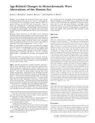
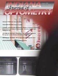
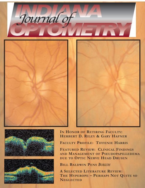
ON THE COVER: Figures 2 (upper left), 4 (middle left), 5 (lower left), and 8 (upper right), from the<br />
article by Tiffenie Harris. See her article on page 6 for further explanation.<br />
Correspondence and manuscripts submitted for publication should be sent to the Editor: David A.<br />
Goss, <strong>School</strong> <strong>of</strong> <strong>Optometry</strong>, <strong>Indiana</strong> <strong>University</strong>, Bloomington, IN 47405 USA (or<br />
dgoss@indiana.edu). Business correspondence should be addressed to the Production Manager:<br />
J. Craig Combs, <strong>School</strong> <strong>of</strong> <strong>Optometry</strong>, <strong>Indiana</strong> <strong>University</strong>, Bloomington, IN 47405 USA (or jocombs<br />
@indiana.edu). Address changes or subscription requests should be sent to Sue Gilmore, <strong>School</strong><br />
<strong>of</strong> <strong>Optometry</strong>, <strong>Indiana</strong> <strong>University</strong>, Bloomington, IN 47405 USA (or sgilmore@indiana.edu).<br />
Our appreciation is extended to Essilor <strong>of</strong> America for<br />
financial support <strong>of</strong> this publication.<br />
Varilux® is a registered trademark <strong>of</strong> Essilor International, S.A
<strong>Spring</strong> <strong>2007</strong><br />
Volume 10, Number 1<br />
Table <strong>of</strong> Contents<br />
<strong>Indiana</strong> <strong>University</strong> <strong>School</strong> <strong>of</strong> <strong>Optometry</strong><br />
Administration:<br />
Gerald E. Lowther, O.D., Ph.D.,<br />
Dean<br />
Clifford W. Brooks, O.D.,<br />
Director,<br />
Optician/Technician Program<br />
Daniel R. Gerstman, O.D., M.S.,<br />
Executive Associate Dean for<br />
Budgetary Planning and<br />
Administration<br />
Joseph A. Bonanno, Ph.D.,<br />
Associate Dean for Research<br />
Steven A. Hitzeman, O.D.,<br />
Director <strong>of</strong> Clinics<br />
Edwin C. Marshall, O.D., M.S.,<br />
M.P.H., Associate Dean for<br />
Academic Affairs<br />
William Swanson,, Ph.D.,<br />
Associate Dean for<br />
Graduate Programs<br />
Sandra L. Pickel, B.G.S., A.S.,<br />
Opt.T.R., Associate Director,<br />
Optician/Technician Program<br />
Cindy Vance,<br />
Director <strong>of</strong> Student Administration<br />
<strong>Indiana</strong> Journal <strong>of</strong> <strong>Optometry</strong><br />
Editor:<br />
David A. Goss, O.D., Ph.D.<br />
Editorial Board:<br />
Arthur Bradley, Ph.D.<br />
Clifford W. Brooks, O.D.<br />
Daniel R. Gerstman, O.D., M.S.<br />
Victor E. Malinovsky, O.D.<br />
Neil A. Pence, O.D.<br />
Production and Layout<br />
J. Craig Combs, M.H.A.<br />
TABLE OF CONTENTS<br />
IN HONOR OF RETIRING FACULTY:<br />
Herbert D. Riley, by David A. Goss and Richard E. Meetz<br />
Gary Hafner, by Paul A. Pietsch ……………………………… 2<br />
FACULTY PROFILE:<br />
Tiffenie Harris, by Victor Malinovsky ………………………… 5<br />
FEATURED REVIEW:<br />
Clinical Findings and Management <strong>of</strong> Pseudopapilledema<br />
due to Optic Nerve Head Drusen<br />
by Tiffenie Harris ……...…………………………………...… 6<br />
BOOK NOTICE: BORISH<br />
Bill Baldwin Pens Biography <strong>of</strong> Irvin Borish<br />
by David A. Goss ………........................................…….…… 10<br />
LITERATURE REVIEW:<br />
The Hyperope – Perhaps Not Quite so Neglected: A<br />
Selected Literature Review<br />
by David A. Goss ………………………………………...…... 12<br />
ANNOUNCEMENT<br />
Optometric Historical Society Launches a New Publication ...... 17<br />
Statement <strong>of</strong> Purpose: The <strong>Indiana</strong> Journal <strong>of</strong> <strong>Optometry</strong> is published by the <strong>Indiana</strong> <strong>University</strong><br />
<strong>School</strong> <strong>of</strong> <strong>Optometry</strong> to provide members <strong>of</strong> the <strong>Indiana</strong> Optometric Association, Alumni <strong>of</strong> the<br />
<strong>Indiana</strong> <strong>University</strong> <strong>School</strong> <strong>of</strong> <strong>Optometry</strong>, and other interested persons with information on the<br />
research and clinical expertise at the <strong>Indiana</strong> <strong>University</strong> <strong>School</strong> <strong>of</strong> <strong>Optometry</strong>, and on new<br />
developments in optometry/vision care.<br />
The <strong>Indiana</strong> Journal <strong>of</strong> <strong>Optometry</strong> and <strong>Indiana</strong> <strong>University</strong> are not responsible for the opinions and<br />
statements <strong>of</strong> the contributors to this journal. The authors and <strong>Indiana</strong> <strong>University</strong> have taken care<br />
that the information and recommendations contained herein are accurate and compatible with the<br />
standards generally accepted at the time <strong>of</strong> publication. Nevertheless, it is impossible to ensure that<br />
all the information given is entirely applicable for all circumstances. <strong>Indiana</strong> <strong>University</strong> disclaims<br />
any liability, loss, or damage incurred as a consequence, directly or indirectly, <strong>of</strong> the use and<br />
application <strong>of</strong> any <strong>of</strong> the contents <strong>of</strong> this journal. This journal is also available on the world wide<br />
web at: http://www.opt.indiana.edu/IndJOpt/home.html
In Honor <strong>of</strong> Retiring Faculty:<br />
Herbert D. Riley, by David A. Goss and Richard E. Meetz<br />
Gary Hafner, by Paul A. Pietsch<br />
Editor’s Note: The close <strong>of</strong> the 2006-<strong>2007</strong> academic<br />
year will mark the retirements <strong>of</strong> two faculty<br />
members who, between them, will have contributed<br />
65 years <strong>of</strong> instruction in the <strong>Indiana</strong> <strong>University</strong><br />
<strong>School</strong> <strong>of</strong> <strong>Optometry</strong>. They are Herbert Riley and<br />
Gary Hafner. Each year retiring faculty on the<br />
Bloomington campus <strong>of</strong> <strong>Indiana</strong> <strong>University</strong> are<br />
honored at a ceremony and banquet. Biographical<br />
sketches <strong>of</strong> each retiree are assembled and<br />
published in a program book associated with that<br />
ceremony. In the past two years these biographical<br />
sketches have also been included at the website <strong>of</strong><br />
the <strong>Indiana</strong> <strong>University</strong> Office <strong>of</strong> Academic Affairs and<br />
Dean <strong>of</strong> the Faculties. Presented here are<br />
biographical sketches prepared for Pr<strong>of</strong>essors Riley<br />
and Hafner.<br />
Herbert D. Riley<br />
Herb Riley is retiring in the <strong>Spring</strong> <strong>of</strong> 2006,<br />
at the completion <strong>of</strong> his 35th year as a faculty<br />
member in the <strong>Indiana</strong> <strong>University</strong> <strong>School</strong> <strong>of</strong><br />
<strong>Optometry</strong>. He leaves behind a<br />
record <strong>of</strong> dedication to teaching<br />
students the rudiments <strong>of</strong> optometric<br />
procedure and clinical care. Herb<br />
was born October 28, 1943 in<br />
Delano, California. Herb’s father<br />
died when he was three, and his<br />
mother moved the family to<br />
Maquoketa, Iowa, where he lived for<br />
the rest <strong>of</strong> his childhood. His undergraduate<br />
education was undertaken in Iowa, and he<br />
earned the Doctor <strong>of</strong> <strong>Optometry</strong> (O.D.) degree<br />
from <strong>Indiana</strong> <strong>University</strong> in 1971. Health<br />
problems dating from his student days didn’t<br />
stop him from completing his education or<br />
from having a career in which he helped<br />
educate well over two thousand optometry<br />
students.<br />
When Herb started optometry school at IU,<br />
classes, labs, and clinics were in various<br />
buildings scattered around the campus.<br />
Construction <strong>of</strong> the current optometry building<br />
was completed in late 1967. Classes began in<br />
the new building in January <strong>of</strong> 1968. Herb<br />
recalls how proud students and faculty were <strong>of</strong><br />
the new building.<br />
After completion <strong>of</strong> optometry school, Herb<br />
joined the <strong>Indiana</strong> <strong>University</strong> optometry faculty.<br />
In his first several years on the faculty, he<br />
served as a full-time clinical instructor in the<br />
general optometry clinic. He also was an<br />
instructor in the contact lens clinic. He<br />
developed a reputation as a well-rounded and<br />
skilled clinician. During this period <strong>of</strong> time,<br />
through the 1970s, optometry was undergoing<br />
an expansion <strong>of</strong> scope. Herb readily<br />
embraced that expansion and helped facilitate<br />
the necessary enhancement <strong>of</strong> training in<br />
ocular disease detection methods.<br />
Later Herb took on responsibility for<br />
teaching in the diagnostic procedures series <strong>of</strong><br />
courses, a role for which he is known by two<br />
decades <strong>of</strong> optometry classes. His work in the<br />
reorganization <strong>of</strong> these courses has been<br />
instrumental in the high ranking <strong>of</strong> <strong>Indiana</strong><br />
<strong>University</strong> optometry students on the Clinical<br />
Skills portion <strong>of</strong> the National Board<br />
Examinations.<br />
When personal computers became<br />
popular, Herb rapidly became adept in their<br />
usage. Integral with his teaching, he<br />
developed a website which contains syllabi,<br />
instructional materials, and photographs <strong>of</strong><br />
ocular disease conditions. The images are <strong>of</strong><br />
high quality and readily illustrate the<br />
conditions and procedures being shown. The<br />
high caliber <strong>of</strong> these pictures and materials is<br />
evidenced by their being used and cited by<br />
other optometry schools and by medical<br />
programs in the United States and across the<br />
globe from the United Kingdom to Thailand.<br />
Appreciation <strong>of</strong> Herb’s teaching skill and<br />
dedication has earned him numerous teaching<br />
awards from students and peers. He has<br />
received awards for teaching both in the clinic<br />
Page 2 ... Vol. 10, No. 1 ... <strong>Spring</strong> <strong>2007</strong> ... <strong>Indiana</strong> Journal <strong>of</strong> <strong>Optometry</strong> ..............................................................................
and in the classroom. A remarkable<br />
consistency <strong>of</strong> fine teaching is evidenced by<br />
the fact that his first teaching recognition came<br />
in 1975 and the most recent in <strong>2007</strong>, a span <strong>of</strong><br />
32 years.<br />
Over the years, Herb has served the<br />
<strong>School</strong> <strong>of</strong> <strong>Optometry</strong> in a variety <strong>of</strong> capacities,<br />
such as Clinic Director in the late 1980s and<br />
Chairman <strong>of</strong> the busy Admissions Committee<br />
in the late 1990s. He has been characterized<br />
as a quiet man who is very capable and who<br />
hasn’t felt compelled to engage in selfpromotion.<br />
Pr<strong>of</strong>essor Emeritus Paul Pietsch,<br />
a long-time colleague, referred to Herb as “a<br />
good human being whom I admire as a<br />
person.” Another faculty member appreciated<br />
Herb’s “deliberate approach” to problems and<br />
noted that in cases <strong>of</strong> disagreement, Herb<br />
would almost apologetically say that he<br />
“wouldn’t do it that way,” rather than openly<br />
criticize.<br />
Herb says that he will miss a lot <strong>of</strong> “great<br />
people” that he has gotten to know in the<br />
<strong>School</strong> <strong>of</strong> <strong>Optometry</strong>. He notes that he has<br />
taught sons and daughters <strong>of</strong> former students.<br />
He recalls mentioning that fact to Henry<br />
H<strong>of</strong>stetter and saying that it was making him<br />
feel old. H<strong>of</strong>stetter replied, “Wait until you<br />
teach their grandchildren.” Herb is retiring<br />
before that happens.<br />
Herb met his wife Phyllis at the <strong>School</strong> <strong>of</strong><br />
<strong>Optometry</strong>. She worked for many years in the<br />
clinic and in the ophthalmic lens laboratory.<br />
They hope to do some traveling during his<br />
retirement and spend some time in Florida.<br />
They enjoy traveling through the United States<br />
by automobile. Herb says that he also wants<br />
to do some fishing, which he hasn’t had time<br />
for in several years.<br />
(Historical note: Dr. Riley’s first name has<br />
been spelled Hurbert for many years in the IU<br />
catalogues, but he learned lately that the<br />
spelling on his birth certificate is actually<br />
Herbert, which he prefers.)<br />
Gary Hafner<br />
Gary Stuart Hafner, Ph.D., Pr<strong>of</strong>essor <strong>of</strong><br />
<strong>Optometry</strong> and Adjunct Pr<strong>of</strong>essor <strong>of</strong> Anatomy<br />
at <strong>Indiana</strong> <strong>University</strong>, is in<br />
the words <strong>of</strong> medical<br />
educator Louis Flexner, an<br />
“anatomist’s anatomist.”<br />
Gary’s teaching, research<br />
and service ran the scope <strong>of</strong><br />
structural biology from<br />
macromolecules in visual<br />
cells to the whole human body. He is among<br />
the world’s authorities on the evolution and<br />
development <strong>of</strong> the crayfish eye. Indeed, his<br />
most recent publication (in Arthropod Structure<br />
and Development, in press), coauthored with<br />
the German zoologist, Steffen Harzsch,<br />
examines a pivotal issue in the evolution <strong>of</strong><br />
invertebrates.<br />
In <strong>Optometry</strong>, his main courses were<br />
Ocular Anatomy and Gross Anatomy. Along<br />
with other major innovations, he introduced<br />
human cadavers into instruction for optometry<br />
students. During the past three years his<br />
teaching included one-third <strong>of</strong> the<br />
neuroscience course for medical students in<br />
the Medical Sciences Program.<br />
Gary was born in Greensboro, North<br />
Carolina in 1943. His family moved to Illinois<br />
where he “grew up on the north shore <strong>of</strong><br />
Chicago.” The Hafners moved again, to<br />
<strong>Indiana</strong>polis, where Gary completed the 8th<br />
grade and went on to pitch baseball for and<br />
graduate from Broad Ripple High <strong>School</strong>. He<br />
earned an A.B. from Hanover College in 1965,<br />
majoring in biology and chemistry and winning<br />
a letter in, <strong>of</strong> course, baseball. (Years later, he<br />
would occasionally enjoy playing catch on<br />
Sunday mornings with friend and academic<br />
role model, Pr<strong>of</strong>essor Emeritus Conrad<br />
Mueller)<br />
After receiving an M.A. in biology at Drake<br />
<strong>University</strong> in 1967, Gary pursued a Ph.D. at IU<br />
in what was then the Department <strong>of</strong> Anatomy<br />
and Physiology. Concentrating on cytology<br />
and neuroanatomy, and assisting in various<br />
basic medical science courses, he earned his<br />
doctorate in 1972. He’d conducted his<br />
dissertation research under C. B. G. (Boyd)<br />
Campbell and H. D. (Dave) Potter.<br />
Before undertaking postdoctoral training, Gary<br />
returned to Hanover College during the fall <strong>of</strong><br />
1972 as an assistant pr<strong>of</strong>essor. In his<br />
............................................................ <strong>Indiana</strong> Journal <strong>of</strong> <strong>Optometry</strong> ... <strong>Spring</strong> <strong>2007</strong> ... Vol. 10, No. 1... page 3
semester there the newly minted cytologist<br />
and neuroanatomist taught not his specialties,<br />
but comparative anatomy and general biology<br />
(again, Flexner’s anatomist’s anatomist).<br />
Gary initiated postdoctoral research at IU<br />
in January <strong>of</strong> 1973 with five-months in Ray<br />
Murray’s lab. In May 1973, he “went west” for<br />
18 months in UCLA’s Jules Stein Eye Institute,<br />
where he collaborated with one <strong>of</strong><br />
neurobiology’s luminaries, Dean Bok. Gary<br />
returned to IU in 1974 for an associateship<br />
with his former mentor, Dave Potter.<br />
In 1976 an outstanding lecture to the<br />
<strong>Optometry</strong> faculty on the ultra-structure <strong>of</strong> the<br />
crayfish eye contributed significantly to Gary’s<br />
successful competition to fill a vacancy<br />
created by the retirement <strong>of</strong> Stanley Rafalko.<br />
Rafalko had taught ocular and general<br />
anatomy from early in the history <strong>of</strong> the<br />
optometric curriculum at IU.<br />
Gary was expected to modernize and <strong>of</strong>fer<br />
those courses, as well as develop instruction<br />
for graduate students. In addition, he was<br />
charged with renovating <strong>Optometry</strong>’s barely<br />
functional electron microscope laboratory and<br />
maintaining a ‘high–tech’ microscopy facility to<br />
support the research and teaching <strong>of</strong> others in<br />
the school. To operate the new lab, Gary<br />
brought in Tom Tokarski, whom he’d known<br />
since graduate school. Although Tom did not<br />
hold a doctorate, Gary treated him as a<br />
colleague. They coauthored 11 papers. At<br />
Gary’s encouraging, Tom also undertook<br />
projects <strong>of</strong> his own and even published with<br />
others. Tom is now retired.<br />
Gary was tenured on schedule; voted<br />
teacher <strong>of</strong> the year for 1976-77; and promoted<br />
to full pr<strong>of</strong>essor in 1993.<br />
How did he treat students? Consider<br />
words <strong>of</strong> Tiffenie Harris: “I first met Dr. Hafner<br />
in 1987 [in] a summer program for<br />
undergraduate minority students … Dr. Hafner<br />
was very patient … and taught me how to<br />
make frozen sections … <strong>of</strong> the crayfish retina.”<br />
Tiffenie would eventually earn an OD degree.<br />
Now a member <strong>of</strong> the <strong>Optometry</strong> faculty<br />
herself, she adds, “His anatomy courses laid<br />
the foundation for us to become competent<br />
optometrists, well-versed in ocular disease<br />
and its systemic associations.”<br />
David Goss, Pr<strong>of</strong>essor <strong>of</strong> <strong>Optometry</strong> and a<br />
colleague <strong>of</strong> Gary’s, sat in on Ocular Anatomy<br />
while was pursuing a Ph.D. Goss still<br />
remembers the course as, “well done, well<br />
organized and authoritative.”<br />
Douglas Freeman, Head <strong>Optometry</strong><br />
Librarian and <strong>Optometry</strong>’s Director <strong>of</strong><br />
Technology volunteers, “Dr. Hafner was one <strong>of</strong><br />
the first people in the <strong>School</strong> to embrace<br />
electronic technologies for instructional<br />
purposes. He introduced specialized s<strong>of</strong>tware<br />
for teaching … and revolutionized the learning<br />
experience for optometry students.”<br />
Gary married Jane Gray in 1978. They’d<br />
both attended Broad Ripple High but only got<br />
to know each other while she was completing<br />
a Ph.D. at IU. Jane is now a retired plant<br />
scientist.<br />
In 1994, while helping with a neighbor’s<br />
renovation project, Gary sustained a major<br />
injury to his right leg. After a prolonged but<br />
futile battle against infection, his lower leg had<br />
to be amputated. Fitted with a prosthetic foot,<br />
and able to walk without a cane, he soon was<br />
back on the job cheerfully, teaching, doing<br />
research and performing service.<br />
Service to the university as well as the<br />
community represented an essential obligation<br />
for Gary. Among other things, he was on the<br />
Monroe County’s Plan Commission. In<br />
addition to several significant school and<br />
university assignments, he chaired<br />
<strong>Optometry</strong>’s tenure and promotions committee<br />
for several years. Dave Goss, who served<br />
with Gary, said “He approached the work <strong>of</strong><br />
the committee with objectivity and common<br />
sense.”<br />
In retirement, would he and Jane travel?<br />
“Some,” he replied. But he intends to put his<br />
ocular anatomy teaching materials into<br />
electronic form. He also has an interest in<br />
woodworking and collecting old hand tools.<br />
But, given the free time, he especially wants<br />
“to restore a 1949 Chevy pickup truck.”<br />
The Hafner Era now closes in <strong>Optometry</strong>,<br />
in tangible ways. Of course, Gary Hafner will<br />
be missed, day-to-day. But his impact will<br />
always exist in the character and conduct <strong>of</strong><br />
his students, his colleagues and their<br />
intellectual descendants.<br />
Page 4 ... Vol 10, No. 1 ... <strong>Spring</strong> <strong>2007</strong> ... <strong>Indiana</strong> Journal <strong>of</strong> <strong>Optometry</strong> ..............................................................
Faculty Pr<strong>of</strong>ile:<br />
Tiffenie Harris, O.D.<br />
by Victor Malinovsky<br />
Dr. Tiffenie Harris attended <strong>Indiana</strong><br />
<strong>University</strong>, receiving her Bachelors <strong>of</strong><br />
Arts in Chemistry in 1989, and<br />
graduating from <strong>Indiana</strong> <strong>University</strong> <strong>School</strong> <strong>of</strong><br />
<strong>Optometry</strong> in 1993. While<br />
attending optometry school,<br />
she taught as an associate<br />
instructor for Gross Anatomy,<br />
Ocular Anatomy,<br />
Neuroanatomy, and Ocular<br />
Pharmacology in the IU<br />
<strong>School</strong> <strong>of</strong> <strong>Optometry</strong>. In<br />
addition, she taught as an<br />
associate instructor for Gross Anatomy in the<br />
<strong>Indiana</strong> <strong>University</strong> <strong>School</strong> <strong>of</strong> Medicine,<br />
Medical Sciences Department. After<br />
graduation, she moved to Southeastern<br />
Michigan and practiced primary care for over<br />
ten years. Her clinical experience includes<br />
working in a variety <strong>of</strong> urban practice settings,<br />
including a multi-discipline medical center.<br />
The demographics <strong>of</strong> this community provided<br />
a wide diversity <strong>of</strong> challenging cases and a<br />
large amount <strong>of</strong> pathology to diagnose, treat,<br />
and manage. She has cared for patients <strong>of</strong> all<br />
ages; the majority have been in the young<br />
adult to mature adult (>50) populations,<br />
including those with physical and mental<br />
disabilities.<br />
In 2004, Dr. Harris joined the IU <strong>School</strong> <strong>of</strong><br />
<strong>Optometry</strong> faculty as an Assistant Clinical<br />
Pr<strong>of</strong>essor and became the Chief <strong>of</strong> Primary<br />
Care. In addition to administrative duties, her<br />
responsibilities include the clinical training and<br />
teaching <strong>of</strong> third year optometry interns,<br />
emphasizing treatment and management <strong>of</strong><br />
ocular pathology, contact lenses, as well as<br />
refractive anomalies. Dr. Harris has served as<br />
an examiner for the clinical skills portion <strong>of</strong> the<br />
National Boards and serves on the school’s<br />
Admissions Committee as well as the Clinical<br />
Care Quality Assurance Committee. Recently,<br />
she was elected to serve as the Faculty<br />
Presider for the school’s faculty meetings.<br />
She attended the American Optometric<br />
Association/American Academy <strong>of</strong> <strong>Optometry</strong><br />
Summer Research Institute in 2006. As a<br />
result <strong>of</strong> this experience, she has been<br />
actively involved in developing a community<br />
based research project. Since joining the<br />
faculty, Dr. Harris was voted the Consultant <strong>of</strong><br />
the Year by the Classes <strong>of</strong> 2006 and <strong>2007</strong>.<br />
She was also awarded the IU Trustees Award<br />
in 2006. Dr. Harris is currently submitting<br />
posters and case reports, with a special<br />
interest in neuro-ophthalmology, in order to<br />
gain the post-graduate distinction <strong>of</strong> fellowship<br />
in the American Academy <strong>of</strong> <strong>Optometry</strong>.<br />
Dr. Harris finds the most rewarding aspect<br />
<strong>of</strong> the educational experience at IU to be the<br />
diversity <strong>of</strong> students and faculty on the<br />
Bloomington Campus. She says studying and<br />
practicing optometry have prepared her to<br />
become a conscientious, goal-oriented<br />
individual both pr<strong>of</strong>essionally and personally.<br />
Academia in optometry has allowed her to<br />
have a positive impact not only on patients,<br />
but also on the next generation <strong>of</strong><br />
optometrists. Her new faculty position has<br />
allowed her to fulfill her passion for teaching<br />
and continue her patient care.<br />
Fellow faculty members appreciate the<br />
excellent rapport that Dr. Harris consistently<br />
demonstrates with her peers, students and<br />
patients. She is also well known for her<br />
dedicated follow-up in patient care.<br />
Tiffenie is married to Walt Harris, who is a<br />
dedicated police <strong>of</strong>ficer in Bloomington and a<br />
former IU football player. They enjoy<br />
spending time and traveling in their van with<br />
their three children: Walter, 9; Joshua, 6; and<br />
Victoria, 4.<br />
............................................................ <strong>Indiana</strong> Journal <strong>of</strong> <strong>Optometry</strong> ... <strong>Spring</strong> <strong>2007</strong> ... Vol 10, No. 1... page 5
Clinical Findings and Management <strong>of</strong><br />
Pseudopapilledema due to Optic Nerve Head<br />
Drusen<br />
by Tiffenie Harris, O.D.<br />
Diagnostic Dilemma<br />
Optic nerve head (ONH) elevation tends to be<br />
an intimidating ocular finding especially when it is<br />
bilateral. In every case <strong>of</strong> ONH elevation, an<br />
evaluation <strong>of</strong> signs and symptoms will help the<br />
practitioner determine effectively if the nerve is in<br />
fact “swollen” or “elevated”. This will provide an<br />
organized approach to the case with logical<br />
differential diagnoses. The optometrist’s role<br />
includes detection, timely and appropriate referral<br />
to specialists, and co-management (monitoring)<br />
once under the care <strong>of</strong> a physician. A clinical case<br />
in which the optic nerves are moderate to severely<br />
swollen is easy to diagnose. A case in which the<br />
patient presents with symptoms <strong>of</strong> headaches and<br />
the nerves appear only mildly elevated makes the<br />
diagnosis very challenging. The diagnostic<br />
dilemma begins with making the clinical decision<br />
and answering the question, “is it or is it not<br />
swollen?”<br />
The foremost clinical goal is to differentiate<br />
congenital causes <strong>of</strong> disc elevation from acquired<br />
disc edema. Papilledema (PE) is acquired bilateral<br />
optic disc swelling due to increased intracranial<br />
pressure (ICP). Pseudopapilledma (PPE) is the<br />
appearance and false impression <strong>of</strong> bilateral disc<br />
swelling that is associated with an underlying<br />
anomalous condition. (Table 1) The most common<br />
cause <strong>of</strong> pseudopapilledema is optic nerve head<br />
drusen (ONHD) which is a congenital anomaly<br />
characterized by disc elevation and blurred<br />
margins, which is 75% bilateral. 1,2 It is critical for<br />
clinicians to differentiate papilledema from ONHD<br />
in order to avoid unnecessary and expensive<br />
neurological testing. It is equally important to avoid<br />
overlooking true, neurological disorders.<br />
autosomal dominant trait occurring in<br />
approximately 1% <strong>of</strong> the population. 1-5 The<br />
scleral canal and optic disc <strong>of</strong> eyes with drusen are<br />
much smaller than average. This is seen clinically<br />
where ONHD predominately occurs in Caucasians<br />
and rarely in African Americans, whose scleral<br />
canal size is <strong>of</strong>ten larger. 2 There are two types <strong>of</strong><br />
ONHD, visible and buried. (Figures 1 and 2)<br />
Visible ONHD protrude from the surface <strong>of</strong> the<br />
disc. They are particularly prominent at the<br />
margins causing irregular borders and elevation.<br />
Buried ONHD are located beneath the disc surface<br />
and are not directly visible. They can cause<br />
elevation <strong>of</strong> the optic disc with or without blurred<br />
margins. ONHD is associated with peripapillary<br />
retinal pigment epithelial changes (33%) and an<br />
absent to very small cup-to-disc ratio. 1-3<br />
Anomalous disc vasculature (10%), including early<br />
branching <strong>of</strong> the retinal vessels and secondary<br />
tortuosity, is also associated with ONHD. 1-5 The<br />
pathogenesis <strong>of</strong> ONHD is not yet fully understood.<br />
Current research suggests an abnormally narrow<br />
opening <strong>of</strong> the scleral canal can cause a stasis <strong>of</strong><br />
axoplasmic flow. 1-6 This leads to abnormal axonal<br />
metabolism and mitochondrial calcifications<br />
creating calcium-like globular deposits within the<br />
papilla.<br />
Figure 1 (left). Visible optic<br />
nerve head drusen (ONHD).<br />
Background on ONHD<br />
ONHD are congenital, inherited as an<br />
Optic nerve head drusen (ONHD)<br />
Congenitally full disc (CFD)<br />
Malinserted oblique insertion<br />
Tilted disc syndrome<br />
Optic nerve hypoplasia<br />
(differential diagnosis for bilateral presentation)<br />
Table 1. Differential diagnosis <strong>of</strong> pseudopapilledema.<br />
Figure 2 (right). Buried ONHD.<br />
This figure also appears in color<br />
on the cover.<br />
Page 6 ... Vol 10, No. 1 ... <strong>Spring</strong> <strong>2007</strong> ... <strong>Indiana</strong> Journal <strong>of</strong> <strong>Optometry</strong> ..............................................................
Drusen <strong>of</strong> the optic nerve head have no<br />
histopathologic connection to retinal drusen and<br />
are not considered age-related, but have a<br />
tendency to become more visible as the patient<br />
ages. In early life, ONHD remain deep in the<br />
nerve and always anterior to the lamina cribosa.<br />
They become more visible at the disc surface in<br />
the second to third decades. 3 In childhood, the<br />
disc may appear papilledema-like with no<br />
physiological cupping, accounting for 75% <strong>of</strong><br />
diagnostically challenging disc anomalies. 3 In<br />
adulthood, ONHD appear as spherical nodules<br />
on the disc surface that are highly reflective by<br />
the ophthalmoscope light and give the disc a<br />
scalloped margin. When illuminated with a redfree<br />
light these refractile bodies will auto<br />
fluoresce.<br />
Most patients with ONHD are asymptomatic.<br />
However, many can present with visual field<br />
defects and rarely with decreased visual acuities.<br />
ONHD can shear and damage the nerve fibers<br />
and vascular supply as they move to the disc<br />
surface. The reduction <strong>of</strong> retinal nerve fiber layer<br />
(RNFL) thickness results in visual field defects<br />
including enlarged blind spots, localized<br />
depressions, arcuate nerve fiber bundle defects,<br />
or constrictions. Visual field defects are present<br />
in 73% <strong>of</strong> visible drusen and 36% <strong>of</strong> buried disc<br />
drusen with no significant difference in the<br />
severity. 5,7 Spontaneous disc hemorrhages can<br />
occur in, around, and over the optic nerve head if<br />
progression <strong>of</strong> the drusen interferes with the<br />
nerve’s blood supply. The incidence <strong>of</strong> retinal<br />
hemorrhage is between 2% and 10 %. 2 Visible<br />
disc drusen may cause peripapillary atrophy and<br />
a break in Bruch’s membrane. As a result, the<br />
patient is at risk <strong>of</strong> the development <strong>of</strong><br />
peripapillary choroidal neovascular membranes,<br />
which may extend to the macular or subfoveal<br />
area and compromise vision. 2,8,9<br />
Conditions known to be associated with<br />
ONHD include Retinitis Pigmentosa (RP) and<br />
Angioid Streaks with or without Pseuodxanthoma<br />
Elasticum (PXE). The ONHD in PXE are similar<br />
but in RP they do not have the same<br />
appearance. 1,2 They tend to be more visible<br />
adjacent to a normal-sized nerve. In addition,<br />
there have been reports <strong>of</strong> non-arteritic anterior<br />
ischemic optic neuropathy (NAION) associated<br />
with ONHD. 1,2,10<br />
Diagnostic Testing<br />
Differential diagnosis <strong>of</strong> ONHD includes any<br />
entity that can cause bilateral optic disc<br />
elevation. This includes increased intracranial<br />
pressure and congenital disc anomalies. Special<br />
investigations for the definitive diagnosis <strong>of</strong><br />
bilateral disc elevation include the following:<br />
Magnetic Resonance Imaging (MRI) and/or<br />
Computerized Tomography (CT) scans <strong>of</strong> the<br />
orbit and brain, Flourescein Angiography, B-scan<br />
ultrasonography, and Optical Coherence<br />
Technology (OCT). The CT scan can detect<br />
intracranial tumors as well as ONHD. However, it<br />
is not sensitive enough to pick up subtle calcific<br />
drusen in the disc and is not reliable for ONHD<br />
diagnosis. 7 The MRI is superior to CT scan for<br />
s<strong>of</strong>t tissue structures and is the preferred<br />
imaging test to rule out the etiology <strong>of</strong> ICP.<br />
Flourescein Angiography (FA) can be helpful.<br />
With papilledema the optic nerve head will show<br />
hyperflouresence and peripapillary leakage <strong>of</strong> the<br />
dye. In ONHD there is no peripapillary leakage<br />
<strong>of</strong> the dye, especially in the late phase. In<br />
addition, the red-free barrier used in the preinjection<br />
phase illuminates the aut<strong>of</strong>lourescent<br />
property <strong>of</strong> disc drusen. Buried ONHD with both<br />
<strong>of</strong> these techniques can be missed. Less<br />
invasive diagnostic investigations are available to<br />
differentiate disc edema and buried disc drusen.<br />
B-Scan ultrasonography has been shown to<br />
be the most sensitive and diagnostically relevant<br />
test to aid in the differential diagnoses. 1,4,7,11<br />
B-scan ultrasonography uses high-frequency<br />
sound waves. The sound waves are reflected<br />
back to the probe, converted into an image,<br />
which is used to make a dynamic evaluation <strong>of</strong><br />
the optic disc. When calcification <strong>of</strong> tissue is<br />
present, there is a very strong reflection <strong>of</strong> the<br />
echo back to the probe. Therefore, B-scan<br />
ultrasonography is the single most important<br />
ancillary test to perform in the diagnosis <strong>of</strong><br />
ONHD. The results for ONHD will show a highly<br />
reflective nodule within the optic nerve even at a<br />
Figure 3. B-scan <strong>of</strong> ONHD.<br />
...........................................<strong>Indiana</strong> Journal <strong>of</strong> <strong>Optometry</strong> ... <strong>Spring</strong> <strong>2007</strong> ... Vol 10, No. 1... page 7
low gain level. (Figure 3) In the presence <strong>of</strong><br />
acquired disc edema, the B-scan will demonstrate<br />
a circle within the optic nerve sheath, separating<br />
the sheath from the optic nerve at a standard gain<br />
level. This is called a “crescent sign” produced by<br />
the increased cerebral spinal fluid transmitted<br />
along the subdural space within the optic nerve. 11<br />
Optical Coherence Tomography (OCT) is<br />
analogous to the B-Scan except light, rather than<br />
sound, is used to provide images <strong>of</strong> the ocular<br />
structures. OCT is an objective, noninvasive<br />
alternative to analyze the optic nerve head. More<br />
importantly, it is useful in quantifying the status <strong>of</strong><br />
the RNFL. 6 In<br />
papilledema, the OCT<br />
shows an elevated<br />
nerve (Figure 4) head<br />
along with excessive<br />
thickening <strong>of</strong> the<br />
RNFL. In ONHD, the<br />
Figure 4. OCT <strong>of</strong> papilledema. This figure also<br />
appears in color on the cover.<br />
Figure 5. OCT <strong>of</strong> ONHD. This figure also<br />
appears in color on the cover.<br />
Figure 6. Retinal nerve fiber layer (RNFL)<br />
analysis <strong>of</strong> buried ONHD. RNFL thickness in<br />
microns is on the y-axis.<br />
Figure 7. Retinal nerve fiber layer analysis <strong>of</strong> early<br />
papilledema.<br />
OCT shows the<br />
elevation <strong>of</strong> the disc<br />
and the underlying<br />
nodular shadows<br />
caused by the drusen.<br />
(Figure 5) The key<br />
differential between<br />
ONHD (Figure 6) and<br />
acquired disc edema<br />
(Figure 7) is that there<br />
is significant thinning<br />
with ONHD and<br />
thickening in disc<br />
edema. In cases <strong>of</strong><br />
papilledema the RNFL<br />
curve falls above the<br />
normative, ageadjusted<br />
scale<br />
exceeding 200<br />
microns. Many cases<br />
<strong>of</strong> early papilledema<br />
can mimic buried<br />
ONHD. (Figure 8) As<br />
seen in Figure 8 the<br />
nasal margins are<br />
blurred and the nerve<br />
appears slightly<br />
hyperemic. It also<br />
appears to be buried<br />
ONHD or CFD. The<br />
OCT results for Figure<br />
8 are shown in Figure<br />
7 and clearly show that<br />
the RNFL layer is<br />
Figure 8. Early papilledema. This<br />
figure also appears in color on the<br />
cover.<br />
significantly thickened as<br />
seen in early papilledema.<br />
In cases <strong>of</strong> Congenitally<br />
Full/Crowded Disc, the<br />
OCT will show a normal<br />
to slightly thickened RNFL<br />
curve that stays within the<br />
age-adjusted scale.<br />
Figures 6 and 7<br />
demonstrate the<br />
application <strong>of</strong> testing<br />
RNFL to aid in<br />
differentiating early PE<br />
from PPE.<br />
Making the Diagnosis<br />
Making the clinical<br />
diagnosis begins with a<br />
comprehensive history.<br />
During the interview, the<br />
practitioner is listening for<br />
symptoms associated with increased intracranial<br />
pressure and any associated neurological defects.<br />
(Table 2) Visual acuities, pupils, color vision,<br />
EOMs, and visual field screening are important to<br />
determine if there is any nerve dysfunction.<br />
Always consider the patient’s age, demographics,<br />
and ocular/medical history including in-<strong>of</strong>fice blood<br />
pressure readings.<br />
A dilated fundus examination including<br />
stereoscopic views <strong>of</strong> the disc provides the most<br />
effective optic nerve evaluation. There are key<br />
characteristics to look for in determining disc<br />
edema from elevation. Start with the overall disc<br />
appearance looking at the size, cup, margins,<br />
neuroretinal rim tissue color, and taking note <strong>of</strong><br />
any spontaneous venous pulsation (SVP). Then<br />
study the vasculature <strong>of</strong> the disc and surrounding<br />
tissue. In the presence <strong>of</strong> papilledema, the ONH<br />
will appear elevated and hyperemic with blurred<br />
margins that will obscure peripapillary vessels as<br />
they leave the disc. The swelling includes no SVP<br />
along with venous congestion with flame-shaped<br />
hemorrhages and cotton wool spots. Buckling or<br />
retinal folds <strong>of</strong> the temporal aspect <strong>of</strong> the disc<br />
(Paton’s lines) may be present. In contrast, ONHD<br />
will appear non-hyperemic and the peripapillary<br />
vessels are not obscured nor will there be any<br />
Headaches (HA)<br />
Transient visual obscurations (TVO)<br />
Tinnitus – “whooshing sound”<br />
Diplopia<br />
(HA and TVO most common)<br />
Table 2. Symptoms <strong>of</strong> papilledema.<br />
Page 8 ... Vol 10, No. 1 ... <strong>Spring</strong> <strong>2007</strong> ... <strong>Indiana</strong> Journal <strong>of</strong> <strong>Optometry</strong> ...........................................................
cotton wool spots (CWS). Paton’s lines are not<br />
associated with pseudopapilledema. The clinician<br />
should look for anomalous vascular patterns<br />
including tortuosity as well as a positive SVP,<br />
which may be present. Clinical diagnosis and<br />
decisions are made based on the appearance <strong>of</strong><br />
the optic nerves and information provided by the<br />
ancillary tests.<br />
Now what to do about it?<br />
Once the clinician determines that the discs are<br />
in fact swollen, the patient must be sent for<br />
neuroimaging within 24 hours to identify the source<br />
<strong>of</strong> increased ICP. Take stereo disc photos, get a<br />
baseline threshold visual field, and order the MRI.<br />
The visual field defects associated with<br />
papilledema start as an enlarged blind spot.<br />
Depending on location and severity <strong>of</strong> a brain<br />
lesion, neurological visual field defects such as<br />
quadrantanopsia or hemianopsia can occur.<br />
In addition to visual field testing, the OCT<br />
provides an available option for long-term follow-up<br />
<strong>of</strong> the changes in RNFL thickness. 6,12 This is<br />
helpful in monitoring the resolution <strong>of</strong> papilledema<br />
as well as monitoring RNFL loss in ONHD.<br />
Patients with ONHD can develop visual field<br />
defects and RNFL loss that resembles glaucoma.<br />
Glaucoma and ONHD can coexist. 1,12-14 The<br />
OCT cannot distinguish nerve fiber layer loss that<br />
occurs from ONHD from that which occurs as a<br />
result <strong>of</strong> glaucoma. Documentation <strong>of</strong> visual field<br />
loss and RNFL reduction provides a baseline for<br />
progression.<br />
The lack <strong>of</strong> significant disc cupping <strong>of</strong> nerves<br />
with drusen contributes to the difficulty in managing<br />
patients who develop glaucoma. Progression <strong>of</strong><br />
cup size is <strong>of</strong>ten undeterminable and the clinician<br />
can only use intraocular pressures as a guide for<br />
treatment and management. In cases with nerve<br />
fiber loss due to ONHD, established from baseline<br />
OCT and Humphrey visual fields (HVF) testing, the<br />
IOP should be lowered to protect the vulnerable<br />
optic nerve fibers. If there is a significant increase<br />
in IOP from baseline readings, the clinician needs<br />
to consider initiating glaucoma treatment,<br />
preferably with Alphagan-P. This ophthalmic drug<br />
will lower the IOP and may have neuroprotective<br />
properties potentially reducing the patient’s risk <strong>of</strong><br />
further damage to the RNFL.<br />
There is no existing treatment for ONHD. An<br />
annual comprehensive eye examination along with<br />
proper diagnosis and patient education is the best<br />
available modality <strong>of</strong> care. Generally, ONHD is<br />
without visual significance. However, patients<br />
need to be aware <strong>of</strong> potential complications that<br />
could affect vision. Patients with ONHD should<br />
undergo regular dilated fundus examinations along<br />
with visual field testing (HVF 24-2), stereoscopic<br />
disc photos, IOP measurement, and nerve fiber<br />
layer examinations for future monitoring. With the<br />
aid <strong>of</strong> diagnostic tests such as B-scan and OCT,<br />
clinicians can avoid unnecessary doubts and<br />
concerns along with expensive neurological<br />
investigations while making the correct diagnosis in<br />
bilateral optic nerve elevation.<br />
References<br />
1. Alexander LJ. Primary Care <strong>of</strong> the Posterior<br />
Segment. New York: McGraw-Hill, 2002:209-315.<br />
2. Auw-Haedrich C, Staubach F, Witschel H. Optic disk<br />
drusen. Survey Ophthalmol 2002; 47:515-532.<br />
3. Wray SH, Paven-Langston D. Neurophthalmology:<br />
visual fields, optic nerve, and pupil. In: Pavan-Langston<br />
D, ed. Manual <strong>of</strong> Ocular Diagnostics and Therapy, 5th<br />
ed. Philadelphia: Lippincott Williams and Wilkins,<br />
2002:365-397.<br />
4. Kanski JJ. Clinical Ophthalmology: A Systematic<br />
Approach, 5th ed. Edinburgh: Butterworth-Heinemann,<br />
2003:613-616.<br />
5. Wilkins J, Pomeranz H. Visual manifestations <strong>of</strong><br />
visible and buried optic disc drusen. J Neuro-<br />
Ophthalmol 2004;24:125-129.<br />
6. Schuman J, Puliafito C. Optical Coherence<br />
Tomography <strong>of</strong> Ocular Diseases. Thor<strong>of</strong>are, NJ: Slack<br />
Incorporated, 2004: 611-651.<br />
7. Kurz-Levin M, Landau K. A comparison <strong>of</strong> imaging<br />
techniques for diagnosing drusen <strong>of</strong> the optic nerve<br />
head. Arch Ophthalmol 1999; 117: 1045-1049.<br />
8. Hordines H. Optic nerve head drusen causing disc<br />
hemorrhage and disc edema. Clin Eye Vision Care 1999;<br />
11:109-113.<br />
9. Sullu Y, Yildiz L. Submacular surgery for choroidal<br />
neovascularization secondary to optic nerve drusen. Am<br />
J Ophthalmol 2003;136: 367-370.<br />
10. Purvin V, King R, Yee R. Anterior ischemic optic<br />
neuropathy in eyes with optic disc drusen. Arch<br />
Ophthalmol 2004;122:48-53.<br />
11. Byrne SF, Green RL. Ultrasound <strong>of</strong> the Eye and<br />
Orbit. St. Louis: Mosby, 2002:434-437.<br />
12. Ocakoglu O, Ustundag C. Long term follow-up <strong>of</strong><br />
retinal nerve fiber layer thickness in eyes with optic nerve<br />
head drusen. Current Eye Res 2003; 26:277-280.<br />
13. Roh S, Noecker R, Schuman J. Effect <strong>of</strong> optic nerve<br />
head drusen on nerve fiber layer thickness. Ophthalmol<br />
1998;105: 878-885.<br />
14. Piccone M, Piltz-Seymour J. Coexisting optic nerve<br />
head drusen and glaucoma. Ophthalmol 1998;105:761-<br />
762.<br />
....................................................................<strong>Indiana</strong> Journal <strong>of</strong> <strong>Optometry</strong> ... <strong>Spring</strong> <strong>2007</strong> ... Vol 10, No. 1 ... page 9
BOOK NOTICE: BORISH<br />
Bill Baldwin Pens Biography <strong>of</strong> Irvin Borish.<br />
by David A. Goss, O.D., Ph.D.<br />
Borish. William R. Baldwin. <strong>Spring</strong>field, MA:<br />
Bassette, 2006. Pages: xiv + 444. ISBN-10: 1-<br />
4243-1888-2. ISBN-13: 978-1-4243-1888-9.<br />
Given the exceptional achievements and<br />
remarkably long career <strong>of</strong> Irvin M. Borish<br />
and the pivotal role he played in<br />
encouraging and nurturing many developments in<br />
optometric education and practice, it is appropriate<br />
that someone should attempt a book-length<br />
biography. Bill Baldwin has achieved that in fine<br />
fashion.<br />
Borish was born in 1913 in Philadelphia. His<br />
childhood was spent in Philadelphia and in Liberty,<br />
New York under humble circumstances. He<br />
matriculated at Temple <strong>University</strong> with thoughts <strong>of</strong><br />
a career in literature. He changed his mind, and<br />
attended Northern Illinois College <strong>of</strong> <strong>Optometry</strong><br />
(NICO) in Chicago, where he graduated with<br />
highest honors in January <strong>of</strong> 1934. After a brief<br />
period <strong>of</strong> time in optometry practice in Chicago, he<br />
became a full-time faculty member at Northern<br />
Illinois College <strong>of</strong> <strong>Optometry</strong> in January <strong>of</strong> 1936.<br />
His teaching and administrative responsibilities<br />
increased at NICO until he started an optometry<br />
practice in Kokomo, <strong>Indiana</strong> in 1944. From 1973 to<br />
1983, Borish was a full-time faculty member at<br />
<strong>Indiana</strong> <strong>University</strong>. In 1983, he became Benedict<br />
Pr<strong>of</strong>essor <strong>of</strong> Optometric Practice at the <strong>University</strong><br />
<strong>of</strong> Houston. In 1989, Borish retired from Houston,<br />
but continued to write, lecture, and work for the<br />
enhancement and advancement <strong>of</strong> the optometric<br />
pr<strong>of</strong>ession.<br />
Into that biographical framework Baldwin<br />
weaves details <strong>of</strong> Borish’s family and friends,<br />
colleagues, pr<strong>of</strong>essional activities, artistic<br />
endeavors, community contributions, travels,<br />
personality, and philosophy to tell an interesting<br />
story. Baldwin captures the insight, intelligence,<br />
and drive that allowed Borish to rise to the top <strong>of</strong><br />
his pr<strong>of</strong>ession.<br />
The fifteen chapters in the book are organized<br />
thematically, each one addressing a common<br />
grouping <strong>of</strong> activities. The successive chapters<br />
form an overlapping chronology. The years<br />
considered in each <strong>of</strong> the chapters are part <strong>of</strong> the<br />
chapter titles, which are as follows: 1) The Early<br />
Years, 1913-34; 2) Courtship and Marriage, 1932-<br />
43; 3) Northern Illinois College <strong>of</strong> <strong>Optometry</strong>, 1936-<br />
44; 4) Early Service to <strong>Optometry</strong>, 1940-50; 5)<br />
Early Years in Kokomo, 1944-50; 6) Two<br />
Triumphs, 1945-51; 7) Achieving Prominence in<br />
the Community, 1950-56; 8) The Advent <strong>of</strong> Corneal<br />
Contact Lenses, 1952-62; 9) National and State<br />
Health Care Legislation, 1962-82; 10) Personal<br />
Health History, 1925-2006; 11) Bloomington, 1973-<br />
83; 12) Houston, 1983-89; 13) <strong>Optometry</strong> Abroad,<br />
1976-87; 14) Essilor, 1977-95; and 15) The Borish<br />
Version <strong>of</strong> Retirement, 1990-2006.<br />
The nature <strong>of</strong> the content <strong>of</strong> each chapter can<br />
probably be guessed from the titles with the<br />
possible exception <strong>of</strong> Chapter 6, “Two Triumphs,<br />
1945-51.” It deals with the efforts to start the<br />
optometry school at <strong>Indiana</strong> <strong>University</strong> and with the<br />
writing <strong>of</strong> the first edition <strong>of</strong> Clinical Refraction.<br />
The book includes three appendices.<br />
Appendix A, “Borish Views His Pr<strong>of</strong>ession,”<br />
contains excerpts <strong>of</strong> essays and articles that he<br />
wrote on the optometric pr<strong>of</strong>ession. Appendix B,<br />
Page 10 ... Vol 10, No. 1 ... <strong>Spring</strong> <strong>2007</strong> ... <strong>Indiana</strong> Journal <strong>of</strong> <strong>Optometry</strong> ..............................................................................
“Borish’s Wit and Wisdom,” consists <strong>of</strong> some<br />
anecdotes that he has used to emphasize<br />
particular points made in his lectures. In Appendix<br />
C, there are tributes from four <strong>of</strong> his friends and<br />
colleagues and a listing <strong>of</strong> some <strong>of</strong> the most<br />
prestigious awards that he has received.<br />
In researching this book, Baldwin used<br />
extensive interviews <strong>of</strong> Borish, interviews <strong>of</strong><br />
several <strong>of</strong> Borish’s colleagues, his own personal<br />
knowledge <strong>of</strong> Borish, and information gained from<br />
a number <strong>of</strong> optometry schools and organizations.<br />
The book contains more than 170 black and white<br />
photographs <strong>of</strong> Borish, his family, friends, and<br />
colleagues, places that he worked, and places that<br />
he visited. There is an index and an eight-page<br />
color section <strong>of</strong> Borish’s paintings. Some <strong>of</strong><br />
Borish’s paintings also adorn the back and the<br />
flaps <strong>of</strong> the dust jacket. Persons who want to learn<br />
more about Irvin M. Borish or about the history <strong>of</strong><br />
optometry in the twentieth century will find much <strong>of</strong><br />
interest in this book. Because Borish played a role<br />
in so many <strong>of</strong> the educational and practice<br />
developments in optometry over several decades,<br />
there is much optometry history that can be<br />
learned from the book.<br />
A copy <strong>of</strong> the book with a greeting and signed<br />
by Borish can be obtained for a contribution to the<br />
Borish Center <strong>of</strong> $100. A book signed and with a<br />
personal message from Borish can be obtained for<br />
$200. The contribution is partially tax deductible<br />
and checks should be made payable to <strong>Indiana</strong><br />
<strong>University</strong> and sent with the order form to:<br />
<strong>Optometry</strong> Budget Office, IU <strong>School</strong> <strong>of</strong> <strong>Optometry</strong>,<br />
800 East Atwater Avenue, Bloomington, IN 47405.<br />
The order form can be found at<br />
www.opt.indiana.edu/bcor/borishbook/BORISH.pdf<br />
Additional information about the book and<br />
ordering a copy can be obtained at<br />
www.opt.indiana.edu/bcor/borishbook/index.htm,<br />
or by contacting Hillary Person (812-855-0351<br />
or hlheflin@indiana.edu) at the <strong>Indiana</strong><br />
<strong>University</strong> <strong>School</strong> <strong>of</strong> <strong>Optometry</strong>.<br />
............................................................ <strong>Indiana</strong> Journal <strong>of</strong> <strong>Optometry</strong> ... <strong>Spring</strong> <strong>2007</strong> ... Vol 10, No. 1... page 11
The Hyperope - Perhaps Not Quite So<br />
Neglected: A Selected Literature Review<br />
by David A. Goss, O.D., Ph.D.<br />
In 1971, Ted Grosvenor published an article<br />
entitled “The Neglected Hyperope.” 1 In it, he<br />
noted that there were many more published<br />
articles on myopia than on hyperopia and there<br />
were more textbook pages devoted to myopia than<br />
to hyperopia. He also observed that there was<br />
evidence that hyperopia was associated with<br />
reduced reading efficiency. Since then, evidence<br />
has continued to accumulate that hyperopia is<br />
associated with reduced reading ability and poor<br />
school performance, as well as with poor<br />
performance on visual perception, visual cognitive,<br />
and visual motor tests. 2-13<br />
Hyperopia is a risk factor for strabismus and<br />
amblyopia. 14 Amblyopia is more likely to be found<br />
with hyperopia than with myopia. Anisometropic<br />
amblyopia is more frequent with lower amounts <strong>of</strong><br />
hyperopic anisometropia than <strong>of</strong> myopic<br />
anisometropia. 15 And isoametropic amblyopia can<br />
be found in high hyperopia in children. 16 Early<br />
correction <strong>of</strong> hyperopia may reduce the risk <strong>of</strong><br />
strabismus and amblyopia. 17 Headaches are<br />
<strong>of</strong>ten thought to be associated with hyperopia,<br />
which has been confirmed in one study in 11 to 13<br />
year old girls. 18<br />
Work on myopia exploded in the 1990s, but<br />
hyperopia was still fairly neglected through the end<br />
<strong>of</strong> the twentieth century. Thirty-three years after<br />
Grosvenor wrote about the neglected hyperope,<br />
Rosner 19 wrote about the “The Still Neglected<br />
Hyperope” and said that it was “time that our<br />
pr<strong>of</strong>ession took a serious, thorough look at the<br />
possible long-term effects <strong>of</strong> ignoring moderate<br />
hyperopia in asymptomatic young children.”<br />
The fact that there has been a smattering <strong>of</strong><br />
papers on hyperopia published in the first few<br />
years <strong>of</strong> the twenty-first century and the fact that<br />
the February, <strong>2007</strong> issue <strong>of</strong> <strong>Optometry</strong> and Vision<br />
Science was devoted to hyperopia in infants and<br />
children suggests that the hyperope may not be<br />
quite as neglected as before. This short review will<br />
give a brief overview <strong>of</strong> some <strong>of</strong> the recent papers<br />
on hyperopia.<br />
Association with Hypertension?<br />
In addition to the associations with the<br />
conditions noted above, one paper has reported an<br />
association <strong>of</strong> hyperopia with hypertension.<br />
Karadayi et al. 20 compared the refractive errors <strong>of</strong><br />
321 patients with hypertension in Turkey to the<br />
refractive errors <strong>of</strong> 188 controls. The mean age <strong>of</strong><br />
the hypertension patients was 53.9 years and the<br />
mean age for the controls was 50.9 years. The<br />
mean blood pressure measurements were<br />
150.8/90.5 for the hypertension patients and<br />
113.6/71.2 for the controls. Refractive errors were<br />
determined by averaging the spherical equivalents<br />
<strong>of</strong> the two eyes found by autorefractor.<br />
The mean refractive errors (+SD) were +0.88 D<br />
(+1.34) for the hypertension patients and -0.26 D<br />
(+1.12) for the controls. The difference was<br />
statistically significant by t test (p
Lyons et al. 21 surveyed pediatric optometrists<br />
and pediatric ophthalmologists concerning the<br />
amounts <strong>of</strong> hyperopia for which they would<br />
prescribe for asymptomatic children at six months,<br />
two years, and four years. The most frequent<br />
responses among optometrists were more than 5<br />
D at six months, more than 3 D at two years, and<br />
more than 3 D at four years, although there was<br />
much variability in the responses. There was also<br />
variability among the ophthalmologists, with the<br />
most frequent responses being more than 5 D at<br />
all three ages. Optometrists were more likely to<br />
prescribe in lower amounts <strong>of</strong> hyperopia at all three<br />
ages. Most survey respondents prescribe less<br />
than the full amount <strong>of</strong> the hyperopia, with half <strong>of</strong><br />
the full amount and two-thirds <strong>of</strong> the full amount<br />
being the most frequent answers.<br />
The Lyons et al. survey was also translated<br />
into German and sent to a sample <strong>of</strong> German<br />
ophthalmologists. 22 The responses from the<br />
German ophthalmologists did not differ significantly<br />
from the responses <strong>of</strong> the American optometrists in<br />
the Lyons et al. survey, but did differ significantly<br />
from the American ophthalmologists.<br />
The February, <strong>2007</strong> issue <strong>of</strong> <strong>Optometry</strong> and<br />
Vision Science contains articles by a leading<br />
pediatric ophthalmologist and a leading pediatric<br />
optometrist in which they present their personal<br />
perspectives on prescribing philosophies for<br />
hyperopia in children. Donahue, 23 the<br />
ophthalmologist, noted that there are several<br />
reasons why prescribing for preschool children is<br />
different from prescribing for adults, including<br />
difficulties in accurately measuring visual acuity,<br />
closer working distance, lesser visual demands,<br />
and risk for strabismus and amblyopia. Donahue<br />
mentioned that the Preferred Practices Patterns <strong>of</strong><br />
the American Academy <strong>of</strong> Ophthalmology<br />
suggests that for children three years <strong>of</strong> age and<br />
older, hyperopia should be corrected when it is<br />
4.50 D or more, and that prescribing at four years<br />
and older should be based on the improvement <strong>of</strong><br />
visual acuity or the alleviation <strong>of</strong> esotropia rather<br />
than a particular numerical threshold. However,<br />
Donahue noted that there may be consensus for<br />
prescribing when the hyperopia exceeds 3.5 D and<br />
that other factors such as family history <strong>of</strong><br />
strabismus and amblyopia should be taken into<br />
account. Both Donahue and Cotter, 24 the<br />
optometrist, expressed the opinion that further<br />
research is needed to justify prescribing patterns.<br />
Cotter 24 noted that the American Optometric<br />
Association Clinical Practice Guideline on<br />
Hyperopia “states that there is no universal<br />
approach to treating hyperopia. Rather, the<br />
patient’s age, degree <strong>of</strong> symptoms, visual acuity,<br />
magnitude <strong>of</strong> hyperopia, accommodative abilities,<br />
and efficiency with visual tasks should all be<br />
considered.” Cotter suggested that the variability<br />
in prescribing philosophies for hyperopia can be<br />
related to the level <strong>of</strong> concern <strong>of</strong> the practitioner<br />
about factors such as the strain on accommodation<br />
and vergence from uncorrected hyperopia and the<br />
effect <strong>of</strong> uncorrected hyperopia on reading ability<br />
and school performance.<br />
Does Prescribing for Hyperopia Disrupt<br />
Emmetropization?<br />
The Dictionary <strong>of</strong> Visual Science and Related<br />
Clinical Terms defines emmetropization as “a<br />
process presumed to be operative in producing a<br />
greater frequency <strong>of</strong> occurrence <strong>of</strong> emmetropia<br />
and near emmetropia than would be expected in<br />
terms <strong>of</strong> chance distribution, as may be explained<br />
by postulating that a mechanism coordinates the<br />
formation and development <strong>of</strong> the various<br />
components <strong>of</strong> the human eye which contribute to<br />
the total refractive power.” 25 It has been observed<br />
that there is a wide range <strong>of</strong> refractive errors<br />
among infants, but that hyperopia and myopia<br />
decrease so that by about five years <strong>of</strong> age,<br />
emmetropia is more common and the variability in<br />
refractive error is less than at any other time in the<br />
life span. 26-29 We will now consider recent<br />
papers that address whether prescribing for<br />
hyperopia could disrupt the emmetropization<br />
process.<br />
Atkinson et al. 30 followed hyperopic (+3.5 to<br />
+6 D) infants from nine months <strong>of</strong> age to 36<br />
months <strong>of</strong> age. Forty-four were treated with<br />
spectacles, and 37 were not. The sphere power in<br />
the spectacle prescriptions was 1 D less than the<br />
least hyperopic meridian. The cylinder in the<br />
spectacle prescriptions was half <strong>of</strong> any<br />
astigmatism that was over 2.5 D up to two years <strong>of</strong><br />
age, and then after two years <strong>of</strong> age, half <strong>of</strong> any<br />
amount <strong>of</strong> astigmatism. Children with strabismus<br />
or with anisometropia <strong>of</strong> more than 1.5 D in parallel<br />
meridians were not included in the study. In the<br />
children who wore spectacles, the mean refractive<br />
error decreased from +4.6 D at nine months to<br />
+3.4 D at 36 months. In those not wearing<br />
glasses, the mean refractive error decreased from<br />
+4.3 at nine months to +3.1 D at 36 months. The<br />
decreases in hyperopia were the same in both<br />
groups, indicating that the spectacles had not<br />
disrupted emmetropization. The authors<br />
performed a sub-analysis in which they considered<br />
only the infants who were compliant to lens wear,<br />
judged as wear <strong>of</strong> the spectacles at least 50% <strong>of</strong><br />
............................................................ <strong>Indiana</strong> Journal <strong>of</strong> <strong>Optometry</strong> ... <strong>Spring</strong> <strong>2007</strong> ... Vol 10, No. 1... page 13
waking hours. The 31 compliant subjects<br />
decreased in refractive error from +4.5 D at nine<br />
months to +3.3 D at 36 months, the same amount<br />
<strong>of</strong> decrease in hyperopia as the infants who did not<br />
wear spectacles. A second study by Atkinson et<br />
al. 13 showed a very similar pattern <strong>of</strong> results,<br />
neither study showing an effect <strong>of</strong> spectacles on<br />
the decrease in hyperopia. Therefore neither <strong>of</strong><br />
the Atkinson et al. studies indicated a disruption <strong>of</strong><br />
emmetropization from wearing <strong>of</strong> spectacles.<br />
In a similar study, Ingram et al. 31 followed<br />
infants initially six months <strong>of</strong> age for periods <strong>of</strong> time<br />
which ranged from about two to four years.<br />
Subjects with more than +5.25 D hyperopia in one<br />
meridian were randomly assigned to spectacle<br />
wear or no spectacle wear groups. The spectacles<br />
undercorrected hyperopia by 2 D in all meridians;<br />
in other words, astigmatism was fully corrected.<br />
The mean changes in refractive error in the two<br />
eyes for subjects without strabismus were -1.29 D<br />
for 89 children who did not wear spectacles, -1.24<br />
D for 55 children who wore spectacles<br />
intermittently, and -0.94 D for 45 children who wore<br />
spectacles consistently. The children who wore<br />
glasses consistently had a significant decrease in<br />
hyperopia, but it was less than the decrease in the<br />
other groups, suggesting a slight disruption in<br />
emmetropization.<br />
The Ingram et al. 31 study also included<br />
strabismic subjects. The mean decreases in<br />
hyperopia in the fixating eyes <strong>of</strong> the subjects with<br />
strabismus were -0.16 D for 53 who did not wear<br />
spectacles, -0.38 D for 25 who wore spectacles<br />
intermittently, and -0.42 D for 22 who wore<br />
spectacles consistently. The mean changes in<br />
refractive error in the deviating eyes <strong>of</strong> the subjects<br />
with strabismus were +0.28 D for those who did not<br />
wear spectacles, +0.01 D for those who wore<br />
spectacles intermittently, and +0.02 D for those<br />
who wore spectacles consistently. In the children<br />
with strabismus the changes in refractive error<br />
were not statistically significant in either eye in any<br />
<strong>of</strong> the treatment groupings.<br />
In another article in the February, <strong>2007</strong> issue <strong>of</strong><br />
<strong>Optometry</strong> and Vision Science, Mutti 32 noted that<br />
as the eye grows, axial length increases and<br />
crystalline lens power decreases.<br />
Emmetropization from hyperopia toward<br />
emmetropia occurs when the dioptric effects <strong>of</strong><br />
increasing axial length exceed the magnitude <strong>of</strong><br />
the decrease in refractive power <strong>of</strong> the crystalline<br />
lens. Data from his studies suggest that most<br />
emmetropization occurs between three and nine<br />
months <strong>of</strong> age and that there is less potential for<br />
emmetropization after about one year <strong>of</strong> age.<br />
Mutti 32 expressed the opinion that “If<br />
emmetropization occurs during the first year and if<br />
little change in refractive error occurs in hyperopes<br />
during childhood, it seems reasonable to conclude<br />
that there is little to no emmetropization potential<br />
with which to interfere. If emmetropization is<br />
complete, refractive error is stable, and the<br />
hyperopic eye is growing at a pace equal to<br />
emmetropes yet it remains hyperopic, then<br />
correction seems unlikely to adversely affect a<br />
system that is already stable. Visual benefit<br />
perhaps should trump concern over interference<br />
with emmetropization when considering correction<br />
<strong>of</strong> the pediatric hyperope.”<br />
Does Prescribing for Hyperopia Prevent<br />
Strabismus and Amblyopia?<br />
The studies by Atkinson et al. 13 have<br />
addressed this question. In the first study, children<br />
with hyperopia <strong>of</strong> 3.5 D or more who did not wear<br />
spectacles had a prevalence <strong>of</strong> strabismus <strong>of</strong> 21%<br />
by four years <strong>of</strong> age. Amblyopia, which they<br />
defined as failure on the Cambridge Crowding<br />
Cards acuity test, was found in 68% at four years<br />
<strong>of</strong> age. In those who wore the undercorrection <strong>of</strong><br />
their hyperopia, the prevalences were significantly<br />
less at the same age: 6.3% had strabismus and<br />
28.6% had amblyopia. 13<br />
In the second study, 33 when hyperopia <strong>of</strong> more<br />
than 4 D was not corrected, 17% developed<br />
strabismus and 68% later had amblyopia. In<br />
comparison, only 0.5% <strong>of</strong> emmetropic controls<br />
developed strabismus and only 0.5% <strong>of</strong><br />
emmetropic controls showed amblyopia. The<br />
hyperopic infants who wore spectacles did not<br />
subsequently have a significantly lower prevalence<br />
<strong>of</strong> strabismus than those who did not wear glasses,<br />
but the prevalence <strong>of</strong> amblyopia was significantly<br />
lower at 17.1%. So both studies answered this<br />
question yes for amblyopia. For strabismus, the<br />
answer was yes from the first study and no from<br />
the second study.<br />
Prevalence and Ethnicity<br />
Kleinstein et al. 34 reported on a multicenter<br />
study <strong>of</strong> the effect <strong>of</strong> ethnicity on the prevalence <strong>of</strong><br />
refractive error in first to eighth grade children.<br />
The study population was 534 African American,<br />
491 Asian, 463 Hispanic, and 1035 white children,<br />
a total <strong>of</strong> 2523. The African Americans were<br />
primarily from Eutaw, Alabama, the Asians<br />
primarily from Irvine, California, the Hispanics<br />
primarily from Houston, Texas, and the whites<br />
primarily from Orinda, California. Ethnicity was<br />
established by parent report. Refractive error was<br />
determined by autorefraction <strong>of</strong> the right eye after<br />
instillation <strong>of</strong> either two drops <strong>of</strong> 1% tropicamide or<br />
Page 14... Vol 10, No. 1 ... <strong>Spring</strong> <strong>2007</strong> ... <strong>Indiana</strong> Journal <strong>of</strong> <strong>Optometry</strong> ..............................................................................
a combination <strong>of</strong> one drop <strong>of</strong> 1% tropicamide and<br />
one drop <strong>of</strong> 1% cyclopentolate. Hyperopia was<br />
defined as +1.25 D or more plus in both principal<br />
meridians. Myopia was defined as -0.75 D or more<br />
minus in both principal meridians. Astigmatism<br />
was defined as at least 1.00 D cylinder<br />
measurement.<br />
The overall prevalence <strong>of</strong> hyperopia reported<br />
by Kleinstein et al. 34 was 12.8%. Whites had the<br />
highest prevalence <strong>of</strong> hyperopia (19.3%), followed<br />
by Hispanics (12.7%), with African Americans<br />
(6.4%) and Asians (6.3%) having the lowest. The<br />
myopia prevalences were Asians, 18.5%; Hispanic,<br />
13.2%; African American, 6.6%; and White, 4.4%.<br />
The prevalences <strong>of</strong> astigmatism were Hispanic,<br />
20.5%; Asian, 18.5%; White, 13.1%; and African<br />
American, 11.1%. The authors concluded that<br />
“…there are a large number <strong>of</strong> children who are<br />
handicapped visually in their everyday classroom,<br />
recreational, and other activities….uncorrected<br />
refractive errors have the potential to make<br />
learning more difficult and to reduce or self-limit the<br />
choices that children make in their daily activities.”<br />
References<br />
1. Grosvenor T. The neglected hyperope. Am J<br />
Optom Arch Am Acad Optom 1971;48:376-382.<br />
2. Rosner J, Gruber J. Differences in the<br />
perceptual skills development <strong>of</strong> young myopes<br />
and hyperopes. Am J Optom Physiol Opt<br />
1985;62:501-504.<br />
3. Grisham JD, Simons HD. Refractive error and<br />
the reading process: a literature analysis. J Am<br />
Optom Assoc 1986;57:44-55.<br />
4. Rosner J, Rosner J. Some observations <strong>of</strong> the<br />
relationship between the visual perceptual skills<br />
development <strong>of</strong> young hyperopes and age <strong>of</strong> first<br />
lens correction. Clin Exp Optom 1986;69:166-168.<br />
5. Rosner J. Comparison <strong>of</strong> visual characteristics<br />
in children with and without learning difficulties. Am<br />
J Optom Physiol Opt 1987;64:531-533.<br />
6. Rosner J. Hyperopia. In: Grosvenor T, Flom MC,<br />
eds. Refractive Anomalies: Research and Clinical<br />
Applications. Boston: Butterworth-Heinemann,<br />
1991:121-130.<br />
7. Garzia RP, Franzel AS. Refractive status,<br />
binocular vision, and reading achievement. In:<br />
Garzia RP, ed. Vision and Reading. St. Louis:<br />
Mosby, 1996:111-131.<br />
8. Rosner J, Rosner J. The relationship between<br />
moderate hyperopia and academic achievement:<br />
how much plus is enough? J Am Optom Assoc<br />
1997;68:648-650.<br />
9. Fulk GW, Goss DA. Relationship between<br />
refractive status and teacher evaluations <strong>of</strong> school<br />
achievement. J Optom Vis Dev 2001;32:80-82.<br />
10. Atkinson J, Anker S, Nardini M, Braddick O,<br />
Hughes C, Rae S, Wattam-Bell J, Atkinson S.<br />
Infant vision screening predicts failures on motor<br />
and cognitive tests up to school age. Strabismus<br />
2002;10:187-198.<br />
11. Grosvenor T. Primary Care <strong>Optometry</strong>, 4th ed.<br />
Boston: Butterworth-Heinemann, 2002:51-53.<br />
12. Simons K. Hyperopia, accommodative<br />
dysfunction and reading. Binoc Vis Strabismus<br />
Quarterly 2004;19:69-70.<br />
13. Atkinson J, Braddick O, Nardini M, Anker S.<br />
Infant hyperopia: Detection, distribution, changes<br />
and correlates – outcomes from the Cambridge<br />
infant screening programs. Optom Vis Sci<br />
<strong>2007</strong>;84:84-96.<br />
14. Tarczy-Hornoch K. The epidemiology <strong>of</strong> early<br />
childhood hyperopia. Optom Vis Sci <strong>2007</strong>;84:115-<br />
123.<br />
15. Tanlamai T, Goss DA. Prevalence <strong>of</strong><br />
monocular amblyopia among anisometropes. Am J<br />
Optom Physiol Opt 1979;56:704-715.<br />
16. Klimek DL, Cruz OA, Scott WE, Davitt BV.<br />
Isoametropic amblyopia due to high hyperopia in<br />
children. J Am Assoc Pediatr Ophthalmol<br />
Strabismus 2004;8:310-313.<br />
17. Atkinson J, Braddick OJ, Bobier B, Anker S,<br />
Ehrlich D, King J, Watson P, Moore A. Two infant<br />
vision screening programmes: Prediction and<br />
prevention <strong>of</strong> strabismus and amblyopia from<br />
photo- and videorefractive screening. Eye<br />
1996;10:189-198.<br />
18. Hendricks TJW, de Brabander J, van der Horst<br />
FG, Hendrikse F, Knottnerus JA. Relationship<br />
between habitual refractive errors and headache<br />
complaints in schoolchildren. Optom Vis Sci<br />
<strong>2007</strong>;84:137-143.<br />
19. Rosner J. The still neglected hyperope. Optom<br />
Vis Sci 2004;81:223-224.<br />
20. Karadayi K, Akin T, Ciftci F, Top C, Keskin O,<br />
Kardesoglu E, Bilge AH. The association between<br />
hypermetropia and essential hypertension. Am J<br />
Ophthalmol 2005;140:446-453.<br />
21. Lyons SA, Jones LA, Walline JJ, Bartolone AG,<br />
Carlson NB, Kattouf V, Harris M, Moore B, Mutti<br />
DO, Twelker JD. Survey <strong>of</strong> clinical prescribing<br />
philosophies for hyperopia. Optom Vis Sci<br />
2004;81:233-237.<br />
22. Reiter C, Leising D, Madsen EM. Survey <strong>of</strong><br />
German clinical prescribing philosophies for<br />
hyperopia. Optom Vis Sci <strong>2007</strong>;84:131-136.<br />
23. Donahue SP. Prescribing spectacles in<br />
children: A pediatric ophthalmologist’s approach.<br />
Optom Vis Sci <strong>2007</strong>;84:110-114.<br />
24. Cotter S. Management <strong>of</strong> childhood hyperopia:<br />
............................................................ <strong>Indiana</strong> Journal <strong>of</strong> <strong>Optometry</strong> ... <strong>Spring</strong> <strong>2007</strong> ... Vol 10, No. 1... page 15
A pediatric optometrist’s perspective. Optom Vis<br />
Sci <strong>2007</strong>;84:103-109.<br />
25. H<strong>of</strong>stetter HW, Griffin JR, Berman MS, Everson<br />
RW. Dictionary <strong>of</strong> Visual Science and Related<br />
Clinical Terms, 5th ed. Boston: Butterworth-<br />
Heinemann, 2000:172.<br />
26. Mohindra I, Held R. Refraction in humans from<br />
birth to five years. Doc Ophthalmol Proc<br />
1981;28:19-27.<br />
27. Grosvenor T. Primary Care <strong>Optometry</strong>, 4th ed.<br />
Boston: Butterworth-Heinemann, 2002:32-34.<br />
28. Zadnik K, Mutti DO. Incidence and distribution<br />
<strong>of</strong> refractive anomalies. In: Benjamin WJ, ed.<br />
Borish’s Clinical Refraction, 2nd ed. St. Louis:<br />
Butterworth-Heinemann-Elsevier, 2006:35-55.<br />
29. Goss DA. Development <strong>of</strong> the ametropias. In:<br />
Benjamin WJ, ed. Borish’s Clinical Refraction, 2nd<br />
ed. St. Louis: Butterworth-Heinemann-Elsevier,<br />
2006:56-92.<br />
30. Atkinson J, Anker S, Bobier W, Braddick O,<br />
Durden K, Nardini M, Watson P. Normal<br />
emmetropization in infants with spectacle correction<br />
for hyperopia. Invest Ophthalmol Vis Sci<br />
2000;41:3726-3731.<br />
31. Ingram RM, Gill LE, Lambert TW. Effect <strong>of</strong><br />
spectacles on changes <strong>of</strong> spherical hypermetropia<br />
in infants who did, and did not, have strabismus. Br<br />
J Ophthalmol 2000;84:324-326.<br />
32. Mutti DO. To emmetropize or not to<br />
emmetropize? The question for hyperopic<br />
development. Optom Vis Sci <strong>2007</strong>;84:97-102.<br />
33. Anker S, Atkinson J, Braddick O, Nardini M,<br />
Ehrlich D. Non-cycloplegic refractive screening can<br />
identify infants whose visual outcome at 4 years is<br />
improved by spectacle correction. Strabismus<br />
2004;12:227-245.<br />
34. Kleinstein RN, Jones LA, Hullett S, Kwon S,<br />
Lee RJ, Friedman NE, Manny RE, Mutti DO, Yu JA,<br />
Zadnik K. Refractive error and ethnicity in children.<br />
Arch Ophthalmol 2003;121:1141-1147.<br />
Page 16 ... Vol 10, No. 1 ... <strong>Spring</strong> <strong>2007</strong> ... <strong>Indiana</strong> Journal <strong>of</strong> <strong>Optometry</strong> ............................................................................
Optometric Historical Society<br />
Launches a New Publication<br />
The Optometric Historical Society has<br />
launched a new quarterly publication, Hindsight:<br />
Journal <strong>of</strong> <strong>Optometry</strong> History. It continues from the<br />
society’s previous publication, Hindsight:<br />
Newsletter <strong>of</strong> the Optometric Historical Society.<br />
Hindsight: Journal <strong>of</strong> <strong>Optometry</strong> History<br />
supports the purposes <strong>of</strong> the Optometric Historical<br />
Society, which are: (1) to encourage the collection<br />
and preservation <strong>of</strong> materials relating to the history<br />
<strong>of</strong> optometry, (2) to assist in securing and<br />
documenting the recollections <strong>of</strong> those who<br />
participated in the development <strong>of</strong> optometry, (3) to<br />
encourage and assist in the care <strong>of</strong> archives <strong>of</strong><br />
optometric interest, (4) to identify and mark sites,<br />
landmarks, monuments, and structures <strong>of</strong><br />
significance in optometric development, and (5) to<br />
shed honor and recognition on persons, groups,<br />
and agencies making notable contributions toward<br />
the goals <strong>of</strong> the society.<br />
The Optometric Historical Society was formed<br />
in 1969 with 33 charter members. The society’s<br />
newsletter was first published in 1970. Editors <strong>of</strong><br />
the newsletter in its first few years were <strong>Indiana</strong><br />
<strong>University</strong> faculty members Henry H<strong>of</strong>stetter and<br />
John Levene.<br />
Hindsight: Journal <strong>of</strong> <strong>Optometry</strong> History<br />
features a regular column by noted optometrist and<br />
vision scientist Jay Enoch. Other articles in<br />
Hindsight explore various aspects <strong>of</strong> optometry<br />
history. For example, the January, <strong>2007</strong>, issue<br />
includes an article on Hans Goldmann by Jay<br />
Enoch; description <strong>of</strong> the development <strong>of</strong><br />
Optometric Management magazine by its long-time<br />
publisher, Irving Bennett; a paper on the status <strong>of</strong><br />
the study <strong>of</strong> optometry history; a narrative on an<br />
interesting document, the 1921 Rochester <strong>School</strong><br />
<strong>of</strong> <strong>Optometry</strong> yearbook; and a review <strong>of</strong> a<br />
biography <strong>of</strong> Thomas Young.<br />
Annual dues for membership in the Optometric<br />
Historical Society are $25 for regular membership<br />
or $50 for patron membership. Lifetime<br />
membership is $250. Membership in the<br />
Optometric Historical Society can be secured by<br />
sending name, address, and dues payment to:<br />
Bridget Kowalczyk; Secretary-Treasurer,<br />
Optometric Historical Society; International Library,<br />
Archives, and Museum <strong>of</strong> <strong>Optometry</strong>; 243 North<br />
Lindbergh Boulevard; St. Louis, MO 63141.<br />
Checks should be made out to the Optometric<br />
Historical Society.<br />
Manuscripts submitted for publication in<br />
Hindsight: Journal <strong>of</strong> <strong>Optometry</strong> History should be<br />
sent to the Editor: David A. Goss, Hindsight Editor,<br />
<strong>School</strong> <strong>of</strong> <strong>Optometry</strong>, <strong>Indiana</strong> <strong>University</strong>,<br />
Bloomington, IN 47405; email:<br />
dgoss@indiana.edu. The Optometric Historical<br />
Society is also seeking Contributing Editors who<br />
will commit to contributing at least one publication<br />
per year to Hindsight. Persons wishing to serve as<br />
Contributing Editors should submit a letter <strong>of</strong> intent<br />
to the Editor at the address noted above.<br />
Membership in the society is required to serve as a<br />
Contributing Editor.<br />
Melvin Wolfberg, former president <strong>of</strong> the<br />
American Optometric Association, is serving as<br />
president <strong>of</strong> the Optometric Historical Society in<br />
<strong>2007</strong>. Additional information about the Optometric<br />
Historical Society can be obtained by visiting its<br />
website, www.opt.indiana.edu/ohs/optohiso.html.<br />
Institutional or library subscriptions to<br />
Hindsight: Journal <strong>of</strong> <strong>Optometry</strong> History can be<br />
obtained by registering membership in the<br />
Optometric Historical Society. Sample copies <strong>of</strong><br />
the journal can be obtained by writing to the Editor<br />
David Goss at the address above or by download<br />
from www.opt.indiana.edu/ohs/hindsightJan07.pdf.<br />
............................................................ <strong>Indiana</strong> Journal <strong>of</strong> <strong>Optometry</strong> ... <strong>Spring</strong> <strong>2007</strong> ... Vol 10, No. 1... page 17
<strong>Indiana</strong> Journal <strong>of</strong> <strong>Optometry</strong><br />
<strong>Indiana</strong> <strong>University</strong> <strong>School</strong> <strong>of</strong> <strong>Optometry</strong><br />
800 East Atwater Avenue<br />
Bloomington, IN 47405<br />
Non-Pr<strong>of</strong>it Org.<br />
U.S. Postage<br />
PAID<br />
Bloomington, IN<br />
Permit #2