Summer 2000 - Indiana University School of Optometry
Summer 2000 - Indiana University School of Optometry
Summer 2000 - Indiana University School of Optometry

Create successful ePaper yourself
Turn your PDF publications into a flip-book with our unique Google optimized e-Paper software.
<strong>Summer</strong> <strong>2000</strong><br />
Volume 3, Number 1<br />
Tribute to Jack Bennett<br />
Pr<strong>of</strong>ile: Arthur Bradley<br />
The Changing Face <strong>of</strong> Refractive Surgery<br />
Short History <strong>of</strong> Keratoscopy and <strong>Indiana</strong><br />
<strong>University</strong> Contributions
In This Issue<br />
Pr<strong>of</strong>iled in this issue is Arthur Bradley, Ph.D., who<br />
has been a faculty member in the IU <strong>School</strong> <strong>of</strong> <strong>Optometry</strong><br />
since 1985. His work on the FDA panel for ophthalmic<br />
devices on the efficacy and safety <strong>of</strong> refractive surgery,<br />
along with his expertise in visual function and visual optics,<br />
make him uniquely suited to objectively evaluate the current<br />
status <strong>of</strong> the most common refractive surgery procedures.<br />
His article, "The Changing Face <strong>of</strong> Refractive Surgery," is<br />
an excellent distillation <strong>of</strong> a wide variety <strong>of</strong> published<br />
literature and source materials. He touches upon a number<br />
<strong>of</strong> issues <strong>of</strong>ten not considered in other discussions <strong>of</strong><br />
refractive surgery.<br />
Quantification <strong>of</strong> corneal contour has been a<br />
necessary element in the development <strong>of</strong> keratorefractive<br />
surgery. The <strong>Indiana</strong> <strong>University</strong> <strong>School</strong> <strong>of</strong> <strong>Optometry</strong> has<br />
made contributions to corneal contour assessment and<br />
quantification methods. A short history <strong>of</strong> the IU<br />
contributions in this area and <strong>of</strong> the development <strong>of</strong><br />
keratoscopy is presented in this issue. Also in this issue<br />
are a review <strong>of</strong> an article on the use <strong>of</strong> progressive addition<br />
lenses for the control <strong>of</strong> childhood myopia progression and<br />
some news items from the IU <strong>School</strong> <strong>of</strong> <strong>Optometry</strong>.<br />
David A. Goss, Editor<br />
<strong>Indiana</strong> Journal <strong>of</strong> <strong>Optometry</strong><br />
<strong>School</strong> <strong>of</strong> <strong>Optometry</strong><br />
<strong>Indiana</strong> <strong>University</strong><br />
Bloomington, IN 47405-3680<br />
(812) 855-5379<br />
dgoss@indiana.edu<br />
Varilux is a registered trademark <strong>of</strong> Essilor International<br />
Appreciation is extended to the Varilux Corporation for financial support <strong>of</strong> this publication <strong>of</strong><br />
the <strong>Indiana</strong> Journal <strong>of</strong> <strong>Optometry</strong>.
<strong>Summer</strong> <strong>2000</strong><br />
Volume 3, Number 1<br />
Table <strong>of</strong> Contents<br />
<strong>Indiana</strong> <strong>University</strong> <strong>School</strong> <strong>of</strong><br />
<strong>Optometry</strong> Administration:<br />
Gerald E. Lowther, O.D., Ph.D.,<br />
Dean<br />
Clifford W. Brooks, O.D., Director,<br />
Optician/Technician Program<br />
Daniel R. Gerstman, O.D., M.S.,<br />
Executive Associate Dean for<br />
Budgetary Planning and<br />
Administration<br />
Steven A. Hitzeman, O.D., Director<br />
<strong>of</strong> Clinics<br />
Edwin C. Marshall, O.D., M.S.,<br />
M.P.H., Associate Dean for<br />
Academic Affairs<br />
Jacqueline S. Olson, B.A., M.A.,<br />
Director <strong>of</strong> Student Affairs<br />
Sandra L. Pickel, B.G.S., A.S.,<br />
Opt.T.R., Associate Director,<br />
Optician/Technician Program<br />
P. Sarita Soni, O.D., M.S.,<br />
Associate Dean for Research<br />
and Graduate Program<br />
Graeme Wilson, O.D., Ph.D.,<br />
Associate Dean for Graduate<br />
Programs<br />
<strong>Indiana</strong> Journal <strong>of</strong> <strong>Optometry</strong><br />
Editor:<br />
David A. Goss, O.D., Ph.D.<br />
Editorial Board:<br />
Arthur Bradley, Ph.D.<br />
Clifford W. Brooks, O.D.<br />
Daniel R. Gerstman, O.D., M.S.<br />
Victor E. Malinovsky, O.D.<br />
Neil A. Pence, O.D.<br />
News Item Editor:<br />
Andrya H. Lowther, M.A.<br />
Production Manager:<br />
J. Craig Combs, M.H.A.<br />
Statement <strong>of</strong> Purpose: The<br />
<strong>Indiana</strong> Journal <strong>of</strong> <strong>Optometry</strong> is<br />
published by the <strong>Indiana</strong><br />
<strong>University</strong> <strong>School</strong> <strong>of</strong> <strong>Optometry</strong><br />
to provide members <strong>of</strong> the<br />
<strong>Indiana</strong> Optometric Association,<br />
Alumni <strong>of</strong> the <strong>Indiana</strong> <strong>University</strong><br />
<strong>School</strong> <strong>of</strong> <strong>Optometry</strong>, and other<br />
interested persons with<br />
information on the research,<br />
clinical expertise, and activities<br />
at the <strong>Indiana</strong> <strong>University</strong> <strong>School</strong><br />
<strong>of</strong> <strong>Optometry</strong>, and on new<br />
developments in<br />
optometry/vision care.<br />
The <strong>Indiana</strong> Journal <strong>of</strong><br />
<strong>Optometry</strong> and <strong>Indiana</strong><br />
<strong>University</strong> are not responsible<br />
for the opinions and statements<br />
<strong>of</strong> the contributors to this<br />
journal. The authors and<br />
<strong>Indiana</strong> <strong>University</strong> have taken<br />
care that the information and<br />
recommendations contained<br />
herein are accurate and<br />
compatible with the standards<br />
generally accepted at the time<br />
<strong>of</strong> publication. Nevertheless, it<br />
is impossible to ensure that all<br />
the information given is entirely<br />
applicable for all<br />
circumstances. <strong>Indiana</strong><br />
<strong>University</strong> disclaims any<br />
liability, loss, or damage<br />
incurred as a consequence,<br />
directly or indirectly, <strong>of</strong> the use<br />
and application <strong>of</strong> any <strong>of</strong> the<br />
contents <strong>of</strong> this journal.<br />
Please contact us with your comments or suggestions by<br />
calling 812-855-4440 or emailing us at<br />
IndJOpt@indiana.edu.<br />
A TRIBUTE to Jack W.<br />
Bennett.................................2<br />
FACULTY PROFILE: Arthur<br />
Bradley,Ph.D........................4<br />
FEATURED REVIEW: The<br />
Changing Face <strong>of</strong> Refractive<br />
Surgery, by Arthur Bradley.... 5<br />
EYE OPENER: The Optical<br />
Science Underlying the<br />
Quantification <strong>of</strong> Corneal<br />
Contour: A Short History <strong>of</strong><br />
Keratoscopy and <strong>Indiana</strong><br />
<strong>University</strong> Contributions, by<br />
David A. Goss and Daniel R.<br />
Gerstman............................13<br />
REVIEW OF ARTICLE OF<br />
INTEREST: Review by David<br />
A. Goss: Progressive Addition<br />
Lenses for Myopia<br />
Control.................................17<br />
NEWS ITEMS: News from the<br />
IU <strong>School</strong> <strong>of</strong> <strong>Optometry</strong>, by<br />
Andrya H. Lowther............... 19<br />
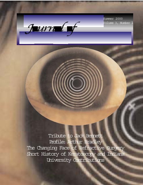
Cover Photo: Photokeratogram<br />
taken with the<br />
Keracorneascope, a<br />
photokeratoscope which was<br />
marketed with an optical device<br />
called a comparator for analysis<br />
<strong>of</strong> the pictures; a short history<br />
<strong>of</strong> keratoscopy starts on page<br />
13.<br />
...........................................................<strong>Indiana</strong> Journal <strong>of</strong> <strong>Optometry</strong> ... <strong>Summer</strong> <strong>2000</strong> ... Vol. 3, No. 1 ... page 1
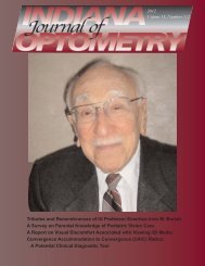
Jack Winn Bennett, 1932-<strong>2000</strong><br />
A Tribute<br />
Earlier this year <strong>Optometry</strong> mourned the loss <strong>of</strong> one <strong>of</strong> its most ardent advocates, Jack W.<br />
Bennett. Anyone who met him would readily note his friendly and jovial Hoosier manner. But<br />
beneath the folksy humor <strong>of</strong> his Granddaddy Barndollar stories and self-deprecating remarks,<br />
there was an man <strong>of</strong> insight and <strong>of</strong> commitment to family and pr<strong>of</strong>ession.<br />
Jack W. Bennett was born on October 23, 1932 in Bloomington, <strong>Indiana</strong>. He graduated in 1950<br />
from Bloomington High <strong>School</strong>. Bennett attended <strong>Indiana</strong> <strong>University</strong> from 1950 to 1952. In 1952, he<br />
married Alice Bauer <strong>of</strong> Bloomington. Alice became an active helpmate to Jack in optometric<br />
activities. For example, she became the president <strong>of</strong> two state optometric auxiliaries and national<br />
president <strong>of</strong> the American Foundation for Vision Awareness.<br />
Bennett served as an optical technician in the United States Army for three years during the<br />
Korean War. He returned to <strong>Indiana</strong> <strong>University</strong> in 1955, and completed his Bachelor <strong>of</strong> Science<br />
degree in 1958 and his Master <strong>of</strong> <strong>Optometry</strong> degree in 1959.<br />
Bennett practiced optometry in Bloomington from 1959 to 1970. During this period <strong>of</strong> time he<br />
was also a part-time Clinical Associate in the IU Division <strong>of</strong> <strong>Optometry</strong> and was very active in the<br />
<strong>Indiana</strong> Optometric Association, serving as its president in 1968-1970. In 1970, he left private<br />
practice to become Associate Pr<strong>of</strong>essor <strong>of</strong> <strong>Optometry</strong> at <strong>Indiana</strong> <strong>University</strong>, a position he held until<br />
1975. Bennett was Director <strong>of</strong> Patient Care for the IU <strong>Optometry</strong> Clinics from 1970 to 1972. While<br />
serving on the IU faculty, Bennett received the<br />
Distinguished Service to <strong>Optometry</strong> Award from the<br />
<strong>Indiana</strong> Optometric Association (1974) and <strong>Indiana</strong><br />
Optometrist <strong>of</strong> the Year award (1975).<br />
In 1975 Bennett left <strong>Indiana</strong> to serve as the Dean for<br />
the new College <strong>of</strong> <strong>Optometry</strong> at Ferris State <strong>University</strong> in<br />
Big Rapids, Michigan. Despite the challenges <strong>of</strong> a state<br />
economy tied to the auto industry, the school survived and<br />
prospered under Bennett’s leadership. He served Ferris<br />
State in various administrative capacities in addition to<br />
being Dean <strong>of</strong> the College <strong>of</strong> <strong>Optometry</strong>, including<br />
Executive Assistant to the President in 1986-87 and Vice<br />
President for Administrative Affairs in 1987-88. He was<br />
president <strong>of</strong> the Michigan Association <strong>of</strong> the Pr<strong>of</strong>essions in<br />
1986-87 and president <strong>of</strong> the Association <strong>of</strong> the <strong>School</strong>s<br />
and Colleges <strong>of</strong> <strong>Optometry</strong> in 1987-89. While working in<br />
Michigan, Bennett received the Pr<strong>of</strong>essional Man <strong>of</strong> the<br />
Year Award from the Michigan Association <strong>of</strong> the<br />
Pr<strong>of</strong>essions (1983) and the Keyman Award from the<br />
Michigan Optometric Association (1984).<br />
In August <strong>of</strong> 1988, Bennett returned to <strong>Indiana</strong><br />
<strong>University</strong> to serve as Dean <strong>of</strong> the <strong>School</strong> <strong>of</strong> <strong>Optometry</strong>.<br />
Jack W. Bennett<br />
This provided opportunities to return to his roots and to<br />
again attend IU basketball and football games regularly,<br />
as well as to make new contributions to optometry. In the<br />
first issue <strong>of</strong> the <strong>Indiana</strong> Journal <strong>of</strong> <strong>Optometry</strong>, Bennett looked back on the developments in the IU<br />
<strong>School</strong> <strong>of</strong> <strong>Optometry</strong> during his years as Dean. 1 He noted changes such as restructuring <strong>of</strong> the<br />
faculty as a unit rather in a departmental structure, the recognition <strong>of</strong> the need for clinical rank<br />
faculty, the encouragement <strong>of</strong> practicing optometrists to mentor bright men and women <strong>of</strong> their<br />
communities concerning optometry as a pr<strong>of</strong>ession, revision and expansion <strong>of</strong> the curriculum,<br />
increases in clinical experience opportunities for students, facility upgrades, increased electronic<br />
technology, investments in research, improved relations with alumni, increased activity <strong>of</strong> faculty in<br />
Page 2 ... Vol. 3, No. 1 ... <strong>Summer</strong> <strong>2000</strong> ... <strong>Indiana</strong> Journal <strong>of</strong> <strong>Optometry</strong> ..........................................................
optometric organizations, and equipment improvements.<br />
During his years as Dean at IU, Bennett continued teaching in the classroom, lecturing on such<br />
topics as optometric ethics, practice management, and optometric history. These years saw him<br />
receive Meritorious Service and Lifetime Achievement Awards from the <strong>Indiana</strong> Optometric<br />
Association, and he was named a Sagamore <strong>of</strong> the Wabash by <strong>Indiana</strong> Governor Frank O’Bannon.<br />
In 1998, Bennett reached the mandatory retirement age for administrators at <strong>Indiana</strong> <strong>University</strong>.<br />
When Bennett stepped down as Dean at IU, he was planning to serve on the IU optometry faculty<br />
for some time and then retire. These plans were put on hold when the <strong>University</strong> <strong>of</strong> Missouri - St.<br />
Louis convinced him to serve as the Dean <strong>of</strong> their <strong>School</strong> <strong>of</strong> <strong>Optometry</strong>. He was Dean there from<br />
January <strong>of</strong> 1999 to April <strong>of</strong> <strong>2000</strong>, when he became ill. His condition rapidly worsened, and he died at<br />
his home in Bloomington on April 28, <strong>2000</strong>. A memorial service, held May 20, <strong>2000</strong> at the First<br />
United Methodist Church in Bloomington, was reflective <strong>of</strong> things that mattered to Jack Bennett: one<br />
<strong>of</strong> his nine grandchildren <strong>of</strong>fering a musical prelude, former optometric colleagues giving words <strong>of</strong><br />
eulogy, and two <strong>of</strong> his four children making touching and humorous tributes. Memorial contributions<br />
can be made to the Jack W. Bennett Endowed Scholarship Fund at <strong>Indiana</strong> <strong>University</strong>, the Jack W.<br />
Bennett Memorial Fund at Ferris State <strong>University</strong>, the Jack W. Bennett Scholarship Fund at<br />
<strong>University</strong> <strong>of</strong> Missouri - St. Louis, or the Creutzfeldt-Jakob’s Disease Foundation.<br />
The Editor<br />
Reference<br />
1. Bennett JW. Reflections. <strong>Indiana</strong> J Optom 1998; 1: 2-5.<br />
Dr. Gordon Heath, Dr, Jack Bennett, and Dr.<br />
Henry H<strong>of</strong>stetter, the first three Deans <strong>of</strong> the<br />
IU <strong>School</strong> <strong>of</strong> <strong>Optometry</strong>.<br />
Dr. Bennett holding the Sagamore <strong>of</strong><br />
the Wabash awarded to him at his<br />
retirement May, 1998<br />
...........................................................<strong>Indiana</strong> Journal <strong>of</strong> <strong>Optometry</strong> ... <strong>Summer</strong> <strong>2000</strong> ... Vol. 3, No. 1 ... page 3
Pr<strong>of</strong>ile: Arthur Bradley, Ph.D.<br />
After what he describes as the longest hitchhiking<br />
trip <strong>of</strong> his life, Dr. Bradley arrived in<br />
Berkeley, California to join the Ph.D.<br />
program in Physiological Optics at the<br />
<strong>School</strong> <strong>of</strong> <strong>Optometry</strong> in 1976. Up to that point in<br />
time he had never met or spoken with an<br />
optometrist, but as an undergraduate at the<br />
<strong>University</strong> <strong>of</strong> Reading, in England he had<br />
developed a deep interest in the human visual<br />
system which spurred him to pursue a research<br />
degree.<br />
He arrived in Berkeley confident in his own<br />
"perfect vision", only to lose a bet with another<br />
recently arrived Ph.D. student, Raymond<br />
Applegate, an IU <strong>School</strong> <strong>of</strong> <strong>Optometry</strong> graduate<br />
who correctly identified Dr. Bradley as a latent<br />
hyperope. Dr. Bradley now describes his<br />
refraction as the "jock" prescription typical <strong>of</strong> one<br />
who spent most <strong>of</strong> his youth chasing soccer, rugby<br />
or cricket balls when he should have been hitting<br />
the books.<br />
At Berkeley, Dr. Bradley studied under<br />
numerous IU alumni (Drs. Tony Adams, Ian Bailey,<br />
Richard VanSluyters, and Russ and Karen<br />
DeValois). He pursued his Ph.D. thesis on human<br />
amblyopia with Ralph Freeman in whose lab he<br />
also studied the neurophysiology <strong>of</strong> primary visual<br />
cortex and vision in amblyopia.<br />
Dr. Bradley financed most <strong>of</strong> is graduate<br />
career by teaching virtually every physiological<br />
optics lab in the curriculum, and lecturing at U.C.<br />
Berkeley and U.C. Santa Cruz on visual optics and<br />
visual perception. After graduating with a Ph.D.,<br />
he worked with the DeValois group on color vision,<br />
after which he joined the faculty <strong>of</strong> <strong>Indiana</strong><br />
<strong>University</strong>. His decision to come to IU was greatly<br />
influenced by his contact with many IU alumni in<br />
Berkeley.<br />
Since arriving at IU in 1985, Dr. Bradley has<br />
developed a world-renown research laboratory<br />
studying visual perception and visual optics. He<br />
has specialized in applying the basic science <strong>of</strong><br />
optics and vision to interesting clinical problems. It<br />
was this expertise that led to his "Glenn A Fry"<br />
award from the American Optometric Foundation,<br />
and his recruitment onto the Federal Drug<br />
Administration (FDA) advisory panel on<br />
Ophthalmic Devices.<br />
In addition to a research career with over 100<br />
publications, Dr. Bradley has continued his longstanding<br />
interest in and commitment to teaching.<br />
He teaches the monocular and binocular visual<br />
function courses within the O.D. curriculum, and<br />
contributes to other courses in visual optics,<br />
contact lenses, and environmental optics. He also<br />
teaches a wide variety <strong>of</strong> courses within the Visual<br />
Sciences program. His commitment and expertise<br />
have been recognized by the students with a<br />
"Pr<strong>of</strong>essor <strong>of</strong> the Year" award, and by the<br />
<strong>University</strong> with two "Teaching Excellence<br />
Recognition Awards".<br />
He was part <strong>of</strong> a special team put together to<br />
advise the Department <strong>of</strong> Defense on the suitability<br />
<strong>of</strong> PRK for service personnel, and within the FDA,<br />
he has advised on numerous refractive devices<br />
and procedures. It is primarily his experience<br />
within this environment that prompted him to write<br />
the article on refractive surgery in the current issue<br />
<strong>of</strong> the <strong>Indiana</strong> Journal.<br />
Arthur Bradley, Ph.D.<br />
Page 4 ... Vol. 3, No. 1 ... <strong>Summer</strong> <strong>2000</strong> ... <strong>Indiana</strong> <strong>University</strong> Journal <strong>of</strong> <strong>Optometry</strong> ........................................
The Changing Face <strong>of</strong> Refractive<br />
Surgery<br />
by Arthur Bradley, Ph.D.<br />
T<br />
he recent diversification and availability <strong>of</strong><br />
refractive surgery has initiated the most<br />
significant change in refractive technology<br />
since the popularization <strong>of</strong> the contact lens<br />
during the 1960s. Just as the contact lens freed the<br />
myope from the spectacle, refractive surgery may<br />
free the myope from spectacles and contact lenses.<br />
In spite <strong>of</strong> its coverage in the popular press (e.g.,<br />
articles in Time magazine and Consumer Reports), it<br />
is not easy to keep abreast <strong>of</strong> the data on and<br />
changes in refractive surgery. Optometrists are <strong>of</strong>ten<br />
provided with pseudo-scholarly publications that are<br />
actually promotional literature 1 published by those<br />
marketing refractive surgery. It is this environment <strong>of</strong><br />
biased and difficult to access information, that<br />
motivated Dr. Bradley, who is a member <strong>of</strong> the FDA<br />
Ophthalmic Devices panel and Pr<strong>of</strong>essor <strong>of</strong><br />
<strong>Optometry</strong> and Vision Science at <strong>Indiana</strong> <strong>University</strong><br />
to write a short summary <strong>of</strong> the recent history and<br />
new developments in this field.<br />
Some prominent ophthalmologists such as<br />
George Waring III are concerned about the mismatch<br />
between the reality <strong>of</strong> refractive surgery and the<br />
promotional marketing literature 2 : "the patient must<br />
have realistic expectations <strong>of</strong> the procedure based on<br />
honest communication from the surgeon and<br />
pr<strong>of</strong>essional staff, regardless <strong>of</strong> portrayal <strong>of</strong> the<br />
procedure in advertising and the popular media".<br />
There are lingering doubts about the reliability,<br />
safety and stability <strong>of</strong> refractive surgery. For<br />
example, Pr<strong>of</strong>essor <strong>of</strong> Ophthalmology, Leo Maguire,<br />
has referred to patients who have undergone<br />
refractive surgery as the "refractive underclass" 3 .<br />
Also, there are sufficient numbers <strong>of</strong> patients<br />
dissatisfied in their refractive surgery results that they<br />
have their own web page. This web page<br />
(http://www.surgicaleyes.org) is full <strong>of</strong> testimonials<br />
and even some computer simulations <strong>of</strong> postrefractive<br />
surgery vision which are worth seeing.<br />
In spite <strong>of</strong> the lingering concerns about refractive<br />
surgery it continues to be promoted and has become<br />
a real option for many patients, some <strong>of</strong> who will seek<br />
advice from their Optometrist prior to deciding on<br />
surgery. This article is designed as an up-to-date<br />
short review <strong>of</strong> this field to help our readers<br />
understand the benefits, shortcomings, and possible<br />
future <strong>of</strong> this approach to correcting ametropia.<br />
1. The optometrists role in laser vision correction: TLC,<br />
Laser Eye Centers, 1999.<br />
2. Waring G III, Future developments in LASIK. In:<br />
Pallikaris I, Siganos D, eds. LASIK, Thoror<strong>of</strong>are, NJ: Slack ,<br />
1998: 367-370.<br />
3. Maguire L, Quoted in Consumer Reports article on<br />
LASIK titled "Zap your myopic eyes", June, 1999.<br />
Introduction:<br />
Although most spherical refractive errors are<br />
caused by anomalous axial length (too long in<br />
myopes and too short in hyperopes), there is a long<br />
history <strong>of</strong> correcting for this anatomical defect by<br />
introducing optical changes at the anterior eye. For<br />
centuries, spectacle lenses were the only option<br />
available to make this change, but during the last half<br />
<strong>of</strong> the 20th century, contact lenses became a<br />
convenient alternative and are currently worn by over<br />
20 million Americans. These lenses work by<br />
changing the curvature at the air-eye interface, where<br />
the refractive index difference is large and most <strong>of</strong> the<br />
eye’s optical power exists. A similar and more<br />
permanent strategy is to change the curvature <strong>of</strong> the<br />
anterior corneal surface directly.<br />
Although refractive surgery (Keratotomy) was<br />
pioneered during the nineteenth century, it was not<br />
widely available until the last quarter <strong>of</strong> the 20th<br />
century. Several methods for implementing corneal<br />
curvature changes were developed during the last<br />
quarter <strong>of</strong> the 20th century and continue to be<br />
developed today. Early methods, e.g., radial<br />
keratotomy (RK) in the 1970’s and 80’s and<br />
photorefractive keratectomy (PRK) in the 1990’s, had<br />
serious shortcomings and they are now being<br />
replaced.<br />
For example, RK, in addition to poor<br />
predictability, produced eyes with unstable refractive<br />
errors that varied diurnally and with altitude and on<br />
average shifted towards hyperopia after surgery (e.g.,<br />
almost 50% shifted by 1 diopter). This article will<br />
describe some <strong>of</strong> the more recent surgical<br />
approaches and in particular will examine the<br />
refractive success and the safety issues associated<br />
with each.<br />
Refractive surgeries designed to reshape the<br />
cornea can be grouped by either the site <strong>of</strong> surgical<br />
intervention or the surgical method. For example, in<br />
treating myopia, RK and PRK differ in both the site <strong>of</strong><br />
intervention and the surgical method. RK makes<br />
incisions deep into the peripheral cornea, while PRK<br />
........................................<strong>Indiana</strong> <strong>University</strong> Journal <strong>of</strong> <strong>Optometry</strong> ... <strong>Summer</strong> <strong>2000</strong> ... Vol. 3, No. 1 ... page 5
emoves tissue from the anterior central cornea using<br />
a high-energy ultraviolet laser.<br />
Photoablative Refractive Surgery<br />
Most photoablative corneal reshaping techniques<br />
employ UVB lasers, e.g., an argon fluoride excimer<br />
laser (λ=193 nm), to produce high-energy radiation<br />
which is highly absorbed by the corneal stroma. This<br />
energy is sufficient to break the chemical bonds that<br />
form the collagen fibers and effectively remove this<br />
tissue from the cornea.<br />
Initial attempts to use UV lasers were based upon<br />
the RK radial incision technique. However, the UV<br />
laser failed as a "knife" because it created wider<br />
incisions than the scalpel and produced more<br />
significant scars. More recently, the UV excimer laser<br />
has been modified to ablate stromal tissue within the<br />
optical zone and thus reshape the optical surface<br />
directly. Two manifestations <strong>of</strong> this approach have<br />
been developed, Photorefractive Keratectomy (PRK)<br />
and laser in situ keratomileusis (LASIK), and both<br />
share a common goal, to reshape the anterior corneal<br />
surface by ablating stromal tissue. However, the<br />
methods for achieving this goal are quite different.<br />
In PRK, anterior stromal tissue is ablated after<br />
the corneal epithelium has been scraped away<br />
(although in rare cases transepithelial PRK was<br />
performed). Of course, this method also ablates the<br />
basement membrane (Bowman’s Layer) upon which<br />
the epithelium grows, and thus has a number <strong>of</strong><br />
undesirable complications associated with loss <strong>of</strong><br />
epithelial function including susceptibility to infection,<br />
post-surgical pain, abnormal epithelial growth, and<br />
reduced optical transparency. These problems are<br />
most pronounced in the period after surgery, and thus<br />
patients did not generally have bilateral PRK, but had<br />
to maintain one untreated eye during the epithelial<br />
recovery period. In spite <strong>of</strong> this protracted recovery<br />
period, PRK surgery has been performed on both<br />
eyes simultaneously.<br />
The problems associated with destruction <strong>of</strong> the<br />
epithelium in PRK have been largely eliminated by<br />
implementing a different pre-ablation surgical<br />
procedure. Instead <strong>of</strong> scraping <strong>of</strong>f the epithelium, a<br />
deep cut into the stromal lamellae is made<br />
approximately parallel to the corneal surface using a<br />
micro-keratome (LASIK). The cut begins temporally<br />
or inferiorly and cuts across the central cornea but<br />
leaves the nasal or superior edge uncut (the flap).<br />
This method produces an anterior corneal flap (70-<br />
160 microns thick), which can be folded back to<br />
expose the corneal stroma. At this point a<br />
photoablative method, the same in principle to that<br />
used in PRK, is employed to remove stromal tissue<br />
and thus reshape the corneal stroma without<br />
destruction or removal <strong>of</strong> the epithelium. Once the<br />
ablation is complete, the flap can be repositioned<br />
over the remaining stroma resulting in a cornea with a<br />
mostly functioning epithelium (some sensory nerve<br />
damage and associated corneal insensitivity occurs,<br />
which remediates after about two weeks). The flap is<br />
a non-rigid structure and when repositioned its shape<br />
is affected by the underlying stromal re-shaping which<br />
is transferred to the anterior corneal surface thus<br />
changing the optical power <strong>of</strong> the cornea.<br />
LASIK is currently the most widely used surgical<br />
method for correcting refractive errors and several<br />
commercial lasers have received FDA approval.<br />
LASIK Efficacy<br />
If refractive surgery is effective, the post-surgical<br />
refractive errors should be the same as the targeted<br />
or intended refractive error. The reason to use<br />
targeted or intended instead <strong>of</strong> emmetropia is that<br />
sometimes emmetropia is not the target. For<br />
example, a patient may elect to have a small amount<br />
<strong>of</strong> myopia to aid in reading.<br />
Many studies report and plot the average postsurgical<br />
refractive error, and in general with more<br />
recent technology this approaches the target<br />
indicating an almost perfect outcome. However,<br />
individual eyes do not achieve the mean post-op Rx,<br />
and therefore, in order to assess efficacy, the postsurgical<br />
refractive errors <strong>of</strong> individual eyes must be<br />
considered.<br />
In order for the FDA to approve a photoablative<br />
laser for LASIK, it must be able to demonstrate<br />
efficacy by having a high percentage <strong>of</strong> the postsurgical<br />
refractions within some range <strong>of</strong> the intended<br />
or target refraction (e.g., 75% must be within 1 diopter<br />
<strong>of</strong> intended and 50% within 0.50 diopters). Most<br />
current systems achieve this goal, with about 60-70%<br />
<strong>of</strong> the eyes ending up within 0.50 D <strong>of</strong> the target and<br />
sometimes more than 90% within 1 diopter.<br />
However, some studies still report less than 75%<br />
within 1 diopter <strong>of</strong> target.<br />
In general, the anticipated residual refractive<br />
errors increase with the magnitude <strong>of</strong> the pre-surgical<br />
refractive error. However, although approximate<br />
emmetropia may not be achieved in some highly<br />
myopic eyes, it can be argued that converting a -10<br />
diopter myope into a -2 D myope is an effective<br />
procedure since their level <strong>of</strong> visual disability while<br />
uncorrected will be greatly reduced.<br />
It is important, therefore, that patients be fully<br />
aware <strong>of</strong> the likely refractive outcome prior to opting<br />
for surgery. Realizing that a patient will typically<br />
expect to leave their eye-care practitioner’s <strong>of</strong>fice<br />
seeing "perfectly", clinicians counseling patients<br />
about refractive surgery should emphasize that this<br />
will probably not happen. Typical results in recent<br />
studies indicate about 80% to 90% <strong>of</strong> patients end up<br />
Page 6 ... Vol. 3, No. 1 ... <strong>Summer</strong> <strong>2000</strong> ... <strong>Indiana</strong> <strong>University</strong> Journal <strong>of</strong> <strong>Optometry</strong> ........................................
with uncorrected VA (UCVA) <strong>of</strong> 20/40 or better, and<br />
between 40 and 70% with 20/20 or better UCVAs.<br />
The FDA requires a new laser system to demonstrate<br />
20/40 UCVA in at least 85% <strong>of</strong> treated eyes to qualify<br />
as effective. That is, perhaps 50% <strong>of</strong> LASIK patients<br />
will have to tolerate uncorrected VAs poorer than<br />
20/20 or wear a spectacle or contact lens to achieve<br />
their pre-surgical VA. As many patients with low<br />
levels <strong>of</strong> refractive error now do, these post LASIK<br />
patients with small residual refractive errors generally<br />
choose to leave them uncorrected making the clear<br />
choice <strong>of</strong> convenience over vision quality.<br />
There is one significant complication associated<br />
with efficacy. Since photoablation removes tissue,<br />
there will always be some wound healing process,<br />
and this can and does lead to post-surgical refractive<br />
instability. Since PRK removed the entire epithelium<br />
and Bowman’s layer, the healing process was very<br />
active, and this was the likely cause <strong>of</strong> much <strong>of</strong> the<br />
post surgical instability. The reduced wound healing<br />
response experienced with LASIK results in less postsurgical<br />
instability in Rx, most eyes (e.g. 95%)<br />
experiencing less than 1 diopter change during the<br />
year post surgery. Recent protocols have reduced<br />
the population mean change in Rx to almost zero.<br />
However, some individual eyes do experience<br />
changes during the 6 months post-surgery.<br />
Although LASIK does not require complete regrowth<br />
<strong>of</strong> the corneal epithelium and the wound<br />
healing is reduced, recent studies have observed<br />
increased epithelial thickness anterior to the ablation<br />
indicating some epithelial response to the surgery or<br />
the ablation.<br />
Of course, efficacy will be compromised by any<br />
change in corneal structure following keratomileusis<br />
or photoablation, and the significant reduction in the<br />
thickness <strong>of</strong> the remaining structurally intact cornea<br />
does seem to have an effect. For example, bowing <strong>of</strong><br />
the posterior corneal surface has been reported and<br />
this may reflect structural changes caused by the<br />
removal <strong>of</strong> more than 100 microns with the keratome<br />
and up to 200 microns with photoablation, reducing<br />
the 500 micron thick cornea to approximately only<br />
200 mechanically integrated microns. A significant<br />
correlation between bowing and residual stromal<br />
thickness has been observed when the thickness is<br />
less than 290 microns. The same study concluded<br />
that inaccuracies in the refractive outcome stem<br />
primarily from a combination <strong>of</strong> secondary bowing<br />
and epithelial thickness changes that develop postsurgically.<br />
Leaving less than 250 microns intact is<br />
generally felt to be unsafe.<br />
The primary determinant <strong>of</strong> efficacy is the amount<br />
and spatial distribution <strong>of</strong> tissue ablated. This <strong>of</strong>ten<br />
depends upon proprietary algorithms, which can be<br />
updated to improve efficacy if a procedure has been<br />
shown to either under or over correct. Very simply, if<br />
the pre-ablation anterior corneal curvature is known,<br />
the desired change in refraction determines the<br />
required new curvature and the amount <strong>of</strong> tissue to<br />
be removed. Studies have shown how much tissue<br />
will be ablated by a given amount <strong>of</strong> laser energy<br />
(e.g. 0.1 microns can be removed by a 50 mJ/cm2<br />
excimer laser pulse), but these values vary slightly<br />
from eye to eye depending upon such things as<br />
stromal hydration. An additional source <strong>of</strong> variability<br />
is eye position and eye movements during surgery.<br />
In response to this concern, some laser systems (e.g.<br />
Autonomous flying spot laser) include an eye position<br />
tracking system to effectively stabilize the eye with<br />
respect to the laser. This system corrects for any eye<br />
movements during the procedure, which can last from<br />
few seconds to 60 seconds depending on the amount<br />
<strong>of</strong> tissue to be ablated.<br />
One major advantage <strong>of</strong> PRK over RK is that,<br />
unlike RK, it did not suffer from significant diurnal<br />
fluctuations or the significant hyperopic shifts<br />
associated with high altitudes that plagued RK.<br />
Recent studies by the US military at 14,000 ft. have<br />
confirmed that LASIK eyes do not suffer from the 1.5<br />
diopter hyperopic shifts seen in RK eyes, but if an eye<br />
has had LASIK recently, a hyperopic shift <strong>of</strong> about 0.5<br />
diopters was observed. However, after six months,<br />
no such shift was observed.<br />
Since the mean post-LASIK Rx has approached<br />
zero, it appears that the tissue ablation algorithms<br />
have been optimized. The fact that the majority <strong>of</strong><br />
eyes do not end up emmetropic results from the eyeto-eye<br />
variability in such factors as epithelial growth,<br />
corneal bowing and reaction to the laser. Therefore,<br />
in order to improve the efficacy still further, a two step<br />
surgery may have to be implemented. The second<br />
ablation will fine tune the small errors left after the<br />
first LASIK. However, the second procedure is nearly<br />
as costly as the first and reduces pr<strong>of</strong>it margins.<br />
Such an approach is already used to correct "poor<br />
outcomes" after the initial LASIK procedure.<br />
LASIK Safety<br />
Evaluation <strong>of</strong> safety is more complicated than<br />
assessing efficacy <strong>of</strong> refractive surgery. We can<br />
consider any change to the eye which compromises<br />
vision as a safety problem. There are five general<br />
categories <strong>of</strong> such problems following LASIK: (1)<br />
infections and pathology in response to the surgical<br />
or/and ablative procedures, (2) undesirable wound<br />
healing responses, (3) photoablative changes that<br />
cannot be corrected with standard spectacle or<br />
contact lenses, (4) effects <strong>of</strong> the high energy laser on<br />
other ocular tissues, and (5) optical problems<br />
.........................................................<strong>Indiana</strong> Journal <strong>of</strong> <strong>Optometry</strong> ... <strong>Summer</strong> <strong>2000</strong> ... Vol. 3, No. 1 ... page 7
associated with the pre-ablation surgery (e.g. flap<br />
irregularities). Due to the invasive nature <strong>of</strong> this<br />
surgery, it is not surprising to find that problems<br />
associated with the flap surgery are the most<br />
significant.<br />
1. Post-surgical pathology:<br />
The incidence <strong>of</strong> infections caused by LASIK is<br />
very low, and includes bacterial keratitis due to poor<br />
ocular hygiene combined with imperfect epithelial<br />
coverage along the flap incision. Vitreous<br />
hemorrhage and retinal detachments following<br />
corneoscleral perforations resulting from the surgical<br />
microkeratome have also been reported, but again,<br />
the incidence is very low (e.g., 2 eyes out <strong>of</strong> 29,916).<br />
Other vitreoretinal pathologies in the post-surgical<br />
LASIK patients were also very rare and may reflect<br />
typical levels experienced by highly myopic eyes.<br />
This emphasizes that, although LASIK may correct<br />
the myopic refractive error, it does not treat or prevent<br />
the other problems associated with and caused by<br />
increased axial length in myopic eyes. Dry eye is a<br />
very common complaint following LASIK, possibly<br />
due to cutting the corneal nerves and decreasing the<br />
primary signal that produces normal tear levels. Dry<br />
eye complaints persist for a long time and individuals<br />
with dry eye prior to surgery should be counseled that<br />
LASIK may exacerbate their existing problem. Those<br />
without dry eye should be counseled that dry eye<br />
complaints are relatively common and can last for<br />
several months to a year following surgery.<br />
2. Wound healing response:<br />
Diffuse interface keratitis, with an accumulation <strong>of</strong><br />
inflammatory cells at the flap interface has been<br />
observed presumably due to a wound healing<br />
response. Also, unusual epithelial growth has been<br />
observed when trauma dislodges the flap. Recent<br />
evidence from animal studies indicates that the<br />
healing process at the flap interface continues for<br />
about 9 months after LASIK. The consequences <strong>of</strong><br />
this prolonged wound healing are unclear.<br />
3. Optical changes uncorrectable with standard<br />
ophthalmic lenses:<br />
There is a genuine concern that photoablative<br />
procedures will result in reduced optical quality <strong>of</strong> the<br />
cornea due to either a loss <strong>of</strong> transparency and<br />
optical scatter or irregular changes in the shape <strong>of</strong> the<br />
optical surface. Both <strong>of</strong> these optical changes are<br />
uncorrectable with standard spectacle lenses.<br />
Aberrations exist in an optical system when, even<br />
with an optimum sphero-cylindrical correction, the<br />
rays forming a point image will not focus to a single<br />
point. Increased optical aberrations reported in post-<br />
PRK and post-LASIK eyes 4 may reflect the<br />
algorithms used to create the ablations, but other<br />
factors must also be involved. For example, myopic<br />
"islands" are <strong>of</strong>ten reported after PRK or LASIK and<br />
for some reason these local under-corrected areas<br />
seem to disappear over time. The cause <strong>of</strong> these<br />
myopic islands and the mechanisms behind their<br />
remediation are not well understood.<br />
As a check for such detrimental changes in the<br />
cornea, the FDA requires that post LASIK VAs be<br />
determined with the optimum spectacle correction in<br />
place (Best Spectacle Corrected Visual Acuity:<br />
BSCVA). If an eye can no longer be corrected to its<br />
pre-surgery levels <strong>of</strong> VA, it is likely that one or both <strong>of</strong><br />
the above optical changes have occurred. The FDA<br />
requires that less than 5% <strong>of</strong> eyes lose more than 2<br />
lines <strong>of</strong> BSCVA, and less than 1% end up with<br />
BSCVA <strong>of</strong> worse than 20/40. One might argue that<br />
any loss <strong>of</strong> BSCVA is unacceptable since it is<br />
essentially an untreatable vision loss. It is, however,<br />
disappointing that after centuries <strong>of</strong> striving to<br />
improve retinal image quality, we are now willing to<br />
accept reduced retinal image quality and significant<br />
loss <strong>of</strong> vision all in the name <strong>of</strong> convenience.<br />
Although current standards tolerate reduced<br />
retinal image quality and the current LASIK protocols<br />
increase the eye’s aberrations, the potential is there<br />
to reduce aberrations and actually improve retinal<br />
image quality. In principle, photoablative techniques<br />
can be used to correct not only the eye’s spherical<br />
and cylindrical refractive errors but also higher order<br />
aberrations such as spherical aberration and coma,<br />
which limit retinal image quality in pre-surgical eyes.<br />
Autonomous Technologies is pioneering this concept,<br />
which requires measurement <strong>of</strong> the eye’s aberrations<br />
in addition to the refractive error typically measured.<br />
We expect to see this approach, referred to as<br />
"custom cornea" to develop rapidly in the next few<br />
years. Of course, in order to correct for the<br />
aberrations, they must first be measured. New<br />
technology borrowed from astronomy has been<br />
successfully employed to measure ocular<br />
aberrations 5 and these can be used to guide<br />
photoablative surgeries. The term "wave-guided<br />
corneal surgery" was recently coined to describe this<br />
procedure.<br />
We shall soon see if wave-guided corneal<br />
surgery can succeed. McDonald presented some <strong>of</strong><br />
the first data earlier this year and showed that the<br />
increase in aberrations and thus reduction in retinal<br />
image quality associated with the standard LASIK<br />
procedure may not occur following a "custom cornea"<br />
approach. Currently, it is not clear how successful<br />
this approach will be. It may be a way to maintain<br />
optical quality at pre-surgical levels, but the potential<br />
is there for actual improvement.<br />
Although the ablation algorithms may be perfect<br />
and corneal transparency maintained, there is<br />
Page 8 ... Vol. 3, No. 1 ... <strong>Summer</strong> <strong>2000</strong> ... <strong>Indiana</strong> Journal <strong>of</strong> <strong>Optometry</strong> ........................................................
another factor that will lead to significant loss <strong>of</strong><br />
retinal image quality in LASIK or PRK. In order to<br />
maintain a mon<strong>of</strong>ocal optical system, the reshaped<br />
cornea must be larger than the eye’s entrance pupil.<br />
However, there are limits to the maximum size <strong>of</strong> the<br />
ablation zone because increased ablation zone size<br />
requires deeper ablations. For example, by<br />
increasing the ablation zone from 4 mm to 7 mm<br />
approximately doubles the necessary ablation depth<br />
in the central cornea when correcting myopia. Thus,<br />
correction <strong>of</strong> large refractive errors requires more<br />
tissue ablation and larger ablation zones also require<br />
deeper ablations. For example, Sher calculated that<br />
300 microns <strong>of</strong> tissue would have to be removed to<br />
correct a -12 diopter myopia over a 7 mm diameter<br />
area. Approximate corneal thinning caused by<br />
photoablation for myopia is 12, 18 and 25 microns<br />
per diopter with 5, 6, and 7 mm ablation zones,<br />
respectively.<br />
The problems associated with leaving too little<br />
attached stroma after ablation are exaggerated with<br />
LASIK since up to 150 microns <strong>of</strong> the anterior cornea<br />
has been removed already in the flap. Ablating<br />
significant amounts <strong>of</strong> the remaining stromal tissue<br />
may compromise the structural abilities <strong>of</strong> the<br />
remaining stroma and result in the observed "bowing"<br />
<strong>of</strong> the posterior corneal surface after surgery.<br />
Since there are limits to how much corneal tissue<br />
can be safely removed, ablation zone size has<br />
typically been smaller than necessary to cover the<br />
entire dilated pupil present at night. Current<br />
standards try to maintain at least 250 microns <strong>of</strong><br />
intact stroma after photoablation. Given this type <strong>of</strong><br />
constraint, the photoablation zone size is limited.<br />
Early PRK photoablations were performed with 4 mm<br />
and 5 mm zones, but the standard now is about 6 mm<br />
with perhaps a 1-2 mm "transition" zone. Because<br />
the pupil <strong>of</strong> many young eyes will be larger than 6<br />
mm under low light conditions, the effective optical<br />
system creating the retinal image will be bifocal. The<br />
central zone will be near to emmetropic and the<br />
marginal zone near to the pre-ablation refractive<br />
error. Although this has obvious parallels to<br />
simultaneous bifocal contact lenses or IOLs, it is not<br />
an effective bifocal correction since the additional add<br />
power in the peripheral optics will vary from eye to<br />
eye and will be too peripheral to be effectiveat high<br />
light levels. This bifocal problem cannot be corrected<br />
with a spectacle lens or easily corrected with a<br />
contact lens and bifocal optics are known to produce<br />
significantly reduced image quality, halos and glare.<br />
Data over the last few years indicate that the<br />
flattening <strong>of</strong> the central cornea by LASIK actually<br />
leads to steepening <strong>of</strong> the peripheral cornea<br />
potentially exaggerating the simultaneous bifocal<br />
effect for larger pupils. Also, by adding transition<br />
zones into the surgical procedure, a dilated pupil<br />
produces multifocal optics.<br />
The impact <strong>of</strong> post-surgical simultaneous bifocal<br />
or multifocal optics would only be manifest at low light<br />
levels, and studies from Europe seem to indicate that<br />
night vision can be significantly compromised by PRK<br />
and LASIK. Visible halos and glare at night are <strong>of</strong>ten<br />
reported, and they increase in frequency with<br />
increased myopic correction, and cases have been<br />
reported in which post LASIK and post PRK night<br />
vision is so poor that night driving has to be<br />
eliminated. It would be wise therefore, as Applegate 6<br />
has been emphasizing for many years now, to<br />
discourage individuals with large night-time pupils<br />
from undergoing this procedure. Simulations <strong>of</strong> these<br />
night vision problems can be visualized on the web at<br />
http://www.surgicaleyes.org.<br />
4. UV damage to other ocular tissue:<br />
The introduction <strong>of</strong> a high intensity UV radiation<br />
source into the eye produces obvious concerns for<br />
other ocular tissue since UV is known to cause<br />
cataractogenesis and may be a significant factor in<br />
age related maculopathy. However, 193 nm UV<br />
radiation does not penetrate more that a few microns.<br />
This is why it is so effective at stromal ablation.<br />
5. Problems with the flap.<br />
The major concern with LASIK stems from the<br />
radical surgery preceding the photoablation. The<br />
entire anterior cornea (epithelium and part <strong>of</strong> the<br />
stroma) is removed across the central cornea<br />
exposing the central stroma. Problems develop due<br />
to poor quality <strong>of</strong> the keratome blade, poor control <strong>of</strong><br />
the cutting speed, failure to complete the cut, leaving<br />
tiny metal fragments from the blade on the flap,<br />
deposition <strong>of</strong> other material (e.g., surgical glove<br />
powder) within the wound, and movement <strong>of</strong> the<br />
tissue during the cut. Expert use and maintenance <strong>of</strong><br />
the micro-keratome is essential to reduce the<br />
incidence <strong>of</strong> these vision-compromising<br />
complications.<br />
It is important to realize that cutting corneal tissue<br />
requires much greater precision and better quality cut<br />
surfaces than cutting tissue in other parts <strong>of</strong> the body.<br />
Errors, such as the micro-chatter marks seen post<br />
LASIK, on the scale <strong>of</strong> the wavelength <strong>of</strong> light, can<br />
become significant. Also, since the stroma is<br />
avascular, there is little opportunity for debris to be<br />
removed by phagocytic inflammatory cells. Reports<br />
<strong>of</strong> tiny metal fragments from the micro-keratome<br />
blade, powder from the surgical gloves, small pieces<br />
<strong>of</strong> sponge as well as corneal tissue remnants have<br />
been seen under the flap post surgically. All <strong>of</strong> these<br />
reduce transparency, and can require a second<br />
procedure in which the flap is opened up and the<br />
.........................................................<strong>Indiana</strong> Journal <strong>of</strong> <strong>Optometry</strong> ... <strong>Summer</strong> <strong>2000</strong> ... Vol. 3, No. 1 ... page 9
tissue cleaned.<br />
LASIK has a unique safety issue not present with<br />
other refractive surgical procedures, which stems<br />
from the structural weakness <strong>of</strong> the corneal flap and<br />
its poor adhesion to the underlying corneal stroma. In<br />
some ways it is remarkable that the flap can<br />
"reattach" so easily without sutures. Initial<br />
reattachment results from hydrostatic pressure due to<br />
the hydrophilic nature <strong>of</strong> the inner cornea. Primary<br />
"reattachment" forces may result from capillary<br />
surface tension. It is therefore quite easy to remove<br />
the flap for additional photoablation, if the initial<br />
surgery was not as effective as desired. However,<br />
the flap can also become dislodged accidentally.<br />
Remarkably, this is very rare, but it can and does<br />
happen, usually following some ocular trauma. A<br />
notable concern exists for patients with dry eye who<br />
may experience adhesion forces between the anterior<br />
corneal surface and the lid. This has led to a patient<br />
waking to find the flap stuck to the lid. Also, because<br />
<strong>of</strong> the reduced sensitivity following surgery (sensory<br />
nerves have been cut) the normal feed-back that<br />
controls corneal insult has been seriously<br />
compromised which must increase the chances <strong>of</strong><br />
elevated mechanical forces on the cornea due to<br />
trauma or lid friction.<br />
In addition to flap displacement, the structural<br />
weakness <strong>of</strong> the flap and its attachment can lead to<br />
structural changes within the flap. Small scale<br />
"ripples" or "wrinkles" in the flap have been reported,<br />
as have larger folds. Flaps are sometimes detached<br />
and reattached to try and remedy flap irregularities.<br />
There is also the problem <strong>of</strong> accurately realigning the<br />
flap and replacing it in the correct location. Flap<br />
decentration has been reported. As with flap<br />
wrinkling, it will lead to reduced optical quality.<br />
The final complication associated with the flap<br />
surgery stems from the pre-incision protocol. In order<br />
for the keratome to make a precise cut, the corneal<br />
tissue must be held firmly by a vacuum ring. During<br />
this procedure, the intraocular pressure spikes to<br />
above 60 mm <strong>of</strong> Hg. There is some concern that this<br />
IOP spike, particularly if it is maintained for more than<br />
a few seconds, can lead to retinal damage. Suction<br />
duration depends upon the speed <strong>of</strong> the procedure<br />
and can vary significantly (e.g., from 6 to 80<br />
seconds). Changes in retinal blood flow and visual<br />
function following this transient elevated IOP have<br />
been reported. In addition to the IOP spike, there is<br />
some globe deformation associated with the vacuum<br />
ring.<br />
In the March <strong>2000</strong> issue <strong>of</strong> Biophotonics<br />
International, a new technology for producing the flap<br />
without a micro-keratome was described. A group at<br />
the <strong>University</strong> <strong>of</strong> Michigan are developing an infra-red<br />
laser to make the flap. This device uses a highly<br />
convergent laser beam with very high energy per<br />
square cm at its focal plane with sufficient energy to<br />
break the collagen fibers. By placing the focal plane<br />
within the stroma and scanning across the eye, the<br />
anterior cornea can be detached from the remaining<br />
posterior stroma and a flap produced. The<br />
advantages are that it requires no mechanical shear<br />
forces, which tend to move and distort the cornea and<br />
lead to variable flap thickness with micro-keratomes.<br />
Also, by optically adjusting the laser focal plane, the<br />
flap depth can be varied across the cornea and flaps<br />
with beveled edges can be produced. The<br />
technology is undergoing trials in Europe and may be<br />
introduced late in <strong>2000</strong> in the US. Interestingly for<br />
optometry, this device removes the necessity for<br />
cutting tissue with a blade, or traditional surgery, and<br />
may, in the classical sense, make LASIK a nonsurgical<br />
procedure.<br />
LASIK Summary<br />
The overall picture emerging from the LASIK<br />
literature indicates that it is a largely safe and<br />
effective treatment for myopia, hyperopia and<br />
astigmatism. However, LASIK is not risk free, and<br />
with current technology final vision quality will<br />
probably be slightly inferior to pre-surgical vision.<br />
Night vision may be significantly impaired. There are<br />
many stories <strong>of</strong> post-PRK and post-LASIK patients<br />
having to modify their night driving behavior because<br />
<strong>of</strong> seriously reduced vision at night. For the patient,<br />
the very small risk <strong>of</strong> serious complications and the<br />
likely small reduction in vision and night driving<br />
problems must be balanced against the obvious<br />
convenience <strong>of</strong> never having to worry about contact<br />
lenses or spectacles. Perhaps more significantly,<br />
highly myopic patients will never have to suffer the<br />
serious handicap that exists when their high myopia<br />
is uncorrected. For many patients, particularly those<br />
who are seriously handicapped by their myopia, and<br />
those for whom highest quality vision is not required,<br />
this may be the surgical treatment <strong>of</strong> choice at this<br />
time. However, it is imperative that all patients are<br />
made aware <strong>of</strong> the risks, particularly the commonly<br />
occurring reduced quality <strong>of</strong> vision and night driving<br />
problems.<br />
Thermokeratoplasty<br />
In addition to the photoablative use <strong>of</strong> short<br />
wavelength UV lasers, corneal irradiation using long<br />
wavelength (1.5 - 2.0 micron) lasers has been<br />
developed to create thermally induced changes in the<br />
corneal stroma. This method, Laser<br />
Thermokeratoplasty (LTK), has some obvious<br />
parallels to radial keratotomy, and it is sometimes<br />
referred to as radial thermokeratoplasty. Unlike RK,<br />
which treated myopia by introducing deep incisions to<br />
Page 10 ... Vol. 3, No. 1 ... <strong>Summer</strong> <strong>2000</strong> ... <strong>Indiana</strong> Journal <strong>of</strong> <strong>Optometry</strong> ........................................................
allow the peripheral cornea to stretch and thus reduce<br />
central corneal curvature, LTK causes peripheral<br />
corneal shrinkage due to thermally induced shrinkage<br />
<strong>of</strong> individual collagen fibers. Thus, LTK has the<br />
opposite effect on the peripheral cornea, and therefore<br />
induces myopic shifts in the central cornea. It has<br />
been suggested and actually tested as a treatment for<br />
hyperopia (either naturally occurring or secondary to<br />
over-correction by PRK or LASIK), but it is still in the<br />
investigational stage and has not received FDA<br />
approval. There are major concerns about its ability to<br />
produce a stable refractive change since large<br />
regressions occur. Also, unlike photoablative<br />
techniques which calculate the desired tissue to be<br />
removed, LTK must rely on empirically determined<br />
nomograms. Predictability with this approach has not<br />
been established and dosimetry studies continue to<br />
examine the impact <strong>of</strong> wavelength, temperature,<br />
penetration <strong>of</strong> the radiation, beam pr<strong>of</strong>ile, and spatial<br />
pattern and duration <strong>of</strong> radiation. There is also<br />
concern that the thermal effects cannot be confined to<br />
the stroma, and damage to the epithelium and<br />
endothelium may occur.<br />
Surgical Implants<br />
In addition to the methods just described in which<br />
the cornea is reshaped by removing tissue or<br />
reshaping the cornea, two new surgical approaches<br />
are being developed that insert foreign bodies into the<br />
eye. The first inserts a ring deep into the peripheral<br />
corneal stroma and the second involves implanting an<br />
intraocular lens (IOL) into a phakic ametropic eye.<br />
1. Intrastromal Corneal Rings<br />
Just as RK and LTK change the curvature <strong>of</strong> the<br />
central cornea by changing the structure <strong>of</strong> the<br />
peripheral cornea, intrastromal corneal rings (ICR) or<br />
intrastromal corneal ring segments (ICRS) are inserted<br />
into the peripheral cornea to treat myopia. The ring or<br />
ring segments are inserted through a small incision<br />
and threaded circumferentially into the deep stromal<br />
lamellae. The structural changes that are produced<br />
translate into curvature changes in the central cornea.<br />
Inserting PMMA annular rings into the deep stromal<br />
lamellae <strong>of</strong> the corneal periphery changes the already<br />
prolate elliptical cornea into an even more prolate<br />
cornea, reducing the overall corneal curvature and<br />
thus producing a hyperopic shift. Studies indicate that<br />
myopia <strong>of</strong> up 3 or 4 diopters can be treated with this<br />
method. The biggest advantage <strong>of</strong> this approach is<br />
that, unlike PRK, RK or LASIK, it is largely reversible<br />
by simply removing the ring (segments). Thicker rings<br />
(0.45 mm diameter) introduce large changes and thus<br />
can correct for more myopia while thinner rings (0.25<br />
mm) are used to correct lower levels <strong>of</strong> myopia.<br />
BSCVAs seem to remain high and thus the method<br />
must not introduce large amounts <strong>of</strong> aberrations or<br />
turbidity in the central cornea. There is some concern<br />
that significant refractive instability exists with this<br />
method including diurnal variations. Peripheral<br />
corneal haze, small lamellae deposits adjacent to the<br />
ring, deep stromal neovascularization, and pannus are<br />
also associated with the ring insertions. Currently the<br />
FDA has approved one ICR (Keravision’s Intacs).<br />
2. Phakic Intraocular Lenses<br />
Unlike the previous methods, which all required<br />
the development <strong>of</strong> new technology, IOL implantation<br />
has a long and successful history as a treatment for<br />
cataract. The major difference with phakic IOL<br />
implantation is that the natural lens is left in place.<br />
The general principle <strong>of</strong> using an IOL to correct for<br />
ametropia has <strong>of</strong> course been part <strong>of</strong> the typical<br />
cataract lens replacement regime for many years. By<br />
manipulating the curvature, refractive index and<br />
thickness <strong>of</strong> an IOL, significant refractive errors can be<br />
corrected by the cataract surgery.<br />
A phakic IOL (PIOL) is placed in either the anterior<br />
or the posterior chamber and anchored in a similar<br />
way to that <strong>of</strong> traditional IOLs. PIOLs are made <strong>of</strong><br />
flexible materials such a silicone and hydrogelcollagen,<br />
and can be anchored with nylon haptics or<br />
other mechanical anchors. The anterior chamber<br />
PIOLs typically anchor in the angle between the<br />
cornea and iris while posterior chamber PIOLs anchor<br />
around the zonules. One beneficial effect <strong>of</strong><br />
transferring the myopic correction from the spectacle<br />
to the iris plane is that there will be significant image<br />
magnification which is responsible for the observed<br />
improvements in VA after this procedure.<br />
The primary concerns with phakic IOLs stem from<br />
the intrusive nature <strong>of</strong> the surgery in an eye that does<br />
not need to be opened and the introduction <strong>of</strong> a<br />
foreign body into the eye. For example, the<br />
acceptably low levels <strong>of</strong> complications associated with<br />
cataract surgery may be unacceptably high for phakic<br />
IOL refractive surgeries. Also, recurring problems with<br />
lenticular and corneal physiology, the development <strong>of</strong><br />
cataracts, and reduced endothelial cell counts cast<br />
doubt on the acceptability <strong>of</strong> this approach for routine<br />
refractive surgery.<br />
The efficacy <strong>of</strong> this approach hinges on the<br />
application <strong>of</strong> thick lens optics and accurate biometric<br />
data on the eye. There is still some uncertainty in<br />
calculating the required PIOL power and therefore the<br />
post surgical refractions are not very accurate with<br />
residual errors <strong>of</strong> up to 6 diopters. These inaccuracies<br />
are, <strong>of</strong> course, affected by the precise position <strong>of</strong> the<br />
lens in the eye, and this can vary significantly from eye<br />
to eye.<br />
There are two primary safety issues that continue<br />
to compromise this approach. First, posterior chamber<br />
..........................................................<strong>Indiana</strong> Journal <strong>of</strong> <strong>Optometry</strong> ... <strong>Summer</strong> <strong>2000</strong> ... Vol. 3, No. 1... page 11
PIOLs that are typically in contact with both the lens<br />
and the iris, routinely lead to cataract development.<br />
Incidence rates <strong>of</strong> up to 80% have been reported, but<br />
other studies report zero incidence <strong>of</strong> cataract.<br />
Anterior chamber PIOLs seem to lead to reduced<br />
endothelial cell counts and thus compromise the<br />
physiology <strong>of</strong> the cornea, and in some cases (20% <strong>of</strong><br />
eyes in one study) have lead to the surgical removal<br />
<strong>of</strong> the PIOL. Also, the posterior chamber PIOLs push<br />
the iris forward and thus lead to reduced anterior<br />
chamber depth (and volume) and narrower angles<br />
with the associated elevated chance <strong>of</strong> angle closure<br />
glaucoma. Also, oval pupils and glare problems have<br />
been reported following insertion <strong>of</strong> anterior chamber<br />
PIOLs.<br />
The major advantage <strong>of</strong> this approach over the<br />
corneal reshaping techniques described previously is<br />
that it can correct for very large refractive errors, and<br />
has been used to correct eyes with up to -30 D <strong>of</strong><br />
myopia and +10 <strong>of</strong> hyperopia. One interesting<br />
combination therapy for the very high myopes has<br />
been to implant a PIOL to correct most <strong>of</strong> the myopia<br />
and then use the more predictable LASIK to further<br />
reduce the myopia towards emmetropia.<br />
One solution to the cataract development<br />
complication associated with posterior chamber<br />
PIOLs is to remove the natural lens and replace it<br />
with one that will correct the refractive error.<br />
PIOLs have not received FDA approval although<br />
several are in the last phases <strong>of</strong> FDA approved<br />
clinicaltrials.<br />
Summary:<br />
Refractive surgery has been widely available for<br />
about three decades now, and it has undergone many<br />
transformations. Overall, the newer techniques have<br />
improved accuracy, stability and reliability, but<br />
continue to be plagued by biological variability leading<br />
to small errors in correction. Although serious<br />
problems rarely occur with PRK or LASIK, minor<br />
problems associated with reduced optical quality are<br />
routinely produced. Eye care practitioners should<br />
advise patients <strong>of</strong> the small risks <strong>of</strong> serious<br />
complications and the high risk <strong>of</strong> slight daytime<br />
vision problems and possible serious night driving<br />
problems. These risks must be balanced with the<br />
tremendous increase in convenience <strong>of</strong> reducing or<br />
eliminating dependence on spectacle or contact<br />
lenses.<br />
The costs associated with excimer lasers and the<br />
imperfect results observed with PRK and LASIK are<br />
the primary driving forces behind the continued<br />
development <strong>of</strong> novel refractive surgical techniques<br />
and products, and we can expect to see more<br />
developed in the future.<br />
Post-script<br />
Most <strong>of</strong> the information reported in this review<br />
article comes directly from the primary literature.<br />
Refractive surgery has proliferated a large number <strong>of</strong><br />
publications. For example, 330 articles were<br />
published on LASIK during the last five years. I used<br />
over 50 such articles identified by searching through<br />
the National Library <strong>of</strong> Medicine’s MEDLINE system<br />
to write this article. I have not included all <strong>of</strong> these<br />
citations, but a comprehensive bibliography on these<br />
topics can be located at<br />
http://www.ncbi.nlm.nih.gov/PubMed/ simply by<br />
searching for PRK, LASIK, PIOLs, etc. Also, the year<br />
<strong>2000</strong> abstract listings from the annual meeting <strong>of</strong> the<br />
Association for Research in Vision and<br />
Ophthalmology (ARVO) proved to be a valuable<br />
resource (http://www.arvo.org).<br />
Acknowledgements:<br />
Earlier drafts were improved with help from Raymond Applegate,<br />
O.D., Ph.D. (<strong>Indiana</strong> Alumnus), Pr<strong>of</strong>essor <strong>of</strong> Ophthalmology,<br />
<strong>University</strong> <strong>of</strong> Texas, San Antonio; Michael Grimmett, M.D. Assistant<br />
Pr<strong>of</strong>essor <strong>of</strong> Ophthalmology, <strong>University</strong> <strong>of</strong> Miami; and by Carolyn<br />
Begley, O.D., M.S., and David Goss, O.D., Ph.D. from the <strong>Indiana</strong><br />
<strong>University</strong> <strong>Optometry</strong> faculty.<br />
Three important papers published by IU faculty<br />
and alumni on refractive surgery:<br />
4. Oshika T, Klyce SD, Applegate RA, Howland HC, El Danasoury<br />
MA, Comparison <strong>of</strong> corneal wavefront aberrations after<br />
photorefractive keratectomy and laser in situ keratomileusis. Am J<br />
Ophthalmol, 1999; 127:1-7.<br />
5. Thibos LN and Hong X Clinical applications <strong>of</strong> the Shack-<br />
Hartmann aberrometer. Optom Vision Sci 1999; 76: 817-825.<br />
6. Applegate RA and Gansel KA The importance <strong>of</strong> pupil size in<br />
optical quality measurements following radial keratotomy. Corneal<br />
Refract Surg 1990; 6:47-54.<br />
Page 12 ... Vol. 3, No. 1 ... <strong>Summer</strong> <strong>2000</strong> ... <strong>Indiana</strong> Journal <strong>of</strong> <strong>Optometry</strong> ........................................................
The Optical Science Underlying the<br />
Quantification <strong>of</strong> Corneal Contour: A<br />
Short History <strong>of</strong> Keratoscopy and <strong>Indiana</strong><br />
<strong>University</strong> Contributions<br />
David Goss, O.D., Ph.D. and Daniel Gerstman, O.D., M.S.<br />
The <strong>Indiana</strong> <strong>University</strong> <strong>School</strong> <strong>of</strong><br />
<strong>Optometry</strong> has been active for a number<br />
<strong>of</strong> years in the optical science underlying<br />
the quantification <strong>of</strong> corneal contour.<br />
The first IU graduate student to earn a<br />
Ph.D. degree in physiological optics, Robert B.<br />
Mandell, did his dissertation research on<br />
instrumentation and measurement <strong>of</strong> corneal<br />
topography. In 1962, Mandell completed his<br />
Ph.D. thesis, "Morphometry <strong>of</strong> the<br />
Human Cornea." Mandell has gone<br />
on to publish material on corneal<br />
topography in books and journals. 1-<br />
3 In 1961 at IU, John R. Levene<br />
completed an M.S. thesis entitled<br />
"An Evaluation <strong>of</strong> the Hand<br />
Keratoscope as a Diagnostic<br />
Instrument for Corneal Astigmatism."<br />
In 1965, Levene published the<br />
definitive work on the history <strong>of</strong> the<br />
invention <strong>of</strong> keratoscopy. 4 Studies<br />
on corneal contour by IU optometry<br />
faculty include a subjective<br />
evaluation <strong>of</strong> keratoscopy images. 5<br />
The <strong>Indiana</strong> <strong>University</strong> <strong>School</strong> <strong>of</strong><br />
<strong>Optometry</strong> got involved early in<br />
videokeratoscopy when it obtained a<br />
Corneal Modeling System in the late<br />
1980s. Purchase <strong>of</strong> this<br />
videokeratoscopic system was made<br />
possible by a grant <strong>of</strong> $89,900 to<br />
Drs. Dan Gerstman, Gordon Heath,<br />
Doug Horner, and Sarita Soni from<br />
the <strong>Indiana</strong> Lions Eye Bank, Inc. 6<br />
The Corneal Modeling System<br />
captures light information reflected<br />
from the cornea in the form <strong>of</strong><br />
concentric rings, and digitizes this<br />
information to produce a color map <strong>of</strong> corneal<br />
dioptric power, thus providing a local radius <strong>of</strong><br />
curvature.<br />
<strong>Indiana</strong> <strong>University</strong> faculty members have<br />
published on reliability, validity, and<br />
mathematical analysis in keratoscopy, 7-9 as<br />
well as on applications <strong>of</strong> videokeratoscopy in<br />
David A. Goss<br />
Daniel R. Gerstman<br />
patient care and clinical research. 10-14 IU<br />
faculty have also written book chapters on<br />
keratoscopy procedures and corneal topography<br />
analysis. 15,16 Tom Salmon, a 1999 IU Ph.D.<br />
graduate, used videokeratoscopy and other<br />
instrumentation to analyze the contributions <strong>of</strong><br />
the cornea to the aberrations <strong>of</strong> the eye,<br />
culminating in his Ph.D. dissertation entitled<br />
"Corneal Contribution to the Wavefront<br />
Aberration <strong>of</strong> the Eye". Recent<br />
instrumentation obtained by IU<br />
includes the Orbtek, Inc., Orbscan,<br />
which yields anterior corneal surface<br />
contour measures, corneal thickness<br />
maps, and back surface curvature<br />
estimates.<br />
A variety <strong>of</strong> factors influenced the<br />
significant gain in popularity <strong>of</strong><br />
photokeratoscopy and<br />
videokeratoscopy in the 1980s and<br />
1990s. Computers have made the<br />
analysis <strong>of</strong> keratoscopy images quick<br />
and simple, thus allowing the use <strong>of</strong><br />
corneal topography measurements<br />
for monitoring keratoconus and<br />
various other corneal conditions in a<br />
timely fashion. Corneal topography<br />
has also been used extensively to<br />
study the effects <strong>of</strong> orthokeratology<br />
and keratorefractive surgery. While it<br />
may seem that photokeratoscopy is a<br />
recent development, the Swedish<br />
ophthalmologist, Allvar Gullstrand<br />
(1862-1930) worked out the optical<br />
concepts <strong>of</strong> photokeratoscopy over a<br />
hundred years ago.<br />
Keratoscopy has its roots in the<br />
development <strong>of</strong> keratometry.<br />
Keratometry is based on the principle that the<br />
radius <strong>of</strong> curvature <strong>of</strong> a convex surface is<br />
proportional to the size <strong>of</strong> an image reflected<br />
from that surface. It appears that this principle<br />
was first applied in 1619 when Christoph<br />
Scheiner measured the radius <strong>of</strong> curvature <strong>of</strong><br />
..........................................................<strong>Indiana</strong> Journal <strong>of</strong> <strong>Optometry</strong> ... <strong>Summer</strong> <strong>2000</strong> ... Vol. 3, No. 1... page 13
the anterior corneal surface by comparing the<br />
sizes <strong>of</strong> images reflected from the cornea to<br />
images reflected from glass balls <strong>of</strong> known<br />
radius. 17,18 The first keratometer was<br />
constructed by Jesse Ramsden, an instrument<br />
maker, in 1769. 19 Subsequently, in the mid 19th<br />
century, Hermann von Helmholtz improved on<br />
Ramsden’s design and made a keratometer (or<br />
ophthalmometer as it was called then) that was<br />
similar to the manual keratometers <strong>of</strong> today.<br />
Whereas the optically centered keratometer<br />
predicts the radius <strong>of</strong> curvature across a span <strong>of</strong><br />
about 3 mm relying on just four localized points<br />
(two per meridian), the centered keratoscope<br />
provides an assessment <strong>of</strong> almost the entire<br />
corneal surface, utilizing thousands <strong>of</strong> localized<br />
points reflected from the cornea and analyzed for<br />
most all meridians.<br />
Levene 4 identified English physician Henry<br />
Goode as the<br />
first to make a<br />
keratoscope.<br />
Goode reflected<br />
a square object<br />
from the<br />
patient’s cornea<br />
and viewed the<br />
reflection from<br />
the side <strong>of</strong> the<br />
keratoscope<br />
target. Goode<br />
was influenced<br />
by George<br />
Biddell Airy’s<br />
description <strong>of</strong><br />
astigmatism,<br />
and in 1847<br />
Goode reported<br />
on his<br />
observations <strong>of</strong><br />
some eyes with<br />
astigmatism<br />
using his<br />
keratoscope.<br />
By studying<br />
publications<br />
and letters to<br />
the editor in 19th century journals, Levene 4<br />
concluded that the Portuguese oculist Antonio<br />
PlÆcido independently reinvented a hand<br />
keratoscope in 1880, and also invented the<br />
photokeratoscope in 1880. PlÆcido’s<br />
keratoscope had black and white concentric<br />
circles and a viewing tube in the center <strong>of</strong> the<br />
keratoscope used for alignment. His pattern <strong>of</strong><br />
alternating black and white rings is used in<br />
modern corneal topographers with the target<br />
<strong>of</strong>ten referred to as a PlÆcido’s disc. French<br />
ophthalmologist Emile Javal was the first to<br />
suggest using auxiliary lenses to magnify the<br />
keratoscope image. Javal talked about attaching<br />
a paper disc with concentric circles either to an<br />
ophthalmoscope along with a plus lens, or to the<br />
Javal-Schiotz ophthalmometer (keratometer), or<br />
to a photographic system. Javal was the first to<br />
use a keratoscope to evaluate a corneal disease,<br />
when he examined an eye with keratoconus.<br />
Gullstrand’s contribution came in 1896 in<br />
being the first to describe the mathematical<br />
analysis <strong>of</strong> photokeratoscopy. 20 Ludlam and<br />
Wittenberg, 20 in their 1966 translation and notes<br />
on Gullstrand’s photo-keratoscopy system,<br />
observed some faults in Gullstrand’s work, but<br />
stated that Gullstrand’s "...work still stands as the<br />
best in photo--keratoscopy. Little has been done<br />
since then<br />
which<br />
approaches<br />
the insights<br />
<strong>of</strong>fered by<br />
Gullstrand..."<br />
Today<br />
computerized<br />
photokeratoscopy<br />
and videokeratoscopy<br />
units have<br />
programs to<br />
calculate<br />
various<br />
parameters <strong>of</strong><br />
corneal<br />
topography.<br />
Gullstrand had<br />
already<br />
worked out a<br />
system for<br />
these<br />
calculations in<br />
1896. But<br />
without rapid<br />
calculation<br />
capability as is made possible today with<br />
computers, Gullstrand recognized that the<br />
necessary calculations would be too tedious for<br />
the typical ophthalmic practice. In talking about<br />
measurements <strong>of</strong> the cornea in his appendices<br />
to Helmholtz’s Treatise on Physiological Optics<br />
published in 1909, Gullstrand stated: " The only<br />
way to do this, when the problem consists in<br />
ascertaining the radii at different points in one<br />
Gullstrand s photokeratoscopy apparatus, Today s videokeratoscopes<br />
look a little different! (Used by permission from: Gullstrand A.<br />
Photographic-ophthalmometric and clinical investigations <strong>of</strong> corneal<br />
refraction, translated by Ludlam WM,, with appendix notes by<br />
Wittenberg S. American Journal <strong>of</strong> <strong>Optometry</strong> and Archives <strong>of</strong> the<br />
American Academy <strong>of</strong> <strong>Optometry</strong>, 43(3): 143-214. ©The American<br />
Academy <strong>of</strong> <strong>Optometry</strong>, 1966.<br />
Page 14 ... Vol. 3, No. 1 ... <strong>Summer</strong> <strong>2000</strong> ... <strong>Indiana</strong> Journal <strong>of</strong> <strong>Optometry</strong> ........................................................
and the same principal section, is by<br />
photographing the reflex image in the cornea.<br />
Such measurements, it must be admitted, take<br />
much time and require special apparatus made<br />
for the purpose. Consequently, they are not<br />
suitable for the general run <strong>of</strong> practice, but on<br />
the other hand they give a resultant accuracy<br />
that previously could not be obtained in any<br />
other way." 21<br />
Gullstrand’s photokeratoscope target was a<br />
series <strong>of</strong> paired concentric circles. Each circle<br />
had another paired circle very close to it with a<br />
thin dark line between them. To measure radii<br />
<strong>of</strong> curvature in the horizontal and vertical<br />
meridians, Gullstrand moved a microscope over<br />
the photographic plates by means <strong>of</strong> a screw<br />
mechanism. A "dividing engine" 22 was used to<br />
determine the amount <strong>of</strong> movement <strong>of</strong> the<br />
microscope after it had been moved to align a<br />
cross hair in the ocular with the dark line<br />
between the paired white circles. These<br />
measurements were converted into radii <strong>of</strong><br />
curvature and then into dioptric powers. In his<br />
1896 paper, Gullstrand gives an example <strong>of</strong> a<br />
photograph taken and analyzed in 1893. He<br />
presented an x,y coordinate plot <strong>of</strong> dioptric<br />
power as a function <strong>of</strong> degrees <strong>of</strong> eccentricity.<br />
This kind <strong>of</strong> plot had been produced previously<br />
with peripheral ophthalmometry (keratometry),<br />
but Gullstrand appears to have made the first<br />
such plot using keratoscopy. Although today’s<br />
keratoscopy is <strong>of</strong>ten thought <strong>of</strong> as a recent<br />
development, Gullstrand had worked out many<br />
<strong>of</strong> the necessary details over a hundred years<br />
ago.<br />
Perhaps because <strong>of</strong> the lack <strong>of</strong> rapid<br />
calculation methodology for the extensive<br />
computations and/or Gullstrand’s statement that<br />
photokeratoscopy measurements "..are not<br />
suitable for the general run <strong>of</strong> practice...," little<br />
work seems to have been done in this area in<br />
the first couple decades <strong>of</strong> the twentieth century.<br />
It appears that the first commercial device for<br />
photokeratoscopy was manufactured by Zeiss in<br />
the 1930s. 23,24 The Zeiss instrument had a flat<br />
target so curvature <strong>of</strong> field would have affected<br />
peripheral measurements. Zeiss did not resume<br />
manufacture <strong>of</strong> the instrument after World War<br />
I.<br />
The next commercially available device for<br />
photokeratoscopy was the Wesley-Jessen<br />
Photo-Electronic Keratoscope or PEK. 25,26 It<br />
was developed in the 1950s, and was<br />
manufactured for about 20 years. Because the<br />
target rings were on an elliptical bowl, there<br />
were less curvature <strong>of</strong> field defects than with the<br />
Allvar Gullstrand (1862-1930) was winner <strong>of</strong><br />
the Nobel Prize in physiology or medicine in<br />
1911. He made many contributions to the<br />
knowledge <strong>of</strong> optics <strong>of</strong> the eye and lenses<br />
and to instrumentation for ophthalmic<br />
clinical practice. This medal, from the<br />
collection <strong>of</strong> Jay M. Galst, was struck by Erik<br />
Lindberg in 1935 for the Royal Swedish<br />
Academy <strong>of</strong> Science. (photo courtesy <strong>of</strong> Jay<br />
M. Galst)<br />
Zeiss instrument. Wesley-Jessen marketed the<br />
PEK as an aid to contact lens fitting. The<br />
practitioner took the keratoscope picture and<br />
mailed it to Wesley-Jessen. Wesley-Jessen<br />
sent back an analysis <strong>of</strong> the corneal topography<br />
and suggested the appropriate contact lens<br />
parameters. Although possibly more accurate<br />
than the Zeiss instrument, the PEK did not<br />
achieve wide acceptance.<br />
Following the PEK, various photokeratoscopes<br />
were available, including the<br />
Corneascope and the Nidek Photokeratoscope.<br />
15,27 The Corneascope was<br />
marketed initially by International Diagnostics<br />
Instruments and later by Kera Corporation. The<br />
practitioner could analyze photokeratograms<br />
taken in the <strong>of</strong>fice with a device called a<br />
Comparator. 28,29 The Comparator is an optical<br />
magnifier with variable magnification. The<br />
Comparator projects the Corneascope<br />
keratogram onto a screen and allows the<br />
practitioner to compare the photokeratogram<br />
rings to a calibrated set <strong>of</strong> concentric rings.<br />
Radii <strong>of</strong> curvature at various points on the<br />
photograph can be determined by varying the<br />
magnification to match the photograph ring size<br />
to the ring size on the comparison pattern.<br />
..........................................................<strong>Indiana</strong> Journal <strong>of</strong> <strong>Optometry</strong> ... <strong>Summer</strong> <strong>2000</strong> ... Vol. 3, No. 1... page 15
The next developments focused on replacing<br />
photographs with computer scanning devices<br />
coupled with video technology. It appears that<br />
the first computerized videokeratoscope was the<br />
Corneal Modeling System by Computed<br />
Anatomy. 16,30 It was this instrument that was<br />
obtained by <strong>Indiana</strong> <strong>University</strong> <strong>School</strong> <strong>of</strong><br />
<strong>Optometry</strong> in the late 1980s. IU now has the<br />
newer generations <strong>of</strong> this and other<br />
videokeratoscopic instruments. There are a<br />
number <strong>of</strong> videokeratoscopes now on the<br />
market. Most use the Placido pattern for the<br />
object and an image analysis system similar to<br />
that <strong>of</strong> Gullstrand’s. Even though computer<br />
technology has allowed keratoscopy to become<br />
more widely accepted and more clinically<br />
friendly, the basic principles underlying the new<br />
technology are the same as those articulated by<br />
Gullstrand. Gullstrand’s approach developed<br />
over a hundred years ago is finally being<br />
incorporated into "the general run <strong>of</strong> practice."<br />
References<br />
1. Mandell RB. Corneal topography. In: Mandell RB,<br />
ed. Contact Lens Practice, 4th ed. Springfield, IL:<br />
Charles C. Thomas, 1988: 107-135.<br />
2. Mandell RB. The enigma <strong>of</strong> the corneal contour.<br />
Contact Lens Assoc Ophthalmol J 1992; 18: 267-273.<br />
3. Mandell RB, Horner D. Alignment <strong>of</strong><br />
videokeratoscopes. In: Sanders DR, Koch DD, eds.<br />
An Atlas <strong>of</strong> Corneal Topography. Thor<strong>of</strong>are, NJ:<br />
Slack, 1993: 197-204.<br />
4. Levene JR. The true inventors <strong>of</strong> the keratoscope<br />
and photo-keratoscope. Brit J Hist Sci 1965; 2: 324-<br />
342.<br />
5. H<strong>of</strong>stetter HW. A keratoscopic survey <strong>of</strong> 13,395<br />
eyes. Am J Optom Arch Am Acad Optom 1959; 36: 3-<br />
11.<br />
6. Anonymous. <strong>School</strong> acquires Corneal Modeling<br />
System. <strong>Optometry</strong> Alumni Focus 1989; 13(2): 1.<br />
7. Heath GG, Gerstman DR, Wheeler WH, Soni PS,<br />
Horner DG. Reliability and validity <strong>of</strong><br />
vidoekeratoscopic measurements. Optom Vis Sci<br />
1991; 68: 946-949.<br />
8. Salmon TO, Horner DG. Comparison <strong>of</strong> elevation,<br />
curvature, and power descriptors for corneal<br />
topographic mapping. Optom Vis Sci 1995; 72: 800-<br />
808.<br />
9. Horner DG, Salmon TO. Accuracy <strong>of</strong> the EyeSys<br />
<strong>2000</strong> in measuring surface elevation <strong>of</strong> calibrated<br />
aspheres. Internat Contact Lens Clin 1998; 25: 171-<br />
177.<br />
10. Horner DG, Soni PS, Heath GG, Gerstman DR.<br />
Management <strong>of</strong> scarred cornea with RGP contact<br />
lens. Internat Contact Lens Clin 1991; 18: 9-12.<br />
11. Soni PS, Gerstman DR, Horner DG, Heath GG.<br />
The management <strong>of</strong> keratoconus using the corneal<br />
modeling system and a piggyback system <strong>of</strong> contact<br />
lenses. J Am Optom Assoc 1991; 62: 593-597.<br />
12. Horner DG, Heck D, Pence NA, Gilmore DM.<br />
Terrien’s marginal degeneration: A case report with<br />
corneal modeling evaluation. Clin Eye Vision Care<br />
1992; 4:64-69.<br />
13. Horner DG, Bryant MK. Take another look at<br />
today’s ortho-k. Rev Optom 1994; 131(6): 43-46.<br />
14. Horner DG, Soni PS, Vyas N, Himebaugh NL.<br />
Longitudinal changes in corneal asphericity in myopia.<br />
Optom Vis Sci <strong>2000</strong>; 77: 198-203.<br />
15. Goss DA. Keratoscopy. In: Eskridge JB, Amos JF,<br />
Bartlett JD, eds. Clinical Procedures in <strong>Optometry</strong>.<br />
Philadelphia: Lippincott, 1991: 379-385.<br />
16. Horner DG, Salmon TO, Soni PS. Corneal<br />
topography. In: Benjamin WJ, ed. Borish’s Clinical<br />
Refraction. Philadelphia: Saunders, 1998: 524-558.<br />
17. Ronchi L, Stefnacci S. An annotated bibliography<br />
on corneal contour. Firenze: Baccini & Chiappi, 1975.<br />
18. Daxecker F. Christoph Scheiner’s eye studies.<br />
Doc Ophthalmol 1992; 81: 27-35.<br />
19. Mandell RB. Jesse Ramsden: inventor <strong>of</strong> the<br />
ophthalmometer. Am J Optom Arch Am Acad Optom<br />
1960; 37: 633-638.<br />
20. Gullstrand A. Photographic-ophthalmometric and<br />
clinical investigations <strong>of</strong> corneal refraction.<br />
(Translated by Ludlam WM, with appendix notes by<br />
Wittenberg S) Am J Optom Arch Am Acad Optom<br />
1966; 43: 143-214.<br />
21. Gullstrand A. Procedure <strong>of</strong> rays in the eye. In:<br />
Southall JPC, ed. Helmholtz’s Treatise on<br />
Physiological Optics, translated from the third German<br />
edition (1909). New York: Dover, 1962; 1: 309.<br />
22. Daumas M. Scientific Instruments <strong>of</strong> the<br />
Seventeenth and Eighteenth Centuries and their<br />
Makers (Translated and edited by Holbrook M).<br />
London: Portman Books, 1989: 194-204.<br />
23. Emsley HH. Optics <strong>of</strong> Vision, Volume 1 <strong>of</strong> Visual<br />
Optics, 5th ed. London: Butterworths, 1953: 330-331.<br />
24. Knoll HA. Photokeratoscopy and corneal contours.<br />
In: Haynes PR, ed. Encyclopedia <strong>of</strong> Contact Lens<br />
Practice. South Bend, IN: International Optics, 1961; 2<br />
(9th supplement): 6-11.<br />
25. Reynolds AE, Kratt HJ. The photo-electronic<br />
keratoscope. Contacto 1959; 3: 53-59.<br />
26. Henson DB. Optometric Instrumentation, 2nd ed.<br />
Oxford: Butterworth-Heinemann, 1996: 132.<br />
27. Rowsey JJ, Reynolds AE, Brown R. Corneal<br />
topography - Corneascope. Arch Ophthalmol 1981;<br />
99: 1093-1100.<br />
28. Reynolds AE. Introduction: History <strong>of</strong> corneal<br />
measurement. In: Schanzlin DJ, Robin JB. Corneal<br />
Topography: Measuring and Modifying the Cornea.<br />
New York: Springer-Verlag, 1992: vii-x.<br />
29. Lundergan MK. The Corneascope-Comparator<br />
method <strong>of</strong> hard contact lens fitting. In: Schanzlin DJ,<br />
Robin JB. Corneal Topography: Measuring and<br />
Modifying the Cornea. New York: Springer-Verlag,<br />
1992: 117-128.<br />
30. Gormley DJ, Gersten M, Koplin RS, Lubkin V.<br />
Corneal modeling. Cornea 1988; 7: 30-35.<br />
Page 16 ... Vol. 3, No. 1 ... <strong>Summer</strong> <strong>2000</strong> ... <strong>Indiana</strong> Journal <strong>of</strong> <strong>Optometry</strong> ........................................................
Article <strong>of</strong> Interest: Progressive Addition<br />
Lenses for Myopia Control<br />
Review by David A. Goss, O.D., Ph.D.<br />
Leung JTM, Brown B. Progression <strong>of</strong> myopia in Hong Kong Chinese schoolchildren is slowed by wearing<br />
progressive lenses. Optom Vis Sci 1999; 76(6): 346-354.<br />
D<br />
ecades <strong>of</strong> research on childhood myopia<br />
progression still haven’t yielded any<br />
definitive answers on how progression rates<br />
can consistently be controlled. However, it<br />
is widely accepted that nearwork plays a role in<br />
myopia development, as evidenced by the<br />
recent publication <strong>of</strong> two books on the<br />
relationship <strong>of</strong> myopia and nearwork. 1,2<br />
Laboratory studies with animals have shown<br />
that myopia can be induced by the defocus <strong>of</strong><br />
retinal imagery. 3-6 The physiological correlate<br />
in humans to these animal models <strong>of</strong> defocusinduced<br />
myopia may be large lag <strong>of</strong><br />
accommodation during nearwork. Myopic<br />
children and young adults tend to have lower<br />
accommodative response levels than<br />
emmetropes and hyperopes. 1,2,7,8 If defocus<br />
associated with high accommodative lag plays a<br />
role in the etiology <strong>of</strong> human myopia, then the<br />
prescription <strong>of</strong> added plus for near becomes a<br />
logical approach to myopia control.<br />
A recent paper by Leung and Brown 9 reports<br />
progressive addition lenses to have a significant<br />
effect in reducing childhood myopia progression<br />
rates. The study was conducted at The Hong<br />
Kong Polytechnic <strong>University</strong> <strong>Optometry</strong> Clinic<br />
where potential subjects were selected by<br />
review <strong>of</strong> the clinic records. Subject inclusion<br />
criteria were: nine to twelve years <strong>of</strong> age, one to<br />
five diopters <strong>of</strong> myopia, astigmatism less than or<br />
equal to 1.50 D, anisometropia less than or<br />
equal to 1.25 D, intraocular pressure less than<br />
20 mm Hg, monocular visual acuities better than<br />
6/9, stereoacuity better than 100 seconds <strong>of</strong> arc,<br />
and myopia progression greater than -0.4<br />
diopters per year. None <strong>of</strong> the subjects had<br />
strabismus or had a correction for large phorias.<br />
All subjects wore spectacles prior to the study.<br />
Seventy-nine subjects started the study, and 68<br />
completed the full two years <strong>of</strong> the study.<br />
Subjects were examined at six month intervals<br />
during the investigation.<br />
The control group wore single vision spectacle<br />
lenses. There were two progressive addition<br />
lens treatment groups: one with +1.50 D reading<br />
additions and one with +2.00 D reading<br />
additions. Subjects were advised to wear their<br />
glasses full-time. When subjects had a change<br />
in spherical equivalent refraction <strong>of</strong> 0.37 D or<br />
greater, they received new lenses with the new<br />
prescription. The examiner was not masked to<br />
the treatment group or to the previous<br />
prescription.<br />
Study Findings<br />
Refractive error measurements were made by<br />
manifest subjective refraction, and the right eye<br />
spherical equivalent was used for analysis. The<br />
mean increase in myopia in two years for the<br />
single vision lens wearing control group was -<br />
1.23 D (n=32; SD=0.51). The subjects who<br />
wore +1.50 D adds had a mean change in<br />
refractive error <strong>of</strong> -0.76 D (n=22; SD=0.43).<br />
This was significantly different from the mean<br />
change for the control group (p=0.0007). The<br />
subjects with the +2.00 D add treatment had a<br />
mean two year progression <strong>of</strong> myopia equal to -<br />
0.66 D (n=14; SD=0.44). This was also<br />
significantly different from the mean for the<br />
control group (p=0.0007). The means for the<br />
+1.50 D and +2.00 D add groups were not<br />
significantly different (p=0.505).<br />
The depth <strong>of</strong> the vitreous chamber was<br />
measured by ultrasonography. The<br />
enlargement <strong>of</strong> eyes <strong>of</strong> control group subjects<br />
was greater than that for subjects who wore<br />
progressive addition lenses. The mean<br />
increases in vitreous chamber depth were 0.63<br />
mm (SD=0.40) in the single vision lens group,<br />
0.46 mm (SD=0.34) for the +1.50 D add group,<br />
and 0.41 mm (SD=0.41) in the group wearing<br />
+2.00 D adds.<br />
Comments<br />
This appears to be the first published paper<br />
on the use <strong>of</strong> progressive addition lenses for<br />
slowing childhood myopia progression. In the<br />
extensive literature on bifocals for myopia<br />
control, study outcomes have been variable.<br />
One consistent result in four studies is that<br />
mean progression rates were about 0.2 diopters<br />
per year less with bifocals than with single vision<br />
lenses in children with esophoria at near on the<br />
von Graefe test. 10,11 Phorias were not reported<br />
..........................................................<strong>Indiana</strong> Journal <strong>of</strong> <strong>Optometry</strong> ... <strong>Summer</strong> <strong>2000</strong> ... Vol. 3, No. 1... page 17
in this paper.<br />
The examiner was not masked to subject<br />
treatment group in this study, so it could be<br />
argued that inadvertent examiner bias could<br />
have affected the refractive error results.<br />
However, the vitreous depth increases show the<br />
same trend <strong>of</strong> less change in the progressive<br />
addition lens groups than in the single vision<br />
lens group.<br />
The amount <strong>of</strong> reduction in myopia<br />
progression rates with progressives in this study<br />
(about a quarter diopter per year) is greater than<br />
that found in most <strong>of</strong> the bifocal studies without<br />
division by phoria status. 10,11 It is unknown<br />
whether this was due to the population studied<br />
or unrecognized variables or whether<br />
progressives may be more effective in myopia<br />
control than bifocals. One possible advantage<br />
<strong>of</strong> progressive addition lenses in this regard is<br />
that parents may more readily accept<br />
progressives than bifocals. Most children adapt<br />
successfully to progressive addition lenses.<br />
Nearpoint plus can be beneficial in nonpresbyopes<br />
with conditions such as<br />
convergence excess and accommodative<br />
insufficiency. This study suggests that<br />
nearpoint plus in the form <strong>of</strong> progressive<br />
addition lenses can also be useful in myopia<br />
control.<br />
7. Goss DA, Zhai H. Clinical and laboratory<br />
investigations <strong>of</strong> the relationship <strong>of</strong><br />
accommodation and convergence function with<br />
refractive error - a literature review. Doc<br />
Ophthalmol 1994; 86: 349-380.<br />
8. Gwiazda J, Bauer J, Thorn F, Held R. A<br />
dynamic relationship between myopia and blurdriven<br />
accommodation in school-age children.<br />
Vis Res 1995; 35: 1299-1304.<br />
9. Leung JTM, Brown B. Progression <strong>of</strong> myopia<br />
in Hong Kong Chinese schoolchildren is slowed<br />
by wearing progressive lenses. Optom Vis Sci<br />
1999; 76: 346-354.<br />
10. Goss DA. Effect <strong>of</strong> spectacle correction on<br />
the progression <strong>of</strong> myopia in children - a<br />
literature review. J Am Optom Assoc 1994; 65:<br />
117-128.<br />
11. Grosvenor T, Goss DA. Clinical<br />
Management <strong>of</strong> Myopia. Boston: Butterworth-<br />
Heinemann, 1999: 113-125.<br />
References<br />
1. Ong E, Ciuffreda KJ. Accommodation,<br />
Nearwork and Myopia. Santa Ana, CA:<br />
Optometric Extension Program, 1997.<br />
2. Rosenfield M, Gilmartin B, eds. Myopia and<br />
Nearwork. Oxford: Butterworth-Heinemann,<br />
1998.<br />
3. Wallman J. Retinal factors in myopia and<br />
emmetropization: clues from research on chicks.<br />
In: Grosvenor T, Flom MC, eds. Refractive<br />
Anomalies: Research and Clinical Applications.<br />
Boston: Butterworth-Heinemann, 1991: 268-<br />
286.<br />
4. Goss DA, Wickham MG. Retinal-image<br />
mediated ocular growth as a mechanism for<br />
juvenile onset myopia and emmetropization.<br />
Doc Ophthalmol 1995; 90: 341-375.<br />
5. Wildsoet CF. Active emmetropization -<br />
evidence for its existence and ramifications for<br />
clinical practice. Ophthal Physiol Opt 1997; 17:<br />
279-290.<br />
6. Smith EL III. Environmentally induced<br />
refractive errors in animals. In: Rosenfield M,<br />
Gilmartin B, eds. Myopia and Nearwork. Oxford:<br />
Butterworth-Heinemann, 1998: 57-90.<br />
Page 18 ... Vol. 3, No. 1 ... <strong>Summer</strong> <strong>2000</strong> ... <strong>Indiana</strong> Journal <strong>of</strong> <strong>Optometry</strong> ........................................................
News from the IU<br />
<strong>School</strong> <strong>of</strong> <strong>Optometry</strong><br />
Dr. Victor Malinovsky recently received<br />
the <strong>Indiana</strong> <strong>University</strong>’s President’s Award for<br />
Excellence in Teaching during the Founder’s Day<br />
ceremony. He was the only recipient from the<br />
Bloomington campus. This is a very competitive<br />
<strong>University</strong> system award and the first time that<br />
an <strong>Optometry</strong> <strong>School</strong> faculty member has<br />
received it. The award is a tribute to Dr.<br />
Malinovsky’s many years <strong>of</strong> hard work in the<br />
classroom, his development <strong>of</strong> the ocular<br />
disease clinic, and his national reputation in<br />
pr<strong>of</strong>essional continuing education.<br />
training, facilities development, and other<br />
programs <strong>of</strong> the Institute.<br />
Thomas Stickel, a fourth year optometry<br />
student, was<br />
recently named<br />
a recipient <strong>of</strong> the<br />
<strong>Indiana</strong><br />
<strong>University</strong> John<br />
H. Edwards<br />
Fellowship.<br />
There were only<br />
five recipients<br />
from the entire<br />
<strong>Indiana</strong><br />
<strong>University</strong>. The<br />
amount <strong>of</strong> the<br />
fellowship was<br />
Tom Stickel<br />
approximately<br />
$14,000. Tom<br />
also was recently<br />
named the Chancellor’s Scholar from the <strong>School</strong><br />
<strong>of</strong> <strong>Optometry</strong>.<br />
U.S. Surgeon General David Satcher, Dr.Norma<br />
Bowyer, HHS Secretary Donna Shalala, and Dr. Ed<br />
Marshall.<br />
Dr. Ed Marshall was recently chosen by the<br />
U.S. Public Health Service to receive the<br />
prestigious <strong>2000</strong> Primary Care Policy Fellow. He<br />
is one <strong>of</strong> 32 individuals from around the country<br />
and world chosen for this fellowship. This is a<br />
very competitive process and Dr. Marshall is the<br />
second optometrist to ever be chosen for this<br />
program. This is a great honor for him, the<br />
<strong>School</strong> <strong>of</strong> <strong>Optometry</strong>, and the optometry<br />
pr<strong>of</strong>ession. The program brings together a multidisciplinary<br />
group <strong>of</strong> primary health care leaders<br />
to work with top government, congressional, and<br />
private sector health care <strong>of</strong>ficials in Washington,<br />
D.C. Dr. Marshall will be making a number <strong>of</strong><br />
additional visits to Washington in conjunction<br />
with this program.<br />
Dr. Sarita Soni was recently appointed to<br />
the National Advisory Eye Council. This group<br />
advises the Secretary <strong>of</strong> Health and Human<br />
Services; the Director, NIH; and the Director,<br />
NEI, on all policies and activities relating to the<br />
conduct and support <strong>of</strong> vision research, research<br />
The Family Health Center <strong>of</strong> Clark County, IN, site<br />
<strong>of</strong> the optometry exam room funded by Dr.<br />
Marshall s grant.<br />
Dr. Ed Marshall recently received a<br />
$99,000 grant from the <strong>Indiana</strong> State Department<br />
<strong>of</strong> Health under the Preventive Health and Health<br />
Services Block Grant program. The application<br />
is to equip an examination room in the Family<br />
Health Center <strong>of</strong> Clark County, the Patoka<br />
Family Healthcare Center in Crawford County,<br />
and the Martin County Health Clinic in Shoals.<br />
These facilities target the under-served, lowincome<br />
residents.<br />
..........................................................<strong>Indiana</strong> Journal <strong>of</strong> <strong>Optometry</strong> ... <strong>Summer</strong> <strong>2000</strong> ... Vol. 3, No. 1... page 19
Dr. Doug Horner was an invited lecturer at<br />
the B.P Koviala Lions Centre for Opthalmic<br />
Studies’ optometry program in Kathmandu,<br />
Nepal recently. Since the library at the Centre<br />
had very few optometric publications, the faculty<br />
<strong>of</strong> IU <strong>School</strong> <strong>of</strong> <strong>Optometry</strong>, under Dr. Horner’s<br />
guidance, is now contributing publications to the<br />
Centre in Nepal.<br />
An <strong>of</strong>ficial ribbon cutting ceremony was<br />
held on March 8 at the opening <strong>of</strong> the new<br />
<strong>Indiana</strong> <strong>University</strong> optometry clinic at the<br />
Hospital General in Guanajuato, Mexico. The<br />
event was presided over by Mrs. Maria Esther<br />
Montes de Martin, President <strong>of</strong> the Department<br />
<strong>of</strong> Infants and Family (DIF); Pr<strong>of</strong>essor Martha<br />
Aguilar Gomez, Director General <strong>of</strong> DIF; and Dr.<br />
Carlos Tena Tamayo, Secretary <strong>of</strong> Health.<br />
Drs. Joe Bonanno, Susana Chung, and<br />
Larry Thibos were recipients <strong>of</strong> sizeable grants<br />
from National Institute <strong>of</strong> Health (NIH) recently.<br />
Drs. Jerry Lowther and Sarita Soni<br />
lectured at the American Academy <strong>of</strong> <strong>Optometry</strong><br />
International meeting in Madrid, Spain in April.<br />
Dr. Sarita Soni will present at the Essilor<br />
200 Presbyopia Conference in Portugal n June<br />
and also at the International Society <strong>of</strong> Contact<br />
Lens Specialists meeting in Switzerland in<br />
September.<br />
Approximately 30 <strong>of</strong> our faculty and<br />
students presented papers, posters, and<br />
continuing education at the American Academy<br />
<strong>of</strong> <strong>Optometry</strong> in Seattle this past December. In<br />
addition, Dr. Sarita Soni and Dr. Jerry<br />
Lowther served on the Executive Council, Dr.<br />
Vic Malinovsky was the chairperson <strong>of</strong> the<br />
Ellerbrock Continuing Education program, and<br />
Dr. Larry Thibos was in charge <strong>of</strong> the AAO web<br />
site. <strong>Indiana</strong> <strong>University</strong> was, indeed, well<br />
represented at the AAO.<br />
Approximately 20 <strong>of</strong> our faculty and<br />
students will present papers and posters at the<br />
Association for Research in Vision and<br />
Ophthalmology (ARVO) annual meeting in Fort<br />
Lauderdale the end <strong>of</strong> April.<br />
Mrs. Maria Esther Montes de Martin, President <strong>of</strong><br />
DIF and Dr. Carlos Tena Tamayo, Guanajuato s<br />
Secretary <strong>of</strong> Health and Medical Director <strong>of</strong> DIF<br />
cut the ribbon to the new Eye Care Center in<br />
Guanajuato.<br />
Representing the IU <strong>School</strong> <strong>of</strong> <strong>Optometry</strong> at the<br />
ceremony were Dr. Cyndee Foster who is the<br />
faculty member in charge <strong>of</strong> the clinic, and Dr.<br />
Doug Horner, who has been instrumental in<br />
establishing the clinic. This clinic will be a<br />
rotation site for fourth year interns from <strong>Indiana</strong><br />
<strong>University</strong> as well as for interns from The Ohio<br />
State <strong>University</strong>.<br />
Hospital General, Guanajuato, Mexico where our Eye clinic is located.<br />
Page 20 ... Vol. 3, No. 1 ... <strong>Summer</strong> <strong>2000</strong> ... <strong>Indiana</strong> Journal <strong>of</strong> <strong>Optometry</strong> ........................................................
<strong>Indiana</strong> Journal <strong>of</strong> <strong>Optometry</strong><br />
<strong>Indiana</strong> <strong>University</strong> <strong>School</strong> <strong>of</strong> <strong>Optometry</strong><br />
800 East Atwater Avenue<br />
Bloomington, IN 47405<br />
Non-Pr<strong>of</strong>it Org.<br />
U.S. Postage<br />
PAID<br />
Bloomington,<br />
IN<br />
Permit #2