Summer 2000 - Indiana University School of Optometry
Summer 2000 - Indiana University School of Optometry
Summer 2000 - Indiana University School of Optometry

Create successful ePaper yourself
Turn your PDF publications into a flip-book with our unique Google optimized e-Paper software.
the anterior corneal surface by comparing the<br />
sizes <strong>of</strong> images reflected from the cornea to<br />
images reflected from glass balls <strong>of</strong> known<br />
radius. 17,18 The first keratometer was<br />
constructed by Jesse Ramsden, an instrument<br />
maker, in 1769. 19 Subsequently, in the mid 19th<br />
century, Hermann von Helmholtz improved on<br />
Ramsden’s design and made a keratometer (or<br />
ophthalmometer as it was called then) that was<br />
similar to the manual keratometers <strong>of</strong> today.<br />
Whereas the optically centered keratometer<br />
predicts the radius <strong>of</strong> curvature across a span <strong>of</strong><br />
about 3 mm relying on just four localized points<br />
(two per meridian), the centered keratoscope<br />
provides an assessment <strong>of</strong> almost the entire<br />
corneal surface, utilizing thousands <strong>of</strong> localized<br />
points reflected from the cornea and analyzed for<br />
most all meridians.<br />
Levene 4 identified English physician Henry<br />
Goode as the<br />
first to make a<br />
keratoscope.<br />
Goode reflected<br />
a square object<br />
from the<br />
patient’s cornea<br />
and viewed the<br />
reflection from<br />
the side <strong>of</strong> the<br />
keratoscope<br />
target. Goode<br />
was influenced<br />
by George<br />
Biddell Airy’s<br />
description <strong>of</strong><br />
astigmatism,<br />
and in 1847<br />
Goode reported<br />
on his<br />
observations <strong>of</strong><br />
some eyes with<br />
astigmatism<br />
using his<br />
keratoscope.<br />
By studying<br />
publications<br />
and letters to<br />
the editor in 19th century journals, Levene 4<br />
concluded that the Portuguese oculist Antonio<br />
PlÆcido independently reinvented a hand<br />
keratoscope in 1880, and also invented the<br />
photokeratoscope in 1880. PlÆcido’s<br />
keratoscope had black and white concentric<br />
circles and a viewing tube in the center <strong>of</strong> the<br />
keratoscope used for alignment. His pattern <strong>of</strong><br />
alternating black and white rings is used in<br />
modern corneal topographers with the target<br />
<strong>of</strong>ten referred to as a PlÆcido’s disc. French<br />
ophthalmologist Emile Javal was the first to<br />
suggest using auxiliary lenses to magnify the<br />
keratoscope image. Javal talked about attaching<br />
a paper disc with concentric circles either to an<br />
ophthalmoscope along with a plus lens, or to the<br />
Javal-Schiotz ophthalmometer (keratometer), or<br />
to a photographic system. Javal was the first to<br />
use a keratoscope to evaluate a corneal disease,<br />
when he examined an eye with keratoconus.<br />
Gullstrand’s contribution came in 1896 in<br />
being the first to describe the mathematical<br />
analysis <strong>of</strong> photokeratoscopy. 20 Ludlam and<br />
Wittenberg, 20 in their 1966 translation and notes<br />
on Gullstrand’s photo-keratoscopy system,<br />
observed some faults in Gullstrand’s work, but<br />
stated that Gullstrand’s "...work still stands as the<br />
best in photo--keratoscopy. Little has been done<br />
since then<br />
which<br />
approaches<br />
the insights<br />
<strong>of</strong>fered by<br />
Gullstrand..."<br />
Today<br />
computerized<br />
photokeratoscopy<br />
and videokeratoscopy<br />
units have<br />
programs to<br />
calculate<br />
various<br />
parameters <strong>of</strong><br />
corneal<br />
topography.<br />
Gullstrand had<br />
already<br />
worked out a<br />
system for<br />
these<br />
calculations in<br />
1896. But<br />
without rapid<br />
calculation<br />
capability as is made possible today with<br />
computers, Gullstrand recognized that the<br />
necessary calculations would be too tedious for<br />
the typical ophthalmic practice. In talking about<br />
measurements <strong>of</strong> the cornea in his appendices<br />
to Helmholtz’s Treatise on Physiological Optics<br />
published in 1909, Gullstrand stated: " The only<br />
way to do this, when the problem consists in<br />
ascertaining the radii at different points in one<br />
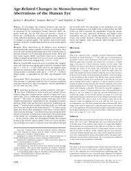
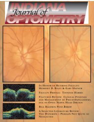
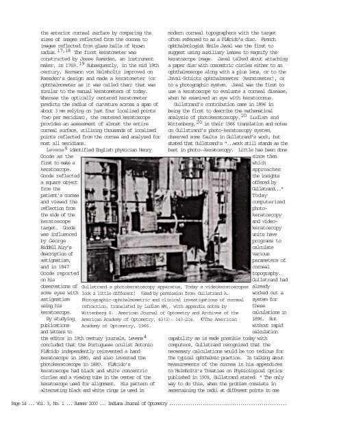
Gullstrand s photokeratoscopy apparatus, Today s videokeratoscopes<br />
look a little different! (Used by permission from: Gullstrand A.<br />
Photographic-ophthalmometric and clinical investigations <strong>of</strong> corneal<br />
refraction, translated by Ludlam WM,, with appendix notes by<br />
Wittenberg S. American Journal <strong>of</strong> <strong>Optometry</strong> and Archives <strong>of</strong> the<br />
American Academy <strong>of</strong> <strong>Optometry</strong>, 43(3): 143-214. ©The American<br />
Academy <strong>of</strong> <strong>Optometry</strong>, 1966.<br />
Page 14 ... Vol. 3, No. 1 ... <strong>Summer</strong> <strong>2000</strong> ... <strong>Indiana</strong> Journal <strong>of</strong> <strong>Optometry</strong> ........................................................