Summer 2000 - Indiana University School of Optometry
Summer 2000 - Indiana University School of Optometry
Summer 2000 - Indiana University School of Optometry
You also want an ePaper? Increase the reach of your titles
YUMPU automatically turns print PDFs into web optimized ePapers that Google loves.
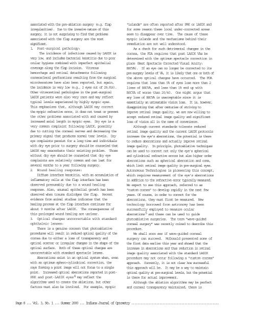
associated with the pre-ablation surgery (e.g. flap<br />
irregularities). Due to the invasive nature <strong>of</strong> this<br />
surgery, it is not surprising to find that problems<br />
associated with the flap surgery are the most<br />
significant.<br />
1. Post-surgical pathology:<br />
The incidence <strong>of</strong> infections caused by LASIK is<br />
very low, and includes bacterial keratitis due to poor<br />
ocular hygiene combined with imperfect epithelial<br />
coverage along the flap incision. Vitreous<br />
hemorrhage and retinal detachments following<br />
corneoscleral perforations resulting from the surgical<br />
microkeratome have also been reported, but again,<br />
the incidence is very low (e.g., 2 eyes out <strong>of</strong> 29,916).<br />
Other vitreoretinal pathologies in the post-surgical<br />
LASIK patients were also very rare and may reflect<br />
typical levels experienced by highly myopic eyes.<br />
This emphasizes that, although LASIK may correct<br />
the myopic refractive error, it does not treat or prevent<br />
the other problems associated with and caused by<br />
increased axial length in myopic eyes. Dry eye is a<br />
very common complaint following LASIK, possibly<br />
due to cutting the corneal nerves and decreasing the<br />
primary signal that produces normal tear levels. Dry<br />
eye complaints persist for a long time and individuals<br />
with dry eye prior to surgery should be counseled that<br />
LASIK may exacerbate their existing problem. Those<br />
without dry eye should be counseled that dry eye<br />
complaints are relatively common and can last for<br />
several months to a year following surgery.<br />
2. Wound healing response:<br />
Diffuse interface keratitis, with an accumulation <strong>of</strong><br />
inflammatory cells at the flap interface has been<br />
observed presumably due to a wound healing<br />
response. Also, unusual epithelial growth has been<br />
observed when trauma dislodges the flap. Recent<br />
evidence from animal studies indicates that the<br />
healing process at the flap interface continues for<br />
about 9 months after LASIK. The consequences <strong>of</strong><br />
this prolonged wound healing are unclear.<br />
3. Optical changes uncorrectable with standard<br />
ophthalmic lenses:<br />
There is a genuine concern that photoablative<br />
procedures will result in reduced optical quality <strong>of</strong> the<br />
cornea due to either a loss <strong>of</strong> transparency and<br />
optical scatter or irregular changes in the shape <strong>of</strong> the<br />
optical surface. Both <strong>of</strong> these optical changes are<br />
uncorrectable with standard spectacle lenses.<br />
Aberrations exist in an optical system when, even<br />
with an optimum sphero-cylindrical correction, the<br />
rays forming a point image will not focus to a single<br />
point. Increased optical aberrations reported in post-<br />
PRK and post-LASIK eyes 4 may reflect the<br />
algorithms used to create the ablations, but other<br />
factors must also be involved. For example, myopic<br />
"islands" are <strong>of</strong>ten reported after PRK or LASIK and<br />
for some reason these local under-corrected areas<br />
seem to disappear over time. The cause <strong>of</strong> these<br />
myopic islands and the mechanisms behind their<br />
remediation are not well understood.<br />
As a check for such detrimental changes in the<br />
cornea, the FDA requires that post LASIK VAs be<br />
determined with the optimum spectacle correction in<br />
place (Best Spectacle Corrected Visual Acuity:<br />
BSCVA). If an eye can no longer be corrected to its<br />
pre-surgery levels <strong>of</strong> VA, it is likely that one or both <strong>of</strong><br />
the above optical changes have occurred. The FDA<br />
requires that less than 5% <strong>of</strong> eyes lose more than 2<br />
lines <strong>of</strong> BSCVA, and less than 1% end up with<br />
BSCVA <strong>of</strong> worse than 20/40. One might argue that<br />
any loss <strong>of</strong> BSCVA is unacceptable since it is<br />
essentially an untreatable vision loss. It is, however,<br />
disappointing that after centuries <strong>of</strong> striving to<br />
improve retinal image quality, we are now willing to<br />
accept reduced retinal image quality and significant<br />
loss <strong>of</strong> vision all in the name <strong>of</strong> convenience.<br />
Although current standards tolerate reduced<br />
retinal image quality and the current LASIK protocols<br />
increase the eye’s aberrations, the potential is there<br />
to reduce aberrations and actually improve retinal<br />
image quality. In principle, photoablative techniques<br />
can be used to correct not only the eye’s spherical<br />
and cylindrical refractive errors but also higher order<br />
aberrations such as spherical aberration and coma,<br />
which limit retinal image quality in pre-surgical eyes.<br />
Autonomous Technologies is pioneering this concept,<br />
which requires measurement <strong>of</strong> the eye’s aberrations<br />
in addition to the refractive error typically measured.<br />
We expect to see this approach, referred to as<br />
"custom cornea" to develop rapidly in the next few<br />
years. Of course, in order to correct for the<br />
aberrations, they must first be measured. New<br />
technology borrowed from astronomy has been<br />
successfully employed to measure ocular<br />
aberrations 5 and these can be used to guide<br />
photoablative surgeries. The term "wave-guided<br />
corneal surgery" was recently coined to describe this<br />
procedure.<br />
We shall soon see if wave-guided corneal<br />
surgery can succeed. McDonald presented some <strong>of</strong><br />
the first data earlier this year and showed that the<br />
increase in aberrations and thus reduction in retinal<br />
image quality associated with the standard LASIK<br />
procedure may not occur following a "custom cornea"<br />
approach. Currently, it is not clear how successful<br />
this approach will be. It may be a way to maintain<br />
optical quality at pre-surgical levels, but the potential<br />
is there for actual improvement.<br />
Although the ablation algorithms may be perfect<br />
and corneal transparency maintained, there is<br />
Page 8 ... Vol. 3, No. 1 ... <strong>Summer</strong> <strong>2000</strong> ... <strong>Indiana</strong> Journal <strong>of</strong> <strong>Optometry</strong> ........................................................


