Summer 2000 - Indiana University School of Optometry
Summer 2000 - Indiana University School of Optometry
Summer 2000 - Indiana University School of Optometry

Create successful ePaper yourself
Turn your PDF publications into a flip-book with our unique Google optimized e-Paper software.
and the same principal section, is by<br />
photographing the reflex image in the cornea.<br />
Such measurements, it must be admitted, take<br />
much time and require special apparatus made<br />
for the purpose. Consequently, they are not<br />
suitable for the general run <strong>of</strong> practice, but on<br />
the other hand they give a resultant accuracy<br />
that previously could not be obtained in any<br />
other way." 21<br />
Gullstrand’s photokeratoscope target was a<br />
series <strong>of</strong> paired concentric circles. Each circle<br />
had another paired circle very close to it with a<br />
thin dark line between them. To measure radii<br />
<strong>of</strong> curvature in the horizontal and vertical<br />
meridians, Gullstrand moved a microscope over<br />
the photographic plates by means <strong>of</strong> a screw<br />
mechanism. A "dividing engine" 22 was used to<br />
determine the amount <strong>of</strong> movement <strong>of</strong> the<br />
microscope after it had been moved to align a<br />
cross hair in the ocular with the dark line<br />
between the paired white circles. These<br />
measurements were converted into radii <strong>of</strong><br />
curvature and then into dioptric powers. In his<br />
1896 paper, Gullstrand gives an example <strong>of</strong> a<br />
photograph taken and analyzed in 1893. He<br />
presented an x,y coordinate plot <strong>of</strong> dioptric<br />
power as a function <strong>of</strong> degrees <strong>of</strong> eccentricity.<br />
This kind <strong>of</strong> plot had been produced previously<br />
with peripheral ophthalmometry (keratometry),<br />
but Gullstrand appears to have made the first<br />
such plot using keratoscopy. Although today’s<br />
keratoscopy is <strong>of</strong>ten thought <strong>of</strong> as a recent<br />
development, Gullstrand had worked out many<br />
<strong>of</strong> the necessary details over a hundred years<br />
ago.<br />
Perhaps because <strong>of</strong> the lack <strong>of</strong> rapid<br />
calculation methodology for the extensive<br />
computations and/or Gullstrand’s statement that<br />
photokeratoscopy measurements "..are not<br />
suitable for the general run <strong>of</strong> practice...," little<br />
work seems to have been done in this area in<br />
the first couple decades <strong>of</strong> the twentieth century.<br />
It appears that the first commercial device for<br />
photokeratoscopy was manufactured by Zeiss in<br />
the 1930s. 23,24 The Zeiss instrument had a flat<br />
target so curvature <strong>of</strong> field would have affected<br />
peripheral measurements. Zeiss did not resume<br />
manufacture <strong>of</strong> the instrument after World War<br />
I.<br />
The next commercially available device for<br />
photokeratoscopy was the Wesley-Jessen<br />
Photo-Electronic Keratoscope or PEK. 25,26 It<br />
was developed in the 1950s, and was<br />
manufactured for about 20 years. Because the<br />
target rings were on an elliptical bowl, there<br />
were less curvature <strong>of</strong> field defects than with the<br />
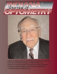
Allvar Gullstrand (1862-1930) was winner <strong>of</strong><br />
the Nobel Prize in physiology or medicine in<br />
1911. He made many contributions to the<br />
knowledge <strong>of</strong> optics <strong>of</strong> the eye and lenses<br />
and to instrumentation for ophthalmic<br />
clinical practice. This medal, from the<br />
collection <strong>of</strong> Jay M. Galst, was struck by Erik<br />
Lindberg in 1935 for the Royal Swedish<br />
Academy <strong>of</strong> Science. (photo courtesy <strong>of</strong> Jay<br />
M. Galst)<br />
Zeiss instrument. Wesley-Jessen marketed the<br />
PEK as an aid to contact lens fitting. The<br />
practitioner took the keratoscope picture and<br />
mailed it to Wesley-Jessen. Wesley-Jessen<br />
sent back an analysis <strong>of</strong> the corneal topography<br />
and suggested the appropriate contact lens<br />
parameters. Although possibly more accurate<br />
than the Zeiss instrument, the PEK did not<br />
achieve wide acceptance.<br />
Following the PEK, various photokeratoscopes<br />
were available, including the<br />
Corneascope and the Nidek Photokeratoscope.<br />
15,27 The Corneascope was<br />
marketed initially by International Diagnostics<br />
Instruments and later by Kera Corporation. The<br />
practitioner could analyze photokeratograms<br />
taken in the <strong>of</strong>fice with a device called a<br />
Comparator. 28,29 The Comparator is an optical<br />
magnifier with variable magnification. The<br />
Comparator projects the Corneascope<br />
keratogram onto a screen and allows the<br />
practitioner to compare the photokeratogram<br />
rings to a calibrated set <strong>of</strong> concentric rings.<br />
Radii <strong>of</strong> curvature at various points on the<br />
photograph can be determined by varying the<br />
magnification to match the photograph ring size<br />
to the ring size on the comparison pattern.<br />
..........................................................<strong>Indiana</strong> Journal <strong>of</strong> <strong>Optometry</strong> ... <strong>Summer</strong> <strong>2000</strong> ... Vol. 3, No. 1... page 15