What You Should Know About Abnormal Blood Clotting - Pathology
What You Should Know About Abnormal Blood Clotting - Pathology
What You Should Know About Abnormal Blood Clotting - Pathology

You also want an ePaper? Increase the reach of your titles
YUMPU automatically turns print PDFs into web optimized ePapers that Google loves.
<strong>What</strong> <strong>You</strong> <strong>Should</strong> <strong>Know</strong> <strong>About</strong><br />
<strong>Abnormal</strong> <strong>Blood</strong> <strong>Clotting</strong><br />
Roger S. Riley, M.D., Ph.D.<br />
<strong>Abnormal</strong> blood clotting (thrombosis) is the major cause of death in the United States and a leading cause of<br />
morbidity, with an annual incidence of about 1 case per 1000 individuals. Nearly 1,000,000 individuals die from<br />
thrombosis in the U.S. each year, in contrast to 500,000 deaths from cancer (Fig. I). Many developments during<br />
the past decade have lead to a greatly improved understanding of the etiology of abnormal blood clotting, and<br />
improvements in clinical diagnosis and therapy have been reported as well. <strong>Blood</strong> clots can develop in the arterial<br />
circulation (arterial thrombosis) or venous circulation (venous thrombosis). Arterial thrombi usually develop in<br />
arteries diseased by the process of atherosclerosis. The factors that lead to venous thrombosis are less well<br />
understood, but inactivity and small injuries to the veins may play a role. <strong>Blood</strong> clots that form in the circulation<br />
often break off and travel to other areas of the circulation, where they can cause major organ damage or death.<br />
Heart attacks and strokes can be caused by these “emboli.” Emboli that lodge in the lungs (pulmonary emboli)<br />
often lead to rapid death. Since thrombosis and embolism occur together, the process is usually referred to as<br />
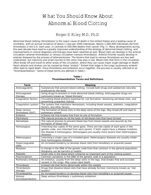
“thromboembolism.” Some of these terms are defined in Table I.<br />
Table I<br />
Thromboembolism Terms and Definitions<br />
Term<br />
Anticoagulants<br />
Anticoagulant<br />
therapy<br />
Coagulation factors<br />
Coagulation system<br />
Deep venous<br />
thrombosis (DVT)<br />
Embolus<br />
Fibrinolysis<br />
Hemostasis<br />
Homozygous<br />
Heterozygous<br />
Hypercoaguable<br />
state<br />
Mutation<br />
Platelets<br />
Polymorphism<br />
Pulmonary embolus<br />
Thrombosis<br />
Thrombolytic<br />
therapy<br />
Thrombophilia<br />
Venous<br />
thromboembolism<br />
Meaning<br />
Substances that prevent blood clotting. Include both drugs and substances naturally<br />
produced by the body<br />
Using drugs to prevent or treat abnormal blood clotting. Anticoagulant drugs are<br />
commonly known as “blood thinners”<br />
Circulating blood proteins essential for clot formation, dissolving old blood clots, and<br />
preventing unwanted clotting<br />
The system that maintains hemostasis, including blood vessels, platelets, coagulation<br />
factors, and other factors<br />
The formation of blood clots in the deep veins of the legs. May break-off and lead to<br />
pulmonary emboli<br />
A blood clot that breaks free from its site of formation<br />
The natural process to rid the body of old blood clots that have formed<br />
The natural process to prevent blood loss from blood vessels and wounds by the<br />
formation of blood clots<br />
Inheriting two copies of the same gene. There are two copies of each gene in the<br />
genetic code, one inherited from each parent. If both copies have a disease mutation,<br />
the disease is homozygous. Homozygous are usually more severe than heterozygous<br />
diseases.<br />
Inheriting a single copy of a mutated gene. Heterozygous diseases are usually milder<br />
than homozygous diseases.<br />
An increased tendency to develop blood clots from hereditary or acquired causes<br />
A change in the DNA of the genetic code from that common in the general population.<br />
Mutations can be harmful or helpful<br />
Small cells in the blood which comprise an important part of the clotting system.<br />
Common variations in the DNA of the genetic code in a population<br />
An embolus, usually from the deep veins in the legs, that breaks free and occludes<br />
vessels in the lungs. May cause rapid death<br />
A blood clot that forms abnormally within the vessels<br />
Drugs that dissolve blood clots that have already formed. Commonly known as “clot<br />
busters”<br />
An increased tendency to develop blood clots from hereditary or acquired causes<br />
Deep venous thrombosis + pulmonary embolism
Deadly <strong>Pathology</strong>/Killer <strong>Blood</strong> Clots<br />
Roger S. Riley, M.D., Ph.D.<br />
2<br />
Most individuals who develop thromboembolism have one or more risk factors. Many of the non-inherited<br />
(acquired) risk factors have been recognized for decades, but a rapid series of scientific discoveries over the past<br />
two decades have lead to the recognition of numerous inherited (genetic) factors that can either increase or<br />
decrease an individuals risk of developing thromboembolic disease. These inherited factors act in conjunction with<br />
the acquired risk factors and involve the vessels, blood platelets, and the chemicals in the blood that are part of<br />
the clotting process. The term thrombophilia refers to individuals who have a tendency to develop thrombosis<br />
from either acquired or inherited causes, or both. In view of the enormous amount of medical resources needed to<br />
care for patients with thromboembolic disease, there is a great interest in the identification and early treatment of<br />
patients who have a high risk of developing thromboembolic disease. Many new laboratory tests and drugs are<br />
available for this purpose. This paper will focus on venous thromboembolism, although much of the information<br />
also pertains to<br />
arterial thrombosis.<br />
1200000<br />
1000000<br />
800000<br />
600000<br />
400000<br />
200000<br />
0<br />
Thrombosis Lung disease Trauma Cancer Other<br />
Causes of Mortality<br />
Fig 1. Leading causes of mortality in the United States. From Fareed et al. Semin. Thromb. Hemost. 26:5-21,<br />
2000.<br />
An Introduction to Hemostasis and Thrombosis<br />
Hemostasis is series of physiologic processes that confine blood to the vascular spaces, maintain the fluidity of<br />
blood, and stop bleeding when injury to a vessel occurs. Hemostasis is a complex process based upon interactions<br />
among the blood vessels and supporting tissues, endothelial cells, platelets, plasma coagulation proteins,<br />
physiologic protease inhibitors, and the fibrinolytic system. Alterations in the hemostatic system can result in<br />
significant pathologic bleeding or clotting.<br />
Resistance to bleeding is provided by: (1) extravascular forces, (i.e., pressure exerted by the skin and supporting<br />
tissue), (2) the physical resistance provided by the blood vessel itself, and (3) substances present within the blood<br />
(e.g. the platelets and coagulation factors). Vessels constrict when injured, limiting the flow of blood to the injured<br />
area. Platelets adhere to collagen fibers exposed by the vascular damage and clump (aggregate) to form a loose,<br />
temporary (primary) plug. The plasma protein fibrinogen helps to stimulate platelet aggregation. Aggregated<br />
platelets undergo a series of mechanical (shape change) and biochemical changes termed activation. Activated<br />
platelets release chemical substances that activate other platelets and initiate the coagulation cascade.<br />
Coagulation generates a fibrin mesh, which stabilizes the platelet plug. The process of healing and recovery is also<br />
initiated to restore normal function to the vessel. The endothelium heals and the blood clot is dissolved through
Deadly <strong>Pathology</strong>/Killer <strong>Blood</strong> Clots<br />
Roger S. Riley, M.D., Ph.D.<br />
3<br />
the action of plasmin and other components of the fibrinolytic system. The four stages of coagulation include: (1)<br />
Vessel constriction, (2) Formation of platelet plug (primary hemostasis), (3) Coagulation and fibrin generation<br />
(secondary hemostasis), (4) Fibrinolysis, healing, and repair.<br />
Fig. 1. Normal process of hemostasis. Platelets are attracted to an injured vessel, stick to the exposed collagen of<br />
the vessel wall (top diagram) and rapidly form a platelet plug (middle diagram) that occludes the gap in the vessel<br />
wall to prevent bleeding. The coagulation system is activated and forms a fibrin mesh that entraps red blood cells<br />
and platelets to form a firm blood clot (bottom diagram). As the vessel wall heals, the clot is gradually removed<br />
by the fibrinolytic system (not shown). The vessel is eventually restored to normal.<br />
The body has an efficient system to assure that unneeded blood clotting does not occur. For example, active<br />
coagulation substances are effective for only very short periods of time (i.e., milliseconds to seconds) and are<br />
rapidly diluted by normal blood flow. The liver and other parts of the body remove activated coagulation factors<br />
and there is an elaborate system of chemical substances that destroy these substances. Lastly, the walls of the<br />
vessels throughout the body (vascular endothelium) release chemical substances (i.e. antithrombin III, protein C<br />
pathway, etc.) that prevent platelet activation and the activation of chemical mediators. Normally, these systems<br />
work together to assure that bleeding does not occur, but also prevent thromboembolic disease. Unfortunately,<br />
defects in the blood vessels (vascular defects), or in any of the processes leading to defective formation of the<br />
hemostatic plug (platelet dysfunction, coagulation defects) may result in a bleeding disorder, while<br />
thromboembolic disease may be caused by inappropriate activation of hemostasis or defective modulation of the<br />
mechanisms which regulate blood clotting.
Deadly <strong>Pathology</strong>/Killer <strong>Blood</strong> Clots<br />
Roger S. Riley, M.D., Ph.D.<br />
4<br />
Risk Factors for Thromboembolic Disease<br />
The major types of venous thromboembolism, deep venous thrombosis (DVT) and pulmonary embolism, are a<br />
leading cause of morbidity and mortality in hospitalized patients, but are being seen with increasing frequency in<br />
outpatients as well. The discovery of a number of acquired and inherited risk factors for thromboembolic disease<br />
has provided a means to predict the risk of these diseases and institute preventive therapy.<br />
Acquired Risk Factors<br />
Acquired risk factors for venous thromboembolism include surgery, smoking, trauma, fractures, immobilization or<br />
venous stasis, inflammatory diseases, pregnancy, the use of oral contraceptives containing synthetic estrogens,<br />
malignancy, congestive heart failure, and other diseases. Venous stasis in the extremities, venous obstruction,<br />
increased blood viscosity, and direct venous damage may cause or contribute to the development of venous<br />
thromboembolism. The increasing trend to early hospital discharge of postsurgical patients may be responsible for<br />
the increasing incidence of venous thromboembolism in outpatients. Venous thromboembolism usually involves<br />
the deep or superficial veins of the legs. <strong>Blood</strong> clots in the superficial veins (superficial thrombophlebitis) leads to<br />
localized tenderness, surrounded by an area of redness (erythema), heat, and edema. A thrombus can often be<br />
palpated in the affected vein. Although usually benign and self-limiting, superficial thromboemboli can cause<br />
serious complications if they extend into the other veins. Deep thrombi confined to calf veins rarely cause clinical<br />
problems, but can lead to pulmonary emboli.<br />
The antiphospholipid syndrome (APS) is another common acquired cause of thrombosis. APS is caused by<br />
autoantibodies that form against certain components of the coagulation system. In addition to venous and arterial<br />
thrombosis, including strokes, APS is associated with cause bleeding, miscarriages, and other medical problems.<br />
The long-term use of certain drugs, including chlorpromazine and phenothiazine, may result in APS. Generally,<br />
patients with APS have a 6-10x risk of developing venous thrombosis than normal individuals, and the risk of<br />
artial thrombosis is increased as well.<br />
Inherited Risk Factors<br />
The majority of patients who develop recurrent venous thromboemboli (thrombophilia) have discernable<br />
abnormalities of the coagulation system, including Factor V Leiden, deficiencies of protein C, protein S,<br />
antithrombin III, the prothrombin G20210A gene mutation, homocystinuria, or abnormalities of the fibrinolytic<br />
system. Most of these abnormalities cause deficiencies of the regulatory substances of clotting. Genetic<br />
abnormalities are especially common in individuals who develop thrombi at an early age (< 40 years) and in those<br />
with a family history of thrombosis. Although no genetic abnormality is detectable in about 15-20% of individuals<br />
with recurrent thromboembolic disease, research in this area is rapidly proceeding, and new genetic abnormalities<br />
may be described in the near future.<br />
Table II<br />
Common Inherited Thrombotic Diseases<br />
<strong>Abnormal</strong>ity<br />
Prevalence in Relative Risk of Venous Thrombosis*<br />
Thrombotic Disease<br />
Factor V Leiden 25-40% Heterozygous – 5 to 10; heterozygous + OCP –<br />
30 to 35; homozygous – 50 - 100, homozygous<br />
+ OCP - >>100<br />
Homocysteine abnormalities 10-25% Coronary artery disease – 2, stroke - 2.5,<br />
venous thrombosis – 7; hyperhomocysteinemia<br />
+ heterozygous Factor V Leiden - 20<br />
Prothrombin gene mutation 6-20% Heterozygous – 3; heterozygous + OCP – 16;<br />
homozygous - ??<br />
ATIII deficiency 3-5% Heterozygous – 5; homozygous – severe<br />
thrombosis at birth<br />
Protein C deficiency 5-6% Heterozygous – 7; homozygous – severe<br />
thrombosis at birth<br />
Protein S deficiency 5-6% Heterozygous – 6; homozygous – lethal prior to<br />
birth<br />
* The relative risk of venous thrombosis in a normal individual (without inherited thromboembolic genes) is 1.0.<br />
Oral contraceptives (birth control pills) increase the relative risk to 4 in a normal individual.
Deadly <strong>Pathology</strong>/Killer <strong>Blood</strong> Clots<br />
Roger S. Riley, M.D., Ph.D.<br />
5<br />
Individuals with decreased levels or abnormal function of naturally occurring anticoagulants such as antithrombin<br />
III and protein C are prone to thrombosis that may present as DVT, thrombophlebitis and/or PE. The primary<br />
inherited causes of thrombosis include resistance to activated protein C (APCR) and deficiencies of protein C,<br />
protein S, and antithrombin III.<br />
Factor V Leiden (Activated Protein C Resistance, APCR)<br />
Activated protein C (APC) is a major regulator of the coagulation system. It inhibits blood clotting by degrading<br />
phospholipid-bound activated factor VIII (VIIIa) and activated factor V (Va). Factor V Leiden, first identified in<br />
February, 1993, is the most common inherited cause of thrombosis known at this time. Factor V Leiden is found in<br />
about 5% of the general population and is responsible for 20-50% cases of inherited thrombosis. Approximately<br />
50,000 individuals die yearly in the United States from this abnormality. Heterozygous individuals are at 5 - 10<br />
times greater risk of thrombosis than the general population, while homozygotes are at a 50-100 times greater.<br />
The use of estrogen or oral contraceptives increases the risk of thrombosis even further. In 90 - 95% of cases,<br />
APCR is a result of a single point mutation in the gene for factor V, inherited as an autosomal dominant trait. This<br />
mutation renders activated factor V (Va) resistant to inactivation by APC. The remaining 5-1 0% of APCR is due to<br />
other genetic abnormalities in the factor V gene.<br />
Prothrombin G20210A Mutation<br />
A mutation in the prothrombin gene that produces elevated levels of prothrombin was discovered in 1996. There<br />
is increasing evidence that the G20210A mutation is an important risk factor for deep venous thrombosis,<br />
myocardial infarction, and stroke. The use of estrogen or oral contraceptives increases the risk of thrombosis even<br />
further in patients with the prothrombin 20210 mutation.<br />
Homocysteine <strong>Abnormal</strong>ities<br />
Hyperhomocysteinemia and homocysteinemia are inherited abnormalities of homocysteine metabolism.<br />
Homocysteine is a naturally occurring substance involved in the metabolism of certain amino acids, including<br />
cysteine and methionine. <strong>Abnormal</strong>ities in three enzymes [methylenetetrahydrofolate reductase (MTHFR),<br />
cystathionine beta-synthase (CBS) and methionine synthase (MS)] associated with homocysteine metabolism in<br />
the body can lead to increased homocysteine levels in the body (hyperhomocysteinemia). Genetic abnormalities in<br />
these enzymes, particularily CBS are the second most common risk factor for thrombotic disease, including heart<br />
disease and stroke. Hyperhomocysteinemia may also be associated with vitamin deficiency, advanced age,<br />
hypothyroidism, impaired kidney function, systemic lupus erythematosus., and the use of certain medications,<br />
including nicotinic acid, theophylline, methotrexate and L-dopa.<br />
Protein S Deficiency<br />
Hereditary protein S deficiency is responsible for 5 - 8% of cases of inherited thrombosis. This disease typically<br />
presents as deep vein thrombosis of the legs, with a median age of onset in the late 20's. The family history is<br />
frequently positive for thrombosis. Unlike protein C deficiency, in which many families of affected individuals are<br />
asymptomatic, no entire kindred with protein S deficiency has been described that is thrombosis free. Acquired<br />
protein S deficiency occurs during pregnancy, oral contraceptive usage, and nephrotic syndrome. Since<br />
approximately 60% of circulating protein S is bound to C4b binding protein, elevations in C4b binding protein are<br />
believed to cause a deficiency of functional protein S.<br />
Protein C Deficiency<br />
Heterozygous protein C deficiency occurs with a prevalence of 1 in 200-300; however, associated thrombosis<br />
probably occurs in less than 1 in 10,000 individuals (2 - 5% prevalence in thrombotic patients). Homozygous<br />
protein C deficiency, which causes life-threatening thrombosis unless treated, typically appears at birth. Patients<br />
with heterozygous protein C deficiency usually present with thrombosis of the leg, mesenteric veins and<br />
iliofemoral veins prior to the age of 35. The initial thrombotic episode is spontaneous in approximately 70% of<br />
patients, while the remaining fraction have other thrombotic risk factors present. Acquired protein C deficiency<br />
occurs in liver disease, disseminated intravascular coagulation (DIC), post-operative state, adult respiratory<br />
distress, nephritic syndrome and in association with L-asparaginase therapy. L-asparaginase is a drug used to<br />
treat certain types of leukemia.
Deadly <strong>Pathology</strong>/Killer <strong>Blood</strong> Clots<br />
Roger S. Riley, M.D., Ph.D.<br />
6<br />
Antithrombin III Deficiency<br />
The prevalence of AT III deficiency is approximately 1 in 2000 to 1 in 5000 individuals. A positive family history of<br />
recurrent thrombosis typically begins in youth and is associated with trauma or surgery. 70% of AT III deficient<br />
individuals develop thrombosis prior to age 35 and 85% develop thrombosis by age 50. Arterial thrombosis is seen<br />
in approximately 20% of symptomatic patients. The use of oral contraceptives and pregnancy increases the risk of<br />
thrombosis. Acquired AT III deficiency may develop in patients following three or more days of intravenous<br />
heparin administration, and is associated with liver disease, DIC, nephrotic syndrome and following L-<br />
asparaginase therapy. Oral contraceptive usage can result in a 10-20% reduction in AT III concentration.<br />
Treatment and Prevention of Thromboembolic Disease<br />
In spite of the effiacy of the natural antithrombotic system, some patients require therapeutic agents to prevent<br />
overactivation of the clotting system. Aspirin, heparin, and coumadin have been available for several decades, but<br />
each of these drugs have many limitations. A wide variety or new drugs are being tested or have been recently<br />
approved by the FDA.<br />
Aspirin<br />
Low-dose aspirin (80 mg) is the most commonly used drug for preventing thrombosis, particularly coronary<br />
thrombosis in patients with atherosclerosis. Aspirin works by inhibiting an enzyme, cyclooxygenase-1, that is<br />
present in platelets and the endothelial cell. A single dose of aspirin works for the life of the platelet (about a<br />
week). However, since platelets are continuously produced, aspirin must be taken daily.<br />
Warfarin (Coumadin)<br />
Crystalline warfarin sodium (Coumadin) is the most widely used oral anticoagulant. Warfarin interferes with the<br />
synthesis of the vitamin-K dependent procoagulants (factors II, VII, IX, X) in the liver by inhibiting the reduction<br />
of oxidized vitamin K. Since functional circulating clotting factors are not affected by warfarin, a week or more of<br />
oral anticoagulation therapy is required to achieve an optimal therapeutic effect. Warfarin is a safe agent for the<br />
prophylaxis of thrombosis if the correct dosage is given and the patient is carefully monitored. However, serious<br />
bleeding complications can occur with excessive anticoagulation, while thromboembolic complications are a risk<br />
with inadequate coagulation. For this reason, accurate laboratory measurements of the prothrombin time (PT) are<br />
critical in the management of patients receiving oral anticoagulation.<br />
Heparin<br />
Heparin is the other major anticoagulant used for therapeutic purposes. Heparin is a negatively charged, highly<br />
sulfated mucopolysaccharide with a molecular weight between 6,000 and 25,000 daltons. It is not absorbed from<br />
the gastrointestinal tract and must be given by injection into the veins (intravenous) or under the skin<br />
(subcutaneous). A single intravenous dose has a half-life of approximately 60 minutes. Heparin exerts its potent<br />
anticoagulant effect by activating a natural anticoagulant termed antithrombin III. Recent drugs derived from<br />
heparin and termed “low molecular weight heparin” act in the same way but have fewer side effects and require<br />
less frequent injections.<br />
New Anti-Thrombotic Drugs<br />
A wide variety of new anti-thrombotic drugs are now available or clinical trials for use in the near future. Unlike<br />
heparin and warfarin, most of these drugs are directed again a specific part of the coagulation system and are<br />
said to be target-specific. They also have fewer side effects and usually do not require laboratory monitoring.
Deadly <strong>Pathology</strong>/Killer <strong>Blood</strong> Clots<br />
Roger S. Riley, M.D., Ph.D.<br />
7<br />
Selective factor Xa inhibitors<br />
Fondaparinux (Synthetic)<br />
Tick anticoagulant peptide (Soft tick)<br />
Antistasin (Mexican leech)<br />
Lefaxin (Mexican leech)<br />
DX-9065a<br />
Direct thrombin inhibitors<br />
Hirudin (Medicinal leech)<br />
Bivalirudin (Synthetic)<br />
Argatroban<br />
Melagatran<br />
Platelet function inhibitors<br />
Abciximab<br />
Dipyridamole<br />
Clopidogrel<br />
Ticlopidine<br />
Eptifibatide<br />
Other anticoagulants<br />
Recombinant human APC<br />
Acitve-site factor IXa blockers<br />
Inhibitory factor IX/IXa Abs<br />
Active-site blocked rVIIa<br />
Table III<br />
New Anti-Thrombotic Drugs<br />
Cost-Effective Diagnosis of <strong>Abnormal</strong> <strong>Blood</strong> <strong>Clotting</strong><br />
The recent identification of inherited and acquired risk factors for thrombosis have greatly improved the ability of<br />
doctors to diagnose thrombotic disorders and to identify individuals at increased risk of thrombosis. There is a<br />
debate about whom to test for thromboembolic disease, but most doctors feel that individuals with a family<br />
history of abnormal blood clotting, as well as those with well-defined acquired risk factors should be screened for<br />
the antiphospholipid antibody syndrome and the common inherited risk factors. This testing can help in estimating<br />
the risk of thromboembolic disease and planning long-term anti-thrombotic management.<br />
The coagulation laboratory performs tests that detect the function and amount of various coagulation factors,<br />
while the molecular diagnosis laboratory looks for specific abnormalities in DNA. Generally, the coagulation tests<br />
are used to screen for the presence of disease and to monitor treatment, while the molecular assays are used for<br />
confirmation. Since the coagulation system is disrupted by the body’s reaction to thromboembolic disease,<br />
coagulation screening in a person who suffers from a blood clot should be delayed for several weeks after<br />
discharge from the hospital and the coagulation system returns to a “steady state.” However, the molecular<br />
assays are not affected by drugs or disease processes, and can be performed at any time. In a person with a<br />
suspected inherited thromboembolic disease, laboratory testing for the most common abnormalities (i.e.,<br />
antiphospholipid antibody syndrome, factor V Leiden, prothrombin G20210A mutation, homocysteine abnormality)<br />
is usually performed first. If these tests are negative, additional testing for less common deficiency states (i.e.,<br />
fibrinolytic abnormalities, plasminogen deficiency, etc.) might be considered.
Deadly <strong>Pathology</strong>/Killer <strong>Blood</strong> Clots<br />
Roger S. Riley, M.D., Ph.D.<br />
8<br />
Table IV<br />
Laboratory Assays for Thrombotic Disorders*<br />
<strong>Abnormal</strong>ity Coagulation Laboratory Assays Molecular Assays<br />
Antiphospholipid antibody Screen - Activated partial thromboplastin None<br />
syndrome<br />
time (aPPT)($25). Confirmation - dilute<br />
Russell Viper Venom assay (dRVVT)($100)<br />
Factor V Leiden APC resistance ($250) Factor V Mutation Assay ($325)<br />
Prothrombin G20210A<br />
mutation<br />
None<br />
Prothrombin G20210A mutation<br />
($325)<br />
Homocysteine abnormalities Plasma homocysteine levels ($175) MTHFR mutation ($325)<br />
Antithrombin III deficiency Antithrombin III function and levels ($200) None<br />
Protein C deficiency Protein C function and levels ($425) None<br />
Protein S deficiency Protein S function and levels ($375) None<br />
Increased factor VIII Factor VIII activity ($175) None<br />
Increased plasminogen Plasminogen activity and levels ($415) None<br />
Plasminogen Activator PAI-1 inhibitor assay ($230)<br />
None<br />
Inhibitor-1<br />
* The most common abnormalities are listed first. The approximate cost is only a guide; laboratory costs may<br />
vary considerably.<br />
Most deficient individuals presenting with thrombosis are managed acutely with heparin therapy, followed by long<br />
term oral anticoagulant therapy. Commercially prepared concentrates are available for use post-surgically and<br />
during parturition in AT III deficient individuals. Protein C concentrates are available on a compassionate use<br />
basis.<br />
Summary<br />
Thromboembolic diseases are presently the leading cause of death, and responsible for more than 1,000,000<br />
deaths/year in the United States alone. Fortunately, thromboembolic disease is preventable, and many recent<br />
scientific discoveries have lead to the ability to estimate a person’s disease risk, so that appropriate preventive<br />
therapy can be instituted. Most doctors agree that persons who have a strong family history of thromboembolic<br />
disease or who develop thrombosis at an early age (i.e.,