article by Drs. Joshi and Ahmad (May 2011) - Pathology Outlines
article by Drs. Joshi and Ahmad (May 2011) - Pathology Outlines
article by Drs. Joshi and Ahmad (May 2011) - Pathology Outlines

Create successful ePaper yourself
Turn your PDF publications into a flip-book with our unique Google optimized e-Paper software.
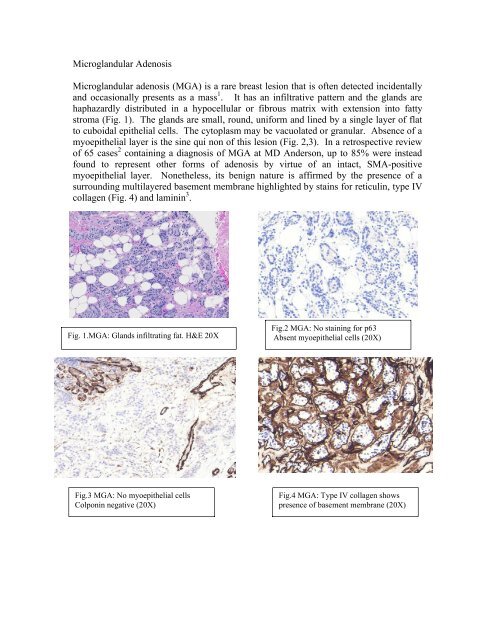
Microgl<strong>and</strong>ular Adenosis<br />
Microgl<strong>and</strong>ular adenosis (MGA) is a rare breast lesion that is often detected incidentally<br />
<strong>and</strong> occasionally presents as a mass 1 . It has an infiltrative pattern <strong>and</strong> the gl<strong>and</strong>s are<br />
haphazardly distributed in a hypocellular or fibrous matrix with extension into fatty<br />
stroma (Fig. 1). The gl<strong>and</strong>s are small, round, uniform <strong>and</strong> lined <strong>by</strong> a single layer of flat<br />
to cuboidal epithelial cells. The cytoplasm may be vacuolated or granular. Absence of a<br />
myoepithelial layer is the sine qui non of this lesion (Fig. 2,3). In a retrospective review<br />
of 65 cases 2 containing a diagnosis of MGA at MD Anderson, up to 85% were instead<br />
found to represent other forms of adenosis <strong>by</strong> virtue of an intact, SMA-positive<br />
myoepithelial layer. Nonetheless, its benign nature is affirmed <strong>by</strong> the presence of a<br />
surrounding multilayered basement membrane highlighted <strong>by</strong> stains for reticulin, type IV<br />
collagen (Fig. 4) <strong>and</strong> laminin 3 .<br />
Fig. 1.MGA: Gl<strong>and</strong>s infiltrating fat. H&E 20X<br />
Fig.2 MGA: No staining for p63<br />
Absent myoepithelial cells (20X)<br />
Fig.3 MGA: No myoepithelial cells<br />
Colponin negative (20X)<br />
Fig.4 MGA: Type IV collagen shows<br />
presence of basement membrane (20X)