article by Drs. Joshi and Ahmad (May 2011) - Pathology Outlines
article by Drs. Joshi and Ahmad (May 2011) - Pathology Outlines
article by Drs. Joshi and Ahmad (May 2011) - Pathology Outlines

You also want an ePaper? Increase the reach of your titles
YUMPU automatically turns print PDFs into web optimized ePapers that Google loves.
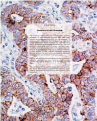
The infiltrative pattern of growth may cause confusion with low-grade invasive ductal<br />
(tubular) carcinomas. The latter is distinguished <strong>by</strong> a stellate “tear-drop” growth pattern<br />
<strong>and</strong> a desmoplastic stromal response 4 . In addition, the gl<strong>and</strong>s of MGA contain a<br />
characteristic PAS-positive, diastase-resistant, eosinophilic material not present in tubular<br />
carcinoma. The immunophenotypic profile is peculiar <strong>and</strong> further helps distinguish<br />
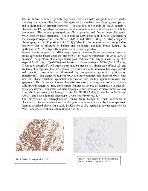
MGA from invasive carcinoma. The gl<strong>and</strong>s are S100 positive (Fig. 5, 10) <strong>and</strong> negative<br />
for estrogen/progesterone receptors (ER/PR) <strong>and</strong> HER-2 (Fig. 6) (triple-negative<br />
phenotype), but EGFR positive (Fig. 7, 8) (Table 1). In contrast to the strong EMApositivity<br />
that is observed in benign <strong>and</strong> malignant gl<strong>and</strong>ular breast lesions, the<br />
epithelium in MGA is typically negative or only focally positive.<br />
Several studies suggest that MGA may represent a non-obligate precursor to invasive<br />
breast carcinoma based upon the presence of an invasive component in up to 27% of<br />
patients 2 . A spectrum of microgl<strong>and</strong>ular proliferations from benign adenosis(Fig 1) to<br />
atypical MGA (Fig. 16)(AMGA) <strong>and</strong> breast carcinoma arising in MGA (MGACA)(Fig.<br />
9) has been described 5 . All three lesions may be present in a single case, (Figs.1-22) <strong>and</strong><br />
are thought to represent true progression <strong>by</strong> virtue of a similar immunophenotypic profile<br />
<strong>and</strong> genetic constitution as illustrated <strong>by</strong> comparative genomic hybridization<br />
experiments 1 . The gl<strong>and</strong>s of atypical MGA are more complex than those of MGA, with<br />
size <strong>and</strong> shape variation, epithelial stratification <strong>and</strong> readily apparent mitoses <strong>and</strong><br />
apoptotic cells. Breast carcinomas that arise from such a background usually exhibit a<br />
solid growth pattern but may demonstrate features as diverse as metaplastic or adenoidcystic<br />
phenotype 5 . Regardless of their cytologic grade, however, invasive tumors arising<br />
from MGA are usually triple-negative for ER/PR/HER2 (Fig.6), similar to MGA <strong>and</strong><br />
AMGA, <strong>and</strong> have a luminal phenotype (CK8/18 positive) (Fig. 13).<br />
The progression of microgl<strong>and</strong>ular lesions from benign to frank carcinoma is<br />
characterized <strong>by</strong> accumulation of complex genetic abnormalities <strong>and</strong> <strong>by</strong> the morphologic<br />
features described above. In a study <strong>by</strong> Khalifeh et al 2 , increasing nuclear reactivity for<br />
MIB-1 <strong>and</strong> p53 reflect this feature (Figs.17,19-22).<br />
Fig 5. MGA: S-100 positive (20X)<br />
Fig. 6 Triple negative carcinoma arising<br />
in MGA. Her 2 negative (20X)