CHAPTER 3 Tumours of the Stomach - Pathology Outlines
CHAPTER 3 Tumours of the Stomach - Pathology Outlines
CHAPTER 3 Tumours of the Stomach - Pathology Outlines

You also want an ePaper? Increase the reach of your titles
YUMPU automatically turns print PDFs into web optimized ePapers that Google loves.
<strong>CHAPTER</strong> 3<br />
<strong>Tumours</strong> <strong>of</strong> <strong>the</strong> <strong>Stomach</strong><br />
The incidence <strong>of</strong> adenocarcinoma <strong>of</strong> <strong>the</strong> stomach is declining<br />
worldwide. In some Western countries, rates have been<br />
reduced to less than one third within just one generation. In<br />
countries with a traditionally high incidence, e.g. Japan and<br />
Korea, <strong>the</strong> reduction is also significant but it will take more<br />
time to diminish <strong>the</strong> still significant disease burden. The main<br />
reasons for <strong>the</strong>se good news is a change in nutrition, in particular<br />
<strong>the</strong> avoidance <strong>of</strong> salt for meat and fish preservation, <strong>the</strong><br />
lowering <strong>of</strong> salt intake from o<strong>the</strong>r sources, and <strong>the</strong> availability<br />
in many countries <strong>of</strong> fresh fruits and vegetables throughout<br />
<strong>the</strong> year. Mortality has been fur<strong>the</strong>r dercreased by significant<br />
advances in <strong>the</strong> early detection <strong>of</strong> stomach cancer.<br />
Infection with Helicobacter pylori appears to play an important<br />
additional aetiological role since it leads to chronic atrophic<br />
gastritis with intestinal metaplasia as an important precursor<br />
lesion.<br />
The stomach is <strong>the</strong> main gastrointestinal site for lymphomas<br />
and most <strong>of</strong> <strong>the</strong>se are also pathogenetically linked to H. pylori<br />
infection. Regression <strong>of</strong> such tumours <strong>of</strong>ten follows H. pylori<br />
eradication.
WHO histological classification <strong>of</strong> gastric tumours 1<br />
Epi<strong>the</strong>lial tumours<br />
Intraepi<strong>the</strong>lial neoplasia – Adenoma 8140/0 2<br />
Carcinoma<br />
Adenocarcinoma 8140/3<br />
intestinal type 8144/3<br />
diffuse type 8145/3<br />
Papillary adenocarcinoma 8260/3<br />
Tubular adenocarcinoma 8211/3<br />
Mucinous adenocarcinoma 8480/3<br />
Signet-ring cell carcinoma 8490/3<br />
Adenosquamous carcinoma 8560/3<br />
Squamous cell carcinoma 8070/3<br />
Small cell carcinoma 8041/3<br />
Undifferentiated carcinoma 8020/3<br />
O<strong>the</strong>rs<br />
Carcinoid (well differentiated endocrine neoplasm) 8240/3<br />
Non-epi<strong>the</strong>lial tumours<br />
Leiomyoma 8890/0<br />
Schwannoma 9560/0<br />
Granular cell tumour 9580/0<br />
Glomus tumour 8711/0<br />
Leiomyosarcoma 8890/3<br />
GI stromal tumour 8936/1<br />
benign 8936/0<br />
uncertain malignant potential 8936/1<br />
malignant 8936/3<br />
Kaposi sarcoma 9140/3<br />
O<strong>the</strong>rs<br />
Malignant lymphomas<br />
Marginal zone B-cell lymphoma <strong>of</strong> MALT-type 9699/3<br />
Mantle cell lymphoma 9673/3<br />
Diffuse large B-cell lymphoma 9680/3<br />
O<strong>the</strong>rs<br />
Secondary tumours<br />
_____________<br />
1<br />
The classification is modified from <strong>the</strong> previous WHO histological classification <strong>of</strong> tumours {2066} taking into account changes in our understanding <strong>of</strong> <strong>the</strong>se lesions. In <strong>the</strong> case <strong>of</strong><br />
endocrine neoplasms, <strong>the</strong> classification is based on <strong>the</strong> recent WHO clinicopathological classification {1784}, but has been simplified to be <strong>of</strong> more practical utility in morphological<br />
classification.<br />
2<br />
Morphology code <strong>of</strong> <strong>the</strong> International Classification <strong>of</strong> Diseases for Oncology (ICD-O) {542} and <strong>the</strong> Systematized Nomenclature <strong>of</strong> Medicine (http://snomed.org). Behaviour is coded<br />
/0 for benign tumours, /3 for malignant tumours, and /1 for unspecified, borderline or uncertain behaviour. Intraepi<strong>the</strong>lial neoplasia does not have a generic code in ICD-O. ICD-O codes<br />
are available only for lesions categorized as glandular intraepi<strong>the</strong>lial neoplaia grade III (8148/2), and adenocarcinoma in situ (8140/2).<br />
TNM classification <strong>of</strong> gastric tumours<br />
TNM classification 1<br />
T – Primary Tumour<br />
TX Primary tumour cannot be assessed<br />
T0 No evidence <strong>of</strong> primary tumour<br />
Tis Carcinoma in situ: intraepi<strong>the</strong>lial tumour<br />
without invasion <strong>of</strong> <strong>the</strong> lamina propria<br />
T1 Tumour invades lamina propria or submucosa<br />
T2 Tumour invades muscularis propria or subserosa 2<br />
T3 Tumour penetrates serosa (visceral peritoneum)<br />
without invasion <strong>of</strong> adjacent structures 2,3,4,5<br />
T4 Tumour invades adjacent structures 2,3,4,5<br />
N – Regional Lymph Nodes<br />
NX Regional lymph nodes cannot be assessed<br />
N0 No regional lymph node metastasis<br />
N1 Metastasis in 1 to 6 regional lymph nodes<br />
N2 Metastasis in 7 to 15 regional lymph nodes<br />
N3 Metastasis in more than 15 regional lymph nodes<br />
M – Distant Metastasis<br />
MX Distant metastasis cannot be assessed<br />
M0 No distant metastasis<br />
M1 Distant metastasis<br />
Stage Grouping<br />
Stage 0 Tis N0 M0<br />
Stage IA T1 N0 M0<br />
Stage IB T1 N1 M0<br />
T2 N0 M0<br />
Stage II T1 N2 M0<br />
T2 N1 M0<br />
T3 N0 M0<br />
Stage IIIA T2 N2 M0<br />
T3 N1 M0<br />
T4 N0 M0<br />
Stage IIIB T3 N2 M0<br />
Stage IV T4 N1, N2, N3 M0<br />
T1, T2, T3 N3 M0<br />
Any T Any N M1<br />
____________<br />
1<br />
{1, 66}. This classification applies only to carcinomas.<br />
2<br />
A help desk for specific questions about <strong>the</strong> TNM classification is available at http://tnm.uicc.org.<br />
3<br />
A tumour may penetrate muscularis propria with extension into <strong>the</strong> gastrocolic or gastrohepatic ligaments or <strong>the</strong> greater and lesser omentum without perforation <strong>of</strong> <strong>the</strong> visceral peritoneum<br />
covering <strong>the</strong>se structures. In this case, <strong>the</strong> tumour is classified as T2. If <strong>the</strong>re is perforation <strong>of</strong> <strong>the</strong> visceral peritoneum covering <strong>the</strong> gastric ligaments or omenta, <strong>the</strong> tumour<br />
is classified as T3.<br />
4<br />
The adjacent structures <strong>of</strong> <strong>the</strong> stomach are <strong>the</strong> spleen, transverse colon, liver, diaphragm, pancreas, abdominal wall, adrenal gland, kidney, small intestine, and retroperitoneum.<br />
5<br />
Intramural extension to <strong>the</strong> duodenum or oesophagus is classified by <strong>the</strong> depth <strong>of</strong> greatest invasion in any <strong>of</strong> <strong>the</strong>se sites including stomach.<br />
38 <strong>Tumours</strong> <strong>of</strong> <strong>the</strong> stomach
Gastric carcinoma<br />
C. Fenoglio-Preiser N. Muñoz<br />
F. Carneiro S.M. Powell<br />
P. Correa M. Rugge<br />
P. Guilford M. Sasako<br />
R. Lambert M. Stolte<br />
F. Megraud H. Watanabe<br />
Definition<br />
A malignant epi<strong>the</strong>lial tumour <strong>of</strong> <strong>the</strong><br />
stomach mucosa with glandular differentiation.<br />
Its aetiology is multifactorial; most<br />
commonly it develops after a long period<br />
<strong>of</strong> atrophic gastritis.<br />
<strong>Tumours</strong> <strong>of</strong> <strong>the</strong> oesophagogastric junction<br />
are dealt with in <strong>the</strong> preceding<br />
chapter.<br />
ICD-O codes<br />
Adenocarcinoma 8140/3<br />
Intestinal type 8144/3<br />
Diffuse type 8145/3<br />
Papillary adenocarcinoma 8260/3<br />
Tubular adenocarcinoma 8211/3<br />
Mucinous adenocarcinoma 8480/3<br />
Signet-ring cell carcinoma 8490/3<br />
Epidemiology<br />
Geographical distribution<br />
Gastric cancer was <strong>the</strong> second commonest<br />
cancer in <strong>the</strong> world in 1990, with an<br />
estimated 800,000 new cases and<br />
650,000 deaths per year; 60% <strong>of</strong> <strong>the</strong>m<br />
occurred in developing countries {1469}.<br />
The areas with <strong>the</strong> highest incidence<br />
rates (> 40/100,000 in males) are in<br />
Eastern Asia, <strong>the</strong> Andean regions <strong>of</strong><br />
South America and Eastern Europe. Low<br />
rates (< 15/100,000) are found in North<br />
America, Nor<strong>the</strong>rn Europe, and most<br />
countries in Africa and in Sou<strong>the</strong>astern<br />
Asia {1471}. There is about a 20-fold difference<br />
in <strong>the</strong> incidence rates when comparing<br />
<strong>the</strong> rates in Japan with those <strong>of</strong><br />
some white populations from <strong>the</strong> US and<br />
those <strong>of</strong> some African countries. A predominance<br />
<strong>of</strong> <strong>the</strong> intestinal type <strong>of</strong> adenocarcinoma<br />
occurs in high-risk areas,<br />
while <strong>the</strong> diffuse type is relatively more<br />
common in low-risk areas {1296}.<br />
Time trends<br />
A steady decline in <strong>the</strong> incidence and<br />
mortality rates <strong>of</strong> gastric carcinoma has<br />
been observed worldwide over <strong>the</strong> past<br />
several decades, but <strong>the</strong> absolute number<br />
<strong>of</strong> new cases per year is increasing<br />
mainly because <strong>of</strong> <strong>the</strong> aging <strong>of</strong> <strong>the</strong> population<br />
{1296}. Analysis <strong>of</strong> time trends by<br />
histological types indicates that <strong>the</strong> incidence<br />
decline results from a decline in<br />
<strong>the</strong> intestinal type <strong>of</strong> carcinoma {1296}.<br />
Age and sex distribution<br />
Gastric carcinoma is extremely rare<br />
below <strong>the</strong> age <strong>of</strong> 30; <strong>the</strong>reafter it increases<br />
rapidly and steadily to reach <strong>the</strong> highest<br />
rates in <strong>the</strong> oldest age groups, both in<br />
males and females. The intestinal type<br />
rises faster with age than <strong>the</strong> diffuse<br />
type; it is more frequent in males than in<br />
females.<br />
Diffuse carcinoma tends to affect<br />
younger individuals, mainly females; it<br />
frequently has hereditary characteristics,<br />
perhaps modulated by environmental<br />
influences {1738, 1633}.<br />
Aetiology<br />
Diet<br />
Epidemiological studies in different populations<br />
show that <strong>the</strong> most consistent<br />
association is diet. This is especially true<br />
<strong>of</strong> intestinal type carcinomas. An adequate<br />
intake <strong>of</strong> fresh fruits and vegetables<br />
lowers <strong>the</strong> risk {1450}, due to <strong>the</strong>ir<br />
antioxidant effects. Ascorbic acid,<br />
carotenoids, folates and tocopherols are<br />
considered active ingredients. Salt intake<br />
strongly associates with <strong>the</strong> risk <strong>of</strong> gastric<br />
carcinoma and its precursor lesions<br />
{869}.<br />
O<strong>the</strong>r foods associated with high risk in<br />
some populations include smoked or<br />
cured meats or fish, pickled vegetables<br />
and chili peppers.<br />
Alcohol, tobacco and occupational<br />
exposures to nitrosamines and inorganic<br />
dusts have been studied in several populations,<br />
but <strong>the</strong> results have been inconsistent.<br />
Bile reflux<br />
The risk <strong>of</strong> gastric carcinoma increases<br />
5-10 years after gastric surgery, especially<br />
when <strong>the</strong> Bilroth II operation, which<br />
increases bile reflux, was performed.<br />
7.4<br />
18.0<br />
45.5<br />
49.1<br />
77.9<br />
7.4<br />
7.4<br />
25.9<br />
10.8<br />
< 6.7 < 11.6 < 17.1 < 25.0 < 77.9<br />
Fig. 3.01 Worldwide annual incidence (per 100,000) <strong>of</strong> stomach cancer in males.<br />
Numbers on <strong>the</strong> map indicate regional average values.<br />
Fig. 3.02 The mortality <strong>of</strong> stomach cancer is decreasing worldwide, including<br />
countries with a high disease burden.<br />
Gastric carcinoma<br />
39
Ascorbic Acid<br />
β-Carotene<br />
Gastritis<br />
iNOS Gene Expression<br />
NO<br />
ONOOH<br />
Fig. 3.03 Pathogenetic scheme <strong>of</strong> carcinogenesis in <strong>the</strong> stomach.<br />
Cell Damage<br />
(DNA, lipids, mitochondria...)<br />
Nitrate Reductase<br />
Nitrite<br />
N 2<br />
O 3<br />
Apoptosis Repair Mutation<br />
Atrophic gastritis<br />
H. Pylori Infection<br />
Diet. Saliva<br />
Acid (HCI)<br />
Antimicrobial<br />
Nitrosamines<br />
CANCER<br />
Helicobacter pylori infection<br />
The most important development in <strong>the</strong><br />
epidemiology <strong>of</strong> adenocarcinoma is <strong>the</strong><br />
recognition <strong>of</strong> its association with<br />
Helicobacter pylori infection. Strong epidemiological<br />
evidence came from three<br />
independent prospective cohort studies<br />
reporting a significantly increased risk in<br />
subjects who 10 or more years before <strong>the</strong><br />
cancer diagnosis had anti-H. pylori antibodies,<br />
demonstrable in stored serum<br />
samples {1371, 1473, 519}. At <strong>the</strong> pathological<br />
level, H. pylori has been shown to<br />
induce <strong>the</strong> phenotypic changes leading<br />
up to <strong>the</strong> development <strong>of</strong> adenocarcinoma<br />
(i.e. mucosal atrophy, intestinal metaplasia<br />
and dysplasia) in both humans<br />
and in experimental animals {1635, 350,<br />
2069}.<br />
A prolonged precancerous process, lasting<br />
decades, precedes most gastric<br />
cancers. It includes <strong>the</strong> following<br />
sequential steps: chronic gastritis, multifocal<br />
atrophy, intestinal metaplasia, and<br />
intraepi<strong>the</strong>lial neoplasia {342}. Gastritis<br />
and atrophy alter gastric acid secretion,<br />
elevating gastric pH, changing <strong>the</strong> flora<br />
and allowing anaerobic bacteria to colonize<br />
<strong>the</strong> stomach. These bacteria produce<br />
active reductases that transform<br />
food nitrate into nitrite, an active molecule<br />
capable <strong>of</strong> reacting with amines,<br />
amides and ureas to produce carcinogenic<br />
N-nitroso compounds {2167}.<br />
H. pylori acts as a gastric pathogen and<br />
it is important in several steps in <strong>the</strong> carcinogenic<br />
cascade. H. pylori is <strong>the</strong> most<br />
frequent cause <strong>of</strong> chronic gastritis. It<br />
decreases acid-pepsin secretion and<br />
interferes with anti-oxidant functions by<br />
decreasing intragastric ascorbic acid<br />
(AA) concentrations. The organisms predominantly<br />
occur in <strong>the</strong> mucus layer<br />
overlying normal gastric epi<strong>the</strong>lium. They<br />
are absent in areas overlying intestinal<br />
metaplasia where neoplasia originates.<br />
Thus, H. pylori’s carcinogenic influences<br />
are exerted from a distance, via soluble<br />
bacterial products or <strong>the</strong> inflammatory<br />
response generated by <strong>the</strong> infection.<br />
H. pylori genome. H. pylori is genetically<br />
heterogeneous, and all strains may not<br />
play <strong>the</strong> same role in <strong>the</strong> development <strong>of</strong><br />
malignancy. Strains containing a group<br />
<strong>of</strong> genes named cag pathogenicity<br />
island {264} induce a greater degree <strong>of</strong><br />
inflammation than strains lacking <strong>the</strong>se<br />
genes. The mechanism involves epi<strong>the</strong>lial<br />
production <strong>of</strong> interleukin 8 via a<br />
nuclear factor KappaB pathway. There is<br />
an association between an infection with<br />
a cag positive H. pylori strain and <strong>the</strong><br />
development <strong>of</strong> gastric carcinoma<br />
{1549}.<br />
The determination <strong>of</strong> <strong>the</strong> complete DNA<br />
sequence <strong>of</strong> two H. pylori strains has<br />
shown o<strong>the</strong>r similar 'islands’ are also<br />
present in <strong>the</strong> H. pylori genome. Research<br />
is ongoing to determine whe<strong>the</strong>r<br />
strain-specific genes located in one <strong>of</strong><br />
<strong>the</strong>se islands named <strong>the</strong> plasticity zone,<br />
or outside on <strong>the</strong> rest <strong>of</strong> <strong>the</strong> chromosome,<br />
could be associated with gastric<br />
carcinogenesis. H. pylori can also produce<br />
a vacuolating cytotoxin named<br />
VacA. This cytotoxin, responsible for<br />
epi<strong>the</strong>lial cell damage, also associates<br />
with gastric carcinogenesis {1771}. The<br />
aetiological role <strong>of</strong> H. pylori in gastric<br />
carcinogenesis was confirmed when<br />
inoculation <strong>of</strong> a cag and VacA positive<br />
strain was able to induce intestinal metaplasia<br />
and gastric carcinoma in<br />
Mongolian gerbils {2069}.<br />
Excessive cell proliferation. Cell replication,<br />
a requisite <strong>of</strong> carcinogenesis, potentiates<br />
action <strong>of</strong> carcinogens targeting<br />
DNA. The higher <strong>the</strong> replication rate, <strong>the</strong><br />
greater <strong>the</strong> chance that replication errors<br />
become fixed and expressed in subsequent<br />
cell generations. Spontaneous<br />
mutations lead to subsequent neoplastic<br />
transformation, but whe<strong>the</strong>r or not <strong>the</strong>y<br />
cause epidemic increases in cancer<br />
rates is debatable. The latter is better<br />
explained by <strong>the</strong> presence <strong>of</strong> external or<br />
endogenous carcinogens. Proliferation is<br />
higher in H. pylori infected than in noninfected<br />
stomachs; it declines significantly<br />
after infection eradication {187}<br />
supporting <strong>the</strong> mitogenic influence <strong>of</strong><br />
H. pylori on gastric epi<strong>the</strong>lium. Ammonia,<br />
a substance stimulating cell replication,<br />
is abundantly liberated by <strong>the</strong> potent urease<br />
activity <strong>of</strong> H. pylori in <strong>the</strong> immediate<br />
vicinity <strong>of</strong> gastric epi<strong>the</strong>lium.<br />
Oxidative stress. Gastritis is associated<br />
with increased production <strong>of</strong> oxidants<br />
and reactive nitrogen intermediates,<br />
including nitric oxide (NO). There is an<br />
increased expression <strong>of</strong> <strong>the</strong> inducible<br />
is<strong>of</strong>orm <strong>of</strong> nitric oxide synthase in gastritis<br />
{1157}. This is<strong>of</strong>orm causes continuous<br />
production <strong>of</strong> large amounts <strong>of</strong> NO.<br />
NO can also be generated in <strong>the</strong> gastric<br />
lumen from non-enzymatic sources.<br />
Acidification <strong>of</strong> nitrite to NO produces <strong>the</strong><br />
reactive nitrogen species dinitrogen trioxide<br />
(N2O3), a potent nitrosating agent<br />
that forms nitrosothiols and nitrosamines<br />
{628}. Nitrosated compounds are recognized<br />
gastric carcinogens in <strong>the</strong> experimental<br />
setting.<br />
Interference with antioxidant functions.<br />
Ascorbic acid (AA), an antioxidant, is<br />
actively transported from blood to <strong>the</strong><br />
gastric lumen by unknown mechanisms.<br />
Its putative anti-carcinogenic role is by<br />
preventing oxidative DNA damage.<br />
H. pylori infected individuals have lower<br />
AA intragastric concentrations than noninfected<br />
subjects. Following H. pylori<br />
40 <strong>Tumours</strong> <strong>of</strong> <strong>the</strong> stomach
treatment, intragastric AA concentrations<br />
increase to levels resembling those <strong>of</strong><br />
non-infected individuals {1613}.<br />
DNA damage. Free radicals, oxidants<br />
and reactive nitrogen species all cause<br />
DNA damage {344}. These usually generate<br />
point mutations, <strong>the</strong> commonest being<br />
G:C→A:T, <strong>the</strong> commonest type <strong>of</strong> transformation<br />
in cancer with a strong link to<br />
chemical carcinogenesis. Peroxynitrite<br />
forms nitro-guanine adducts that induce<br />
DNA damage, generating ei<strong>the</strong>r DNA<br />
repair or apoptosis. The latter process<br />
removes cells containing damaged DNA<br />
from <strong>the</strong> pool <strong>of</strong> replicating cells in order<br />
to avoid introduction <strong>of</strong> mutations into <strong>the</strong><br />
genome and an associated heightened<br />
cancer risk. NO impairs DNA repair by<br />
compromising <strong>the</strong> activity <strong>of</strong> Fpg, a DNA<br />
repair protein. Thus, NO not only causes<br />
DNA damage but it also impairs repair<br />
mechanisms designed to prevent <strong>the</strong> formation<br />
<strong>of</strong> genetic mutations.<br />
As noted, cell proliferation increases in<br />
H. pylori infection. This increased replication<br />
is balanced by increased cell death.<br />
It is likely that <strong>the</strong> increased mitoses are a<br />
response to increased epi<strong>the</strong>lial loss.<br />
However, <strong>the</strong> replicative rate exceeds<br />
apoptotic rates in patients infected with<br />
<strong>the</strong> virulent cagA vacA s1a H. pylori<br />
{1481} suggesting that cell loss also<br />
occurs via desquamation in patients<br />
infected by toxigenic H. pylori strains.<br />
Antitoxin derived from H. pylori also<br />
induces apoptosis. In patients with<br />
H. pylori gastritis, treatment with anti-oxidants<br />
attenuates <strong>the</strong> degree <strong>of</strong> apoptosis<br />
and peroxynitrite formation {1481}.<br />
It seems more than coincidental that<br />
dietary nitrite, nitrosamines and H. pyloriinduced<br />
gastritis share so much chemistry<br />
and <strong>the</strong>ir association with cancer. As<br />
this process is chronic, <strong>the</strong> opportunity<br />
for random hits to <strong>the</strong> genome to occur at<br />
critical sites increases dramatically.<br />
Localization<br />
The most frequent site <strong>of</strong> sub-cardial<br />
stomach cancer is <strong>the</strong> distal stomach,<br />
i.e. <strong>the</strong> antro-pyloric region. Carcinomas<br />
in <strong>the</strong> body or <strong>the</strong> corpus <strong>of</strong> <strong>the</strong> stomach<br />
are typically located along <strong>the</strong> greater or<br />
lesser curvature.<br />
Clinical features<br />
Symptoms and signs<br />
Early gastric cancer <strong>of</strong>ten causes no<br />
symptoms, although up to 50% <strong>of</strong><br />
patients may have nonspecific gastrointestinal<br />
complaints such as dyspepsia.<br />
Among patients in Western countries who<br />
have endoscopic evaluations for dyspepsia,<br />
however, gastric carcinoma is found<br />
in only 1-2% <strong>of</strong> cases (mostly in men over<br />
<strong>the</strong> age <strong>of</strong> 50). Symptoms <strong>of</strong> advanced<br />
carcinoma include abdominal pain that is<br />
<strong>of</strong>ten persistent and unrelieved by eating.<br />
Ulcerated tumours may cause bleeding<br />
and haematemesis, and tumours that<br />
obstruct <strong>the</strong> gastric outlet may cause<br />
vomiting. Systemic symptoms such as<br />
anorexia and weight loss suggest disseminated<br />
disease.<br />
The lack <strong>of</strong> early symptoms <strong>of</strong>ten delays<br />
<strong>the</strong> diagnosis <strong>of</strong> gastric cancer.<br />
Consequently, 80- 90% <strong>of</strong> Western<br />
patients with gastric cancers present to<br />
<strong>the</strong> physician with advanced tumours that<br />
have poor rates <strong>of</strong> curability. In Japan,<br />
where gastric cancer is common, <strong>the</strong><br />
government has encouraged mass<br />
screening <strong>of</strong> <strong>the</strong> adult population for this<br />
tumour. Approximately 80% <strong>of</strong> gastric<br />
malignancies detected by such screening<br />
programs are early gastric cancers.<br />
However, many individuals do not choose<br />
to participate in <strong>the</strong>se screening programs,<br />
and consequently only approximately<br />
50% <strong>of</strong> all gastric cancers in<br />
Japan are diagnosed in an early stage.<br />
Imaging and endoscopy<br />
Endoscopy is widely regarded as <strong>the</strong><br />
most sensitive and specific diagnostic<br />
test for gastric cancer. With high resolution<br />
endoscopy, it is possible to detect<br />
slight changes in colour, relief, and architecture<br />
<strong>of</strong> <strong>the</strong> mucosal surface that suggest<br />
early gastric cancer. Endoscopic<br />
detection <strong>of</strong> <strong>the</strong>se early lesions can be<br />
improved with chromoendoscopy (e.g.<br />
using indigo carmine solution at 0.4 %).<br />
Even with <strong>the</strong>se procedures, a substantial<br />
number <strong>of</strong> early gastric cancers can<br />
be missed {745A}.<br />
Type I<br />
Protruded<br />
Type IIa<br />
Elevated<br />
Type IIb<br />
Flat<br />
Type IIc<br />
Depressed<br />
Type III<br />
Excavated<br />
Fig. 3.04 Growth features <strong>of</strong> early gastric carcinoma.<br />
Gastric cancers can be classified endoscopically<br />
according to <strong>the</strong> growth pattern<br />
{1298, 63} The patterns I. II and III <strong>of</strong><br />
superficial cancer (Fig. 3.03) reflect <strong>the</strong><br />
gross morphology <strong>of</strong> <strong>the</strong> operative specimen.<br />
The risk <strong>of</strong> deep and multifocal penetration<br />
into <strong>the</strong> submucosa and <strong>the</strong> risk<br />
<strong>of</strong> lymphatic invasion is higher in type IIc,<br />
<strong>the</strong> depressed variant <strong>of</strong> type II. Infiltration<br />
<strong>of</strong> <strong>the</strong> gastric wall (linitis plastica) may not<br />
be apparent endoscopically. This lesion<br />
may be suspected if <strong>the</strong>re is limited flexibility<br />
<strong>of</strong> <strong>the</strong> gastric wall. Diagnosis may<br />
require multiple, jumbo biopsies. The<br />
depth <strong>of</strong> invasion <strong>of</strong> <strong>the</strong> tumour is staged<br />
with endoscopic ultrasound. A 5-layer<br />
image is obtained at 7.5/12 MHz: in<br />
superficial (T1) cancer <strong>the</strong> second hyperechoic<br />
layer is not interrupted.<br />
Radiology with barium meal is still used<br />
in mass screening protocols in Japan,<br />
followed by endoscopy if an abnormality<br />
has been detected. For established gas-<br />
A<br />
B<br />
Fig. 3.05 Endoscopic views <strong>of</strong> early, well differentiated adenocarcinoma. A Polypoid type. B Elevated type.<br />
Gastric carcinoma<br />
41
A<br />
B<br />
A<br />
C<br />
Fig. 3.06 Endoscopic views <strong>of</strong> gastric cancer (A, C) and corresponding images with dye enhancement (B, D).<br />
A, B Depressed early gastric cancer. C, D Deep ulcer scar surrounded by superficial early gastric cancer infiltrating<br />
<strong>the</strong> mucosa and submucosa.<br />
D<br />
B<br />
Fig. 3.08 Gastric adenocarcinoma <strong>of</strong> (A) polypoid<br />
and (B) diffusely infiltrative type.<br />
tric cancers, radiology usually is not necessary,<br />
but may complement endoscopic<br />
findings in some cases. Tumour staging<br />
prior to treatment decision involves<br />
percutaneous ultrasound or computerized<br />
tomography to detect liver metastases<br />
and distant lymph node metastases.<br />
Laparoscopic staging may be <strong>the</strong><br />
only way to exclude peritoneal seeding in<br />
<strong>the</strong> absence <strong>of</strong> ascites.<br />
Type I<br />
Polypoid<br />
Type III<br />
Ulcerated<br />
Type II<br />
Fungating<br />
Type IV<br />
Infiltrative<br />
Fig. 3.07 Borrmann classification <strong>of</strong> advanced gastric<br />
carcinoma.<br />
Macroscopy<br />
Dysplasia may present as a flat lesion<br />
(difficult to detect on conventional endoscopy,<br />
but apparent on dye-staining<br />
endoscopy) or polypoid growth. Appearances<br />
intermediate between <strong>the</strong>m<br />
include a depressed or reddish or discolored<br />
mucosa. The macroscopic type <strong>of</strong><br />
early gastric carcinoma is classified using<br />
critera similar to those in endoscopy (Fig.<br />
3.03) {1298, 63}. The gross appearance<br />
<strong>of</strong> advanced carcinoma forms <strong>the</strong> basis<br />
<strong>of</strong> <strong>the</strong> Borrmann classification (Fig. 3.06)<br />
{63, 175}.<br />
Ulcerating types II or III are common.<br />
Diffuse (infiltrative) tumours (type IV)<br />
spread superficially in <strong>the</strong> mucosa and<br />
submucosa, producing flat, plaque-like<br />
lesions, with or without shallow ulcerations.<br />
With extensive infiltration, a linitis<br />
plastica or ‘lea<strong>the</strong>r bottle’ stomach results.<br />
Mucinous adenocarcinomas appear gelatinous<br />
with a glistening cut surface.<br />
Tumour spread and staging<br />
Gastric carcinomas spread by direct<br />
extension, metastasis or peritoneal dissemination.<br />
Direct tumour extension<br />
involves adjacent organs. <strong>Tumours</strong> invading<br />
<strong>the</strong> duodenum are most <strong>of</strong>ten <strong>of</strong> <strong>the</strong><br />
diffuse type and <strong>the</strong> frequency <strong>of</strong> serosal,<br />
lymphatic, and vascular invasion and<br />
lymph node metastases in <strong>the</strong>se lesions<br />
is high. Duodenal invasion may occur<br />
through <strong>the</strong> submucosa or subserosa or<br />
via <strong>the</strong> submucosal lymphatics.<br />
Duodenal invasion occurs more frequently<br />
than expected based on gross<br />
examination. Therefore, resection margins<br />
should be monitored by intraoperative<br />
consultation.<br />
Intestinal carcinomas preferentially metastasize<br />
haematogenously to <strong>the</strong> liver,<br />
whereas diffuse carcinomas preferentially<br />
metastasize to peritoneal surfaces {1273,<br />
245}. An equal incidence <strong>of</strong> lymph node<br />
metastases occurs in both types <strong>of</strong><br />
tumours with T2 or higher lesions. Mixed<br />
tumours exhibit <strong>the</strong> metastatic patterns <strong>of</strong><br />
both intestinal and diffuse types. When<br />
carcinoma penetrates <strong>the</strong> serosa, peritoneal<br />
implants flourish. Bilateral massive<br />
ovarian involvement (Krukenberg tumour)<br />
can result from transperitoneal or haematogenous<br />
spread.<br />
The principal value <strong>of</strong> nodal dissection is<br />
<strong>the</strong> detection and removal <strong>of</strong> metastatic<br />
disease and appropriate tumour staging.<br />
The accuracy <strong>of</strong> pathological staging is<br />
proportional to <strong>the</strong> number <strong>of</strong> regional<br />
lymph nodes examined and <strong>the</strong>ir location.<br />
When only nodes close to <strong>the</strong><br />
tumour are assessed, many cancers are<br />
classified incorrectly.<br />
Histopathology<br />
Gastric adenocarcinomas are ei<strong>the</strong>r<br />
gland-forming malignancies composed<br />
42 <strong>Tumours</strong> <strong>of</strong> <strong>the</strong> stomach
A<br />
B<br />
C<br />
D<br />
E<br />
Fig. 3.09 A Depressed adenocarcinoma. B Depressed signet ring cell carcinoma. C Gastric cancer, dye sprayed (pale area). D, E, F Advanced gastric carcinoma<br />
with varying degrees <strong>of</strong> infiltration.<br />
F<br />
<strong>of</strong> tubular, acinar or papillary structures,<br />
or <strong>the</strong>y consist <strong>of</strong> a complex mixture <strong>of</strong><br />
discohesive, isolated cells with variable<br />
morphologies, sometimes in combination<br />
with glandular, trabecular or alveolar solid<br />
structures {243}. Several classification<br />
systems have been proposed, including<br />
Ming, Carniero, and Goseki {1623}, but<br />
<strong>the</strong> most commonly used are those <strong>of</strong><br />
WHO and Laurén {419, 87}.<br />
WHO classification<br />
Despite <strong>the</strong>ir histological variability, usually<br />
one <strong>of</strong> four patterns predominates.<br />
The diagnosis is based on <strong>the</strong> predominant<br />
histological pattern.<br />
Tubular adenocarcinomas<br />
These contain prominent dilated or slitlike<br />
and branching tubules varying in<br />
<strong>the</strong>ir diameter; acinar structures may be<br />
present. Individual tumour cells are<br />
columnar, cuboidal, or flattened by intraluminal<br />
mucin. Clear cells may also be<br />
present. The degree <strong>of</strong> cytological atypia<br />
varies from low to high-grade {466,<br />
1362}. A poorly differentiated variant is<br />
sometimes called solid carcinoma.<br />
<strong>Tumours</strong> with a prominent lymphoid stroma<br />
are sometimes called medullary carcinomas<br />
or carcinomas with lymphoid<br />
stroma {2063}. The degree <strong>of</strong> desmoplasia<br />
varies and may be conspicuous.<br />
Papillary adenocarcinomas<br />
These are well-differentiated exophytic<br />
carcinomas with elongated finger-like<br />
processes lined by cylindrical or<br />
cuboidal cells supported by fibrovascular<br />
connective tissue cores. The cells<br />
tend to maintain <strong>the</strong>ir polarity. Some<br />
tumours show tubular differentiation<br />
(papillotubular). Rarely, a micropapillary<br />
architecture is present. The degree <strong>of</strong><br />
cellular atypia and mitotic index vary;<br />
<strong>the</strong>re may be severe nuclear atypia. The<br />
invading tumour edge is usually sharply<br />
demarcated from surrounding structures;<br />
<strong>the</strong> tumour may be infiltrated by acute<br />
and chronic inflammatory cells.<br />
Mucinous adenocarcinomas<br />
By definition, > 50% <strong>of</strong> <strong>the</strong> tumour contains<br />
extracellular mucinous pools. The<br />
two major growth patterns are (1) glands<br />
lined by a columnar mucous-secreting<br />
epi<strong>the</strong>lium toge<strong>the</strong>r with interstitial mucin<br />
and (2) chains or irregular cell clusters<br />
floating freely in mucinous lakes. There<br />
may also be mucin in <strong>the</strong> interglandular<br />
stroma. Scattered signet-ring cells, when<br />
present, do not dominate <strong>the</strong> histological<br />
picture. Grading mucinous adenocarci-<br />
A B C<br />
Fig. 3.10 Features <strong>of</strong> tubular adenocarcinoma. A Well differentiated tumour with invasion into <strong>the</strong> muscularis propria. B Solid variant. C Clear cell variant.<br />
Gastric carcinoma<br />
43
A<br />
Fig. 3.11 A, B Tubular adenocarcinoma.<br />
A<br />
Fig. 3.12 A Papillary adenocarcinoma. B Well differentiated mucinous adenocarcinoma.<br />
nomas is unreliable in tumours containing<br />
only a few cells. The term ‘mucin-producing’<br />
is not synonymous with mucinous in<br />
this context.<br />
Signet-ring cell carcinomas<br />
More than 50% <strong>of</strong> <strong>the</strong> tumour consists <strong>of</strong><br />
isolated or small groups <strong>of</strong> malignant<br />
cells containing intracytoplasmic mucin.<br />
A<br />
C<br />
Superficially, cells lie scattered in <strong>the</strong> lamina<br />
propria, widening <strong>the</strong> distances<br />
between <strong>the</strong> pits and glands. The tumour<br />
cells have five morphologies: (1) Nuclei<br />
push against cell membranes creating a<br />
classical signet ring cell appearance due<br />
to an expanded, globoid, optically clear<br />
cytoplasm. These contain acid mucin<br />
and stain with Alcian blue at pH 2.5; (2)<br />
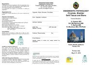
Fig. 3.13 Signet-ring cell carcinomas. A Overview showing Infiltration <strong>of</strong> <strong>the</strong> lamina propria. B Dispersed<br />
signet-ring cells. C Accumulation <strong>of</strong> neoplastic signet ring cells in <strong>the</strong> mucosa. D Alcian green positive<br />
signet-ring cells expanding <strong>the</strong> lamina propria in this Movat stain.<br />
B<br />
B<br />
B<br />
D<br />
o<strong>the</strong>r diffuse carcinomas contain cells<br />
with central nuclei resembling histiocytes,<br />
and show little or no mitotic activity; (3)<br />
small, deeply eosinophilic cells with<br />
prominent, but minute, cytoplasmic granules<br />
containing neutral mucin; (4) small<br />
cells with little or no mucin, and (5)<br />
anaplastic cells with little or no mucin.<br />
These cell types intermingle with one<br />
ano<strong>the</strong>r and constitute varying tumour<br />
proportions. Signet-ring cell tumours may<br />
also form lacy or delicate trabecular glandular<br />
patterns and <strong>the</strong>y may display a<br />
zonal or solid arrangement.<br />
Signet-ring cell carcinomas are infiltrative;<br />
<strong>the</strong> number <strong>of</strong> malignant cells is<br />
comparatively small and desmoplasia<br />
may be prominent. Special stains,<br />
including mucin stains (PAS, mucicarmine,<br />
or Alcian blue) or immunohistochemical<br />
staining with antibodies to<br />
cytokeratin, help detect sparsely dispersed<br />
tumour cells in <strong>the</strong> stroma. Cytokeratin<br />
immunostains detect a greater<br />
percentage <strong>of</strong> neoplastic cells than do<br />
mucin stains. Several conditions mimic<br />
signet-ring cell carcinoma including<br />
signet-ring lymphoma, lamina propria<br />
muciphages, xanthomas and detached<br />
or dying cells associated with gastritis.<br />
Laurén classification<br />
The Laurén classification {1021} has<br />
proven useful in evaluating <strong>the</strong> natural<br />
history <strong>of</strong> gastric carcinoma, especially<br />
with regard to its association with environmental<br />
factors, incidence trends and<br />
its precursors. Lesions are classified into<br />
one <strong>of</strong> two major types: intestinal or diffuse.<br />
<strong>Tumours</strong> that contain approximately<br />
equal quantities <strong>of</strong> intestinal and diffuse<br />
components are called mixed carcinomas.<br />
Carcinomas too undifferentiated to<br />
fit neatly into ei<strong>the</strong>r category are placed<br />
in <strong>the</strong> indeterminate category.<br />
Intestinal carcinomas<br />
These form recognizable glands that<br />
range from well differentiated to moderately<br />
differentiated tumours, sometimes<br />
with poorly differentiated tumour at <strong>the</strong><br />
advancing margin. They typically arise<br />
on a background <strong>of</strong> intestinal metaplasia.<br />
The mucinous phenotype <strong>of</strong> <strong>the</strong>se cancers<br />
is intestinal, gastric and gastrointestinal.<br />
Diffuse carcinomas<br />
They consist <strong>of</strong> poorly cohesive cells diffusely<br />
infiltrating <strong>the</strong> gastric wall with little<br />
44 <strong>Tumours</strong> <strong>of</strong> <strong>the</strong> stomach
Fig. 3.14 Undifferentiated gastric carcinoma.<br />
Fig. 3.15 Hepatoid variant <strong>of</strong> gastric carcinoma.<br />
Fig. 3.16 Gastric choriocarcinoma composed <strong>of</strong> syncytiotrophoblastic and cytotrophoblastic cells next to<br />
thin-walled vascular structures. A Papillary carcinoma component is adjacent to <strong>the</strong> choriocarcinoma.<br />
B High magnification <strong>of</strong> <strong>the</strong> choriocarcinoma.<br />
B<br />
or no gland formation. The cells usually<br />
appear round and small, ei<strong>the</strong>r arranged<br />
as single cells or clustered in abortive,<br />
lacy gland-like or reticular formations.<br />
These tumours resemble those classified<br />
as signet-ring cell tumours in <strong>the</strong> WHO<br />
classification. The mitotic rate is lower in<br />
diffuse carcinomas than in intestinal<br />
tumours. Small amounts <strong>of</strong> interstitial<br />
mucin may be present. Desmoplasia is<br />
more pronounced and associated inflammation<br />
is less evident in diffuse cancers<br />
than in <strong>the</strong> intestinal carcinomas.<br />
Rare variants<br />
Several o<strong>the</strong>r carcinomas exist that are<br />
not an integral part <strong>of</strong> <strong>the</strong> Laurén or WHO<br />
classifications.<br />
Adenosquamous carcinoma<br />
This lesion combines an adenocarcinoma<br />
and squamous cell carcinoma; nei<strong>the</strong>r<br />
quantitatively prevails. Transitions<br />
exist between both components. A<br />
tumour with a distinct boundary between<br />
<strong>the</strong> two components may represent a<br />
collision tumour. <strong>Tumours</strong> containing discrete<br />
foci <strong>of</strong> benign-appearing squamous<br />
metaplasia are termed adenocarcinomas<br />
with squamous differentiation<br />
(synonymous with adenoacanthoma).<br />
Squamous cell carcinoma<br />
Pure squamous cell carcinomas develop<br />
rarely in <strong>the</strong> stomach; <strong>the</strong>y resemble<br />
squamous cell carcinomas arising elsewhere<br />
in <strong>the</strong> body.<br />
Undifferentiated carcinoma<br />
These lesions lack any differentiated features<br />
beyond an epi<strong>the</strong>lial phenotype<br />
(e.g. cytokeratin expression). They fall<br />
into <strong>the</strong> indeterminate group <strong>of</strong> Laurén’s<br />
scheme. Fur<strong>the</strong>r analysis <strong>of</strong> this heterogeneous<br />
group using histochemical methods<br />
may allow <strong>the</strong>ir separation into o<strong>the</strong>r<br />
types.<br />
O<strong>the</strong>r rare tumours include mixed adenocarcinoma-carcinoid<br />
(mixed exocrineendocrine<br />
carcinoma), small cell<br />
carcinoma, parietal cell carcinoma, choriocarcinoma,<br />
endodermal sinus tumour,<br />
embryonal carcinoma, Paneth cell richadenocarcinoma<br />
and hepatoid adenocarcinoma.<br />
A<br />
Fig. 3.17 A, B Adenocarcinoma, poorly differentiated. These two lesions show both intestinal and diffuse<br />
components (Laurén classification).<br />
B<br />
Early gastric cancer<br />
Early gastric cancer (EGC) is a carcinoma<br />
limited to <strong>the</strong> mucosa or <strong>the</strong> mucosa<br />
and submucosa, regardless <strong>of</strong> nodal status.<br />
Countries in which asymptomatic<br />
patients are screened have a high incidence<br />
<strong>of</strong> EGCs ranging from 30-50%<br />
{1410, 908, 718}, contrasting with a<br />
smaller fraction <strong>of</strong> 16-24% {620, 253,<br />
627} in Western countries. The follow-up<br />
<strong>of</strong> dysplastic lesions does appear to<br />
increase <strong>the</strong> prevalence <strong>of</strong> EGC. The<br />
cost effectiveness <strong>of</strong> such an integrated<br />
Gastric carcinoma<br />
45
A B C<br />
Fig. 3.18 Tubular adenocarcinoma. A Well differentiated; intramucosal invasion. B Moderately differentiated. C Poorly differentiated.<br />
A<br />
B<br />
Fig. 3.19 A, B Tubular adenocarcinoma, well differentiated.<br />
endoscopic/biopsy approach remains to<br />
be evaluated {1634, 1638}. Histologically,<br />
most subtypes <strong>of</strong> carcinoma occur in<br />
EGC in ei<strong>the</strong>r pure or mixed forms.<br />
Elevated carcinomas with papillary, granular<br />
or nodular patterns and a red colour<br />
are more <strong>of</strong>ten well or moderately differentiated,<br />
tubular or papillary tumours<br />
with intestinal features; sometimes a preexisting<br />
adenoma is recognizable. Flat,<br />
depressed, poorly differentiated carcinomas<br />
may contain residual or regenerative<br />
mucosal islands. Ulcerated lesions are<br />
ei<strong>the</strong>r intestinal or diffuse cancers.<br />
Adenocarcinoma limited to <strong>the</strong> mucosal<br />
thickness has also been divided into<br />
small mucosal (< 4cm=SM) and superficial<br />
(> 4cm=SUPER) {950}. Both <strong>of</strong> <strong>the</strong>m<br />
may be strictly confined at <strong>the</strong> mucosal<br />
level (small mucosal M and superficial M)<br />
or focally infiltrate <strong>the</strong> sub-mucosa (small<br />
mucosal SM and superficial SM). In <strong>the</strong><br />
penetrating variant, (including two subcategories:<br />
PenA and PenB) <strong>the</strong> invasion<br />
<strong>of</strong> <strong>the</strong> submucosa is more extensive than<br />
in <strong>the</strong> two above-mentioned variants.<br />
PenA is defined by a pushing margin,<br />
and is less frequent than PenB, which<br />
penetrates muscularis mucosae at multiple<br />
sites.<br />
The prognosis is worse in PenA carcinomas<br />
(in contrast to adenocarcinomas <strong>of</strong><br />
<strong>the</strong> colon, where a pushing margin is<br />
associated with a better prognosis). The<br />
coexistence <strong>of</strong> more than one <strong>of</strong> <strong>the</strong><br />
described patterns results in <strong>the</strong> mixed<br />
variant {950}.<br />
Stromal reactions<br />
The four common stromal responses to<br />
gastric carcinoma are marked desmoplasia,<br />
lymphocytic infiltrates, stromal<br />
eosinophilia and a granulomatous response.<br />
The granulomatous reaction is<br />
characterized by <strong>the</strong> presence <strong>of</strong> single<br />
and confluent small sarcoid-like granulomas,<br />
<strong>of</strong>ten accompanied by a moderately<br />
intense mononuclear cell infiltrate. The<br />
lymphoid response is associated with an<br />
improved survival.<br />
Grading<br />
Well differentiated: An adenocarcinoma<br />
with well-formed glands, <strong>of</strong>ten resembling<br />
metaplastic intestinal epi<strong>the</strong>lium.<br />
Moderately differentiated: An adenocarcinoma<br />
intermediate between well differentiated<br />
and poorly differentiated.<br />
Poorly differentiated: An adenocarcinoma<br />
composed <strong>of</strong> highly irregular glands<br />
that are recognized with difficulty, or single<br />
cells that remain isolated or are<br />
arranged in small or large clusters with<br />
mucin secretions or acinar structures.<br />
They may also be graded as low-grade<br />
(well and moderately differentiated) or<br />
high-grade (poorly differentiated). Note<br />
that this grading system applies primarily<br />
to tubular carcinomas. O<strong>the</strong>r types <strong>of</strong><br />
gastric carcinoma are not graded.<br />
Precursor lesions<br />
Gastritis and intestinal metaplasia<br />
Chronic atrophic gastritis and intestinal<br />
metaplasia commonly precede and/or<br />
accompany intestinal type adenocarcinoma,<br />
particularly in high-incidence<br />
areas {780}. H. pylori associated gastritis<br />
is <strong>the</strong> commonest gastric precursor<br />
lesion.<br />
However, autoimmune gastritis also<br />
associates with an increased carcinoma<br />
risk. If gastritis persists, gastric atrophy<br />
occurs followed by intestinal metaplasia,<br />
beginning a series <strong>of</strong> changes that may<br />
result in neoplasia, especially <strong>of</strong> intestinal<br />
type cancers. In contrast, diffuse gastric<br />
cancers <strong>of</strong>ten arise in a stomach<br />
lacking atrophic gastritis with intestinal<br />
metaplasia.<br />
Fig. 3.20 Intestinal metaplasia. The two glands on<br />
<strong>the</strong> left exhibit complete intestinal metaplasia,<br />
o<strong>the</strong>rs show <strong>the</strong> incomplete type.<br />
46 <strong>Tumours</strong> <strong>of</strong> <strong>the</strong> stomach
There are two main types <strong>of</strong> intestinal<br />
metaplasia: ‘complete’ (also designated<br />
as ‘small intestinal type’ or type I), and<br />
‘incomplete’ (types II and III) {843}.<br />
Different mucin expression patterns characterize<br />
<strong>the</strong> metaplasias: complete shows<br />
decreased expression <strong>of</strong> ‘gastric’ (MUC1,<br />
MUC5AC and MUC6) mucins and<br />
expression <strong>of</strong> MUC2, an intestinal mucin.<br />
In incomplete intestinal metaplasia, ‘gastric’<br />
mucins are co-expressed with MUC2<br />
mucin. These findings show that incomplete<br />
intestinal metaplasia has a mixed<br />
gastric and intestinal phenotype reflecting<br />
an aberrant differentiation program<br />
not reproducing any normal adult gastrointestinal<br />
epi<strong>the</strong>lial phenotype {1574}.<br />
Intraepi<strong>the</strong>lial neoplasia<br />
Intraepi<strong>the</strong>lial neoplasia (dysplasia) arises<br />
in ei<strong>the</strong>r <strong>the</strong> native gastric or <strong>of</strong> intestinalized<br />
gastric epi<strong>the</strong>lia. Pyloric gland adenoma<br />
is a form <strong>of</strong> intraepi<strong>the</strong>lial neoplasia<br />
arising in <strong>the</strong> native mucosa {2066, 1885}.<br />
In <strong>the</strong> multi-stage <strong>the</strong>ory <strong>of</strong> gastric oncogenesis,<br />
intraepi<strong>the</strong>lial neoplasia lies<br />
between atrophic metaplastic lesions<br />
and invasive cancer (Table 3.01).<br />
Problems associated with diagnosing<br />
gastric intraepi<strong>the</strong>lial neoplasia include<br />
<strong>the</strong> distinction from reactive or regenerative<br />
changes associated with active<br />
Fig. 3.21 Reactive gastritis with marked foveolar<br />
hyperplasia.<br />
inflammation, and <strong>the</strong> distinction between<br />
intraepi<strong>the</strong>lial and invasive carcinoma<br />
{1683, 1025}. Several proposals have<br />
been made for <strong>the</strong> terminology <strong>of</strong> <strong>the</strong><br />
morphological spectrum <strong>of</strong> lesions that lie<br />
between non-neoplastic changes and<br />
early invasive cancer, including <strong>the</strong><br />
recent international Padova classification<br />
{1636}.<br />
Indefinite for intraepi<strong>the</strong>lial neoplasia<br />
Sometimes, doubts arise as to whe<strong>the</strong>r a<br />
lesion is neoplastic or non-neoplastic (i.e.<br />
reactive or regenerative), particularly in<br />
small biopsies. In such cases, <strong>the</strong> dilemma<br />
is usually solved by cutting deeper<br />
levels <strong>of</strong> <strong>the</strong> block, by obtaining additional<br />
biopsies, or after removing possible<br />
sources <strong>of</strong> cellular hyperproliferation. One<br />
important source <strong>of</strong> a potentially alarming<br />
lesion is <strong>the</strong> regeneration associated with<br />
NSAID-induced injury or superficial erosion/ulceration<br />
caused by gastric acid.<br />
Cases lacking all <strong>the</strong> attributes required<br />
for a definitive diagnosis <strong>of</strong> intraepi<strong>the</strong>lial<br />
neoplasia may be placed into <strong>the</strong> category<br />
‘indefinite for intraepi<strong>the</strong>lial neoplasia’.<br />
In native gastric mucosa, foveolar hyperproliferation<br />
may be indefinite for dysplasia,<br />
showing irregular and tortuous tubular<br />
structures with epi<strong>the</strong>lial mucus depletion,<br />
a high nuclear-cytoplasmic ratio and loss<br />
<strong>of</strong> cellular polarity. Large, oval/round,<br />
hyperchromatic nuclei associate with<br />
prominent mitoses, usually located near<br />
<strong>the</strong> proliferative zone in <strong>the</strong> mucous neck<br />
region.<br />
In intestinal metaplasia, areas indefinite<br />
for intraepi<strong>the</strong>lial neoplasia exhibit a<br />
hyperproliferative metaplastic epi<strong>the</strong>lium.<br />
The glands may appear closely packed,<br />
lined by cells with large, hyperchromatic,<br />
rounded or elongated, basally located<br />
nuclei. Nucleoli are an inconsistent finding.<br />
The cyto-architectural alterations tend<br />
to decrease from <strong>the</strong> base <strong>of</strong> <strong>the</strong> glands to<br />
<strong>the</strong>ir superficial portion.<br />
Intraepi<strong>the</strong>lial neoplasia<br />
It has flat, polypoid, or slightly depressed<br />
growth patterns; <strong>the</strong> flat pattern may lack<br />
any endoscopic changes on conventional<br />
endoscopy, but shows an irregular<br />
appearance on dye endoscopy. In<br />
Western countries, <strong>the</strong> term adenoma is<br />
applied when <strong>the</strong> proliferation produces<br />
a macroscopic, usually discrete, protruding<br />
lesion. However, in Japan, adenomas<br />
include all gross types (i.e. flat, elevated<br />
and depressed). Gastric adenomas are<br />
less common than hyperplastic polyps;<br />
overall, <strong>the</strong>y account for approximately<br />
10% <strong>of</strong> gastric polyps {1843}. They tend<br />
to arise in <strong>the</strong> antrum or mid stomach in<br />
areas <strong>of</strong> intestinal metaplasia.<br />
Morphologically, adenomas can be<br />
described as tubular (<strong>the</strong> most common),<br />
tubulovillous, or villous; <strong>the</strong> latter<br />
two have also been called papillotubular<br />
and papillary. Most have epi<strong>the</strong>lium <strong>of</strong><br />
intestinal type, but some have gastric<br />
foveolar features.<br />
Low-grade intraepi<strong>the</strong>lial neoplasia<br />
This lesion shows a slightly modified<br />
mucosal architecture, including <strong>the</strong> presence<br />
<strong>of</strong> tubular structures with budding<br />
and branching, papillary enfolding, crypt<br />
leng<strong>the</strong>ning with serration, and cystic<br />
changes. Glands are lined by enlarged<br />
columnar cells with minimal or no mucin.<br />
Homogeneously blue vesicular, rounded<br />
or ovoid nuclei are usually pseudostratified<br />
in <strong>the</strong> proliferation zone located at<br />
<strong>the</strong> superficial portion <strong>of</strong> <strong>the</strong> dysplastic<br />
tubules.<br />
High-grade intraepi<strong>the</strong>lial neoplasia<br />
There is increasing architectural distortion<br />
with glandular crowding and prominent<br />
cellular atypia. Tubules can be irregular in<br />
shape, with frequent branching and fold-<br />
Fig. 3.22 Tubular adenoma <strong>of</strong> gastric antrum.<br />
Uninvolved pyloric glands below <strong>the</strong> lesion show<br />
cystic dilatation.<br />
Gastric carinoma<br />
47
Polyps<br />
Hyperplastic polyps<br />
Hyperplastic polyps are one <strong>of</strong> <strong>the</strong> commonest<br />
gastric polyps. They are sessile<br />
or pedunculated lesions, usually < 2.0<br />
cm in diameter, typically arising in <strong>the</strong><br />
antrum on a background <strong>of</strong> H. pylori gastritis.<br />
They contain a proliferation <strong>of</strong> surface<br />
foveolar cells lining elongated, distorted<br />
pits extending deep into <strong>the</strong><br />
stroma. They may contain pyloric glands,<br />
chief cells and parietal cells. The surface<br />
<strong>of</strong>ten erodes. In a minority <strong>of</strong> cases, carcinoma<br />
develops within <strong>the</strong> polyps in<br />
areas <strong>of</strong> intestinal metaplasia and dysplasia.<br />
A<br />
B<br />
Fig. 3.23 A, B Examples <strong>of</strong> low-grade intraepi<strong>the</strong>lial neoplasia <strong>of</strong> flat gastric mucosa. The atypia extends to<br />
<strong>the</strong> surface.<br />
ing; <strong>the</strong>re is no stromal invasion. Mucin<br />
secretion is absent or minimal. The pleomorphic,<br />
hyperchromatic, usually pseudostratified<br />
nuclei <strong>of</strong>ten are cigar-shaped.<br />
Prominent amphophilic nucleoli are common.<br />
Increased proliferative activity is<br />
present throughout <strong>the</strong> epi<strong>the</strong>lium.<br />
Progression <strong>of</strong> intraepi<strong>the</strong>lial neoplasia to<br />
carcinoma<br />
Carcinoma is diagnosed when <strong>the</strong> tumour<br />
invades into <strong>the</strong> lamina propria (intramucosal<br />
carcinoma) or through <strong>the</strong> muscularis<br />
mucosae. Some gastric biopsies<br />
contain areas suggestive <strong>of</strong> true invasion<br />
(such as isolated cells, gland-like structures,<br />
or papillary projections). The term<br />
‘suspicious for invasion’ is appropriate<br />
when <strong>the</strong> histological criteria for an invasive<br />
malignancy are equivocal.<br />
Up to 80% <strong>of</strong> intraepi<strong>the</strong>lial neoplasias<br />
may progress to invasion. Indeed, invasive<br />
cancer already may be present in<br />
patients found to have high-grade intraepi<strong>the</strong>lial<br />
neoplasia with no obvious<br />
tumour mass. The extent <strong>of</strong> intestinal<br />
metaplasia associated with intraepi<strong>the</strong>lial<br />
neoplasia, toge<strong>the</strong>r with a sulphomucinsecreting<br />
phenotype <strong>of</strong> <strong>the</strong> intestinalized<br />
mucosa (type III intestinal metaplasia),<br />
correlate with an increased risk <strong>of</strong> carcinoma<br />
development.<br />
Adenomas<br />
Adenomas are circumscribed, benign<br />
lesions, composed <strong>of</strong> tubular and/or villous<br />
structures showing intraepi<strong>the</strong>lial<br />
neoplasia. The frequency <strong>of</strong> malignant<br />
transformation depends on size and histological<br />
grade. It occurs in approximately<br />
2% <strong>of</strong> lesions measuring < 2 cm and in<br />
40-50% <strong>of</strong> lesions > 2 cm. Flat adenomas<br />
may have a greater tendency to progress<br />
to carcinoma.<br />
Fundic gland polyps<br />
Fundic gland polyps are <strong>the</strong> commonest<br />
gastric polyp seen in Western populations.<br />
They occur sporadically, without a<br />
relationship to H. pylori gastritis. They<br />
also affect patients on long-term proton<br />
pump inhibitors or patients with familial<br />
adenomatous polyposis (FAP), who may<br />
have hundreds <strong>of</strong> fundic gland polyps<br />
{2064, 2065}.<br />
The lesions consist <strong>of</strong> a localized hyperplasia<br />
<strong>of</strong> <strong>the</strong> deep epi<strong>the</strong>lial compartment<br />
<strong>of</strong> <strong>the</strong> oxyntic mucosa, particularly<br />
<strong>of</strong> mucous neck cells, with variable<br />
degrees <strong>of</strong> cystic dilatation. Sporadic<br />
fundic gland polyps have no malignant<br />
potential. Exceptionally, patients with<br />
attentuated FAP may develop dysplasia<br />
and carcinoma in <strong>the</strong>ir fundic gland<br />
polyps {2214, 1204}<br />
Polyposis syndromes<br />
Peutz-Jeghers polyps, juvenile polyps,<br />
and Cowden polyps generally do not<br />
occur spontaneously, but ra<strong>the</strong>r as part<br />
<strong>of</strong> hereditary polyposis syndromes. In <strong>the</strong><br />
stomach, Peutz-Jeghers polyps are characterized<br />
histologically by branching<br />
bands <strong>of</strong> smooth muscle derived from<br />
A B C<br />
Fig. 3.24 High-grade intraepi<strong>the</strong>lial neoplasia in flat gastric mucosa (flat adenoma). A Architectal distortion <strong>of</strong> <strong>the</strong> gastric glands. B High degree <strong>of</strong> cellular atypia.<br />
C Papillary pattern.<br />
48 <strong>Tumours</strong> <strong>of</strong> <strong>the</strong> stomach
Table 3.01<br />
Histological follow-up studies <strong>of</strong> gastric intraepi<strong>the</strong>lial neoplasia. Proportion progressing to carcinoma and<br />
mean interval.<br />
muscularis mucosae, and hyperplasia,<br />
elongation and cystic change <strong>of</strong> foveolar<br />
epi<strong>the</strong>lium; <strong>the</strong> deeper glandular components<br />
tend to show atrophy.<br />
Genetic susceptibility<br />
Most gastric carcinomas occur sporadically;<br />
only about 8-10% have an inherited<br />
familial component {996}. Familial clustering<br />
occurs in 12 to 25% with a dominant<br />
inheritance pattern {597, 864}.<br />
Case-control studies also suggest a<br />
small but consistent increased risk in<br />
first-degree relatives <strong>of</strong> gastric carcinoma<br />
patients {2200}.<br />
A<br />
C<br />
Reports Low-grade dysplasia High-grade dysplasia<br />
Saraga, 1987 {2355} 2% (1/64) 4 yr. 81% (17/21) 4 mos.<br />
Lansdown, 1990 {2356} 0 (0/7) 85% (11/13) 5 mos.<br />
Rugge, 1991 {2008} 17% (12/69) 1yr. 75% (6/8) 4 mos.<br />
Fertitta, 1993 {2357} 23% (7/30) 10 mos. 81% (25/31) 5 mos.<br />
Di Gregorio, 1993 {2358} 7% (6/89) 2 yr. 60% (6/10) 11 mos.<br />
Rugge, 1994 {2009} 14% (13/90) 2 yr. 78% (14/18) 9 mos.<br />
Kokkola, 1996 {2359} 0% (0/96) 67% (2/3) 1.5 yr.<br />
Gastric carcinoma occasionally develops<br />
in families with germline mutations in<br />
ATM5, TP53 (Li Fraumeni syndrome)<br />
{2001, 743, 1652}, and BRCA2 {1934}.<br />
Rare site-specific gastric carcinoma predisposition<br />
traits have been reported in<br />
several families {1147, 2130}, including<br />
that <strong>of</strong> Napoleon.<br />
Hereditary diffuse gastric carcinoma<br />
Germline mutations in <strong>the</strong> gene encoding<br />
<strong>the</strong> cell adhesion protein E-cadherin<br />
(CDH1) lead to an autosomal dominant<br />
predisposition to gastric carcinoma,<br />
referred to as hereditary diffuse gastric<br />
Fig. 3.25 A Large hyperplastic polyp <strong>of</strong> <strong>the</strong> stomach. B, C Typical histology <strong>of</strong> gastric hyperplastic polyp. D<br />
Hyperplastic polyp with florid epi<strong>the</strong>lial hyperplasia.<br />
B<br />
D<br />
carcinoma (HDGC) {640, 568}. Predisposing<br />
germline CDH1 mutations generally<br />
resulting in truncated proteins are<br />
spread throughout <strong>the</strong> gene with no<br />
apparent hotspots {641, 640, 568, 1581}.<br />
HDGC has an age <strong>of</strong> onset ranging<br />
upwards from 14 years and a penetrance<br />
<strong>of</strong> approximately 70% {641, 568}.<br />
Histologically, HDGC tumours are diffuse,<br />
poorly differentiated infiltrative adenocarcinomas<br />
with occasional signetring<br />
cells {641, 640, 568}.<br />
HNPCC<br />
Gastric carcinomas can develop as part<br />
<strong>of</strong> <strong>the</strong> hereditary nonpolyposis colon<br />
cancer (HNPCC) syndrome {1130, 922}.<br />
They are intestinal type cancers, without<br />
an association with H. pylori infection;<br />
most exhibit microsatellite instability<br />
(MSI) {4} with a trend that is opposite to<br />
that found in tumours arising in young<br />
patients {1739}.<br />
Gastrointestional polyposis syndromes<br />
Gastric carcinomas also occur in<br />
patients with gastrointestinal polyposis<br />
syndromes including FAP and Peutz-<br />
Jeghers syndrome.<br />
Overall, gastric carcinoma is rare in<br />
<strong>the</strong>se settings, and <strong>the</strong> exact contribution<br />
<strong>of</strong> <strong>the</strong> polyposis and underlying germline<br />
alterations <strong>of</strong> APC and LKB1/STK11 to<br />
cancer development is unclear.<br />
Blood group A<br />
The blood group A phenotype associates<br />
with gastric carcinomas {27, 649}.<br />
H. pylori adhere to <strong>the</strong> Lewis b blood<br />
group antigen and <strong>the</strong> latter may be an<br />
important host factor facilitating this<br />
chronic infection {244} and subsequent<br />
cancer risk.<br />
Molecular genetics<br />
Loss <strong>of</strong> heterozygosity studies and comparative<br />
genomic hybridization (CGH)<br />
analyses have identified several loci with<br />
significant allelic loss, indicating possible<br />
tumour suppressor genes important<br />
in gastric carcinoma. Common target(s)<br />
<strong>of</strong> loss or gain include chromosomal<br />
regions 3p, 4, 5q, (30 to 40% at or near<br />
APC’s locus) {1656, 1577}, 6q {255}, 9p,<br />
17p (over 60 percent at TP53’s locus)<br />
{1656}, 18q (over 60 percent at DCC’s<br />
locus) {1981}, and 20q {1287, 449,<br />
2192}. Similar LOH losses at 11p15<br />
occur in proximal and distal carcinomas,<br />
suggesting common paths <strong>of</strong> develop-<br />
Gastric carcinoma<br />
49
A<br />
Fig. 3.26 A, B Fundic gland polyp. Cystic glands are typical.<br />
B<br />
ment {1288}. Loss <strong>of</strong> a locus on 7q<br />
(D7S95) associates with peritoneal<br />
metastasis.<br />
The frequency <strong>of</strong> MSI in sporadic gastric<br />
carcinoma ranges from 13% to 44%<br />
{1713}. MSI+ tumours tend to be<br />
advanced intestinal-type cancers. The<br />
degree <strong>of</strong> genome-wide instability varies<br />
with more significant instability (e.g.,<br />
MSI-H: > 33% abnormal loci) occurring<br />
in only 16% <strong>of</strong> gastric carcinoma, usually<br />
<strong>of</strong> <strong>the</strong> subcardial intestinal or mixed type,<br />
with less frequent lymph node or vessel<br />
invasion, prominent lymphoid infiltration,<br />
and better prognosis {430}. Loss <strong>of</strong> ei<strong>the</strong>r<br />
hMLH1 or hMSH2 protein expression<br />
affects all MSI-H cases {654} suggesting<br />
Fig. 3.27<br />
glands.<br />
Peutz-Jeghers polyp with hyperplastic<br />
inactivation <strong>of</strong> both alleles by mechanisms<br />
such as hypermethylation {1050,<br />
510}.<br />
Genes with simple tandem repeat<br />
sequences within <strong>the</strong>ir coding regions<br />
that are altered in MSI+ tumours include<br />
<strong>the</strong> TGF-β II receptor, BAX, IGFRII,<br />
hMSH3, hMSH6, and E2F-4. A study <strong>of</strong><br />
gastric cancers displaying <strong>the</strong> MSI-H<br />
phenotype reveal that a majority contain<br />
mutated TGF-β type II receptors in a<br />
polyadenine tract {1420, 1462}. Altered<br />
TGF-β II receptor genes can also be<br />
found in MSI-lesions.<br />
Allelic loss <strong>of</strong> TP53 occurs in > 60% <strong>of</strong><br />
cases and mutations are identified in<br />
approximately 30-50% <strong>of</strong> cases depending<br />
on <strong>the</strong> mutational screening method<br />
and sample sizes {729, 1937}. TP53<br />
mutations are identifiable in some intestinal<br />
metaplasias; {497} most alterations<br />
affect advanced tumours. TP53 mutations<br />
in gastric lesions resemble those<br />
seen in o<strong>the</strong>r cancers with a predominance<br />
<strong>of</strong> base transitions, especially at<br />
CpG dinucleotides. Immunohistochemical<br />
analyses to detect TP53 overexpression<br />
can indirectly identify TP53 mutations<br />
but do not have consistent<br />
prognostic value in gastric carcinoma<br />
patients {557, 766}. Finally, with respect<br />
to TP53, <strong>the</strong>re is a polymorphism in<br />
codon 72 encoding a proline ra<strong>the</strong>r than<br />
an arginine that strongly associates with<br />
antral cancers {1735}.<br />
Sporadic gastric carcinomas, especially<br />
diffuse carcinomas, exhibit reduced or<br />
abnormal E-cadherin expression {1196,<br />
1135}, and genetic abnormalities <strong>of</strong> <strong>the</strong><br />
E-cadherin gene and its transcripts.<br />
Reduced E-cadherin expression is associated<br />
with reduced survival {848}.<br />
E-cadherin splice site alterations produce<br />
exon deletion and skipping. Large<br />
deletions including allelic loss and missense<br />
point mutations also occur; some<br />
tumours exhibit alterations in both alleles<br />
{135}. Somatic E-cadherin gene alterations<br />
also affect <strong>the</strong> diffuse component<br />
<strong>of</strong> mixed tumours {1136}. Alpha-catenin,<br />
which binds to <strong>the</strong> intracellular domain <strong>of</strong><br />
E-cadherin and links it to actin-based<br />
cytoskeletal elements, shows reduced<br />
immunohistochemical expression in<br />
many tumours and correlates with infiltrative<br />
growth and poor differentiation<br />
{1189}. Beta catenin may also be abnormal<br />
in gastric carcinoma.<br />
There is evidence <strong>of</strong> a tumour suppressor<br />
locus on chromosome 3p in gastric<br />
carcinomas {893, 1688}. This area<br />
encodes <strong>the</strong> FHIT gene. Gastric carcinomas<br />
develop abnormal transcripts, deleted<br />
exons {1411}, a somatic missense<br />
mutation in exon 6 and loss <strong>of</strong> FHIT protein<br />
expression {102}.<br />
Somatic APC mutations, mostly missense<br />
in nature and low in frequency,<br />
affect Japanese patients with in situ and<br />
invasive neoplasia {1309}. Significant<br />
allelic loss (30%) at <strong>the</strong> APC loci suggest<br />
that <strong>the</strong>re is a tumour suppressor gene<br />
important in gastric tumourigenesis nearby.<br />
Indeed, alternative loci have been<br />
mapped to commonly deleted regions in<br />
gastric carcinomas {1891}.<br />
Amplification and overexpression <strong>of</strong> <strong>the</strong><br />
c-met gene encoding a tyrosine kinase<br />
receptor for <strong>the</strong> hepatocyte growth factor<br />
occurs in gastric carcinoma {976}. O<strong>the</strong>r<br />
growth factor and receptor signal systems<br />
that may be involved include epidermal<br />
growth factor, TGF-alpha, interleukin-1-a,<br />
cripto, amphiregulin, platelet-derived<br />
50 <strong>Tumours</strong> <strong>of</strong> <strong>the</strong> stomach
A B C<br />
Fig. 3.28 E-cadherin expression in gastric adenocarcinoma. A Intestinal type <strong>of</strong> adenocarcinoma showing a normal pattern <strong>of</strong> membranous staining. B Diffuse type<br />
<strong>of</strong> adenocarcinoma with reduced E-cadherin expression. Normal expression can be seen in <strong>the</strong> non-neoplastic gastric epi<strong>the</strong>lium overlying <strong>the</strong> tumour. C Undifferentiated<br />
gastric carcinoma with highly reduced membranous expression and dot-like cytoplasmic expression.<br />
growth factor, and K-sam {1879}. Amplification<br />
<strong>of</strong> c-erbB-2, a transmembrane<br />
tyrosine kinase receptor oncogene,<br />
occurs in approximately 10% <strong>of</strong> lesions<br />
and overexpression associates with a<br />
poor prognosis {375}. Telomerase activity<br />
has been detected by a PCR-based<br />
assay frequently in <strong>the</strong> late stages <strong>of</strong> gastric<br />
tumours and observed to be associated<br />
with a poor prognosis {719}.<br />
Prognosis and predictive factors<br />
Early gastric cancer<br />
In early gastric cancers, small mucosal<br />
(< 4 cm), superficial (> 4 cm) and Pen B<br />
lesions have a low incidence <strong>of</strong> vessel<br />
invasion and lymph node metastasis and<br />
a good prognosis after surgery (about<br />
90% <strong>of</strong> patients survive 10 years). In contrast,<br />
penetrating lesions <strong>of</strong> <strong>the</strong> Pen A<br />
type are characterized by a relatively<br />
high incidence <strong>of</strong> vessel invasion and<br />
lymph node metastasis and a poor prognosis<br />
after surgery (64.8% 5-year survival).<br />
advanced cases. Lymph node status,<br />
which is part <strong>of</strong> <strong>the</strong> TNM system, is also<br />
an important prognostic indicator. The 5th<br />
edition <strong>of</strong> <strong>the</strong> UICC TNM Classification <strong>of</strong><br />
Malignant <strong>Tumours</strong> {66} and <strong>the</strong> AJCC<br />
Manual for <strong>the</strong> Staging <strong>of</strong> Cancer {1} published<br />
in 1997, have a number-based<br />
classification scheme for reporting nodal<br />
involvement in gastric cancer.<br />
Roder et al recently published data supporting<br />
<strong>the</strong> value <strong>of</strong> this reporting system.<br />
These authors found that for<br />
patients who had nodal involvement in<br />
1-6 lymph nodes (pN1), <strong>the</strong> 5-year sur-<br />
Advanced gastric cancer<br />
Staging. The TNM staging system for<br />
gastric cancer is widely used and it provides<br />
important prognostic information.<br />
Lymphatic and vascular invasion carries<br />
a poor prognosis and is <strong>of</strong>ten seen in<br />
Fig. 3.29 CGH analysis <strong>of</strong> a poorly differentiated gastric adenocarcinoma: copy number gains at chromosomes<br />
3q21, 7p15, 8q, 10p12-15, 11q13, 12q24, 13q13-14, 15q23-25, 17q24, 20 and 21q21. Copy number losses<br />
at chromosomes 4q12-28 and 5.<br />
Gastric carcinoma<br />
51
Fig. 3.30 TP53 mutations in gastric carcinoma. The<br />
mutations are shown by both single-strand conformation<br />
polymorphisms (SSCP) as well as direct<br />
sequencing. There is a G to A substitution indicated<br />
by <strong>the</strong> right hand panel.<br />
vival rate was 44% compared with a 30%<br />
survival rate in patients with 7-15 lymph<br />
nodes involved with tumour (pN2).<br />
Patients with more than 15 lymph nodes<br />
involved by metastatic tumour (pN3) had<br />
an even worse 5-year survival <strong>of</strong> 11%<br />
{1602}. Gastric carcinoma with obvious<br />
invasion beyond <strong>the</strong> pyloric ring, those<br />
with invasion up to <strong>the</strong> pyloric ring, and<br />
those without evidence <strong>of</strong> duodenal invasion<br />
have 5-year survival rates <strong>of</strong> 8%,<br />
22%, and 58%, respectively {671}.<br />
Patients with T1 cancers limited to <strong>the</strong><br />
mucosa and submucosa have a 5-year<br />
survival <strong>of</strong> approximately 95%. <strong>Tumours</strong><br />
that invade <strong>the</strong> muscularis propria have a<br />
60-80% 5-year survival, whereas tumours<br />
invading <strong>the</strong> subserosa have a 50%<br />
5-year survival {2181}. Unfortunately,<br />
most patients with advanced carcinoma<br />
already have lymph node metastases at<br />
<strong>the</strong> time <strong>of</strong> diagnosis.<br />
Histological features. The value <strong>of</strong> <strong>the</strong> histological<br />
type <strong>of</strong> tumour in predicting<br />
tumour prognosis is more controversial.<br />
This relates in part to <strong>the</strong> classification<br />
scheme that is used to diagnose <strong>the</strong> cancers.<br />
Using <strong>the</strong> Laurén classification,<br />
some believe that diffuse lesions generally<br />
carry a worse prognosis than intestinal<br />
carcinomas. The prognosis is particularly<br />
bad in children and young adults, in<br />
whom <strong>the</strong> diagnosis is <strong>of</strong>ten delayed<br />
{1986, 1554} and likely fit into <strong>the</strong> category<br />
<strong>of</strong> HDGC. However, o<strong>the</strong>rs have not<br />
found <strong>the</strong> Laurén classification to predict<br />
prognosis {1788, 1177}. One study found<br />
that only <strong>the</strong> Goseki classification {610}<br />
added additional prognostic information<br />
to <strong>the</strong> TNM stage {610}. 5-year survival <strong>of</strong><br />
patients with mucus rich (Goseki II and<br />
IV) T3 tumours was significantly worse<br />
than that <strong>of</strong> patients with mucus poor<br />
(Goseki I and III) T3 tumours (18% vs.<br />
53% p
Endocrine tumours <strong>of</strong> <strong>the</strong> stomach<br />
C. Capella<br />
E. Solcia<br />
L.H. Sobin<br />
R. Arnold<br />
Definition<br />
Most endocrine tumours <strong>of</strong> <strong>the</strong> stomach<br />
are well differentiated, nonfunctioning<br />
enterochromaffin-like (ECL) cell carcinoids<br />
arising from oxyntic mucosa in <strong>the</strong><br />
corpus or fundus. Three distinct types<br />
have are recognized: (1) Type I, associated<br />
with autoimmune chronic atrophic<br />
gastritis (A-CAG); (2) type II, associated<br />
with muliple endocrine neoplasia type 1<br />
(MEN-1) and Zollinger-Ellison syndrome<br />
(ZES); type III, sporadic, i.e. not associated<br />
with hypergastrinaemia or A-CAG.<br />
ICD-O Code<br />
Carcinoid 8240/3<br />
Small cell carcinoma 8041/3<br />
Epidemiology<br />
In <strong>the</strong> past, carcinoid tumours <strong>of</strong> <strong>the</strong><br />
stomach have been reported to occur<br />
with an incidence <strong>of</strong> 0.002-0.1 per<br />
100,000 population per year and to<br />
account for 2-3 % <strong>of</strong> all gastrointestinal<br />
carcinoids {587} and 0.3 percent <strong>of</strong> gastric<br />
neoplasms {1132}. More recent studies,<br />
however, based on endoscopic techniques<br />
and increased awareness <strong>of</strong> such<br />
lesions, have shown a much higher incidence<br />
<strong>of</strong> gastric carcinoids, which may<br />
now account for 11-41% <strong>of</strong> all gastrointestinal<br />
carcinoids {1588, 1764, 1782}.<br />
The incidence <strong>of</strong> gastric carcinoids is<br />
higher in Japan, where <strong>the</strong>y re-present<br />
30% <strong>of</strong> all gastrointestinal carcinoids,<br />
which may be due to <strong>the</strong> high incidence<br />
<strong>of</strong> chronic atrophic gastritis in this country<br />
{1277}.<br />
Age and sex distribution<br />
Type I gastric ECL-cell carcinoids have<br />
been reported to represent 74% <strong>of</strong> gastric<br />
endocrine tumours and to occur most<br />
<strong>of</strong>ten in females (M:F ratio, 1:2.5). The<br />
mean age at biopsy is 63 years (range<br />
15-88 years). Type II ECL-cell carcinoids<br />
represent 6% <strong>of</strong> all gastric endocrine<br />
tumours and show no gender predilection<br />
(M:F ratio, 1:1) at a mean age <strong>of</strong> 50<br />
years (range 28-67 years) {1590}. Type<br />
III ECL-cell carcinoids constitute 13% <strong>of</strong><br />
all gastric endocrine tumours and are<br />
observed mainly in male patients (M:F<br />
ratio, 2.8:1) at a mean age <strong>of</strong> 55 years<br />
(range 21-38 years) {1590}.<br />
Small cell carcinoma (poorly differentiated<br />
endocrine carcinoma) accounts for<br />
6% <strong>of</strong> gastric endocrine tumours and prevails<br />
in men (M:F ratio, 2:1) at a mean age<br />
<strong>of</strong> 63 years (range 41-61 years) {1590}.<br />
Gastrin cell tumours represent less than<br />
1% <strong>of</strong> gastric endocrine tumours {1590}<br />
and are reported in adults (age range<br />
55-77).<br />
Aetiology<br />
Gastrin has a trophic effect on ECL-cells<br />
both in humans and experimental animals<br />
{172, 652}. Hypergastrinaemic<br />
states, resulting ei<strong>the</strong>r from unregulated<br />
hormone release by a gastrinoma or from<br />
a secondary response <strong>of</strong> antral G cells to<br />
achlorhydria, are consistently associated<br />
with ECL-cell hyperplasia {172}.<br />
Autoimmune chronic atrophic gastritis<br />
(A-CAG)<br />
This disease is caused by antibodies to<br />
parietal cells <strong>of</strong> <strong>the</strong> oxyntic mucosa. It<br />
leads to chronic atrophic gastritis (with or<br />
without pernicious anaemia) which leads<br />
to an increase in gastrin production.<br />
Zollinger-Ellison syndrome<br />
This disease results from hypergastrinaemia<br />
due to gastrin-producing neoplasms<br />
that are preferentially located in<br />
<strong>the</strong> small intestine and pancreas. ECLcell<br />
proliferation is usually limited to<br />
hyperplastic lesions <strong>of</strong> <strong>the</strong> simple linear<br />
type {1042, 1777}.<br />
MEN-1<br />
This inherited tumour syndrome causes a<br />
variety <strong>of</strong> endocrine neoplasms, including<br />
gastrinomas. In patients with MEN-1<br />
associated ZES (MEN-1/ZES), ECL-cell<br />
lesions are usually dysplastic or overtly<br />
carcinoid in nature {1779}. In <strong>the</strong> MEN-1<br />
syndrome, <strong>the</strong> mutation or deletion <strong>of</strong> <strong>the</strong><br />
suppressor MEN-1 oncogene in 11q13<br />
may be involved {394} as an additional<br />
pathogenetic factor. In A-CAG, achlorhydria<br />
or associated mucosal changes may<br />
Fig. 3.31 Chromogranin A immunostain demonstrates<br />
hyperplasia <strong>of</strong> endocrine cells at <strong>the</strong> base <strong>of</strong><br />
glandular tubules.<br />
contribute to tumourigenesis {1785}.<br />
Several growth factors, including transforming<br />
growth factor-α (TGFα) and<br />
basic fibroblast growth factor (bFGF)<br />
seem to be involved in tumour development<br />
and progression as well as stromal<br />
and vascular proliferation <strong>of</strong> ECL-cell<br />
carcinoids {171}.<br />
Localization<br />
Type I, II, and III ECL-cell carcinoids are<br />
all located in <strong>the</strong> mucosa <strong>of</strong> <strong>the</strong> bodyfundus<br />
<strong>of</strong> <strong>the</strong> stomach, whereas <strong>the</strong> rare<br />
G-cell tumours are located in <strong>the</strong> antropyloric<br />
region. Small cell carcinomas<br />
prevail in <strong>the</strong> body/fundus, but some are<br />
located in <strong>the</strong> antrum {1590}.<br />
Clinical features<br />
The three distinct types <strong>of</strong> ECL-cell carcinoids<br />
are well differentiated growths<br />
but with variable and poorly predictable<br />
behaviour.<br />
Type I ECL-cell carcinoids<br />
These are associated with A-CAG involving<br />
<strong>the</strong> corpus and fundus mucosa.<br />
Clinical signs include achlorhydria and,<br />
less frequently, pernicious anemia.<br />
Hypergastrinaemia or evidence <strong>of</strong> antral<br />
gastrin-cell hyperplasia is observed in all<br />
cases <strong>of</strong> A-CAG. In patients with a carcinoid,<br />
ECL-cell hyperplastic changes are<br />
a constant feature and dysplastic<br />
growths are frequently observed {1590}.<br />
A-CAG associated carcinoids are typically<br />
small (usually less than 1 cm), mul-<br />
Endocrine tumours<br />
53
tiple and multicentric. Of 152 cases studied<br />
by endoscopy, 57% had more than<br />
two growths {1561}.<br />
Type II ECL-cell carcinoids<br />
Hypertrophic, hypersecretory gastropathy<br />
and high levels <strong>of</strong> circulating gastrin<br />
are critical diagnostic findings. In all<br />
cases, ECL-cell hyperplasia and/or dysplasia<br />
were noted in <strong>the</strong> fundic peritumoural<br />
mucosa {1590}. These gastric<br />
carcinoids are usually multiple and smaller<br />
than 1.5 cm in size in <strong>the</strong> majority <strong>of</strong><br />
cases {1590}.<br />
Type III (sporadic) ECL-cell carcinoids<br />
These lesions are not associated with<br />
hypergastinaemia or A-CAG. They are<br />
generally solitary growths, and arise in <strong>the</strong><br />
setting <strong>of</strong> gastric mucosa devoid <strong>of</strong><br />
ECL-cell hyperplasia/dysplasia and <strong>of</strong><br />
significant pathologic lesions except for<br />
gastritis (o<strong>the</strong>r than A-CAG). Rare multiple<br />
tumours have been observed {1590}.<br />
Clinically, type III tumours present (1) as a<br />
mass lesion with no evidence <strong>of</strong> endocrine<br />
symptoms (nonfunctioning carcinoid)<br />
and with clinical findings similar to<br />
those <strong>of</strong> adenocarcinoma, including gastric<br />
haemorrhage, obstruction and metastasis,<br />
or (2) with endocrine symptoms <strong>of</strong><br />
an ‘atypical carcinoid syndrome’ with red<br />
cutaneous flushing and absence <strong>of</strong> diarrhoea,<br />
usually coupled with liver metastases<br />
and production <strong>of</strong> histamine and<br />
5-hydroxytryptophan {1386, 1598}.<br />
Non ECL-cell gastric carcinoids.<br />
These uncommon tumours may present<br />
with ZES due to <strong>the</strong>ir gastrin production<br />
(which is more frequently found in duodenal<br />
gastrinomas) or with Cushing syndrome<br />
due to secretion <strong>of</strong> adrenocorticotrophic<br />
hormone (ACTH) {711, 1791}.<br />
Macroscopy<br />
Type I ECL-cell carcinoids are multiple in<br />
57% <strong>of</strong> cases {1590}, usually appearing<br />
as small tan nodules or polyps that are<br />
circumscribed in <strong>the</strong> mucosa or, more<br />
<strong>of</strong>ten, to <strong>the</strong> submucosa. Most tumours<br />
(77%) are < 1 cm in maximum diameter<br />
and 97% <strong>of</strong> tumours are < 1.5 cm. The<br />
muscularis propria is involved in only a<br />
minority <strong>of</strong> cases (7%) {1590}.<br />
The stomachs with type II tumours are<br />
enlarged and show a thickened gastric<br />
wall (0.6-4.5 cm) due to severe hypertrophic-hypersecretory<br />
gastropathy and<br />
multiple mucosal-submucosal nodules<br />
which, though larger than those <strong>of</strong> type I,<br />
are generally smaller than 1.5 cm in size<br />
in 75% <strong>of</strong> cases {1590}.<br />
Type III ECL-cell tumours are usually single<br />
and in 33% <strong>of</strong> <strong>the</strong> cases larger than 2<br />
cm in diameter. Infiltration <strong>of</strong> <strong>the</strong> muscularis<br />
propria is found in 76%, and <strong>of</strong> <strong>the</strong><br />
serosa in 53% <strong>of</strong> cases {1590}.<br />
Histopathology<br />
The histopathological categorization <strong>of</strong><br />
endocrine tumours <strong>of</strong> <strong>the</strong> stomach<br />
described here, is a modification <strong>of</strong> <strong>the</strong><br />
WHO classification <strong>of</strong> endocrine tumours<br />
{1784}.<br />
Carcinoid tumour<br />
A carcinoid is defined morphologically<br />
as a well differentiated neoplasm <strong>of</strong> <strong>the</strong><br />
diffuse endocrine system.<br />
ECL-cell carcinoid<br />
The majority <strong>of</strong> type I and type II<br />
ECL-cell carcinoids are characterized<br />
by small, microlobular-trabecular aggregates<br />
formed by regularly distributed,<br />
<strong>of</strong>ten aligned cells (mosaic-like pattern),<br />
with regular, monomorphic nuclei, usually<br />
inapparent nucleoli, ra<strong>the</strong>r abundant,<br />
fairly eosinophilic cytoplasm, almost<br />
absent mitoses, and infrequent angioinvasion.<br />
<strong>Tumours</strong> with <strong>the</strong>se features (grade 1<br />
according to Rindi et al {1589}) are generally<br />
limited to mucosa or submucosa<br />
{1589} and can be considered as<br />
tumours with benign behaviour. The ECL<br />
nature <strong>of</strong> <strong>the</strong> tumours is confirmed by<br />
strong argyrophilia by Grimelius or<br />
Sevier Munger techniques and positive<br />
immunoreactivity for chromogranin A, in<br />
<strong>the</strong> absence <strong>of</strong> reactivity for <strong>the</strong><br />
argentaffin or diazonium tests for serotonin,<br />
and no or only occasional<br />
immunoreactivity for hormonal products<br />
{1591}. Minor cell sub-populations expressing<br />
serotonin, gastrin, somatostatin,<br />
pancreatic polypeptide (PP), or<br />
α-hCG have been detected in a minority<br />
<strong>of</strong> tumours {1591}. A few ECL-cell<br />
tumours produce histamine and<br />
5-hydroxy-tryptophan; <strong>the</strong>se lesions,<br />
when <strong>the</strong>y metastasize, can produce<br />
‘atypical’ carcinoid syndrome {1591}<br />
Vesicular monoamine transporter type 2<br />
(VMAT-2) is a suitable and specific marker<br />
for ECL-cell tumours {1592} while histamine<br />
or histidine decarboxylase immunohistochemical<br />
analysis, although specific,<br />
is less suitable for routinely processed<br />
Fig. 3.32 Sporadic (type III) ECL-cell carcinoid <strong>of</strong> <strong>the</strong><br />
gastric body. The surrounding mucosa is normal.<br />
specimens {1865}. The ECL-cell nature <strong>of</strong><br />
argyrophil tumours is ultimately assessed<br />
by demonstrating ECL-type granules by<br />
electron microscopy {232, 1591}.<br />
Sporadic ECL-cell carcinoids are usually<br />
more aggressive than those associated<br />
with A-CAG or MEN-1. Histopathologically,<br />
<strong>the</strong>se tumours show a prevalence<br />
<strong>of</strong> solid cellular aggregates and large trabeculae,<br />
crowding, and irregular distribution<br />
<strong>of</strong> round to spindle and polyhedral<br />
tumour cells, fairly large vesicular nuclei<br />
with prominent eosinophilic nucleoli, or<br />
smaller, hyperchromatic nuclei with irregular<br />
chromatin clumps and small nucleoli,<br />
considerable mitotic activity, sometimes<br />
with atypical mitotic figures and<br />
scarce necrosis.<br />
<strong>Tumours</strong> with <strong>the</strong>se histological features<br />
or grade 2 features {1589} show a higher<br />
mitotic rate (mean <strong>of</strong> 9 per 10 HPF), a frequent<br />
expression <strong>of</strong> p53 (60%), a higher<br />
Table 3.02.<br />
Histological classification <strong>of</strong> endocrine neoplasms<br />
<strong>of</strong> <strong>the</strong> stomach 1<br />
1. Carcinoid –<br />
well differentiated endocrine neoplasm<br />
1.1 ECL-cell carcinoid<br />
1.2 EC-cell, serotonin-producing<br />
carcinoid<br />
1.3 G-cell, gastrin-producing tumour<br />
1.4 O<strong>the</strong>rs<br />
2. Small cell carcinoma –<br />
poorly differentiated endocrine neoplasm<br />
3. Tumour-like lesions<br />
Hyperplasia<br />
Dysplasia<br />
1<br />
Benign behaviour <strong>of</strong> ECL-cell carcinoid is associated<br />
with <strong>the</strong> following: tumour confined to mucosa-submucosa,<br />
nonangioinvasive, < 1cm in size, nonfunctioning;<br />
occurring in CAG or MEN-1/ ZES. Aggressive<br />
behaviour <strong>of</strong> ECL-cell carcinoid is associated with <strong>the</strong><br />
following: tumour invades muscularis propria or<br />
beyond, > 1cm in size, angioinvasive, functioning, and<br />
sporadic occurrence.<br />
54 <strong>Tumours</strong> <strong>of</strong> <strong>the</strong> stomach
A<br />
Fig. 3.33 A Type I ECL-cell carcinoid in a patient with pernicious anaemia. B Type II ECL-cell carcinoid in a patient with MEN1 and ZES.<br />
B<br />
Ki67 labelling index (above 1000 per 10<br />
HPF) and more frequent lymphatic and<br />
vascular invasion than well differentiated<br />
ECL-cell carcinoids {1589}. In addition,<br />
deeply invasive tumours are associated<br />
with local and/or distant metastases in<br />
most cases.<br />
EC-cell, serotonin-producing carcinoid<br />
This is a very rare tumour in <strong>the</strong> stomach<br />
{1591}. It is formed by rounded nests <strong>of</strong><br />
closely packed small tumour cells, <strong>of</strong>ten<br />
with peripheral palisading, reminiscent <strong>of</strong><br />
<strong>the</strong> typical type A histologic pattern <strong>of</strong><br />
<strong>the</strong> argentaffin EC-cell carcinoid <strong>of</strong> <strong>the</strong><br />
midgut. The tumour cells are argentaffin,<br />
intensely argyrophilic and reactive with<br />
chromogranin A and anti-serotonin antibodies.<br />
Electron microscopic examination<br />
confirms <strong>the</strong> EC-cell nature by<br />
detecting characteristic pleomorphic,<br />
intensely osmiophilic granules similar to<br />
those <strong>of</strong> normal gastric EC-cells.<br />
Gastrin-cell tumours<br />
Most well differentiated gastrin-cell<br />
tumours are small mucosal-submucosal<br />
nodules, found incidentally at endoscopy<br />
or in a gastrectomy specimen. They may<br />
show a characteristic thin trabeculargyriform<br />
pattern or a solid nest pattern.<br />
The cells are uniform with scanty cytoplasm<br />
and show predominant immunoreactivity<br />
for gastrin.<br />
Small cell carcinoma (poorly differentiated<br />
endocrine neoplasm)<br />
These are identical to small cell carcinomas<br />
<strong>of</strong> <strong>the</strong> lung. They correspond to<br />
grade 3 tumours according to Rindi et al.<br />
{1589}, and are particularly aggressive,<br />
malignant tumours {1591}.<br />
Large cell neuroendocrine carcinoma is a<br />
malignant neoplasm composed <strong>of</strong> large<br />
cells having organoid, nesting, trabecular,<br />
rosette-like and palisading patterns that<br />
suggest endocrine differentiation, and in<br />
which <strong>the</strong> last can be confirmed by<br />
immunohistochemistry and electron<br />
microscopy. In contrast to small cell carcinoma,<br />
cytoplasm is more abundant,<br />
nuclei are more vesicular and nucleoli are<br />
prominent {1954}. These tumours have<br />
not been well described in <strong>the</strong> gastrointestinal<br />
tract because <strong>of</strong> <strong>the</strong>ir apparent<br />
low frequency {1188}.<br />
Fig. 3.34 ECL-cell carcinoid showing immunoexpression<br />
<strong>of</strong> chromogranin A.<br />
Mixed exocrine-endocrine carcinomas<br />
These consist <strong>of</strong> neoplastic endocrine<br />
cells composing more than 30% <strong>of</strong> <strong>the</strong><br />
whole tumour cell population. They are<br />
relatively rare in <strong>the</strong> stomach, despite <strong>the</strong><br />
frequent occurrence <strong>of</strong> minor endocrine<br />
components inside <strong>the</strong> ordinary adenocarcinoma.<br />
They should generally be<br />
classified as adenocarcinomas.<br />
Precursor lesions<br />
ECL-cell carcinoids arising in hypergastrinaemic<br />
conditions (types I and II)<br />
develop through a sequence <strong>of</strong> hyperplasia-dysplasia-neoplasia<br />
that has been<br />
well documented in histopathological<br />
studies {1777}. The successive stages <strong>of</strong><br />
hyperplasia are termed simple, linear,<br />
micronodular, and adenomatoid. Dysplasia<br />
is characterized by relatively atypical<br />
cells with features <strong>of</strong> enlarging or fusing<br />
micronodules, micro-invasion or newly<br />
formed stroma. When <strong>the</strong> nodules<br />
increase in size to > 0.5 mm or invade<br />
into <strong>the</strong> submucosa, <strong>the</strong> lesion is classified<br />
as a carcinoid. The entire spectrum<br />
<strong>of</strong> ECL-cell growth, from hyperplasia to<br />
dysplasia and neoplasia has been<br />
observed in MEN-1/ZES and autoimmune<br />
chronic atrophic gastritis (A-CAG). A similar<br />
sequence <strong>of</strong> lesions has been shown<br />
in experimental models <strong>of</strong> <strong>the</strong> disease,<br />
mostly based on hypergastrinaemia secondary<br />
to pharmacological inhibition <strong>of</strong><br />
acid secretion in rodents {1896}.<br />
Genetic susceptibility<br />
ECL-cell carcinoids are integral components<br />
<strong>of</strong> <strong>the</strong> MEN-1 syndrome {1042}. In<br />
patients with familial MEN-1/ZES, type II<br />
gastric carcinoids arise in 13-30% <strong>of</strong><br />
cases {854, 1042}. However, patients<br />
Endocrine tumours<br />
55
A<br />
B<br />
Fig. 3.35 Sporadic (type III) ECL carcinoid. A Tumour extends from mucosa into submucosa with well delineated inferior border. B The carcinoid (left) has round,<br />
regular, isomorphic nuclei.<br />
with sporadic ZES rarely develop gastric<br />
carcinoids despite serum gastrin levels,<br />
which persist 10 fold above normal for a<br />
prolonged time.<br />
Diagnostic criteria <strong>of</strong> MEN-1<br />
This rare dominantly inherited disorder is<br />
characterized by <strong>the</strong> synchronous or<br />
metachronous development <strong>of</strong> multiple<br />
endocrine tumours in different endocrine<br />
organs by <strong>the</strong> third decade <strong>of</strong> life. The<br />
parathyroid glands are involved in<br />
90-97%, endocrine pancreas in 30-82%,<br />
duodenal gastrinomas in 25%, pituitary<br />
adenomas in more than 60%, and foregut<br />
carcinoids (stomach, lung, thymus) in<br />
5-9% <strong>of</strong> cases {394}. O<strong>the</strong>r, so-called<br />
non-classical MEN-1 tumours, such as<br />
cutaneous and visceral lipomas, thyroid<br />
and adrenal adenomas, and skin angi<strong>of</strong>ibromas,<br />
may occur {394, 1444}.<br />
MEN-1 gene<br />
MEN-1 has been mapped to chromosome<br />
11q13 {107, 1015}. It encodes for a<br />
610 amino acid nuclear protein, termed<br />
‘menin’, whose suppressor function<br />
involves direct binding to JunD and inhibition<br />
<strong>of</strong> JunD activated transcription<br />
{271, 18}. The tumour suppressor function<br />
<strong>of</strong> <strong>the</strong> gene has been proposed based on<br />
<strong>the</strong> results <strong>of</strong> combined tumour deletion<br />
and pedigree analysis {107, 271, 394}.<br />
High rates <strong>of</strong> loss <strong>of</strong> heterozygosity (LOH)<br />
at <strong>the</strong> MEN-1 gene locus have been<br />
reported in classic tumours <strong>of</strong> <strong>the</strong> MEN-1,<br />
such as endocrine pancreatic, pituitary<br />
and parathyroid neoplasms {1553, 1923}.<br />
LOH at 11q13 <strong>of</strong> type II gastric carcinoids<br />
was found in 9 <strong>of</strong> 10 MEN-1 patients<br />
investigated {123, 173, 219, 394}.<br />
These findings support <strong>the</strong> concept that<br />
<strong>the</strong>se gastric tumours are integral components<br />
<strong>of</strong> <strong>the</strong> MEN-1 phenotype, sharing<br />
with parathyroid and islet cell<br />
tumours <strong>the</strong> highest frequency <strong>of</strong> LOH at<br />
11q13. In multiple carcinoids from <strong>the</strong><br />
same stomach, <strong>the</strong> deletion size in <strong>the</strong><br />
wild-type allele differed from one tumour<br />
to ano<strong>the</strong>r, suggesting a multiclonal origin<br />
{394}. One <strong>of</strong> <strong>the</strong> type II tumours<br />
showing LOH at 11q13 was in a patient<br />
who had nei<strong>the</strong>r ZES nor hypergastrinaemia<br />
{173}, suggesting that inactivation<br />
<strong>of</strong> <strong>the</strong> MEN-1 gene alone is capable<br />
Fig. 3.36 Gastrin cell tumour (gastrinoma) <strong>of</strong> <strong>the</strong><br />
pylorus with trabecular growth pattern.<br />
<strong>of</strong> causing ECL-cell tumours without<br />
requiring <strong>the</strong> promoting effect <strong>of</strong> hypergastrinaemia.<br />
The role <strong>of</strong> MEN-1 in non MEN-associated<br />
gastric carcinoids is more controversial.<br />
Analysing six type I gastric carcinoids,<br />
Debelenko et al. {394} found<br />
11q13 LOH in one tumour while D’Adda<br />
et al. {363} detected 11q13 LOH in 12<br />
out <strong>of</strong> 25 cases (48%). Large deletions in<br />
both <strong>the</strong> 11q13 and 11q14 regions were<br />
observed in two poorly differentiated<br />
endocrine carcinomas {363}.<br />
Prognosis and predictive factors<br />
The prognosis <strong>of</strong> carcinoids is highly<br />
variable, ranging from slowly growing<br />
benign lesions to malignant tumours with<br />
extensive metastatic spread.<br />
Benign behaviour <strong>of</strong> ECL-cell carcinoids<br />
is associated with <strong>the</strong> following: tumour<br />
confined to mucosa-submucosa, nonangioinvasive,<br />
< 1 cm in size, nonfunctioning;<br />
occurring in CAG or MEN-1/ ZES.<br />
Type I, A-CAG associated tumours, have<br />
an excellent prognosis, as do most type<br />
II MEN-1/ZES tumours.<br />
Aggressive behaviour <strong>of</strong> ECL-cell carcinoid<br />
is associated with <strong>the</strong> following:<br />
tumour invades muscularis propria or<br />
beyond, is > 1 cm in size, angioinvasive,<br />
functioning, with high mitotic activity and<br />
sporadic occurrence {1591, 1590, 1589}.<br />
Metastasis. Lymph node metastases are<br />
detected in 5% <strong>of</strong> type I and 30% <strong>of</strong> type<br />
II cases, while distant (liver) metastases<br />
are found respectively in 2.5% and 10%<br />
<strong>of</strong> cases. No tumour-related or only<br />
exceptional death was observed among<br />
patients with type I carcinoid, while only<br />
1/10 patients died <strong>of</strong> type II carcinoid. On<br />
56 <strong>Tumours</strong> <strong>of</strong> <strong>the</strong> stomach
Fig. 3.37 Small cell carcinoma <strong>of</strong> <strong>the</strong> stomach.<br />
<strong>the</strong> o<strong>the</strong>r hand, lymph node metastases<br />
are found in 71% and distant metastases<br />
in 69% <strong>of</strong> patients with type III tumours;<br />
death from <strong>the</strong> tumour occurs in 27% <strong>of</strong><br />
patients with a mean survival <strong>of</strong> 28<br />
months {1590}.<br />
Therapy<br />
Polypoid type I carcinoids < 1cm, fewer<br />
than 3-5 in number, associated with<br />
A-CAG can be endoscopically excised<br />
and have an excellent prognosis. If larger<br />
than 1 cm or more than 3-5 lesions are<br />
present, antrectomy and local excision <strong>of</strong><br />
all accessible fundic lesions is recommended.<br />
In type II carcinoids <strong>the</strong> clinical evolution<br />
depends on <strong>the</strong> behaviour <strong>of</strong> associated<br />
pancreatic and duodenal gastrinomas<br />
more than on <strong>the</strong> behaviour <strong>of</strong> gastric<br />
tumours, although some aggressive<br />
ECL-cell carcinomas may be fatal {173}.<br />
In such patients, careful search for associated<br />
pancreatic, duodenal, parathyroid,<br />
or o<strong>the</strong>r tumours and family investigation<br />
for <strong>the</strong> MEN-1 gene mutation are needed.<br />
Type III (sporadic) ECL-cell carcinoids<br />
> 1 cm generally require surgical resection<br />
even when <strong>the</strong>y are histologically<br />
well differentiated.<br />
Lymphoma <strong>of</strong> <strong>the</strong> stomach<br />
A. Wo<strong>the</strong>rspoon<br />
A. Chott<br />
R.D. Gascoyne<br />
H.K. Müller-Hermelink<br />
Definition<br />
Primary gastric lymphomas are defined<br />
as lymphomas originating from <strong>the</strong> stomach<br />
and contiguous lymph nodes.<br />
Lymphomas at this site are considered<br />
primary if <strong>the</strong> main bulk <strong>of</strong> disease is<br />
located in <strong>the</strong> stomach. The majority <strong>of</strong><br />
gastric lymphomas are high-grade B-cell<br />
lymphomas, some <strong>of</strong> which have developed<br />
through progression from<br />
low-grade lymphomas <strong>of</strong> mucosa associated<br />
lymphoid tissue (MALT). The lowgrade<br />
lesions are almost exclusively<br />
B-cell MALT lymphomas.<br />
Historical annotation<br />
Classically, primary gastric lymphomas<br />
have been considered to be lymphomas<br />
that are confined to <strong>the</strong> stomach and <strong>the</strong><br />
contiguous lymph nodes {378}. While<br />
this excludes cases <strong>of</strong> secondary<br />
involvement <strong>of</strong> <strong>the</strong> stomach by nodaltype<br />
lymphomas – which may occur in<br />
up to 25% <strong>of</strong> nodal lymphomas {508} –<br />
this definition is excessively restrictive<br />
and excludes more disseminated, higher<br />
stage lymphomas arising within <strong>the</strong><br />
stomach as well as those with bone marrow<br />
involvement. Today, stomach lymphomas<br />
are considered primary if <strong>the</strong><br />
main bulk <strong>of</strong> disease is present in <strong>the</strong><br />
stomach. Recognition <strong>of</strong> morphological<br />
features characteristic <strong>of</strong> primary extranodal<br />
lymphomas <strong>of</strong> mucosa-associated<br />
lymphoid tissue-type helps in defining<br />
<strong>the</strong>se lesions as primary to <strong>the</strong> stomach<br />
irrespective <strong>of</strong> <strong>the</strong> degree <strong>of</strong> dissemination.<br />
Epidemiology<br />
Approximately 40% <strong>of</strong> all non-Hodgkin<br />
lymphomas arise at extranodal sites<br />
{1438, 527}, with <strong>the</strong> gastrointestinal tract<br />
as <strong>the</strong> commonest extranodal site,<br />
accounting for about 4-18% <strong>of</strong> all<br />
non-Hodgkin lymphomas in Western<br />
countries and up to 25% <strong>of</strong> cases in <strong>the</strong><br />
Middle East. Within <strong>the</strong> gastrointestinal<br />
tract, <strong>the</strong> stomach is <strong>the</strong> most frequent<br />
site <strong>of</strong> involvement in Western countries<br />
while <strong>the</strong> small intestine is most frequently<br />
affected in Middle Eastern countries.<br />
Lymphoma constitutes up to 10% <strong>of</strong> all<br />
gastric malignancies; its incidence<br />
appears to be increasing but this may, at<br />
least in part, be due to <strong>the</strong> recognition <strong>of</strong><br />
<strong>the</strong> neoplastic nature <strong>of</strong> lesions previously<br />
termed ‘pseudolymphoma’ {677}.<br />
Gastric lymphoma has a worldwide distribution;<br />
somewhat higher incidences<br />
have been reported for some Western<br />
communities with a high prevalence <strong>of</strong><br />
Helicobacter pylori infection {420}.<br />
Primary Hodgkin disease is very rare in<br />
<strong>the</strong> gastrointestinal tract.<br />
Age and sex distribution<br />
Incidence rates are similar in men and<br />
women. The age range is wide but <strong>the</strong><br />
majority <strong>of</strong> patients are over 50 years at<br />
presentation.<br />
Aetiology<br />
Helicobacter pylori infection<br />
Initial studies <strong>of</strong> low-grade MALT lymphoma<br />
suggested that <strong>the</strong> tumour was<br />
associated with H. pylori in 92-98% <strong>of</strong><br />
cases {447, 2135}; subsequent studies<br />
have suggested an association in<br />
62-77% {1316, 583, 2146, 890, 178}.<br />
H. pylori infection is seen less frequently<br />
in high-grade lymphomas with a lowgrade<br />
component (52-71%) and in pure<br />
high-grade lymphomas (25-38%) {583,<br />
Lymphoma<br />
57
890, 178}. The organism has been shown<br />
to be present in 90% <strong>of</strong> cases limited to<br />
<strong>the</strong> mucosa and submucosa, falling to<br />
76% when deep submucosa is involved,<br />
and is present in only 48% <strong>of</strong> cases with<br />
extension beyond <strong>the</strong> submucosa<br />
{1316}. It has been shown that <strong>the</strong> infection<br />
by H. pylori precedes <strong>the</strong> development<br />
<strong>of</strong> lymphoma, both by sequential<br />
serological studies {1474} and by retrospective<br />
studies <strong>of</strong> archival gastric biopsy<br />
material {2211, 1314}.<br />
There is some controversy surrounding<br />
<strong>the</strong> role <strong>of</strong> <strong>the</strong> organism’s genetic features<br />
and <strong>the</strong> risk <strong>of</strong> lymphoma development.<br />
Studies <strong>of</strong> <strong>the</strong> association between<br />
MALT lymphoma and cagA bearing<br />
H. pylori strains have produced conflicting<br />
results, ranging from a lack <strong>of</strong> association<br />
between cagA and lymphoma<br />
{1492, 384} to a strong association {441}.<br />
One study claimed no association with<br />
low-grade lymphoma but a high frequency<br />
<strong>of</strong> cagA strains in high-grade lesions<br />
{1492}. Recently, a truncated form <strong>of</strong> an<br />
H. pylori associated protein, fldA, has<br />
been shown to be closely associated<br />
with gastric MALT lymphoma. All strains<br />
<strong>of</strong> H. pylori associated with MALT lymphoma<br />
showed a nucleotide G insertion<br />
at position 481 <strong>of</strong> <strong>the</strong> fldA gene, compared<br />
to 6/17 stains unassociated with<br />
lymphoma. This mutation causes a short<br />
truncation in <strong>the</strong> protein and antibodies<br />
to this truncated protein could be detected<br />
in 70% <strong>of</strong> <strong>the</strong> patients studied with<br />
MALT lymphoma, compared to 17% <strong>of</strong><br />
control patients {274}.<br />
Immunosuppression<br />
Lymphomas may arise or involve <strong>the</strong><br />
stomach in patients with both congenital<br />
and acquired immunodeficiencies. In<br />
general, <strong>the</strong> incidence, clinical features<br />
and <strong>the</strong> histology <strong>of</strong> <strong>the</strong> lesions is indistinguishable<br />
from those that develop outside<br />
<strong>the</strong> stomach. Up to 23% <strong>of</strong> gastrointestinal<br />
tract non-Hodgkin lymphomas<br />
arising in HIV infected patients occur in<br />
<strong>the</strong> stomach and <strong>the</strong> vast majority <strong>of</strong><br />
<strong>the</strong>se are large B-cell or Burkitt/Burkittlike<br />
lymphomas, {122} although occasional<br />
low-grade MALT lymphomas are<br />
described {2132}.<br />
Clinical features<br />
Symptoms and signs<br />
Patients with low-grade lymphomas <strong>of</strong>ten<br />
present with a long history <strong>of</strong> non-specific<br />
symptoms, including dyspepsia, nausea<br />
and vomiting. High-grade lesions<br />
may appear as a palpable mass in <strong>the</strong><br />
epigastrium and can cause severe<br />
symptoms, including weight loss.<br />
Imaging<br />
Low-grade MALT lymphomas present as<br />
intragastric nodularity with preferential<br />
location in <strong>the</strong> antrum {2180}. A more<br />
precise assessment is obtained with spiral<br />
CT, particularly if this is used in conjunction<br />
with distension <strong>of</strong> <strong>the</strong> stomach<br />
by water. This technique can identify up<br />
to 88% <strong>of</strong> cases, most <strong>of</strong> which have<br />
nodularity or enlarged rugal folds, and it<br />
can assess <strong>the</strong> submucosal extent <strong>of</strong> <strong>the</strong><br />
tumour {1493}. High-grade lymphomas<br />
are usually larger and more frequently<br />
associated with <strong>the</strong> presence <strong>of</strong> a mass<br />
and with ulceration. In some cases, <strong>the</strong><br />
radiological features may mimic diffuse<br />
adenocarcinoma {1059}. Endoscopic<br />
ultrasound is emerging as <strong>the</strong> investigation<br />
<strong>of</strong> choice in <strong>the</strong> assessment <strong>of</strong> <strong>the</strong><br />
extent <strong>of</strong> lymphoma infiltration through<br />
<strong>the</strong> gastric wall. Local lymph node<br />
involvement can also be assessed by<br />
this technique.<br />
Endoscopy<br />
Some cases show enlarged gastric folds,<br />
gastritis, superficial erosions or ulceration.<br />
In <strong>the</strong>se cases <strong>the</strong> surrounding normal<br />
appearing gastric mucosa may harbour<br />
lymphoma, and accurate mapping<br />
<strong>of</strong> <strong>the</strong> lesion requires multiple biopsies<br />
from all sites including areas appearing<br />
macroscopically normal. In a proportion<br />
<strong>of</strong> cases, endoscopic examination shows<br />
very minor changes such as hyperaemia<br />
and in a few cases random biopsies <strong>of</strong><br />
apparently entirely normal mucosa may<br />
reveal lymphoma. High-grade lymphoma<br />
is usually associated with more florid<br />
lesions, ulcers and masses. It is <strong>of</strong>ten<br />
impossible to distinguish lymphoma from<br />
carcinoma endoscopically.<br />
MALT lymphomas<br />
Pathogenesis<br />
The normal gastric mucosa contains<br />
scattered lymphocytes and plasma cells<br />
but is devoid <strong>of</strong> organised lymphoid tissue.<br />
The initial step in <strong>the</strong> development <strong>of</strong><br />
primary gastric lymphoma is <strong>the</strong> acquisition<br />
<strong>of</strong> organised lymphoid tissue from<br />
within which <strong>the</strong> lymphoma can develop.<br />
In most cases, this is associated with<br />
infection by H. pylori {572}, although it<br />
has also been seen following infection by<br />
Fig. 3.38 Multifocal malignant lymphoma <strong>of</strong> <strong>the</strong><br />
stomach. The two larger lesions are centrally ulcerated.<br />
Helicobacter heilmannii {1842} and in<br />
association with coeliac disease {227}.<br />
This organised lymphoid tissue shows all<br />
<strong>the</strong> features <strong>of</strong> MALT, including <strong>the</strong> infiltration<br />
<strong>of</strong> <strong>the</strong> epi<strong>the</strong>lium by B-lymphocytes<br />
reminiscent <strong>of</strong> <strong>the</strong> lymphoepi<strong>the</strong>lium<br />
seen in Peyer patches {2135}.<br />
The cellular basis <strong>of</strong> <strong>the</strong> interaction<br />
between H. pylori and MALT lymphoma<br />
cells has been studied in detail. When<br />
unseparated cells isolated from lowgrade<br />
gastric MALT lymphomas are incubated<br />
in vitro with heat treated whole cell<br />
preparations from H. pylori, <strong>the</strong> tumour<br />
cells proliferate while those cultured in<br />
<strong>the</strong> absence <strong>of</strong> <strong>the</strong> organism or stimulating<br />
chemical mitogen rapidly die {768}.<br />
The proliferative response appeared to<br />
be strain specific for individual tumours<br />
but varied between tumours from different<br />
patients {768}. When T-cells were<br />
removed from <strong>the</strong> culture system <strong>the</strong> proliferative<br />
response was not seen and this<br />
could not be induced if <strong>the</strong> T-cells were<br />
replaced by supernatant from o<strong>the</strong>r cultures<br />
containing unseparated tumour<br />
derived cells {769}. Toge<strong>the</strong>r <strong>the</strong>se studies<br />
show that <strong>the</strong> proliferation <strong>of</strong> <strong>the</strong><br />
MALT lymphoma is driven by <strong>the</strong> presence<br />
<strong>of</strong> <strong>the</strong> H. pylori but that this, ra<strong>the</strong>r<br />
than being a direct effect on <strong>the</strong> tumour<br />
Fig. 3.39 Low-grade B-cell MALT lymphoma.<br />
Perifollicular distribution <strong>of</strong> centrocyte-like cells<br />
with a predominant monocytoid morphology.<br />
58 <strong>Tumours</strong> <strong>of</strong> <strong>the</strong> stomach
Fig. 3.40 Low-grade B-cell MALT lymphoma. Small<br />
lymphoid cells form a diffuse infiltrate extending<br />
into <strong>the</strong> submucosa.<br />
cells, is due to a mechanism mediated<br />
via T-cells and that this help is contact<br />
dependant. Fur<strong>the</strong>r studies have shown<br />
that <strong>the</strong> T-cells responsible for <strong>the</strong> proliferative<br />
drive are specifically those found<br />
within <strong>the</strong> tumour and <strong>the</strong>ir function cannot<br />
be replaced by T-cells derived from<br />
elsewhere (e.g. <strong>the</strong> spleen) in <strong>the</strong> same<br />
patient {769}.<br />
Histopathology<br />
The organisation <strong>of</strong> <strong>the</strong> lymphoma mimics<br />
that <strong>of</strong> normal MALT and <strong>the</strong> cellular<br />
morphology and immunophenotype is<br />
essentially that <strong>of</strong> <strong>the</strong> marginal zone<br />
B-cell. The neoplastic cells infiltrate<br />
between pre-existing lymphoid follicles,<br />
initially loca-lised outside <strong>the</strong> follicular<br />
mantle zone in a marginal zone pattern.<br />
As <strong>the</strong> lesion progresses, <strong>the</strong> neoplastic<br />
cells erode, colonize and eventually<br />
overrun <strong>the</strong> lymphoid follicles resulting in<br />
a vague nodularity to an o<strong>the</strong>rwise diffuse<br />
lymphomatous infiltrate {800}. The<br />
morphology <strong>of</strong> <strong>the</strong> neoplastic cell can be<br />
variable even within a single case.<br />
Characteristically, <strong>the</strong> cell is <strong>of</strong> intermediate<br />
size with pale cytoplasm and an<br />
irregular nucleus. The resemblance <strong>of</strong><br />
<strong>the</strong>se cells to <strong>the</strong> centrocyte <strong>of</strong> <strong>the</strong> follicle<br />
centre has led to <strong>the</strong> term ‘centrocyte-like<br />
(CCL)’ cell being applied to <strong>the</strong> neoplastic<br />
component <strong>of</strong> MALT lymphomas. In<br />
some cases, <strong>the</strong> CCL cell may be more<br />
reminiscent <strong>of</strong> a mature small B lymphocyte<br />
while in o<strong>the</strong>r cases, <strong>the</strong> cell may<br />
have a monocytoid appearance with<br />
more abundant, pale cytoplasm and a<br />
well defined cell border. Plasma cell differentiation<br />
is typical and may be very<br />
prominent. Dutcher bodies may be identified.<br />
The CCL cells infiltrate and destroy<br />
adjacent gastric glands to form lymphoepi<strong>the</strong>lial<br />
lesions. Lympho-epi<strong>the</strong>lial<br />
lesions typical for MALT lymphoma are<br />
defined as infiltration <strong>of</strong> <strong>the</strong> glandular<br />
epi<strong>the</strong>lium by clusters <strong>of</strong> neoplastic lymphoid<br />
cells with associated destruction<br />
<strong>of</strong> gland architecture and morphological<br />
changes within <strong>the</strong> epi<strong>the</strong>lial cells,<br />
including increased eosinophilia.<br />
Immunohistochemistry<br />
The immunophenotype <strong>of</strong> <strong>the</strong> CCL cell is<br />
similar to that <strong>of</strong> <strong>the</strong> marginal zone B-cell.<br />
There is expression <strong>of</strong> pan-B-cell antigens<br />
such as CD20 and CD79a and <strong>the</strong><br />
more mature B-cell markers CD21 and<br />
CD35. The cells do not express CD10.<br />
They are usually positive for bcl-2 protein<br />
and may express CD43 but do not<br />
express CD5 or CD23. They express surface<br />
and, to a lesser extent, cytoplasmic<br />
immunoglobulin (usually IgM or IgA,<br />
rarely IgG) and show light chain restriction.<br />
Immunostaining with anti-cytokeratin<br />
antibodies is useful in demonstrating<br />
lymphoepi<strong>the</strong>lial lesions. Immunostaining<br />
with antibodies that highlight follicular<br />
dendritic cells (anti-CD21, anti-CD23 or<br />
anti-CD35) help to demonstrate underlying<br />
follicular dendritic cell networks in<br />
those cases in which <strong>the</strong> lymphoid follicles<br />
have been completely overrun by<br />
<strong>the</strong> lymphoma.<br />
Differential diagnosis<br />
The distinction between florid gastritis<br />
and low-grade MALT lymphoma may be<br />
difficult. In such cases it is essential to<br />
have sufficient biopsy material (up to<br />
eight biopsies from endoscopically suspicious<br />
areas) with good preservation <strong>of</strong><br />
morphology and correct orientation <strong>of</strong><br />
<strong>the</strong> biopsy specimen. For <strong>the</strong> distinction<br />
between reactive and neoplastic infiltrates,<br />
histological evaluation remains <strong>the</strong><br />
gold standard, but accessory studies<br />
may be helpful. In both reactive and neoplastic<br />
cases, lymphoid follicles are present<br />
and <strong>the</strong>se may be associated with<br />
active inflammation, crypt abscesses<br />
and reactive epi<strong>the</strong>lial changes. In gastritis,<br />
<strong>the</strong> infiltrate surrounding <strong>the</strong> lymphoid<br />
follicles in <strong>the</strong> lamina propria is<br />
plasma cell predominant while in MALT<br />
lymphoma <strong>the</strong> infiltrate contains a dominant<br />
population <strong>of</strong> lymphocytes with CCL<br />
cell morphology, infiltrating through <strong>the</strong><br />
lamina propria and around glands.<br />
Prominent lymphoepi<strong>the</strong>lial lesions,<br />
Dutcher bodies and moderate cytological<br />
atypia are associated only with lymphoma.<br />
All <strong>of</strong> <strong>the</strong>se features may not be<br />
present in biopsy material from a single<br />
case. In some cases it is justifiable to<br />
make <strong>the</strong> diagnosis <strong>of</strong> low-grade MALT<br />
lymphoma in <strong>the</strong> absence <strong>of</strong> one or more<br />
<strong>of</strong> <strong>the</strong>se features if <strong>the</strong> overall histological<br />
appearances are those <strong>of</strong> lymphoma.<br />
Rare or questionable lymphoepi<strong>the</strong>lial<br />
lesions, dense lymphoid infiltration, mild<br />
cytological atypia and muscularis mucosae<br />
invasion are features more <strong>of</strong>ten<br />
associated with, but not limited to, lymphoma<br />
{2212}.<br />
In some cases it will not be possible to<br />
make a definite distinction between reactive<br />
infiltrates and lymphoma and in<br />
<strong>the</strong>se cases a diagnosis <strong>of</strong> ‘atypical lymphoid<br />
infiltrate <strong>of</strong> uncertain nature’ is<br />
appropriate.<br />
Effect <strong>of</strong> H. pylori eradication<br />
The histological appearances <strong>of</strong> gastric<br />
biopsies from patients showing complete<br />
regression <strong>of</strong> lymphoma after H. pylori<br />
A<br />
B<br />
Fig. 3.41 Low-grade B-cell MALT lymphoma. The<br />
centrocyte-like cells show prominent plasma cell<br />
differentiation with (A) extracellular immunoglobulin<br />
deposition, and (B) prominent Dutcher bodies.<br />
Lymphoma<br />
59
A<br />
B<br />
C<br />
Fig. 3.42 A, B, C Low-grade B-cell MALT lymphoma. A, B Lymphoepi<strong>the</strong>lial lesions. C Immunostaining for cytokeratin highlights lymphoepi<strong>the</strong>lial lesions. D Diffuse<br />
large B-cell lymphoma; <strong>the</strong> neoplastic cells focally infiltrate glandular epi<strong>the</strong>lium to form structures reminiscent <strong>of</strong> lymphoepi<strong>the</strong>lial lesions.<br />
D<br />
eradication are characteristic. The lamina<br />
propria appears ‘empty’ with gland<br />
loss. Scattered lymphocytes and plasma<br />
cells are seen within <strong>the</strong> lamina propria<br />
and <strong>the</strong>re are usually focal nodular collections<br />
<strong>of</strong> small lymphocytes. These collections<br />
frequently contain a mixture <strong>of</strong><br />
B- and T-cells and may be based on follicular<br />
dendritic cell networks.<br />
In most cases, <strong>the</strong> appearances are<br />
insufficient for a diagnosis <strong>of</strong> residual<br />
lymphoma. The significance <strong>of</strong> <strong>the</strong>se<br />
lymphoid nodules remains uncertain. In<br />
cases showing partial regression or no<br />
change following H. pylori eradication,<br />
<strong>the</strong> lamina propria contains an infiltrate<br />
morphologically indistinguishable from<br />
that seen at diagnosis, but in <strong>the</strong>se treated<br />
cases lymphoepi<strong>the</strong>lial lesions may<br />
be very scanty or absent. In some cases<br />
<strong>of</strong> partial regression and in cases with<br />
relapsed low-grade MALT lymphoma following<br />
H. pylori eradication, <strong>the</strong> lymphoma<br />
may be largely confined to <strong>the</strong> submucosa<br />
with only minimal involvement <strong>of</strong><br />
<strong>the</strong> mucosa.<br />
PCR based diagnosis<br />
The role <strong>of</strong> genetic analyses in <strong>the</strong> diagnosis<br />
and follow up <strong>of</strong> low-grade MALT<br />
lymphoma remains controversial. Up to<br />
10% <strong>of</strong> well characterized cases <strong>of</strong> MALT<br />
lymphoma identified as clonal through<br />
demonstration <strong>of</strong> rearrangement <strong>of</strong> <strong>the</strong><br />
immunoglobulin heavy chain gene by<br />
Sou<strong>the</strong>rn blot fail to show a clonal pattern<br />
when examined for immunoglobulin<br />
heavy chain gene rearrangement by PCR<br />
using fresh frozen tissue {418}. This false<br />
negative rate increases if paraffin embedded<br />
material is studied {417}. Several<br />
studies have revealed by PCR <strong>the</strong> presence<br />
<strong>of</strong> clonal B-cell populations in biopsies<br />
from patients with uncomplicated<br />
chronic gastritis and no morphological<br />
evidence <strong>of</strong> lymphoma {1677, 225, 388}.<br />
In conjunction with histological assessment,<br />
PCR studies may be useful in monitoring<br />
regression <strong>of</strong> MALT lymphomas<br />
following conservative <strong>the</strong>rapy {25}.<br />
However, PCR detected clonal B-cell<br />
populations may still be detected in<br />
cases showing complete histological<br />
regression. Some, but no all <strong>of</strong> <strong>the</strong>se will<br />
eventually show molecular regression but<br />
<strong>the</strong>re may be a prolonged time lag<br />
between histological and molecular<br />
regression {1677}. In <strong>the</strong> absence <strong>of</strong> histological<br />
evidence <strong>of</strong> residual lymphoma,<br />
<strong>the</strong> clinical significance <strong>of</strong> a persistent<br />
clonal population remains uncertain.<br />
Progression to high-grade lymphoma<br />
The emergence <strong>of</strong> clusters <strong>of</strong> large transformed<br />
‘blastic’ B-cells reflects transformation<br />
to high-grade lymphoma {383}.<br />
Eventually, <strong>the</strong>se areas become confluent<br />
to form sheets <strong>of</strong> cells indistinguishable<br />
from <strong>the</strong> cells <strong>of</strong> a diffuse large<br />
B-cell lymphoma. As long as a low-grade<br />
component remains, <strong>the</strong>se tumours may<br />
be termed high-grade MALT lymphomas<br />
but during fur<strong>the</strong>r progression, all traces<br />
<strong>of</strong> <strong>the</strong> pre-existing low-grade lymphoma<br />
are lost, making it impossible to distinguish<br />
<strong>the</strong> lesion from a diffuse large<br />
B-cell lymphoma <strong>of</strong> unspecified type. In<br />
cases with both low- and high-grade<br />
components, genetic studies have con-<br />
60 <strong>Tumours</strong> <strong>of</strong> <strong>the</strong> stomach
firmed <strong>the</strong> transformation <strong>of</strong> low-grade to<br />
high-grade lymphoma in <strong>the</strong> majority <strong>of</strong><br />
cases {1263} while in o<strong>the</strong>r cases both<br />
components appear clonally unrelated,<br />
suggesting <strong>the</strong> development <strong>of</strong> a second<br />
primary lymphoma {1184, 1491}.<br />
Molecular genetics <strong>of</strong> MALT lymphomas<br />
Early studies confirmed <strong>the</strong> presence <strong>of</strong><br />
immunoglobulin gene rearrangement in<br />
each case {1803} and suggested that<br />
<strong>the</strong>re was no involvement <strong>of</strong> <strong>the</strong> bcl-1 or<br />
bcl-2 oncogenes {2136}. The translocation<br />
t(11;18)(q21;q21) has been identified<br />
in a significant number <strong>of</strong> low-grade<br />
MALT lymphomas and may be <strong>the</strong> sole<br />
genetic alteration in <strong>the</strong>se cases. However,<br />
this translocation appears to be less<br />
common in high-grade lesions {1435, 95}.<br />
Trisomy 3 has been detected in up to 60%<br />
<strong>of</strong> cases in some studies using both<br />
metaphase and interphase techniques<br />
{2134, 2137}, but this finding has not<br />
been confirmed by o<strong>the</strong>r studies {1434}.<br />
The translocation t(1;14) (p22; q32) has<br />
also been described in a small proportion<br />
<strong>of</strong> cases {2138} and this is associated<br />
with increased survival <strong>of</strong> tumour cells in<br />
unstimulated cell culture. Cloning <strong>of</strong> <strong>the</strong><br />
breakpoint involved in this translocation<br />
has led to <strong>the</strong> discovery <strong>of</strong> a novel gene,<br />
bcl-10, on chromosome 1 that may be<br />
significant in determining <strong>the</strong> behaviour <strong>of</strong><br />
MALT lymphomas {2116}.<br />
Studies <strong>of</strong> <strong>the</strong> immunoglobulin gene <strong>of</strong><br />
MALT lymphoma cells has shown <strong>the</strong><br />
sequential accumulation <strong>of</strong> somatic<br />
mutations, consistent with an ongoing,<br />
antigen driven selection and proliferation<br />
{279, 434, 1546}. Study <strong>of</strong> <strong>the</strong> third complementary<br />
determining region <strong>of</strong> <strong>the</strong><br />
immunoglobulin heavy chain gene shows<br />
a pattern <strong>of</strong> changes associated with <strong>the</strong><br />
generation <strong>of</strong> antibody diversity and<br />
increased antigen binding affinity {131}.<br />
Transformation <strong>of</strong> low-grade MALT lymphoma<br />
to a high-grade lesion has been<br />
associated with several genetic alterations.<br />
While <strong>the</strong> t(11;18) chromosomal<br />
translocation is not seen in high-grade<br />
MALT lymphoma and may be protective<br />
against transformation, alterations in <strong>the</strong><br />
genes coding for p53, p16, c-myc and<br />
trisomy 12 have all been identified in<br />
high-grade lesions {1489, 1490, 1341,<br />
270, 435, 1992}. Bcl-6 protein has also<br />
been described in high-grade lymphomas<br />
while being absent from lowgrade<br />
lesions {1425}. Some studies have<br />
shown a high level <strong>of</strong> bcl-6 gene hypermutations<br />
in diffuse large B-cell lymphomas<br />
independent <strong>of</strong> a rearrangement<br />
<strong>of</strong> <strong>the</strong> gene {1070}. Epstein-Barr virus is<br />
not associated with low-grade lymphomas<br />
and has only been seen in some<br />
high-grade lymphomas {1038, 1437}.<br />
Mantle cell lymphoma<br />
Mantle cell lymphoma <strong>of</strong> <strong>the</strong> stomach is<br />
typically a component <strong>of</strong> multiple lymphomatous<br />
polyposis <strong>of</strong> <strong>the</strong> gastrointestinal<br />
tract and infrequently encountered<br />
outside this clinical context {1380}.<br />
Morphologically and immunophenotypically,<br />
<strong>the</strong> lymphoma is indistinguishable<br />
from mantle cell lymphomas <strong>of</strong> lymph<br />
nodes, with a diffuse and monotonous<br />
infiltrate <strong>of</strong> cells with scanty cytoplasm<br />
and irregular nuclei that express B-cell<br />
markers toge<strong>the</strong>r with CD5 and cyclinD1.<br />
O<strong>the</strong>r low-grade B-cell lymphomas<br />
Although <strong>the</strong> lymphoid tissue in <strong>the</strong> stomach<br />
contains all <strong>the</strong> B-cell populations<br />
encountered in nodal lymphoid tissue,<br />
o<strong>the</strong>r low-grade B-cell lymphomas, such<br />
as follicle centre cell lymphomas, are<br />
very rare and usually indistinguishable<br />
from <strong>the</strong>ir nodal counterparts.<br />
Diffuse large B-cell lymphoma<br />
These lymphomas are morphologically<br />
indistinguishable from diffuse large B-cell<br />
lymphomas that arise within lymph<br />
nodes. There is complete destruction <strong>of</strong><br />
<strong>the</strong> gastric glandular architecture by<br />
large cells with vesicular nuclei and<br />
prominent nucleoli. Variants <strong>of</strong> large B-<br />
cell lymphoma (e.g. plasmablastic lymphoma)<br />
may also be encountered {1541}.<br />
Burkitt lymphoma<br />
Although rare, classical Burkitt lymphomas<br />
may be encountered in <strong>the</strong><br />
stomach {55}. The morphology is identical<br />
to that <strong>of</strong> Burkitt lymphoma encountered<br />
elsewhere, with diffuse sheets <strong>of</strong><br />
medium sized cells with scanty cytoplasm<br />
and round/oval nuclei containing<br />
small nucleoli. Within <strong>the</strong> sheets <strong>the</strong>re are<br />
numerous macrophages, giving a ‘starrysky’<br />
appearance. Mitoses are frequent<br />
and apoptotic debris abundant. The<br />
cells express CD10 in addition to<br />
pan-B-cell markers. Close to 100% <strong>of</strong><br />
nuclei are immunoreactive for Ki-67.<br />
T-cell lymphoma<br />
Primary gastric T-cell lymphomas are<br />
rare. Most have been reported from<br />
areas <strong>of</strong> endemic HTLV-1 infection and<br />
probably represent gastric manifestations<br />
<strong>of</strong> adult T-cell leukemia/lymphoma<br />
(ATLL). In <strong>the</strong>se regions, T-cell lymphoma<br />
may represent up to 7% <strong>of</strong> gastric lymphomas<br />
{1741}. Most <strong>of</strong> <strong>the</strong> remainder<br />
are similar to peripheral T-cell lymphomas<br />
encountered in lymph nodes but<br />
occasionally, gastric NK cell lymphomas<br />
are also seen {1741}. It has recently<br />
been demonstrated that some gastric<br />
T-cell lymphomas display features <strong>of</strong><br />
intraepi<strong>the</strong>lial T lymphocyte differentiation<br />
(e.g. expression <strong>of</strong> <strong>the</strong> human<br />
mucosal lymphocyte 1 antigen, CD103),<br />
similar to those seen in intestinal T-cell<br />
lymphomas {520}.<br />
Hodgkin disease<br />
Hodgkin disease may involve <strong>the</strong> gastrointestinal<br />
tract but this is usually secondary<br />
to nodal disease. Primary gastric<br />
Hodgkin disease is very rare {2210}.<br />
Prognosis and predictive factors<br />
Studies on <strong>the</strong> regression <strong>of</strong> low-grade<br />
MALT lymphoma through H. pylori eradication<br />
have shown remission in 67-84%<br />
<strong>of</strong> cases {1926, 1520, 2133}, but this<br />
applies only to low-grade lesions and is<br />
most effective for lesions showing superficial<br />
involvement <strong>of</strong> <strong>the</strong> gastric wall.<br />
Although remission following H. pylori<br />
eradication has occasionally been seen<br />
in advanced tumours, <strong>the</strong> highest success<br />
rate <strong>of</strong> 90-100% is seen in tumours<br />
confined to <strong>the</strong> mucosa and superficial<br />
submucosa. The time taken to achieve<br />
remission in <strong>the</strong>se patients varies from<br />
4-6 weeks to 18 months. The stability <strong>of</strong><br />
<strong>the</strong>se remissions remains to be determined;<br />
one study has reported a relapse<br />
in 10% <strong>of</strong> patients after a mean follow-up<br />
period <strong>of</strong> 24 months {1338} while o<strong>the</strong>rs<br />
have found sustained remissions for up<br />
to six years {801}.<br />
Surgical resection is associated with prolonged<br />
survival {552} in many cases.<br />
Involvement <strong>of</strong> <strong>the</strong> resection margins and<br />
advanced stage are poor prognostic features,<br />
but not with <strong>the</strong> addition <strong>of</strong><br />
chemo<strong>the</strong>rapy {1262}. Irrespective <strong>of</strong><br />
treatment modality, <strong>the</strong> only significant<br />
independent prognostic variables are<br />
stage and tumour-grade {260, 1653,<br />
1262, 320, 383}.<br />
Lymphoma<br />
61
Mesenchymal tumours <strong>of</strong> <strong>the</strong> stomach<br />
M. Miettinen<br />
J.Y. Blay<br />
L.H. Sobin<br />
Definition<br />
Most gastrointestinal mesenchymal neoplasms<br />
are gastrointestinal stromal<br />
tumours (GIST) or smooth muscle types.<br />
They are predominantly located in <strong>the</strong><br />
stomach. The definitions <strong>of</strong> o<strong>the</strong>r mesenchymal<br />
lesions follow <strong>the</strong> WHO histological<br />
classification <strong>of</strong> s<strong>of</strong>t tissue<br />
tumours {2086}.<br />
Terminology<br />
The designation GIST was originally introduced<br />
as a neutral term for tumours that<br />
were nei<strong>the</strong>r leiomyomas nor schwannomas.<br />
The term GIST is now used for a<br />
specific group <strong>of</strong> tumours comprising <strong>the</strong><br />
majority <strong>of</strong> all gastrointestinal mesenchymal<br />
tumours. These tumours encompass<br />
most gastric and intestinal mesenchymal<br />
tumours earlier designated as leiomyoma,<br />
cellular leiomyoma, leiomyoblastoma and<br />
leiomyosarcoma {80, 76, 78, 79, 1227}.<br />
Currently, <strong>the</strong> terms leiomyoma and<br />
leiomyosarcoma are reserved for those<br />
tumours that show smooth muscle differentiation,<br />
histologically or by immunohistochemistry,<br />
e.g. with strong and diffuse<br />
actin and desmin positivity. Most tumours<br />
historically called leiomyosarcoma {31,<br />
1559, 1750} are now classified as GISTs;<br />
hence <strong>the</strong> old literature on gastric (and<br />
intestinal) leiomyosarcomas largely<br />
reflects GISTs.<br />
Epidemiology<br />
GIST accounts for 2.2% <strong>of</strong> malignant gastric<br />
tumours in SEER data. There is no gender<br />
preference (M:F, 1.1:1), in contrast to<br />
carcinomas which have a M:F <strong>of</strong> 2:1<br />
{1928}. Adults between <strong>the</strong> 6th and 8th<br />
decade are primarily affected. The ratio <strong>of</strong><br />
<strong>the</strong> age-adjusted incidence rates for<br />
Blacks and Whites is greater for sarcomas<br />
(3 to 1) than for carcinomas (2 to 1). Black<br />
women are affected six times more frequently<br />
than white women (0.6 versus 0.1<br />
per 100,000 per year, analogous to <strong>the</strong><br />
ratio for uterine leiomyosarcomas) {1584}.<br />
Localization<br />
GISTs occur at every level <strong>of</strong> <strong>the</strong> tubular<br />
gastrointestinal tract and additionally<br />
may be primary in <strong>the</strong> omentum and<br />
mesentery. They are most common in <strong>the</strong><br />
stomach (60-70%), followed by small<br />
intestine (20-30%), colorectum and<br />
oesophagus (toge<strong>the</strong>r < 10%) {1227}.<br />
Clinical features<br />
GISTs present a spectrum from clinically<br />
benign, small to medium-sized tumours,<br />
to frank sarcomas. According to our estimate,<br />
approximately 30% <strong>of</strong> GISTs are<br />
clinically malignant, and a substantial<br />
number <strong>of</strong> patients with apparent radical<br />
surgery will relapse {1344, 462}. Typical<br />
<strong>of</strong> <strong>the</strong> malignant GISTs at all locations is<br />
intra-abdominal spread as multiple<br />
tumour nodules, and distant metastases<br />
most commonly to liver followed by lung<br />
and bone in decreasing frequency<br />
{478A, 1984, 1855}. Vague abdominal<br />
discomfort is <strong>the</strong> usual complaint in<br />
symptomatic tumours. Both benign and<br />
sarcomatous GISTs that project into <strong>the</strong><br />
lumen may ulcerate and be a source <strong>of</strong><br />
bleeding {80, 78, 79}.<br />
Fig. 3.43 Cajal cells immunoexpress KIT antigen<br />
(CD117) in fetal small intestine.<br />
Macroscopy<br />
Small gastric GISTs appear as serosal,<br />
submucosal or intramural nodules that<br />
are usually incidental findings during<br />
abdominal surgery or endoscopy. Some<br />
tumours may ulcerate, especially <strong>the</strong><br />
epi<strong>the</strong>lioid stromal tumours. The larger<br />
tumours protrude intraluminally or to <strong>the</strong><br />
serosal side, and may have a massive<br />
extragastric component that masks <strong>the</strong><br />
gastric origin. Intraluminal tumours are<br />
<strong>of</strong>ten lined by intact mucosa, but ulceration<br />
occurs in 20-30% <strong>of</strong> cases. Infiltration<br />
by direct extension to <strong>the</strong> pancreas<br />
or liver occurs. On sectioning GISTs vary<br />
from slightly firm to s<strong>of</strong>t, tan, <strong>of</strong>ten with<br />
foci <strong>of</strong> haemorrhage. Larger tumours<br />
may undergo massive haemorrhagic<br />
necrosis and cyst formation leaving only<br />
a narrow rim <strong>of</strong> peripheral viable tissue;<br />
malignant tumours may form complex<br />
cystic masses. Multinodular peritoneal<br />
seeding is typical <strong>of</strong> malignant GISTs.<br />
Histopathology<br />
Typically GISTs are immunohistochemically<br />
positive for KIT tyrosine kinase<br />
receptor (stem cell factor receptor),<br />
which is perhaps <strong>the</strong>ir single best defining<br />
feature {920, 713, 1665, 1762}.<br />
The c-kit positivity <strong>of</strong> GISTs parallels that<br />
seen in <strong>the</strong> interstitial cells <strong>of</strong> Cajal, <strong>the</strong><br />
pacemaker cells regulating autonomic<br />
motor activity {1139, 1654}. Based on<br />
this, and on <strong>the</strong> expression <strong>of</strong> an embryonic<br />
form <strong>of</strong> smooth muscle myosin<br />
heavy chain in GISTs and Cajal cells<br />
{1648} <strong>the</strong> origin from Cajal cells has<br />
been proposed {920, 1762}. However,<br />
considering <strong>the</strong> origin <strong>of</strong> Cajal cells and<br />
smooth muscle from a common precursor<br />
cell {1035, 2186}, <strong>the</strong> hybrid Cajal<br />
cell and smooth muscle differentiation<br />
seen in many GISTs, and <strong>the</strong> occurrence<br />
<strong>of</strong> GISTs in <strong>the</strong> omentum and mesentery<br />
{1225}, <strong>the</strong>ir origin from such a precursor<br />
cell pool with differentiation towards a<br />
Cajal cell phenotype is more likely.<br />
Electron microscopic observations showing<br />
hybrid autonomic nerve and smooth<br />
muscle features in many GISTs are also<br />
consistent with origin from a multipotential<br />
precursor cell {474, 1227}.<br />
Morphology. GISTs may resemble<br />
smooth muscle tumours histologically as<br />
well as grossly. The majority <strong>of</strong> gastric<br />
GISTs are spindle cell tumours that show<br />
a variety <strong>of</strong> histological patterns {1866}.<br />
Some, including many <strong>of</strong> <strong>the</strong> smaller<br />
ones, are collagen-rich and paucicellular.<br />
A perinuclear vacuolization pattern is<br />
common. <strong>Tumours</strong> with moderate cellularity<br />
and focal nuclear palisading can<br />
resemble nerve sheath tumours. Peri-<br />
62 <strong>Tumours</strong> <strong>of</strong> <strong>the</strong> stomach
Fig. 3.44 Radiograph demonstrating mass defect in<br />
stomach due to a stromal tumour.<br />
vascular hyalinization can accompany<br />
myxoid change. The epi<strong>the</strong>lioid pattern<br />
occurs in approximately one-third <strong>of</strong> gastric<br />
GISTs and corresponds to tumours<br />
previously designated as leio-myoblastoma<br />
or epi<strong>the</strong>lioid leiomyosarcoma.<br />
Some <strong>of</strong> <strong>the</strong> epi<strong>the</strong>lioid tumours show<br />
mild pleomorphism. Marked pleomorphism<br />
is rare.<br />
Immunohistochemistry. Most GISTs are<br />
positive for KIT (CD117), which may show<br />
membrane, diffuse cytoplasmic or a perinuclear<br />
accentuation pattern. Approximately<br />
70-80% <strong>of</strong> GISTs are positive for<br />
CD34 (typically membrane pattern).<br />
30-40% are focally or diffusely positive for<br />
α-smooth muscle actin, very few show<br />
reactivity for desmin (< 5%), and very few<br />
for S100-protein (< 5%, usually weak reactivity)<br />
{526, 1229, 1260, 1991, 1227, 1232}.<br />
Assessment <strong>of</strong> malignancy and grading.<br />
Histological assessment <strong>of</strong> malignancy is<br />
essentially based on mitotic counts and<br />
size <strong>of</strong> <strong>the</strong> lesion. <strong>Tumours</strong> less than 5 cm<br />
are usually benign. Different limits have<br />
been applied for low-grade malignant<br />
tumours. This designation has been used<br />
for tumours showing mitotic counts<br />
greater than 5 per 50 HPF, or tumours<br />
showing as many as 5 mitoses per 10<br />
HPF. <strong>Tumours</strong> over 5 cm, but with fewer<br />
than 5 mitoses per 50hpf, are <strong>of</strong>ten<br />
assigned to <strong>the</strong> category <strong>of</strong> ‘uncertain<br />
malignant potential’. However, large<br />
tumours (especially over 10 cm) with no<br />
detected mitotic activity may develop<br />
late recurrences and even metastases.<br />
DNA-aneuploidy, high proliferative index<br />
(over > 10%) by proliferation markers<br />
(especially Ki67 analogs, such as MIB1)<br />
may reflect higher malignant potential<br />
{338, 362, 929, 525, 1048, 1632, 461,<br />
462}.<br />
Histological grading follows <strong>the</strong> systems<br />
commonly used for s<strong>of</strong>t tissue sarcomas.<br />
Mitotic activity is <strong>the</strong> main criterion,<br />
namely those tumours with over 10<br />
mitoses per 10 hpf are considered highgrade.<br />
Lower mitotic activity (over 1-5<br />
mitoses/10 HPH) is considered lowgrade.<br />
Genetics<br />
Both benign and malignant GISTs commonly<br />
show losses in chromosomes 14<br />
and 22 in cytogenetic studies and by<br />
comparative genomic hybridization. Losses<br />
in 1p and chromosome 15 have been<br />
shown less frequently. Gains and high<br />
level amplifications occur in malignant<br />
GISTs in 3q, 8q, 5p and Xp {450, 451}.<br />
A proportion <strong>of</strong> GISTs, more commonly<br />
<strong>the</strong> malignant examples, show mutations<br />
in <strong>the</strong> regulatory juxtamembrane domain<br />
(exon 11) <strong>of</strong> <strong>the</strong> c-kit gene. A family with<br />
germline KIT mutations and GISTs has<br />
also been described. These c-kit<br />
mutations have been shown to represent<br />
gain-<strong>of</strong>-function mutations leading to ligand-independent<br />
activation (autophos-<br />
A<br />
B<br />
C<br />
Fig. 3.46 Benign stromal tumours. A Vague palisading<br />
pattern reminiscent <strong>of</strong> a nerve sheath tumour.<br />
B Spindle cells with prominent cytoplasmic vacuolation.<br />
C An epi<strong>the</strong>lioid pattern corresponding to <strong>the</strong><br />
previous designation <strong>of</strong> leiomyoblastoma.<br />
A<br />
Fig. 3.45 Gastrointestinal stromal tumour. A Ulceration is present at <strong>the</strong> summit <strong>of</strong> <strong>the</strong> lesion. B Cut surface showing transmural extension.<br />
B<br />
Mesenchymal tumours<br />
63
Fig. 3.47 Examples <strong>of</strong> mutations <strong>of</strong> <strong>the</strong> exon 11 <strong>of</strong> <strong>the</strong> c-kit gene in gastrointestinal stromal tumours. A Nucleotide sequence <strong>of</strong> <strong>the</strong> c-kit gene. B Predicted amino<br />
acid sequences <strong>of</strong> <strong>the</strong> mutant KIT. The top line in each figure represents <strong>the</strong> germline I and <strong>the</strong> wild type KIT protein, respectively. Each line below <strong>the</strong>m re-presents<br />
one case. The codons are indicated by numbers. The shaded areas correspond to deletions (black) or point mutations (gray). Courtesy <strong>of</strong> Dr. J. Lasota,<br />
Washington D.C.<br />
phorylation) <strong>of</strong> <strong>the</strong> tyrosine kinase and<br />
fur<strong>the</strong>r <strong>the</strong> phosphorylation cascade that<br />
leads into mitogenic activation {928, 713,<br />
1310, 1356}. The most common mutations<br />
appear to be in-frame deletions <strong>of</strong><br />
3-21 base pairs, followed by point mutations<br />
and occasionally described insertions<br />
{475, 713, 1018, 1289}. Association<br />
<strong>of</strong> neur<strong>of</strong>ibromatosis type I has been<br />
described in rare cases; <strong>the</strong>se tumours<br />
represent phenotypical GISTs, but<br />
molecular genetic studies are not available<br />
{1681A}. The rare combination <strong>of</strong><br />
pulmonary chondroma, gastric epi<strong>the</strong>lioid<br />
GIST and paraganglioma in <strong>the</strong><br />
Carney triad has probably a common yet<br />
unknown genetic link {246}.<br />
Prognosis and predictive factors<br />
The prognosis <strong>of</strong> GISTs is largely<br />
dependent on <strong>the</strong> mitotic rate, size,<br />
depth <strong>of</strong> invasion, and presence or<br />
absence <strong>of</strong> metastasis {462}. Although<br />
race and gender did not play a role in<br />
survival rates in <strong>the</strong> SEER data for gastric<br />
carcinomas, <strong>the</strong> 5-year survival rates for<br />
sarcomas varied considerably, e.g. 49%<br />
5-year survival for males versus 74% for<br />
females; 37% for Blacks versus 66% for<br />
Whites {1928}.<br />
O<strong>the</strong>r mesenchymal tumours<br />
Gastrointestinal autonomic nerve tumour<br />
(GANT)<br />
Gastrointestinal autonomic nerve tumour<br />
(GANT), or <strong>the</strong> previous designation plexosarcoma,<br />
has been applied to mesenchymal<br />
tumours that have shown ultrastructural<br />
features <strong>of</strong> autonomic neurons:<br />
A<br />
Fig. 3.48 Malignant gastrointestinal stromal tumours. A Tumour cells form perivascular collars surrounded by necrosis. B Numerous mitotic figures are present.<br />
B<br />
64 <strong>Tumours</strong> <strong>of</strong> <strong>the</strong> stomach
Fig. 3.49 Glomus tumour. Uniform tumour cells and<br />
dilated thin-walled blood vessels.<br />
Fig. 3.50 Gastric schwannoma including part <strong>of</strong> <strong>the</strong><br />
lymphoid cuff.<br />
Fig. 3.51 Gastric lipoma.<br />
cell processes with neurosecretory type<br />
dense core granules and arrays <strong>of</strong> microtubules<br />
{702, 701, 1023, 2038}. Histologically,<br />
such tumours have shown a variety<br />
<strong>of</strong> spindle cell and epi<strong>the</strong>lioid patterns<br />
similar to those seen in GISTs; at least<br />
some <strong>of</strong> <strong>the</strong>se tumours are positive for<br />
KIT. It <strong>the</strong>refore appears that GANT and<br />
GIST groups overlap, and may even<br />
merge. Because electron microscopy is<br />
currently applied less widely for tumour<br />
diagnosis than before, GAN-type differentiation<br />
in gastrointestinal tumours is probably<br />
underestimated. Correlative light<br />
microscopic, ultrastructural, immunohistochemical<br />
and molecular genetic<br />
studies are needed to resolve <strong>the</strong> question<br />
<strong>of</strong> <strong>the</strong> relationship <strong>of</strong> GANT and GIST.<br />
Leiomyoma and leiomyosarcoma<br />
Well-documented true gastric leiomyomas<br />
and leiomyosarcomas are so infrequent<br />
that <strong>the</strong>re is no significant data on<br />
demographic, clinical or gross features.<br />
Leiomyomas are composed <strong>of</strong> bland<br />
spindle cells showing low or moderate<br />
cellularity and slight if any mitotic activity.<br />
There may be focal nuclear atypia. The<br />
cells have eosinophilic, fibrillary, <strong>of</strong>ten<br />
clumped cytoplasm. Leiomyosarcomas<br />
are tumours that show histologically and<br />
immunohistochemically evident smooth<br />
muscle differentiation. They usually present<br />
in older age and are typically <strong>of</strong> highgrade<br />
malignancy. As defined here,<br />
leiomyomas and leiomyosarcomas are<br />
typically globally positive for desmin and<br />
smooth muscle actin, and are negative<br />
for CD34 and CD117 (KIT). <strong>Tumours</strong> with<br />
mitotic counts exceeding 10 mitoses per<br />
10 high power fields are classed as highgrade.<br />
Glomus tumours<br />
Lesions similar to glomus tumours <strong>of</strong><br />
peripheral s<strong>of</strong>t tissue occur predominantly<br />
in <strong>the</strong> gastric antrum as small intramural<br />
masses (1-4 cm in diameter, average<br />
2 cm). They occur in older adults (mean<br />
6th decade) with equal sex incidence<br />
{77}. One-third manifests as ulcer,<br />
one-third as bleeding, and one-third is<br />
asymptomatic. The lesions are <strong>of</strong>ten surrounded<br />
by hyperplastic smooth muscle<br />
and have sheets <strong>of</strong> rounded or epi<strong>the</strong>lioid<br />
cells with sharp cell borders outlined<br />
by well-defined basement membranes<br />
demonstrable by PAS-stain or immunostaining<br />
for basement membrane proteins<br />
such as laminin and collagen type IV.<br />
The tumour cells have small, uniform<br />
nuclei and mitotic activity is virtually<br />
absent. The tumour cells are positive for<br />
smooth muscle actin and negative for<br />
keratins. Multiple glomus tumours with<br />
apparent intravascular spread have<br />
been described {666}.<br />
Schwannomas<br />
These lesions are rare in <strong>the</strong> gastrointestinal<br />
tract, but <strong>the</strong> stomach is <strong>the</strong>ir<br />
most common site within <strong>the</strong> digestive<br />
system. They are not associated with<br />
neur<strong>of</strong>ibromatosis types I or II and occur<br />
predominantly in older adults (average<br />
58 years in <strong>the</strong> largest series). They<br />
grossly and clinically resemble GISTs.<br />
Schwannomas are usually covered by<br />
intact mucosa and principally involve <strong>the</strong><br />
muscularis propria. The tumours vary<br />
from 0.5-7 cm (mean 3 cm) in diameter,<br />
and are spherical or ovoid, occasionally<br />
showing a plexiform multinodular pattern.<br />
Histologically, gastrointestinal schwannomas<br />
usually show a spindle cell pattern<br />
like cellular schwannoma with vague<br />
nuclear palisading. The tumours <strong>of</strong>ten<br />
have sprinkled lymphocytes and a nodular<br />
lymphoid cuff {366, 1666}. The distinction<br />
between schwannoma and GIST<br />
is important because <strong>the</strong> former is<br />
benign even when large and mitotically<br />
active. Schwannomas are positive for<br />
S100-protein and negative for desmin,<br />
actin and KIT.<br />
Lipoma<br />
Lipomas composed <strong>of</strong> mature adipose<br />
tissue may be observed in <strong>the</strong> stomach.<br />
They typically protrude into <strong>the</strong> lumen.<br />
Granular cell tumour<br />
Lesions similar to those in peripheral s<strong>of</strong>t<br />
tissues are occasionally encountered in<br />
<strong>the</strong> stomach, where <strong>the</strong>y principally<br />
occur as small submucous nodules and<br />
less commonly as intramural or subserous<br />
masses. These lesions occur predominantly<br />
in middle age, and show a<br />
strong predilection for Blacks. Associated<br />
gastric ulcer symptoms are common.<br />
See chapter on mesenchymal tumours <strong>of</strong><br />
<strong>the</strong> oesophagus for pathological features<br />
{862}.<br />
Fig. 3.52 Kaposi sarcoma <strong>of</strong> <strong>the</strong> stomach.<br />
Kaposi sarcoma<br />
Kaposi sarcoma may occur in <strong>the</strong> stomach<br />
as a mucosal lesion or, less commonly,<br />
as a mural mass, usually in HIVpositive<br />
patients.<br />
Mesenchymal tumours<br />
65
Secondary tumours <strong>of</strong> <strong>the</strong> stomach<br />
C. Niederau<br />
L.H. Sobin<br />
Definition<br />
<strong>Tumours</strong> <strong>of</strong> <strong>the</strong> stomach that originate<br />
from an extra-gastric neoplasm or which<br />
are discontinuous with a primary tumour<br />
elsewhere in <strong>the</strong> stomach.<br />
Incidence<br />
Metastatic disease involving <strong>the</strong> stomach<br />
is unusual. An autopsy study from <strong>the</strong><br />
USA found 17 metastases to <strong>the</strong> stomach<br />
in 1010 autopsies <strong>of</strong> cancer patients, giving<br />
a frequency <strong>of</strong> 1.7% {1220}. In a large<br />
series <strong>of</strong> autopsies from Malmö (Table<br />
3.02), 92 gastric metastases were found<br />
in 7165 patients (1.28%) who had cancer<br />
at <strong>the</strong> time <strong>of</strong> death {130}.<br />
Clinical features<br />
Gastrointestinal symptoms may occur in<br />
up to 50% <strong>of</strong> patients with gastric metastases.<br />
Bleeding and abdominal pain are<br />
<strong>the</strong> most common clinical features, followed<br />
by vomiting and anorexia. Intestinal<br />
and gastric metastases were found<br />
after a median interval <strong>of</strong> 6 years (range,<br />
0.12-12.5 years) following <strong>the</strong> diagnosis<br />
<strong>of</strong> primary breast cancer {1700}. Gastric<br />
metastasis from a breast cancer has<br />
occurred up to 30 years after diagnosis<br />
<strong>of</strong> <strong>the</strong> primary neoplasm {1148}. Occasionally,<br />
metastatic breast cancer in <strong>the</strong><br />
stomach is detected before <strong>the</strong> primary<br />
tumour is diagnosed.<br />
Imaging and endoscopy<br />
An upper gastrointestinal endoscopy<br />
study identified 14 metastatic tumours in<br />
<strong>the</strong> upper gastrointestinal tract, 13 <strong>of</strong><br />
which were in <strong>the</strong> stomach {873}. Many<br />
Fig. 3.53 Multiple gastric metastases from rhabdomyosarcoma<br />
<strong>of</strong> <strong>the</strong> spermatic cord in a 15-year<br />
old boy.<br />
metastases are described as volcanolike<br />
ulcers {618; 1108}. On endoscopy,<br />
pigmentation may not be evident in some<br />
melanomas {1069}. In patients with metastatic<br />
lobular breast carcinoma <strong>the</strong> endoscopic<br />
appearance may be that <strong>of</strong> linitis<br />
plastica. In such cases, conventional<br />
biopsies may be too superficial to<br />
include diagnostic tissue in <strong>the</strong> submucosa.<br />
Endosonography may help direct<br />
attention to <strong>the</strong> deeper infiltrate {1097}.<br />
Gastric melanomas <strong>of</strong>ten appear as<br />
polypoid or target lesions on barium<br />
X-ray studies {1718} and, less commonly,<br />
as a submucosal mass {1148}.<br />
Origin<br />
In a large Swedish autopsy series {130} ,<br />
most gastric metastases were from primary<br />
breast cancer, followed by melanoma<br />
and lung cancer (Table 3.02).<br />
There were gastric metastases in 25 <strong>of</strong><br />
695 (3.6%) patients with breast cancer,<br />
whereas gastric metastases were found<br />
Fig. 3.54 Metastatic lobular carcinoma <strong>of</strong> <strong>the</strong><br />
breast. Typical single file growth pattern.<br />
in 10 <strong>of</strong> 747 (1.3%) <strong>of</strong> patients with lung<br />
cancer (see Table 4.01) {1220}. Several<br />
studies have shown lung, breast, o<strong>the</strong>r<br />
gastrointestinal carcinomas, and melanoma<br />
to be <strong>the</strong> most frequent primary<br />
lesions {1220, 158, 873, 618}. Less frequently,<br />
cancers <strong>of</strong> <strong>the</strong> ovary, testis, liver,<br />
colon, and parotid metastasize to <strong>the</strong><br />
stomach {1220; 618; 1148; 1872}.<br />
Of all <strong>the</strong> primary cancers that can lead<br />
to gastric metastasis, breast cancer<br />
does so most frequently. Some reports<br />
show that between 50% and 75% <strong>of</strong><br />
patients with breast cancer develop gastric<br />
metastases {1148; 455}. However, in<br />
a Dutch study covering a 15-year-period,<br />
<strong>the</strong>re were only 27 patients with gastric<br />
metastases from primary breast cancer<br />
{1872}.<br />
There is no preferential localization <strong>of</strong><br />
metastases to subsites in <strong>the</strong> stomach.<br />
Cancers at any site can produce gastric<br />
metastases through haematogeneous<br />
spread. Lesions <strong>of</strong> <strong>the</strong> pancreas, oeso-<br />
Table 3.02<br />
Metastases to <strong>the</strong> stomach, small intestine, colon and appendix. Data are from 16,294 autopsies {130}.<br />
Site <strong>of</strong> metastasis No. <strong>of</strong> cases with metastasis % <strong>of</strong> all autopsies Most frequent primary cancer Next most frequent primary cancer<br />
<strong>Stomach</strong> 92 0.58% Breast (25 cases) Melanoma (19)<br />
Small intestine 125 0.78% Lung (33 cases) Melanoma (33)<br />
Colon 62 0.39% Lung (14 cases) Breast (10)<br />
Appendix 7 0.04% Breast (2 cases) Various<br />
66 <strong>Tumours</strong> <strong>of</strong> <strong>the</strong> stomach