CHAPTER 3 Tumours of the Stomach - Pathology Outlines
CHAPTER 3 Tumours of the Stomach - Pathology Outlines
CHAPTER 3 Tumours of the Stomach - Pathology Outlines

Create successful ePaper yourself
Turn your PDF publications into a flip-book with our unique Google optimized e-Paper software.
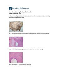
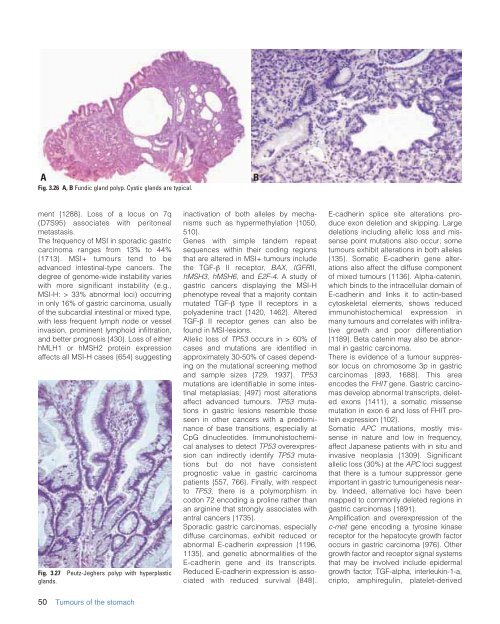
A<br />
Fig. 3.26 A, B Fundic gland polyp. Cystic glands are typical.<br />
B<br />
ment {1288}. Loss <strong>of</strong> a locus on 7q<br />
(D7S95) associates with peritoneal<br />
metastasis.<br />
The frequency <strong>of</strong> MSI in sporadic gastric<br />
carcinoma ranges from 13% to 44%<br />
{1713}. MSI+ tumours tend to be<br />
advanced intestinal-type cancers. The<br />
degree <strong>of</strong> genome-wide instability varies<br />
with more significant instability (e.g.,<br />
MSI-H: > 33% abnormal loci) occurring<br />
in only 16% <strong>of</strong> gastric carcinoma, usually<br />
<strong>of</strong> <strong>the</strong> subcardial intestinal or mixed type,<br />
with less frequent lymph node or vessel<br />
invasion, prominent lymphoid infiltration,<br />
and better prognosis {430}. Loss <strong>of</strong> ei<strong>the</strong>r<br />
hMLH1 or hMSH2 protein expression<br />
affects all MSI-H cases {654} suggesting<br />
Fig. 3.27<br />
glands.<br />
Peutz-Jeghers polyp with hyperplastic<br />
inactivation <strong>of</strong> both alleles by mechanisms<br />
such as hypermethylation {1050,<br />
510}.<br />
Genes with simple tandem repeat<br />
sequences within <strong>the</strong>ir coding regions<br />
that are altered in MSI+ tumours include<br />
<strong>the</strong> TGF-β II receptor, BAX, IGFRII,<br />
hMSH3, hMSH6, and E2F-4. A study <strong>of</strong><br />
gastric cancers displaying <strong>the</strong> MSI-H<br />
phenotype reveal that a majority contain<br />
mutated TGF-β type II receptors in a<br />
polyadenine tract {1420, 1462}. Altered<br />
TGF-β II receptor genes can also be<br />
found in MSI-lesions.<br />
Allelic loss <strong>of</strong> TP53 occurs in > 60% <strong>of</strong><br />
cases and mutations are identified in<br />
approximately 30-50% <strong>of</strong> cases depending<br />
on <strong>the</strong> mutational screening method<br />
and sample sizes {729, 1937}. TP53<br />
mutations are identifiable in some intestinal<br />
metaplasias; {497} most alterations<br />
affect advanced tumours. TP53 mutations<br />
in gastric lesions resemble those<br />
seen in o<strong>the</strong>r cancers with a predominance<br />
<strong>of</strong> base transitions, especially at<br />
CpG dinucleotides. Immunohistochemical<br />
analyses to detect TP53 overexpression<br />
can indirectly identify TP53 mutations<br />
but do not have consistent<br />
prognostic value in gastric carcinoma<br />
patients {557, 766}. Finally, with respect<br />
to TP53, <strong>the</strong>re is a polymorphism in<br />
codon 72 encoding a proline ra<strong>the</strong>r than<br />
an arginine that strongly associates with<br />
antral cancers {1735}.<br />
Sporadic gastric carcinomas, especially<br />
diffuse carcinomas, exhibit reduced or<br />
abnormal E-cadherin expression {1196,<br />
1135}, and genetic abnormalities <strong>of</strong> <strong>the</strong><br />
E-cadherin gene and its transcripts.<br />
Reduced E-cadherin expression is associated<br />
with reduced survival {848}.<br />
E-cadherin splice site alterations produce<br />
exon deletion and skipping. Large<br />
deletions including allelic loss and missense<br />
point mutations also occur; some<br />
tumours exhibit alterations in both alleles<br />
{135}. Somatic E-cadherin gene alterations<br />
also affect <strong>the</strong> diffuse component<br />
<strong>of</strong> mixed tumours {1136}. Alpha-catenin,<br />
which binds to <strong>the</strong> intracellular domain <strong>of</strong><br />
E-cadherin and links it to actin-based<br />
cytoskeletal elements, shows reduced<br />
immunohistochemical expression in<br />
many tumours and correlates with infiltrative<br />
growth and poor differentiation<br />
{1189}. Beta catenin may also be abnormal<br />
in gastric carcinoma.<br />
There is evidence <strong>of</strong> a tumour suppressor<br />
locus on chromosome 3p in gastric<br />
carcinomas {893, 1688}. This area<br />
encodes <strong>the</strong> FHIT gene. Gastric carcinomas<br />
develop abnormal transcripts, deleted<br />
exons {1411}, a somatic missense<br />
mutation in exon 6 and loss <strong>of</strong> FHIT protein<br />
expression {102}.<br />
Somatic APC mutations, mostly missense<br />
in nature and low in frequency,<br />
affect Japanese patients with in situ and<br />
invasive neoplasia {1309}. Significant<br />
allelic loss (30%) at <strong>the</strong> APC loci suggest<br />
that <strong>the</strong>re is a tumour suppressor gene<br />
important in gastric tumourigenesis nearby.<br />
Indeed, alternative loci have been<br />
mapped to commonly deleted regions in<br />
gastric carcinomas {1891}.<br />
Amplification and overexpression <strong>of</strong> <strong>the</strong><br />
c-met gene encoding a tyrosine kinase<br />
receptor for <strong>the</strong> hepatocyte growth factor<br />
occurs in gastric carcinoma {976}. O<strong>the</strong>r<br />
growth factor and receptor signal systems<br />
that may be involved include epidermal<br />
growth factor, TGF-alpha, interleukin-1-a,<br />
cripto, amphiregulin, platelet-derived<br />
50 <strong>Tumours</strong> <strong>of</strong> <strong>the</strong> stomach